image analysis workshop: practicals overview and … · problems •ihc analysis was never that...
TRANSCRIPT
Image Analysis Workshop: Practicals Overview and WISH LIST
Jeffrey L. Fine, M.D. Advanced Imaging and Image Analysis
Breast/Gyne Pathologist University of Pittsburgh Medical Center

Objectives
• Present Image Analysis as it is
• Talk about Practical Issues
• Show Potential for Image Analysis Future
(pathologists’ Computer Assisted Diagnosis pCAD)

Jargon!
• pathologists’ Computer Assisted Diagnosis (pCAD)
• Whole Slide Image (WSI)
• Regions of Interest (ROIs)
• Immunohistochemistry (IHC)
• Turnaround Time (TAT)

Current State of Image Analysis
Image Analysis
• Functional background analysis – Slide scanners (they “see” tissue, skip whitespace) – Digital photo software (camera focus gauge)
• Computer-assisted* interpretation of stains – Is this Her2/NEU stain positive? *(I use the term assisted very loosely)
• Automated slide interpretation (pCAD) – Which lymph nodes are positive? – Is tumor near any margin?

Current Systems
• Mostly limited to immunostain interpretation – Her2, ER, PR, Ki-67, p53, EGFR, etc.
– FDA cleared or home brewed
– Very manual workflow
– Novel algorithms do exist, but in a vacuum
• WSI integrated
• Stand-alone (either ROIs or WSI)
• Legacy – Dedicated non WSI device

Why Immunostains?
• Technically easy (“brown-o-meter”)
• High profile markers – oncologists know few stains explicitly—they know about Her2, et al.
• IHC Analysis is (was?) a billable test – CPT code 88361 (quant IHC computer assisted)
– Vendors and Management can grasp this easily
– IHC reimbursements now targeted by payors

Problems
• IHC analysis was never that automated – Low bar, and vendors stopped when bare
minimum for reimbursement was achieved
– FDA clearance expensive and a barrier to changes
• Technical billing often done thru APLIS or other system—not the analysis system – Integration can be difficult
• IHC analysis almost always slower than manual method, and requires supervision

Adult Supervision Required

Pre-WSI Advantages
• Easy to know cost per test
– WSI systems require infrastructure, servers, $$
• Easy to administer (shared account)
• Easier informatics (print-out or PDF)
– Not always, with bar coded schemes
• Single workstation, easier to maintain/fix
– Not easy though—either new vendors, or established vendors with new competencies

WSI or modern System Advantages
• Pathologist can do work from own office
• Pre-scanned slide means no need for glass in hand
– IHC lab consolidation or send-out
– Better TAT for cheaper (no overnight shipping)

Pitfalls
• Training
– Biggest issue in my experience
• Supervision of Computer
– Blind rubberstamping versus manually doing the interpretation then comparing with the computer
• Supervision of Users
– Proper use and good results?
– Proper billing?

Adult Supervision Required

Computer Supervision II

Examples of Computer Supervision
• Overcalling of Her-2 (2+ vs 3+) – Newer antibody less linear—very dark 2+ patterns
– One system – 6% of “3+” were FISH negative
• Overcalling of Ki-67 – Hard for computer to count negative nuclei
• ER/PR issue – No one does Allred score or H score (optional per
guidelines but widely expected by Oncology)
– Percentage staining AND intensity judgement
– This is an artifact of original billing code requirement

User Supervision/Training
• It’s OK to disagree with computer
– never blindly accept a computer-assisted result (at least in 2014)
– Pathologists may not understand this
• Mechanism for auditing
– If billed, was image analysis done?
– If not billed, was it done anyway?

Image Analysis Process
• Imaging and/or ROI Selection
• Image analysis occurs in one or more steps
– Find tumor within the ROI (tumor/tissue classifier)
– Measurement of staining (stain classifier)
• Show result to operator (supervision)
– Usually ROI by ROI
• Generate a result
– Average ROI results; Reconcile with Pathologist
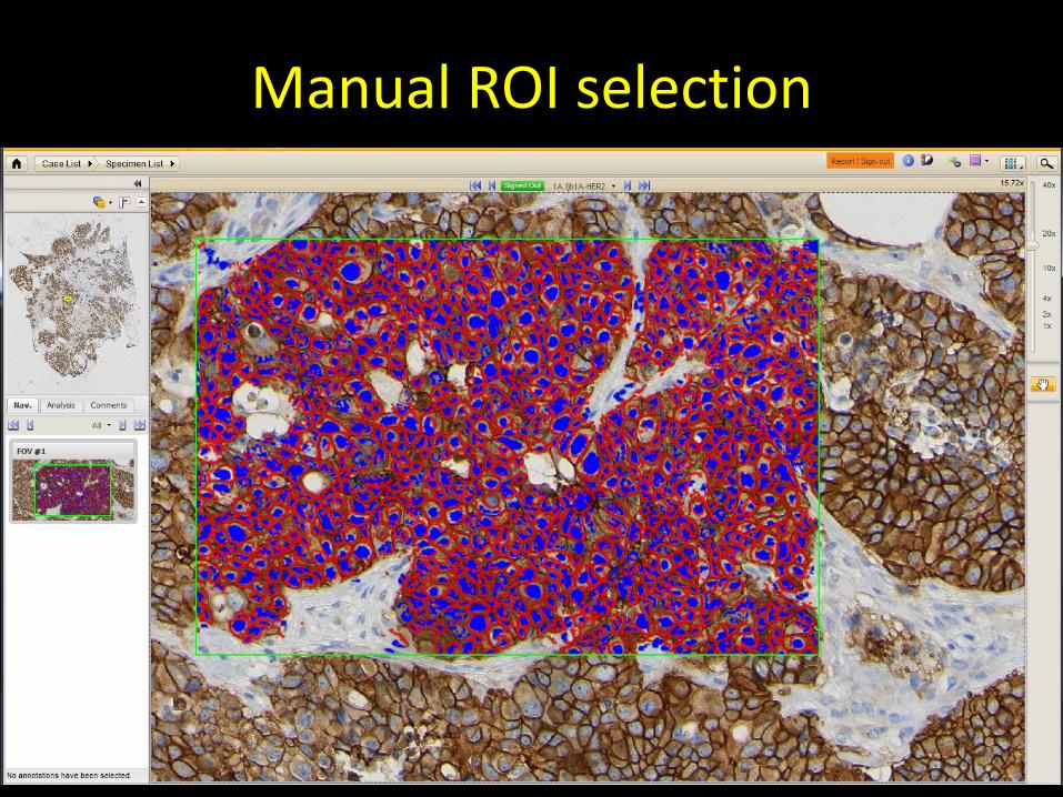
Manual ROI selection

Image analysis results mask (supervision)


FUTURE IMAGE ANALYSIS (21ST CENTURY WORKFLOW PROPOSAL)
What if the most important finding is buried at the end of the case?
Problem
• Current Digital Pathology systems fail to leverage the full power of whole slide images
– They propose that we will do exactly the same thing as before, looking at “virtual slides” in a linear fashion from A to Z
• Development focuses on good virtual microscope ergonomics and replicating the working draft information electronically

Proposal
• Shatter the slide images into small useful fragments
called “regions of interest” (ROIs)
• Triage these ROIs and present them to the pathologist in context of sign-out tasks
• Automatically generate report data from this work
• Eventually automate review so that all ROIs need NOT be reviewed by the pathologist (a la Pap test imaging)

Simplify specimens into a concrete task or set of tasks
• Sentinel lymph node; endocervical curettage; excisional breast biopsy following a benign but risky core biopsy finding
• Post-Partum Tubal Ligation
• What do we need to do, concretely – Give the engineers tangible targets
• Identify where the “art” is; this is work pathologists need to spend most time on

Example

Lymph Nodes
• Current – Pathologists must crawl through lymph node
sections; tumor may be a rare event
– Nodes must be counted
• Future – Computer “reads” the gross (synoptic grossing?)
– Presents ‘suspicious’ ROIs to the pathologist for review

Glass
Dinosaur Med Ctr S17-0000335
Flintstomp, Ferd A1 H&E

Levels as Images…
UPMC S14-97035 John Doe 1A H&E

Stack the levels
UPMC S14-97035 John Doe 1A H&E

Break the level into objects (Lymph Nodes)
UPMC S14-97035 John Doe 1A H&E

The case’s nodes (from all slides) (levels stacked)
(IHC stains are levels)

How to break up the object?

What are the compartments?
FAT
NODE (interior)
SUBCAPSULAR SINUS

Break compartments into ROIs
FAT
NODE (interior)
SUBCAPSULAR SINUS

A series of ROIs
Most suspicious or Highest-yield
Least suspicious or Lowest-yield


Interactive ROI Review
• Show ROIs to Pathologist
• Ask Pathologist to agree/disagree with finding
– (“is this tumor”)
• Automatically catalogue findings into a report
– (“3 of 12 lymph nodes are positive”)
– Measurements can be done in real-time

In the future
• Computer algorithms will be “trustworthy”
– Not all ROIs need be reviewed
• Lymph node fat; non-suspicious lymph node parenchyma

Bottom Line
• How much time would it save if:
– The computer screened lymph nodes first
– The computer kept track of counting and measuring
• Pathologist being chauffeured rather than driving
– Jumping straight to ROIs w/o driving there

Summary
• Current Image Analysis is a gadget
– Manual labor
– Reimbursement no longer a no brainer
• Pathologists must know what they want
– Automation (real delegation of easy decisions)
– Decision Support
– Productivity increases with ever-cheaper IT

Do you really need the computer to tell you this? (Score 3+)