inhibition of wnt signaling by icat, a novel Я-catenin-interacting
TRANSCRIPT

Inhibition of Wnt signaling by ICAT,a novel �-catenin-interacting proteinKen-ichi Tago,1,2,5 Tsutomu Nakamura,1,5 Michiru Nishita,3 Junko Hyodo,3 Shin-ichi Nagai,3
Yoji Murata,1 Shungo Adachi,1 Susumu Ohwada,2 Yasuo Morishita,2 Hiroshi Shibuya,3
and Tetsu Akiyama1,4,6
1Laboratory of Molecular and Genetic Information, Institute for Molecular and Cellular Biosciences, The Universityof Tokyo, Bunkyo-ku, Tokyo 113, Japan; 2Second Department of Surgery, Gunma University School of Medicine, Maebashi,Gunma 371, Japan; 3Division of Morphogenesis, Department of Developmental Biology, National Institute for Basic Biology,Okazaki 444, Japan; 4Department of Oncogene Research, Institute for Microbial Diseases, Osaka University, Suita,Osaka 565, Japan
Wnt signaling has an important role in both embryonic development and tumorigenesis. �-Catenin, a keycomponent of the Wnt signaling pathway, interacts with the TCF/LEF family of transcription factors andactivates transcription of Wnt target genes. Here, we identify a novel �-catenin-interacting protein, ICAT, thatwas found to inhibit the interaction of �-catenin with TCF-4 and represses �-catenin–TCF-4-mediatedtransactivation. Furthermore, ICAT inhibited Xenopus axis formation by interfering with Wnt signaling.These results suggest that ICAT negatively regulates Wnt signaling via inhibition of the interaction between�-catenin and TCF and is integral in development and cell proliferation.
[Key Words: Wnt; �-catenin; TCF; ICAT; signaling]
Received Feburary 28, 2000; revised version accepted May 24, 2000.
The Wnt/Wingless signaling transduction pathway is in-volved in many developmental processes via the regula-tion of Wnt-responsive genes (Miller and Moon 1996;Cadigan and Nusse 1997; Clevers and van de Wetering1997; Bienz 1998; Eastman and Grosschedl 1999). Ex-pression of these genes is regulated by the TCF/LEF fam-ily of transcription factors, whose activity is promotedby their association with �-catenin (Behrens et al. 1996;Molenaar et al. 1996; Brunner et al. 1997; Riese et al.1997; van de Wetering et al. 1997; Hsu et al. 1998; Gal-ceran et al. 1999). The stability of �-catenin is in turndetermined by its association with Axin, glycogen syn-thase kinase-3� (GSK-3�), and the tumor suppressor ad-enomatous polyposis coli (APC) (Munemitsu et al. 1995;Behrens et al. 1998; Hart et al. 1998; Ikeda et al. 1998;Itoh et al. 1998; Nakamura et al. 1998; Sakanaka et al.1998; Hamada et al. 1999; Willert et al. 1999), the mu-tation of which is responsible for familial adenomatouspolyposis (FAP) and sporadic colorectal tumors (Kinzlerand Vogelstein 1996; Polakis 1997; Bienz 1999). Wnt sig-naling promotes the stabilization of �-catenin by nega-tively regulating the activity of GSK-3�. Intact APC nor-mally induces the degradation of �-catenin, but the mu-tant APCs found in most colon cancers are defective inthis activity. Furthermore, of those tumors that contain
intact APC, many have stabilizing mutations in�-catenin itself (Morin et al. 1997; Rubinfeld et al. 1997).Therefore, regulation of �-catenin stability and, conse-quently, �-catenin-TCF/LEF-mediated transactivationare critical for Wnt signaling during development andtumorigenesis. In this study we show that a novel�-catenin-interacting protein, termed ICAT (inhibitor of�-catenin and TCF-4), interferes with the interaction be-tween �-catenin and TCF-4 and antagonizes Wnt signal-ing.
Results and Discussion
In an attempt to identify �-catenin-interacting proteins,we performed a yeast two-hybrid screen of a mouse em-bryo cDNA library using the Armadillo repeat domain of�-catenin as bait and found a new protein, ICAT. Se-quence analysis of the ICAT cDNA showed that it en-codes a protein of 81 amino acids with no homology toother known proteins (Fig. 1A). Highly conserved or-thologs were identified as EST clones from human, rat,and zebrafish, and Xenopus ICAT was isolated from aXenopus oocyte library. Northern blot analysis detectedan mRNA of 2.6 kb, which is expressed at high levels inmouse heart, brain, liver, and skeletal muscle, at lowlevels in kidney, testis, and lung, and at undetectablelevels in spleen (data not shown). ICAT mRNA was ex-pressed at fairly constant levels during development ofthe mouse embryo (data not shown).
To confirm that ICAT and �-catenin interact directly,we examined the ability of ICAT fused to glutathione
5These authors contributed equally to the work.6Corresponding author.E-MAIL [email protected]; FAX 81 35841 8482.
GENES & DEVELOPMENT 14:1741–1749 © 2000 by Cold Spring Harbor Laboratory Press ISSN 0890-9369/00 $5.00; www.genesdev.org 1741
Cold Spring Harbor Laboratory Press on April 2, 2018 - Published by genesdev.cshlp.orgDownloaded from

S-transferase (GST) to interact with �-catenin producedby in vitro translation. GST–ICAT, but not GST alone,associated with in vitro-translated �-catenin (Fig. 1B).Two-hybrid assays using various deletion fragments ofICAT revealed that the central region of ICAT is in-volved in the interaction with �-catenin (Fig. 1C). It hasbeen reported that the �-catenin binding regions of cad-herins, APC, and TCF family members are all acidic(Huber et al. 1997). Therefore, we generated a mutantICAT–E37–39A, in which Glu-37, Glu-38, and Glu-39were all replaced with Ala and found that this mutant isnegative for interaction with �-catenin (Fig. 1C). On theother hand, the minimal region of �-catenin required forbinding to ICAT was found to reside in the fragmentspanning from the carboxy-terminal portion of repeat 10to repeat 12 (Fig. 1C). In contrast, Armadillo repeats 1–9did not exhibit any affinity to ICAT.
Next, we examined whether ICAT is associated with�-catenin in vivo. For this purpose, we generated anti-bodies to the carboxy-terminal portion of ICAT and con-
firmed that the antibodies react specifically with GST–ICAT (data not shown). When a lysate from mouse brainwas subjected to immunoprecipitation and subsequentimmunoblotting with anti-ICAT antibodies, we detecteda 9-kD protein, and precipitation of this protein was in-hibited by preincubation of the antibodies with antigen(Fig. 1D). In addition, a protein of the same mobility wasexpressed prominently when COS-7 cells were trans-fected with ICAT cDNA (data not shown). These resultssuggest that ICAT gene product is a 9-kD protein. Thenwe subjected a lysate from mouse brain to immunopre-cipitation with anti-ICAT antibodies and immunoblot-ted the precipitates with anti-�-catenin antibody. ICATwas found to coprecipitate with �-catenin, and coprecipi-tation was inhibited by preincubation of anti-ICAT an-tibodies with antigen (Fig. 1D). Also, immunoprecipita-tion of the lysate with anti-�-catenin antibody, followedby immunoblotting with anti-ICAT antibodies, revealedan association between ICAT and �-catenin. Preincuba-tion of the anti-�-catenin antibody with the antigen pre-
Figure 1. Association of ICAT with �-catenin. (A) Predicted amino acid sequences of ICATs. Identical and similar residues arehighlighted in black and gray, respectively. Vertical bars indicate the region critical for binding to �-catenin (see C). (B) Associationof ICAT with �-catenin in vitro. In vitro-translated 35S-labeled proteins indicated (Input) were incubated with GST–ICAT–Sepharose(+GST–ICAT). The bound proteins were analyzed by SDS-PAGE. (C) Mapping of regions in ICAT and �-catenin required for interac-tion. Deletion constructs of ICAT (left) or �-catenin (right) were analyzed for their ability to interact with GAL4–�-catenin orGAL4–ICAT, respectively, in the two-hybrid system. (+) Detectable activity; (±) marginal activity; (−) no detectable activity. (D)Association of ICAT with �-catenin in vivo. Lysates prepared from mouse brain were subjected to immunoprecipitation with theantibodies indicated, fractionated by SDS-PAGE, and immunoblotted with the antibodies indicated. (Ag+) Antibodies were preincu-bated with antigen before use in immunoprecipitation.
Tago et al.
1742 GENES & DEVELOPMENT
Cold Spring Harbor Laboratory Press on April 2, 2018 - Published by genesdev.cshlp.orgDownloaded from

vented coprecipitation of �-catenin and ICAT. These re-sults suggest that ICAT is associated with �-catenin inliving cells. On the other hand, ICAT–E37–39A ectopi-cally expressed in COS-7 cells failed to coprecipitatewith �-catenin (data not shown). Consistent with theseresults, ICAT was found to colocalize with �-catenin inthe nucleus of the human colorectal tumor cell lineSW480 (data not shown). ICAT was also detected in thecytoplasm and nucleus of mouse colon epithelial cells.ICAT colocalized with �-catenin in the cytoplasm butnot at the plasma membrane in the epithelial cells of thecolon.
The TCF family of proteins is known to form a com-plex with �-catenin that binds to specific DNA se-quences and transactivates target genes (Behrens et al.1996; Molenaar et al. 1996; Brunner et al. 1997; Riese et
al. 1997; van de Wetering et al. 1997; Hsu et al. 1998;Galceran et al. 1999). Therefore, we asked whether ICATaffects the DNA-binding properties of the �-catenin–TCF-4 complex. As reported previously (Korinek et al.1997), an electrophoretic mobility-shift assay (EMSA)showed that �-catenin produced by the baculovirus sys-tem and TCF-4 generated by in vitro translation boundto an oligonucleotide containing a TCF-4-binding site asa ternary complex (Fig. 2A). However, when in vitro-translated TCF-4 and �-catenin were preincubated withGST–ICAT, this ternary complex was not detected. In-stead, a band migrating with the mobility of the TCF-4–DNA complex was detected. Addition of anti-TCF-4 an-tibodies, but not anti-�-catenin and/or anti-ICAT anti-bodies, induced supershift of this band, suggesting thatthe band represents the TCF-4–DNA complex. In a par-
Figure 2. ICAT inhibits the formation of the �-catenin–TCF-4 complex. (A) The �-catenin–TCF-4–DNA complex is not detected inthe presence of ICAT. An oligonucleotide containing a potential binding site for TCF-4 was incubated with in vitro-translated TCF-4,�-catenin produced by the baculovirus system, and GST–ICAT as indicated. DNA–protein interactions were analyzed by EMSAs.Unlabeled oligonucleotides containing either consensus (competitor-TOP) or mutated (competitor-FOP) sites were used as competi-tors in some reactions. (B) ICAT inhibits the interaction between �-catenin and TCF-4 in vitro. �-Catenin (3 ng) and TCF-4 (10 ng)produced by the baculovirus system were incubated in the presence of 100 ng or 1 µg of GST–ICAT, GST–ICAT–E37–39A, orGST–ICAT–�42–61, respectively. �-Catenin was immunoprecipitated with anti-�-catenin antibody, and the immunoprecipitates weresubjected to immunoblotting analysis with anti-TCF-4 antibodies. (C) ICAT inhibits the interaction between �-catenin and TCF-4 invivo. �-catenin and TCF-4 were transfected along with ICAT into human kidney epithelial 293 cells. �-Catenin was immunoprecipi-tated with anti-�-catenin antibody, and the immunoprecipitates were subjected to immunoblotting analysis with antibodies indicated.(D) ICAT–�42–61 abrogates the inhibitory effect of wild-type ICAT on the interaction between �-catenin and TCF-4. HA-taggedICAT–�42–61 was transfected along with HA-tagged ICAT, �-catenin, and TCF-4 into 293 cells. �-Catenin was immunoprecipitated,and coprecipitating TCF-4 was detected by immunoblotting with anti-TCF-4 antibodies.
Inhibition of Wnt signaling by ICAT
GENES & DEVELOPMENT 1743
Cold Spring Harbor Laboratory Press on April 2, 2018 - Published by genesdev.cshlp.orgDownloaded from
allel pull-down experiment, we found that the amountsof TCF-4 that coimmunoprecipitated with anti-�-catenin decreased in a dose-dependent manner with in-creasing amounts of ICAT (Fig. 2B). Furthermore, when�-catenin and TCF-4 were transfected along with ICATinto human kidney epithelial 293 cells, the amounts ofTCF-4 coimmunoprecipitating with �-catenin also de-creased as the expressed amount of ICAT increased (Fig.2C). These results suggest that ICAT interferes with theformation of the �-catenin–TCF-4 complex. In addition,ICAT was also found to interfere with the interactionbetween �-catenin and Xenopus TCF-3 (data not shown).On the other hand, the mutant ICAT–E37–39A did notinhibit the interaction between �-catenin and TCF-4(Fig. 2B) and had no effect on the formation of a�-catenin–TCF-4–DNA complex (Fig. 2A).
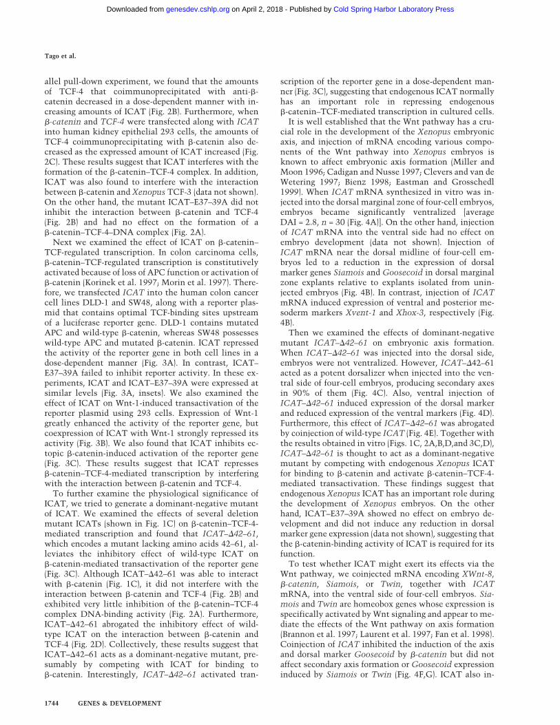
Next we examined the effect of ICAT on �-catenin–TCF-regulated transcription. In colon carcinoma cells,�-catenin–TCF-regulated transcription is constitutivelyactivated because of loss of APC function or activation of�-catenin (Korinek et al. 1997; Morin et al. 1997). There-fore, we transfected ICAT into the human colon cancercell lines DLD-1 and SW48, along with a reporter plas-mid that contains optimal TCF-binding sites upstreamof a luciferase reporter gene. DLD-1 contains mutatedAPC and wild-type �-catenin, whereas SW48 possesseswild-type APC and mutated �-catenin. ICAT repressedthe activity of the reporter gene in both cell lines in adose-dependent manner (Fig. 3A). In contrast, ICAT–E37–39A failed to inhibit reporter activity. In these ex-periments, ICAT and ICAT–E37–39A were expressed atsimilar levels (Fig. 3A, insets). We also examined theeffect of ICAT on Wnt-1-induced transactivation of thereporter plasmid using 293 cells. Expression of Wnt-1greatly enhanced the activity of the reporter gene, butcoexpression of ICAT with Wnt-1 strongly repressed itsactivity (Fig. 3B). We also found that ICAT inhibits ec-topic �-catenin-induced activation of the reporter gene(Fig. 3C). These results suggest that ICAT represses�-catenin–TCF-4-mediated transcription by interferingwith the interaction between �-catenin and TCF-4.
To further examine the physiological significance ofICAT, we tried to generate a dominant-negative mutantof ICAT. We examined the effects of several deletionmutant ICATs (shown in Fig. 1C) on �-catenin–TCF-4-mediated transcription and found that ICAT–�42–61,which encodes a mutant lacking amino acids 42–61, al-leviates the inhibitory effect of wild-type ICAT on�-catenin-mediated transactivation of the reporter gene(Fig. 3C). Although ICAT–�42–61 was able to interactwith �-catenin (Fig. 1C), it did not interfere with theinteraction between �-catenin and TCF-4 (Fig. 2B) andexhibited very little inhibition of the �-catenin–TCF-4complex DNA-binding activity (Fig. 2A). Furthermore,ICAT–�42–61 abrogated the inhibitory effect of wild-type ICAT on the interaction between �-catenin andTCF-4 (Fig. 2D). Collectively, these results suggest thatICAT–�42–61 acts as a dominant-negative mutant, pre-sumably by competing with ICAT for binding to�-catenin. Interestingly, ICAT–�42–61 activated tran-
scription of the reporter gene in a dose-dependent man-ner (Fig. 3C), suggesting that endogenous ICAT normallyhas an important role in repressing endogenous�-catenin–TCF-mediated transcription in cultured cells.
It is well established that the Wnt pathway has a cru-cial role in the development of the Xenopus embryonicaxis, and injection of mRNA encoding various compo-nents of the Wnt pathway into Xenopus embryos isknown to affect embryonic axis formation (Miller andMoon 1996; Cadigan and Nusse 1997; Clevers and van deWetering 1997; Bienz 1998; Eastman and Grosschedl1999). When ICAT mRNA synthesized in vitro was in-jected into the dorsal marginal zone of four-cell embryos,embryos became significantly ventralized [averageDAI = 2.8, n = 30 (Fig. 4A)]. On the other hand, injectionof ICAT mRNA into the ventral side had no effect onembryo development (data not shown). Injection ofICAT mRNA near the dorsal midline of four-cell em-bryos led to a reduction in the expression of dorsalmarker genes Siamois and Goosecoid in dorsal marginalzone explants relative to explants isolated from unin-jected embryos (Fig. 4B). In contrast, injection of ICATmRNA induced expression of ventral and posterior me-soderm markers Xvent-1 and Xhox-3, respectively (Fig.4B).
Then we examined the effects of dominant-negativemutant ICAT–�42–61 on embryonic axis formation.When ICAT–�42–61 was injected into the dorsal side,embryos were not ventralized. However, ICAT–�42–61acted as a potent dorsalizer when injected into the ven-tral side of four-cell embryos, producing secondary axesin 90% of them (Fig. 4C). Also, ventral injection ofICAT–�42–61 induced expression of the dorsal markerand reduced expression of the ventral markers (Fig. 4D).Furthermore, this effect of ICAT–�42–61 was abrogatedby coinjection of wild-type ICAT (Fig. 4E). Together withthe results obtained in vitro (Figs. 1C, 2A,B,D,and 3C,D),ICAT–�42–61 is thought to act as a dominant-negativemutant by competing with endogenous Xenopus ICATfor binding to �-catenin and activate �-catenin–TCF-4-mediated transactivation. These findings suggest thatendogenous Xenopus ICAT has an important role duringthe development of Xenopus embryos. On the otherhand, ICAT–E37–39A showed no effect on embryo de-velopment and did not induce any reduction in dorsalmarker gene expression (data not shown), suggesting thatthe �-catenin-binding activity of ICAT is required for itsfunction.
To test whether ICAT might exert its effects via theWnt pathway, we coinjected mRNA encoding XWnt-8,�-catenin, Siamois, or Twin, together with ICATmRNA, into the ventral side of four-cell embryos. Sia-mois and Twin are homeobox genes whose expression isspecifically activated by Wnt signaling and appear to me-diate the effects of the Wnt pathway on axis formation(Brannon et al. 1997; Laurent et al. 1997; Fan et al. 1998).Coinjection of ICAT inhibited the induction of the axisand dorsal marker Goosecoid by �-catenin but did notaffect secondary axis formation or Goosecoid expressioninduced by Siamois or Twin (Fig. 4F,G). ICAT also in-
Tago et al.
1744 GENES & DEVELOPMENT
Cold Spring Harbor Laboratory Press on April 2, 2018 - Published by genesdev.cshlp.orgDownloaded from

hibited Wnt-8-induced axis duplication (Fig. 4F). Thus,ICAT appears to exert its effect by interfering with sig-naling through the Wnt pathway at a point downstream
of �-catenin, and upstream of Siamois and Twin. In ad-dition, we found that ICAT failed to block the inductionof the secondary axis caused by ventral injection of
Figure 3. Effect of ICAT on �-catenin–TCF-regulated transcription. (A) ICAT repressesconstitutively activated �-catenin–TCF-me-diated transcription in colorectal cancercells. The human colorectal cancer cell linesDLD-1 and SW48 were transfected with theluciferase reporter plasmid TOPtkLuciferase(solid bars) or FOPtkLuciferase (shaded bars),and increasing amounts of ICAT, ICAT–E37–39A, and luciferase activity were mea-sured (15). TOPtkLuciferase contains opti-mal and FOPtkLuciferase contains mutatedTCF-binding sites placed upstream of a lu-ciferase reporter gene. Luciferase activitiesare expressed relative to samples containingno ICAT and ICAT–E37–39A. (Insets) Detec-tion of endogenous ICAT and exogenouslyexpressed ICAT and ICAT–E37–39A (indi-cated by closed arrow). Lysates preparedfrom DLD-1 or SQ48 cells transfected withthe indicated plasmids (1.0 µg) were sub-jected to immunoprecipitation and subse-quent immunoblotting with anti-ICAT anti-bodies. (Ag+) Antibodies were preincubatedwith antigen before use in immunnoprecipi-tation. (B) ICAT represses Wnt-1-induced�-catenin–TCF-mediated transactivation.293 cells were transfected with luciferase re-porter plasmids, ICAT, and Wnt-1, and lucif-erase activity was measured. (C) ICAT–�42–61 abrogates the inhibitory effect of wild-type ICAT on �-catenin–TCF-mediatedtranscription. 293 cells were transfectedwith the luciferase reporter (0.05 µg),�-catenin (0.01 µg), ICAT, and ICAT–�42–61, as indicated. (Inset) Detection of endog-enous ICAT, exogenously expressed ICAT(indicated by solid arrow), and ICAT–�42–61(open arrow). Lysates prepared from 293 cellstransfected with the indicated plasmids (0.5µg) were subjected to immunoprecipitationand subsequent immunoblotting with anti-ICAT antibodies. (Ag+) Antibodies were pre-incubated with antigen before use in immu-noprecipitation. (D) ICAT–�42–61 activatesendogeneous �-catenin–TCF-mediated tran-scription. 293 cells were transfected with theluciferase reproter (0.05 µg) and increasingamounts of ICAT–�42–61 (as indicated).
Inhibition of Wnt signaling by ICAT
GENES & DEVELOPMENT 1745
Cold Spring Harbor Laboratory Press on April 2, 2018 - Published by genesdev.cshlp.orgDownloaded from

mRNA encoding Noggin, a natural inhibitor of BMPs ora dominant-negative truncated BMP receptor (�BMPR-IA; Fig. 4F). Thus, ICAT is thought to act as a negativeregulator specifically on the Wnt signaling pathway.
When cells are stimulated with the Wnt signal,
�-catenin is stabilized and accumulates within the cell(Miller and Moon 1996; Cadigan and Nusse 1997; Clev-ers and van de Wetering 1997; Bienz 1998; Eastman andGrosschedl 1999), whereas ICAT levels do not changesignificantly (data not shown). Thus, the amount of
Figure 4. Effects of ICAT on axis formation in Xenopus em-bryos. (A) Dorsal injection of ICAT-induced ventralization ofXenopus embryos. Synthetic ICAT mRNA (2 ng) was injectedinto each of two dorsal blastomeres at the four-cell stage andphenotypes were evaluated at the tadpole stage. (B) Dorsal in-jection of ICAT reduced expression of the dorsal markersGoosecoid and Siamois and induced expression of the ventralmarkers Xhox-3 and Xvent-1. DMZ fragments injected withICAT mRNA (2 ng) were cultured until gastrula stage 11, andtotal RNA was analyzed by RT–PCR for the presence of theindicated transcripts: (lane 1) Whole embryo; (lane 2) unin-jected; (lane 3) ICAT (2 ng); (lane 4) RT. Histone was used as aloading control. (C) Ventral injection of ICAT–�42–61 mRNA(500 pg) induced ectopic axis formation in Xenopus embryos.(D) Ventral injection of ICAT–�42–61 induced expression ofthe dorsal marker and reduced expression of the ventral mark-ers. VMZ fragments injected with ICAT–�42–61 mRNA (500pg) were analyzed as described in B. (Lane 1) Whole embryo;(lane 2) uninjected; (lane 3) ICAT–�42–61 (500 pg); (lane 4) RT.(E) ICAT–�42–61-induced ectopic axis formation in Xenopusembryos was not observed when wild-type ICAT was coin-jected with ICAT�42–61. (F) (Left) Ventral coinjection of ICATmRNA inhibits ectopic axis formation by �-catenin but not bydownstream targets (Siamois and Twin) of the Wnt pathway, orby Noggin or �BMPR-IA. mRNA encoding the indicated dor-salizing factor was injected into two ventral blastomeres at thefour-cell stage with or without 2 ng of ICAT, and embryos wereexamined for axial duplications at the tadpole stage. The frac-tion of embryos with duplicated axes is indicated above eachbar. mRNAs were injected in the minimal amounts needed toinduce ectopic axes at high frequency: (5 pg) Siamois; (5 pg)Twin; (1 pg) Noggin; or (50 pg) �BMPR-IA. (Right) ICAT (2 ng)also inhibits Wnt-8 (10 pg)-induced axis duplication. WhenICAT plus �-catenin plus Wnt-8 were expressed, ICAT did notexhibit a strong effect. This may be because the amounts of�-catenin in cells overexpressing both exogenous �-catenin andWnt-8 were higher than that of ICAT. (G) Coinjection of ICATmRNA inhibits induction of the dorsal marker Goosecoid by�-catenin but not by Siamois or Twin. Synthetic mRNAs con-taining the indicated DNA sequences were injected into theequatorial region of blastomeres at the two-cell stage. Animalcaps coinjected with �-catenin (200 pg), Siamois (5 pg), or Twin(5 pg), with or without ICAT (2 ng) mRNA, were cultured untilthe gastrula stage, and total RNA was analyzed by RT–PCR forthe presence of the indicated transcripts: (Lane 1) Whole em-bryo; (lane 2) uninjected; (lane 3) ICAT; (lane 4) �-catenin; (lane5) �-catenin and ICAT; (lane 6) Siamois; (lane 7) Siamois andICAT; (lane 8) Twin; (lane 9) Twin and ICAT; (lane 10) RT.
Tago et al.
1746 GENES & DEVELOPMENT
Cold Spring Harbor Laboratory Press on April 2, 2018 - Published by genesdev.cshlp.orgDownloaded from

�-catenin not associated with ICAT may increase, lead-ing to transcriptional activation. The importance ofICAT in vivo was confirmed by experiments using Xeno-pus embryos, which suggested that ICAT has an impor-tant role in negatively regulating the Wnt signaling path-way in Xenopus development. We found that XenopusICAT (XICAT) transcripts are expressed maternally andthroughout development from the egg to the tailbudstage, with a decline in expression during gastrulation,and that XICAT coprecipitates with �-catenin from alysate prepared from Xenopus embryos (stage 10.5) (datanot shown). XICAT transcripts are expressed ubiqui-tously and are not localized to any specific region in theearly gastrula stage. However, XICAT transcripts be-come localized to the nervous system at the end of neu-rulation and are restricted to the central nervous system,eye, and head neural crest cell populations by the tadpolestages. These expression patterns are consistent with itsputative function in developmental processes, includingdorsoventral axis formation. Although it is well knownthat endogenous �-catenin is enriched in the dorsal,compared to the ventral, regions, there is a certainamount of �-catenin present in ventral regions (Larabellet al. 1997). We speculate that �-catenin in the ventralregions is associated with ICAT, and the amount of freeactive �-catenin is insufficient to induce transactivationin the absence of a Wnt signal. Consistent with this no-tion, ventral expression of the dominant-negative mu-tant ICAT–�42–61 induced axis duplication, presumablyby competing with endogenous ICAT for binding to�-catenin. The amount of active �-catenin generated bythe action of ICAT–�42–61 may be enough to inducetranscriptional activation in Xenopus embryos. Takentogether, we speculate that ICAT may function to estab-lish a threshold to prevent premature and inappropriatesignaling events. On the other hand, it has been reportedrecently that in the absence of �-catenin, TCF is associ-ated with members of the Groucho family of proteinsand acts as a transcriptional repressor of Wnt/Winglesstarget genes (Cavallo et al. 1998; Roose et al. 1998). Thus,the function of �-catenin may be regulated by the bal-ance among ICAT, �-catenin, and the Groucho family ofproteins.
Constitutive activation of �-catenin–TCF-mediatedtranscription due to inactivation of the tumor suppressorAPC or gain-of function mutations in �-catenin isthought to be important in colorectal tumorigenesis(Kinzler and Vogelstein 1996; Korinek et al. 1997; Morinet al. 1997; Polakis 1997; Rubinfeld et al. 1997; Bienz1999). Because ICAT is a negative regulator of Wnt sig-naling, its inactivation could also induce inappropriateactivation of the transcription. It is therefore interestingto speculate that ICAT may function as a tumor suppres-sor and its inactivation may lead to tumorigenesis.Given its function in inhibiting �-catenin–TCF-medi-ated transcription, ICAT may be of interest as a genetherapy agent. Drugs that mimic the effects of ICAT maybe useful as antitumor reagents as well, and elucidationof the three-dimensional structure of ICAT may provideinsights into the development of such drugs.
Materials and methods
Two-hybrid system
Two-hybrid experiments were performed as described (Hamadaet al. 1999) using the Armadillo domain of mouse �-catenin(amino acids 128–683) as bait (The GenBank accession numberof ICAT is AB021261.)
In vitro binding assays35S-Labeled proteins were synthesized by in vitro transcriptiontranslation in the presence of [35S]methionine using the TNT-coupled reticulocyte lysate system (Promega). GST fusion pro-teins immobilized to glutathione–Sepharose were mixed within vitro-translated proteins in buffer A (10 mM Tris-HCl at pH8.0, 140 mM NaCl, 1 mM EDTA, 10 µg/ml leupeptin, 10 µg/mlaprotinin) containing 0.1% Triton X-100 for 2 hr at 4°C andthen washed extensively with buffer A.
Antibodies
Antibodies to ICAT were prepared by immunizing rabbits witha peptide containing amino acids 70–81 of ICAT. Antibodies toTCF-4E were generated with a peptide corresponding to the car-boxy-terminal 20 amino acids of TCF-4E. Mouse monoclonalantibody to �-catenin was purchased from Transduction Labo-ratories. GST–�-catenin was used to block anti-�-catenin anti-body.
Immunoprecipitation and immunoblotting
Immunoprecipitation and immunoblotting were performed asdescribed previously (Matsumine et al. 1996). To examine theeffect of ICAT on the interaction between �-catenin and TCF-4in vitro, �-catenin (3 ng) and TCF-4 (10 ng) produced by thebaculovirus system were incubated in 150 µl of buffer A for 1 hrat 4°C in the presence of 100 ng or 1 µg of GST–ICAT, GST–ICAT–E37–39A, or GST–ICAT–�42–61, respectively. �-Cateninwas immunoprecipitated with anti-�-catenin antibody and pro-tein G–Sepharose, and the immunoprecipitates were subjectedto immunoblotting analysis with anti-TCF-4 antibodies.
EMSAs
As the optimal TCF probe, we used a double-stranded 26-nucleotide oligomer containing a potential TCF/LEF bindingsite derived from TOPtkLuciferase reporter (Korinek et al.1997). The mutant probe was derived from FOPtkLuciferase re-porter. In vitro-translated TCF-4, �-catenin produced by thebaculovirus system (0.1 µg) and GST–ICAT (0.1, 0.3, or 1 µg)were incubated for 2 hr in binding buffer (10 mM Tris at pH 7.5,1 mM EDTA, 60 mM KCl, 12% glycerol, 1.0 mM DTT, 1.5 µgBSA) in a total volume of 30 µl. Probe (6.8 ng) end-labeled to7.6 × 106 cpm/µg, poly[d(I-C)] (1 µg) and herring sperm DNA (1.5µg) were added and incubated for an additional 20 min. Com-petition analyses were performed with an excess amount (680ng) of unlabeled probe.
Luciferase assays
Cells (6 × 10⁵ cells/60-mm dish) were transfected by Lipofect-amine with a total of 4 µg of the various combinations of plas-mids: 0.5 µg of reporter plasmid (TOPtkLuciferase or FOPtkLu-ciferase); 0.05 µg of internal control pRL-TK (Promega); the in-dicated amount of wild-type and/or mutant ICAT expressionvector (pMKITNeoICAT), and empty pMKITNeo vector as
Inhibition of Wnt signaling by ICAT
GENES & DEVELOPMENT 1747
Cold Spring Harbor Laboratory Press on April 2, 2018 - Published by genesdev.cshlp.orgDownloaded from

stuffer. Luciferase activities were measured 24 hr after transfec-tion using the Dual-Luciferase Reporter Assay System (Pro-mega).
Embryo manipulations
ICAT, ICAT–�42–61, and ICAT–E37–39A cDNAs were clonedinto the pCS2+ vector (Rupp et al. 1994). RNAs were then in-jected into the animal poles or marginal zones of early-stageembryos as described (Moon and Christian 1989). Dorsal mar-ginal zone (DMZ) assay, ventral marginal zone (VMZ) assay, andanimal cap assay were performed as described (Shibuya et al.1998). Total RNA was then extracted and analyzed with RT–PCR as described (Wilson and Melton 1994; Wilson and Hem-mati-Brivanlou 1995).
Acknowledgments
We thank N. Ueno and T. Ishidate for discussion and encour-agement. We also thank V. Korinek and H. Clevers for TOPtk-Luciferase and FOPtkLuciferase. This work was supported byGrants-in-Aid for Scientific Research on Priority Areas and theOrganization for Pharmaceutical Safety and Research.
The publication costs of this article were defrayed in part bypayment of page charges. This article must therefore be herebymarked “advertisement” in accordance with 18 USC section1734 solely to indicate this fact.
References
Behrens, J., von Kries, J.P., Kuhl, M., Bruhn, L., Wedlich, D.,Grosschedl, R., and Birchmeier, W. 1996. Functional inter-action of �-catenin with the transcription factor LEF-1. Na-ture 382: 638–642.
Behrens, J., Jerchow, B.A., Wurtele, M., Grimm, J., Asbrand, C.,Wirtz, R., Kuhl, M., Wedlich, D., and Birchmeier, W. 1998.Functional interaction of an axin homolog, conductin, with�-catenin, APC, and GSK3�. Science 280: 596–599.
Bienz, M. 1998. TCF: Transcriptional activator or repressor?Curr. Opin. Cell Biol. 10: 366–372.
———. 1999. APC: The plot thickens. Curr. Opin. Genet. Dev.9: 595–603.
Brannon, M., Gomperts, M., Sumoy, L., Moon, R.T., and Kimel-man, D. 1997. A �-catenin/XTcf-3 complex binds to the Sia-mois promoter to regulate dorsal axis specification in Xeno-pus. Genes & Dev. 11: 2359–2370.
Brunner, E., Peter, O., Schweizer, L., and Basler, K. 1997. pango-lin encodes a Lef-1 homologue that acts downstream of Ar-madillo to transduce the Wingless signal in Drosophila. Na-ture 385: 829–833.
Cadigan, K.M. and Nusse, R. 1997. Wnt signaling: A commontheme in animal development. Genes & Dev. 11: 3286–3305.
Cavallo, R.A., Cox, R.T., Moline, M.M., Roose, J., Polevoy,G.A., Clevers, H., Peifer, M., and Bejsovec, A. 1998. Dro-sophila Tcf and Groucho interact to repress Wingless signal-ling activity. Nature 395: 604–608.
Clevers, H. and van de Wetering, M. 1997. TCF/LEF factor earntheir wings. Trends Genet. 13: 485–489.
Eastman, Q. and Grosschedl, R. 1999. Regulation of LEF-1/TCFtranscription factors by Wnt and other signals. Curr. Opin.Cell Biol. 11: 233–240.
Fan, M.J., Gruning, W., Walz, G., and Sokol, S.Y. 1998. Wntsignaling and transcription control of Siamois in Xenopus
embryo. Proc. Natl. Acad. Sci. 95: 5626–5631.Galceran, J., Farinas, I., Depew, M.J., Clevers, H., and Gross-
chedl, R. 1999. Wnt3a−/−-like phenotype and limb deficiencyin Lef1−/−Tcf1−/− mice. Genes & Dev. 13: 709–717.
Hamada, F., Tomoyasu, Y., Takatsu, Y., Nakamura, M., Nagai,S., Suzuki, A., Fujita, F., Shibuya, H., Toyoshima, K., Ueno,N., et al. 1999. Negative regulation of Wingless signaling byD-axin, a Drosophila homolog of axin. Science 283: 1739–1742.
Hart, M.J., de los Santos, R., Albert, I.N., Rubinfeld, B., andPolakis, P. 1998. Downregulation of �-catenin by humanAxin and its association with the APC tumor suppressor,�-catenin and GSK3�. Curr Biol. 8: 573–581.
Hsu, S.C., Galceran, J., and Grosschedl, R. 1998. Modulation oftranscriptional regulation by LEF-1 in response to Wnt-1 sig-naling and association with �-catenin. Mol. Cell. Biol.18: 4807–4818.
Huber, A.H., Nelson, W.J., and Weis, W.I. 1997. Three-dimen-sional structure of the armadillo repeat region of �-catenin.Cell 90: 871–882.
Ikeda, S., Kishida, S., Yamamoto, H., Murai, H., Koyama, S., andKikuchi, A. 1998. Axin, a negative regulator of the Wnt sig-naling pathway, forms a complex with GSK-3� and�-catenin and promotes GSK-3�-dependent phosphorylationof �-catenin. EMBO J. 17: 1371–1384.
Itoh, K., Krupnik, V.E., and Sokol, S.Y. 1998. Axis determina-tion in Xenopus involves biochemical interactions of axin,glycogen synthase kinase 3 and �-catenin. Curr. Biol. 8: 591–594.
Kinzler, K.W. and Vogelstein, B. 1996. Lessons from hereditarycolorectal cancer. Cell 87: 159–170.
Korinek, V., Barker, N., Morin, P.J., van Wichen, D., de Weger,R., Kinzler, K.W., Vogelstein, B., and Clevers, H. 1997. Con-stitutive transcriptional activation by a �-catenin-Tcf com-plex in APC−/− colon carcinoma. Science 275: 1784–1787.
Larabell, C.A., Torres, M., Rowning, B.A., Yost, C., Miller, J.R.,Wu, M., Kimelman, D., and Moon, R.T. 1997. Establishmentof the dorso-ventral axis in Xenopus embryos is presaged byearly asymmetries in �-catenin that are modulated by theWnt signaling pathway. J. Cell Biol. 136: 1123–1136.
Laurent, M.N., Blitz, I.L., Hashimoto, C., Rothbacher, U., andCho, K.W.-Y. 1997. The Xenopus gene Twin mediates Wntinduction of Goosecoid in establishment of Spemann’s orga-nizer. Development 124: 4905–4916.
Matsumine, A., Ogai, A., Senda, T., Okumura, N., Satoh, K.,Baeg, G.H., Kawahara, T., Kobayashi, S., Okada, M.,Toyoshima, K., and Akiyama, T. 1996. Binding of APC to thehuman homolog of the Drosophila discs large tumor sup-pressor protein. Science 272: 1020–1023.
Miller, J.R. and Moon, R.T. 1996. Signal transduction through�-catenin and specification of cell fate during embryogen-esis. Genes & Dev. 10: 2527–2539.
Molenaar, M., van de Wetering, M., Oosterwegel, M., Peterson-Maduro, J., Godsave, S., Korinek, V., Roose, J., Destree, O.,and Clevers, H. 1996. XTcf-3 transcription factor mediates�-catenin-induced axis formation in Xenopus embryos. Cell86: 391–399.
Moon, R.T. and Christian, J.L. 1989. Microinjection and expres-sion of synthetic mRNAs in Xenopus embryos. Technique1: 76–89.
Morin, P.J., Sparks, A.B., Korinek, V., Barker, N., Clevers, H.,Vogelstein, B., and Kinzler, K.W. 1997. Activation of�-catenin-Tcf signaling in colon cancer by mutations in�-catenin or APC. Science 275: 1787–1790.
Munemitsu, S., Albert, I., Souza, B., Rubinfeld, B., and Polakis,P. 1995. Regulation of intracellular �-catenin levels by the
Tago et al.
1748 GENES & DEVELOPMENT
Cold Spring Harbor Laboratory Press on April 2, 2018 - Published by genesdev.cshlp.orgDownloaded from

adenomatous polyposis coli (APC) tumor-suppressor pro-tein. Proc. Natl. Acad. Sci. 92: 3046–3050.
Nakamura, T., Hamada, F., Ishidate, T., Anai, K., Kawahara, K.,Toyoshima, K., and Akiyama, T. 1998. Axin, an inhibitor ofthe Wnt signalling pathway, interacts with �-catenin, GSK-3� and APC and reduces the �-catenin level. Genes Cells3: 395–403.
Polakis, P. 1997. The adenomatous polyposis coli (APC) tumorsuppressor. Biochim. Biophys. Acta 1332: F127–F147.
Riese, J., Yu, X., Munnerlyn, A., Eresh, S., Hsu, S.C., Gross-chedl, R., and Bienz, M. 1997. LEF-1, a nuclear factor coor-dinating signaling inputs from wingless and decapentaplegic.Cell 88: 777–787.
Roose, J., Molenaar, M., Peterson, J., Hurenkamp, J., Brantjes,H., Moerer, P., van de Wetering, M., Destree, O., and Clev-ers, H. 1998. The Xenopus Wnt effector XTcf-3 interactswith Groucho-related transcriptional repressors. Nature395: 608–612.
Rubinfeld, B., Robbins, P., El-Gamil, M., Albert, I., Porfiri, E.,and Polakis, P. 1997. Stabilization of �-catenin by geneticdefects in melanoma cell lines. Science 275: 1790–1792.
Rupp, R.A., Snider, L., and Weintraub, H. 1994. Xenopus em-bryos regulate the nuclear localization of XMyoD. Genes &Dev. 8: 1311–1323.
Sakanaka, C., Weiss, J.B., and Williams, L.T. 1998. Bridging of�-catenin and glycogen synthase kinase-3� by axin and in-hibition of �-catenin-mediated transcription. Proc. Natl.Acad. Sci. 95: 3020–3023.
Shibuya, H., Iwata, H., Masuyama, N., Gotoh, Y., Yamaguchi,K., Irie, K., Matsumoto, K., Nishida, E., and Ueno, N. 1998.Role of TAK1 and TAB1 in BMP signaling in early Xenopusdevelopment. EMBO J. 17: 1019–1028.
van de Wetering, M., Cavallo, R., Dooijes, D., van Beest, M., vanEs, J., Loureiro, J., Ypma, A., Hursh, D., Jones, T., Bejsovec,A. et al. 1997. Armadillo coactivates transcription driven bythe product of the Drosophila segment polarity gene dTCF.Cell 88: 789–799.
Willert, K., Logan, C.Y., Arora, A., Fish, M., and Nusse, R. 1999.A Drosophila Axin homolog, Daxin, inhibits Wnt signaling.Development 126: 4165–4173.
Wilson, P.A. and Hemmati-Brivanlou, A. 1995. Induction of epi-dermis and inhibition of neural fate by Bmp-4. Nature376: 331–333.
Wilson, P.A. and Melton, D.A. 1994. Mesodermal patterning byan inducer gradient depends on secondary cell-cell commu-nication. Curr. Biol. 4: 676–686.
Inhibition of Wnt signaling by ICAT
GENES & DEVELOPMENT 1749
Cold Spring Harbor Laboratory Press on April 2, 2018 - Published by genesdev.cshlp.orgDownloaded from

10.1101/gad.14.14.1741Access the most recent version at doi: 14:2000, Genes Dev.
Ken-ichi Tago, Tsutomu Nakamura, Michiru Nishita, et al. protein
-catenin-interactingβInhibition of Wnt signaling by ICAT, a novel
References
http://genesdev.cshlp.org/content/14/14/1741.full.html#ref-list-1
This article cites 40 articles, 20 of which can be accessed free at:
License
ServiceEmail Alerting
click here.right corner of the article or
Receive free email alerts when new articles cite this article - sign up in the box at the top
Cold Spring Harbor Laboratory Press
Cold Spring Harbor Laboratory Press on April 2, 2018 - Published by genesdev.cshlp.orgDownloaded from