manual of surgery - wordpress.com college of pharmacy – baghdad university – department of...
TRANSCRIPT
1
College of Pharmacy – Baghdad University –
Department of Clinical Pharmacy
Manual of Surgery
ردهة الجراحية \المرحلة الخامسة \خاص بتدريب طلبة كلية الصيدلة
-أعداد :
فرع الصيدلة السريرية2016
2016
2
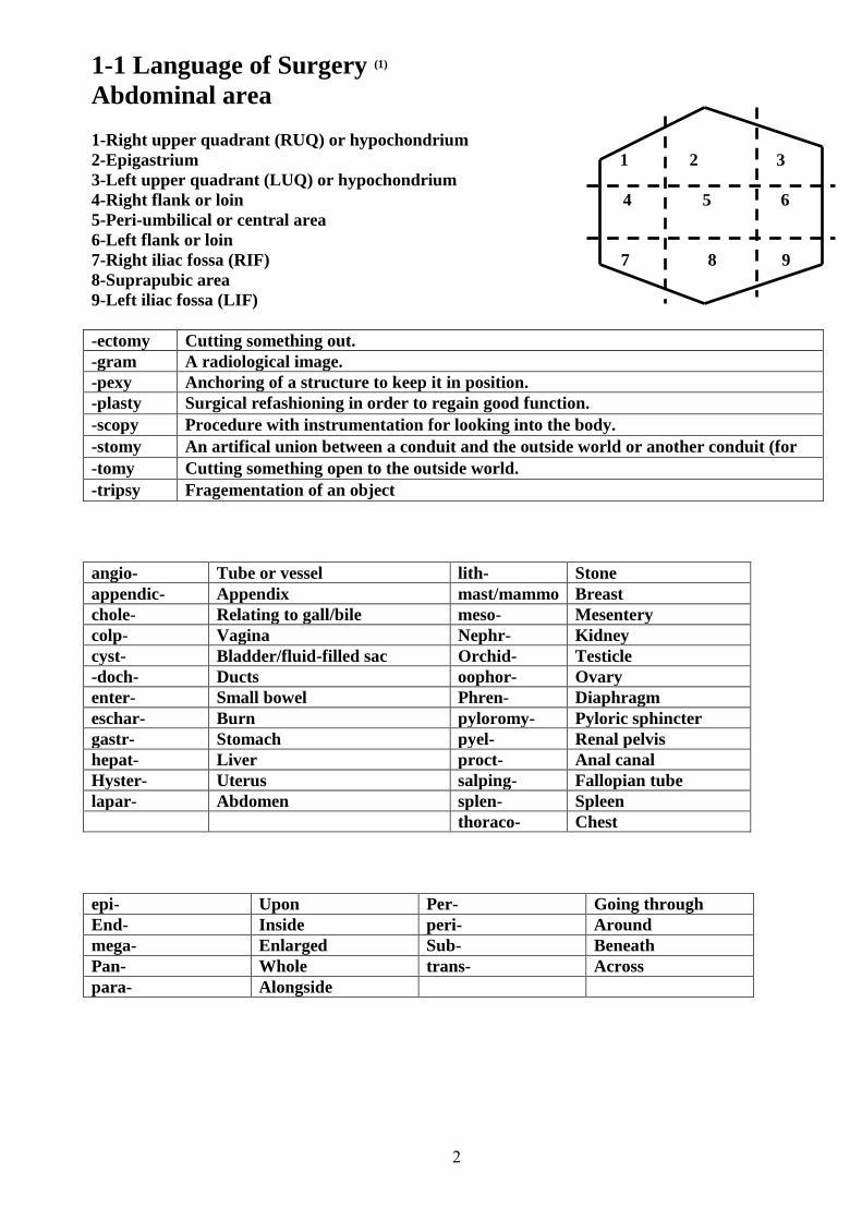
1-1 Language of Surgery (1)
Abdominal area
1-Right upper quadrant (RUQ) or hypochondrium
2-Epigastrium 1 2 3
3-Left upper quadrant (LUQ) or hypochondrium
4-Right flank or loin 4 5 6
5-Peri-umbilical or central area
6-Left flank or loin
7-Right iliac fossa (RIF) 7 8 9
8-Suprapubic area
9-Left iliac fossa (LIF)
-ectomy Cutting something out.
-gram A radiological image.
-pexy Anchoring of a structure to keep it in position.
-plasty Surgical refashioning in order to regain good function.
-scopy Procedure with instrumentation for looking into the body.
-stomy An artifical union between a conduit and the outside world or another conduit (for
-tomy Cutting something open to the outside world.
-tripsy Fragementation of an object
angio- Tube or vessel lith- Stone
appendic- Appendix mast/mammo Breast
chole- Relating to gall/bile meso- Mesentery
colp- Vagina Nephr- Kidney
cyst- Bladder/fluid-filled sac Orchid- Testicle
-doch- Ducts oophor- Ovary
enter- Small bowel Phren- Diaphragm
eschar- Burn pyloromy- Pyloric sphincter
gastr- Stomach pyel- Renal pelvis
hepat- Liver proct- Anal canal
Hyster- Uterus salping- Fallopian tube
lapar- Abdomen splen- Spleen
thoraco- Chest
epi- Upon Per- Going through
End- Inside peri- Around
mega- Enlarged Sub- Beneath
Pan- Whole trans- Across
para- Alongside

3
abscess A cavity containing pus. For different types consult the index. Remember the
aphorism: if there is pus about, let it out.
fistula An abnormal connection between two epithelial surfaces. Fistulae often close
spontaneously, but will not do so in the presence of malignant tissue, distal
obstruction, foreign bodies, chronic inflammation, and the formation of a muco-
cutaneous junction (eg stoma).
hernia Any structure passing through another and so ending up in the wrong place.
ileus Used in this book as a term for adynamic bowel.
sinus A blind-ending tract, typically lined by epithelial or granulation tissue, which opens to
an epithelial surface.
stent An artificial tube placed in a biological tube to keep it open.
stoma An artificial union between conduits or a conduit and the outside.
ulcer An abnormal break in an epithelial surface.
volvulus Twisting of a structure around itself. Common GI sites include the sigmoid colon and
caecum, and more rarely the stomach.
References :
1-Longmore, Murray; Wilkinson, Ian B; Turmezei, Tom; Cheung, Chee Kay. Oxford Handbook of
Clinical Medicine, 7th Edition . Copyright ©2007 Oxford University Press.
1-2 Surgical Antibiotic Prophylaxis Definition :
1-Antibiotics administered prior to contamination of previously uninfected tissues or fluids are
considered prophylactic. The goal for prophylactic antibiotics is to prevent a surgical-site infection
(SSI) from developing (1) .
Common surgical pathogens *The predominant organisms causing SSIs after clean procedures are skin flora, including S. aureus
and coagulase-negative staphylococci (e.g., Staphylococcus epidermidis)
*In clean-contaminated procedures, including abdominal procedures and heart, kidney, and liver
transplantations, the predominant organisms include gram negative rods and enterococci in addition
to skin flora(5)
Microbiology:
1-The choice of the prophylactic antimicrobial depends on the type of surgical procedure, most
likely pathogenic organisms, safety and efficacy of the antimicrobial (1)
2-Typically, gram-positive coverage is included in the choice of surgical prophylaxis, because
organisms such as S. aureus and Staphylococcus epidermidis are common skin flora (1. )
3-First-generation cephalosporins (particularly cefazolin) are the preferred choice, particularly for
clean surgical procedures (1.)
4-In cases where broader gram-negative and anaerobic coverage is desired, the antianaerobic
cephalosporins such as cefoxitin, or cefotetan, are appropriate (1.)

4
1-3 Types of Surgical Operations Surgical operations are classified as clean, clean -contaminated, contaminated, or dirty.
Antimicrobial prophylaxis is appropriate for clean, clean-contaminated, and contaminated
operations. Dirty operations take place in situations of existing infection and antimicrobials are used
for treatment, not prophylaxis (2). (Table 1).
Table -1. Wound Classification, Risk of SSIb, and Indication for Antibiotics (1,2,5)
Classificati
on Description
SSI
risk
Antibiotics
Clean An uninfected operative wound in which
no inflammation is encountered and the
respiratory, alimentary, genital, or
uninfected urinary tracts are not entered.
In addition, clean wounds are primarily
closed and, if necessary, drained with
closed drainage.
Low Not indicated
unless high-
risk
procedure
Clean-
contaminated
Operative wounds in which the
respiratory, alimentary, genital, or urinary
tracts are entered under controlled
conditions and without unusual
contamination. Specifically, operations
involving the biliary tract, appendix,
vagina, and oropharynx are included in
this category, provided no evidence of
infection or major break in techniques
encountered .also when Clean procedures
performed emergently or with major
technique breaks.
Medim Prophylactic
antibiotics
indicated
Contaminatd Open, fresh, accidental wounds. In
addition, operations with major breaks in
sterile technique (e.g., open cardiac
massage) or gross spillage from the
gastrointestinal tract and incisions in
which acute, nonpurulent inflammation is
encountered are included in this category.
Technique break during clean-
contaminated procedure.
High Prophylactic
antibiotics
indicated
Dirty Obvious preexisting infection present
(abscess, pus, or necrotic tissue present). ______ Therapeutic
antibiotics
required

5
Principles of Antimicrobial Prophylaxis
1-Route of Administration Intravenous administration is preferred because it produces a more reliable and predictable serum
and tissue concentration than intramuscular administration (3).
Oral administration is also used in some bowel operations. Non-absorbable compounds like
erythromycin base and neomycin are given up to 24 hours prior to surgery to cleanse the bowel.
Note that oral agents are used adjunctively and do not replace IV agents (2).
2-Timing of First Dose Correct timing of antibiotic administration is imperative to preventing SSI. It is recommended to
start infusing antimicrobials for surgical prophylaxis within 60 minutes of the first incision (2). (A
single dose of antibiotic should be administered within 30 minutes to one hour before incision (3))
(They are given 15-60min prior to the procedure)(4). ( وكلها معناها واحد تقريبا)
Exceptions to this rule are fluoroquinolones and vancomycin, which can be infused 120 minutes
prior to avoid infusion-related reactions. Beginning the infusion after the first incision is of little
value in preventing SSI (2).
3-Dosing and Redosing The goal of antimicrobial dosing for surgical prophylaxis is to maintain antibiotic concentrations
above the MIC of suspected organisms for the duration of the operation(2).
Guidelines suggest that if an operation exceeds two half-lives of the selected antimicrobial, then
another dose should be administered. Repeat dosing has been shown to lower rates of SSI. For
example, cefazolin has a half-life of about 2 hours, thus another dose should be given if the
operation exceeds 4 hours. The clinician should have extra doses of antibiotic ready in case
an operation lasts longer than planned (2).
4-Duration Evidence suggest that the continuation of antimicrobial prophylaxis beyond wound closure is
unnecessary. Studies have not shown benefit for additional doses of antibiotic and the duration of
antimicrobial prophylaxis should not exceed 24 hours (2) .
There is little evidence to support the practice of administering antibiotics until all drains are
removed. Continuing the antibiotic does not necessarily reduce the infection rate. Moreover, it can
encourage proliferation of resistant micro-organisms and subject patients to increased antibiotic-
associated morbidity. Prolonged prophylaxis using antibiotics is also unnecessarily expensive (3).
Longer durations of antibiotic prophylaxis are advocated by some guidelines(2) .
\

6
TABLE 2. Most Likely Pathogens and Specific Recommendations for Surgical Prophylaxis (1) .لالطالع

7
References: 1- Joseph T. DiPiro, Robert L. Pharmacotherapy: A Pathophysiologic Approach, Sixth Edition. Copyright 2005,
by The McGraw-Hill Companies, Inc.
2-Marie A. Chisholm-Burns , Barbara G. Wells. Pharmacotherapy principles and practice . 2007.
3- Mohamed H. Rahman and James Anson . Peri-operative antibacterial prophylaxis. Pharmaceutical Journal .12
June 2004 The (Vol 272) : 743 -745.
4-Longmore, Murray; Wilkinson, Ian B; Turmezei, Tom; Cheung, Chee Kay. Oxford Handbook of Clinical
Medicine, 7th Edition . Copyright 2007©آ Oxford University Press.
5- Re/Dale W. Bratzler, e. Patchen Dellinger, Keith M. Olsen, trish M. Perl. Clinical practice guidelines for
antimicrobial prophylaxis in surgery. Am J Health-Syst Pharm. 2013; 70:195-283.
1-4 Thromboprophylaxis
Deep venous thrombosis (DVT) is most common in patients over 40 years of age who undergo major
surgery. A postoperative increase in platelets coupled with venous endothelial trauma and stasis all
contribute. If no prophylaxis is given, 30% of these patients will develop DVT and 0.1-0.2% will die
from pulmonary thromboembolism (PTE) (1).
Types of thromboprophylaxis (1).
1-Mechanical devices :Thromboembolic deterrent stockings (TEDS).
2-Drugs acting on the clotting cascade :Heparin and Low molecular weight heparin (LMWH).
Regimen : heparin 5000U SC 2h pre-op, then every 8-12h SC for 7d or until ambulant. Low molecular weight
heparin (LMWH) may be better (less bleeding, no monitoring needed).: eg enoxaparin 20mg/d SC,
increased to 40mg/d in major-risk surgery (2).
Fondaparinux (a factor Xa inhibitor) and ximelagatran may be better than LMWH . (2).
Risk groups (1).
All patients are -at risk of developing deep vein thrombosis just as is the general population. Certain
factors increase this risk and warrant specific interventions. It is usual to divide patients according to
estimated risk.
1-Low risk (TEDS only) Day case surgery, minor orthopaedic procedures, and surgery after which patients mobilize immediately.
2-Medium risk (TEDS and prophylactic dose LMWH) Examples include minor surgery where mobilization is expected to be slow; abdominal, thoracic, upper
limb orthopaedic surgery; low risk procedures with associated comorbid risk factors (diabetes, obesity,
cardiorespiratory disease, malignancy, oral contraceptive pill, previous history of thromboembolic
disease).
3-High risk (TEDS and treatment dose LMWH or IV heparin) Examples include pelvic surgery, major lower limb orthopaedic procedures, surgery for malignancy,
medium risk procedures with associated comorbid risk factors (diabetes, obesity, cardiorespiratory
disease, malignancy, oral contraceptive pill, previous history of thromboembolic disease).
References :
1-McLatchie, Greg; Borley, Neil; Chikwe, Joanna . Oxford Handbook of Clinical Surgery, 4th Edition
.Copyright 2013 .Oxford University Press.

8
2-Longmore, Murray; Wilkinson, Ian B; Turmezei, Tom; Cheung, Chee Kay. Oxford Handbook of
Clinical Medicine, 9th Edition . Copyright 2014 .Oxford University Press.
1-5 : Preoperative prophylaxis against aspiration
pneumonia Obesity, DM, pregnancy, peptic ulcer, stress, elderly, pediatric, trauma and emergency surgery are risk
factors which may lead to delayed gastric emptying, increase gastric volume, and decrease esophageal
sphincter result in regurgitation and aspiration of gastric contents causing potentially fatal condition
called aspiration pneumonitis, therefore such patient require special pharmaceutical care to prevent
aspiration by:
30 min before -15 they should be given as a single dose 30 ml approximately: Antacid agents -A
: advantagesinduction of anesthesia, antacids has two major
1-Rapid onset of action .
2-Effective on the fluid already present in the stomach.
are: disadvantagesThe major
1-Their effect may not last as long as the surgical procedure.
2-Their administration adds fluid volume to the stomach.
)Gastric motility stimulants (prokinetic agents -B
They act by promoting gastric emptying therefore reducing gastric volume, these agents should be given
60min before induction of anesthesia when given orally, 30min when given IV.
H2 receptor antagonists -C They act by reducing gastric acidity and volume by inhibition of gastric secretion.H2 blockers has no
action on gastric contents already present in the stomach therefore oral dose of H2 blockers is given at
the evening before surgery followed by an oral or parenteral dose on the morning of surgery, these
agents do not produce an immediate effect.
Proton pump inhibitors -D They are effective in suppressing acid secretion .
1-6 Preoperative bowel preparation
A-Elective colon operation: The human colon and distal small intestine contain a numerous reservoir of aerobic and anaerobic
bacteria that are excluded from the body by a mucous membrane barrier, if this barrier is disturbed by
disease, trauma, or if the colon is opened to the peritoneal cavity during operation, bacteria may escape
into adjacent tissues and causes serious infection, this risk can be minimized by two ways:
Mechanical preparation:-1
This is done by one or both of the following procedures:
A-Whole gut lavage with an electrolyte solution, mannitol 10%, or poly ethylene glycol the day before
surgery.
B-Standard mechanical cleansing, which utilizes dietary restriction, catheters, and sometimes enemas 1-
2 days before the operation.
ibiotic preparation:Ant-2 Either oral or parenteral antibiotic ,
: are now used Two oral regimens
A-An aminoglycoside with erythromycin base.
B-An aminoglycoside with metronidazole .
hesia.IV before induction of anestcefoxitin that is now used is Parenteral regimen
Combination of parenteral and oral antibiotics show low incidence of infection.

9
B-Emergency colon preparation: The following is recommended:
1-Intraoperative lavage performed by introducing of saline in the colon through balloon catheter.
2-Parenteral antibiotic, they should be given IV shortly before operation and continues for 1-7 days
postoperatively.
1-7 Intravenous fluid therapy If fluids cannot be given orally, they are normally given intravenously. However, remember that all
cannulas carry a risk of infection (1).
Three principles of fluid therapy
1-Maintain normal daily requirements: About 2500mL fluid containing roughly 100mmol sodium
and 70mmol potassium per 24h are required. A good regimen is 2L of 5% dextrose and 1L of 0.9%
saline every 30h with 20-30mmol of potassium per litre of fluid. Post-operative patients may need more
fluid and more saline depending on operative losses. If the serum sodium is rising, then more dextrose
and less saline is required (1).
2-Replace additional losses: The amount and type of fluid lost is a guide. Remember that febrile
patients have increased insensible losses. In practice, the problem is usually whether to give saline or
dextrose. Most body fluids (eg vomit) contain salt, but less than plasma, and thus replacement will
require a mixture of saline and dextrose. Shocked patients require resuscitation with saline, or a colloidal
plasma expander, eg Dextranآ®, but not dextrose (caution in liver failure, see below). Note that
Dextranآ® interferes with platelet function and may prolong bleeding. Patients with acute blood loss
require transfusion with packed cells or whole blood. As a holding measure, colloid or saline may be
used while blood is being cross-matched. If more than 1L is required then group O-negative or group-
specific blood should be used (1) .
3-Special cases Patients with heart failure and the elderly are at greater risk of pulmonary oedema if
given too much fluid. They also tolerate saline less well since Na+ retention accompanies heart failure.
If IV fluids must be given, use with care. Patients with liver failure, despite being oedematous and often
hyponatraemic, have increased total body sodium, and saline should not be used in resuscitation; salt-
poor albumin solution or blood should be given. Fluid maintenance for children is calculated as:
100mL/kg for the first 10kg; 50mL/kg for the next 10kg; and 20mL/kg thereafter-all per 24hrs. Usually
given as dextrose-saline (4% dextrose 0.18% saline) (1).
Types of fluid according to isotonicity A- Isotonic: Isotonic crystalloids have a tonicity equal to the body plasma. When administered to a
normally hydrated patient, isotonic crystalloids do not cause a significant shift of water between the
blood vessels and the cells. Thus, there is no (or minimal) osmosis occurring (4).
B-Hypertonic: crystalloids have a tonicity higher than the body plasma. The administration of a
hypertonic crystalloid causes water to shift from the extravascular spaces into the bloodstream,
increasing the intravascular volume. This osmotic shift occurs as the body attempts to dilute the higher
concentration of electrolytes contained within the IV fluid by moving water into the intravascular
space(4).
C-Hypotonic: crystalloids have a tonicity lower than the body plasma. The administration of a
hypotonic crystalloid causes water to shift from the intravascular space to the extravascular space, and
10
eventually into the tissue cells. Because the IV solution being administered is hypotonic, it creates an
environment where the extravascular spaces have higher concentrations of electrolytes. (4).
Types of fluid
A-Crystalloids: Resuscitation fluids that are composed of dissolved electrolytes (3).
1- 0.9% saline (normal saline) Has about the same sodium content as plasma (150mmol/L) and is isotonic with plasma (1) .
2- 5% dextrose Is isotonic, but only contains 278mmol/L glucose, i.e. 50g/L (dextrose is glucose), and is a way of
giving water, since the liver rapidly metabolizes all the glucose leaving only water. It provides little
energy. 5% once DW enters the body, the cells rapidly consume the glucose. This leaves primarily water
and causes IV fluid to become hypotonic in relation to the plasma surrounding the cells. Accordingly,
the now hypotonic solution causes an osmotic shift of water to and from the bloodstream and into the
cells.
More concentrated glucose solutions exist, and may be used in the treatment of hypoglycemia. They are
hypertonic and irritant to veins. Therefore, care in their use is needed, and infusion sites should be
inspected regularly, and flushed with saline after use (1).
3-Dextrose-saline (one-fifth normal saline) Is also isotonic, containing 0.18% saline (30mmol/L of sodium) and 4% glucose (222mmol/L). It has
roughly the concentration of saline required for normal fluid maintenance, when given 10 hourly (1).
Intravenous 0.18% saline/4% glucose solution (‘hypotonic saline’) in children: reports of fatal
hyponatraemia – do not use in children aged 16 years or less, except in specialist settings under
expert medical supervision such as renal, cardiac, liver, high dependency and intensive care
units.(5)
4-Hartmann's solution contains: Na+ 131mmol/L, Cl- 111mmol/L, lactate 29mmol/L, K+
5mmol/L, HCO 29 mmol/L, and Ca2+ 2mmol/L. Some consider it more "physiological" (1)
technically the closest fluid to serum composition although theoretical : Ringer's lactate solution-5. Lactated Ringer's solution is often used for fluid )(2advantages are of limited practical value
resuscitation after a blood loss due to trauma, surgery, or a burn injury. [4] It has been used to induce
urine output in patients with renal failure. [4]
Lactated Ringer's solution is used because the by-products of lactate metabolism in the liver counteract
acidosis, which is a chemical imbalance that occurs with acute fluid loss or renal failure [4]. Lactated
Ringer's solution should also not be used in patients with a pH level above 7.5 (alkalosis) and in anuria
or renal failure due to accumulation of K (4)
Note:
1-The maximum concentration of K+ that is safe to infuse via a peripheral line is 80mmol/L, at a
maximum rate of 40mmol/h. Higher concentrates risk phlebitis, and faster rates dysrhythmias.
Give more concentrated solutions via a central line (1).
B-Colloids: Resuscitation fluids that restore and/or increase the intravascular oncotic
pressure (3). Colloids (especially blood) produce a more lasting expansion of intravascular volume than crystalloid,
which rapidly enters the interstitial tissues (2) :
1-Gelofusine is succinylated gelatin (a bovine collagen), which has a half-life of about 2h in plasma,
and is associated with increased bleeding times in postoperative patients.
2-Dextran is a glucose polymer mixture that has a plasma half-life of about 2h: it has been associated
with anaphylactic reactions and profound coagulopathy.

11
3-HES preparations are derived from hydroxyethyl starch: they have widely differing plasma half-lives
and effects on plasma expansion.
4-Albumin is a naturally occurring plasma protein, sterilized by ultrafiltration: 5% albumin is
isotonic; 20% albumin is hypertonic. Indications for use of albumin as a volume expander are very
limited.
5-Blood, platelets, FFP (fresh Frozen Plasma), and cryoprecipitate.
Intravenous Fluid Packaging Most IV fluids are packaged in soft plastic or vinyl bags of various sizes (10, 50, 100, 250, 500, 1,000,
2,000, and 3,000 milliliters).
IV fluids on the surgical ward
Notes:
1* Fluid required = pre-existing deficit + normal maintenance + ongoing losses
2*In the prehospital setting, LR and NS are commonly used for fluid replacement
because of their immediate ability to expand the volume of circulating blood. However,
over the course of about 1 hour, approximately two-thirds of these IV fluids eventually
leave the blood vessels and move into the cells. Some authorities recommend that for
every 1liter of blood lost, 3 liters of an isotonic crystalloid be administered for
replacement. This is only a guide, and the volume of IV fluid administered should be
based on medical direction or local protocol, as well as the patient’s clinical response to
fluid administration.(4 )
3*Too many fluids can lead to (4):
- Lungs stiffer - > gas exchange impaired
- Cardiac failure -Peripheral oedema-
- Inhibition of wound healing
4*Not enough fluid can lead to :
- Renal damage
- Cardiovascular damage
-Tissue hypoperfusion
What fluids to use
1-Haemorrhagic/hypovolaemic shock :
Insert 2 large IV cannulae, for fast fluid infusion. Start with crystalloid (eg 0.9% saline) or colloid (eg
Gelofusineآ®) until blood is available. The advantage of crystalloids is that they are cheap but they do
not stay as long in the intravascular compartment as colloids, as they equilibrate with the total
extracellular volume (dextrose is useless for resuscitation as it rapidly equilibrates with the enormous
intracellular volume). In practice, the best results are achieved by combining crystalloids and colloids.
Aim to keep the haematocrit at ~0.3, and urine flowing at >30mL/h. Monitor pulse and BP often (1).
2- Septicaemic shock: e.g. Gelofusine like substance
3-Heart or liver failure:
Avoid sodium loads: use 5% dextrose (1). Or one-fifth normal saline (4)
4-Excessive vomiting:
Use 0.9% saline: replace losses, including K+ (1) /.

12
Complications of I.V Fluid Therapy:
infection of I.V is usually local causing easily visible redness , and fever.the bacteria : Infection-1
may spread via blood stream causing life threatening septicemia .
presence of foreign is ian irritation of vein that is not caused by infection but from the Phlebitis-2
body ( the I.V catheter, the fluid , or medication ) . Symptom are swelling, pain and redness around the
vein site .
: this occurs when the tip of the I.V catheter withdraws from the vein or pokes through Infiltration-3
the vein into surrounding tissue or when the vein wall becomes permeable and leaks fluid . It requires
replacement of the I.V catheter at different location.
this occurs when fluids are given at higher rate or in larger volume than the : Fluid overload-4
system can absorb or excrete . Possible consequences includes : Hypertension , CHF , and pulmonary
edema .
5-Embolism : A blood clot or other solid mass , or an air bubble can delivered into the circulation
through an I.V line and end up with blocking vessel . Peripheral I.V have a lower risk of embolism than
the central I.V line .
References :
1-Longmore, Murray; Wilkinson, Ian B; Turmezei, Tom; Cheung, Chee Kay. Oxford Handbook of Clinical
Medicine, 7th Edition . Copyright 2007©آ Oxford University Press.
2- McLatchie, Greg; Borley, Neil; Chikwe, Joanna . Oxford Handbook of Clinical Surgery, 3rd Edition
.Copyright 2007©آ Oxford University Press
3-Marie A. Chisholm-Burns , Barbara G. Wells. Pharmacotherapy principles and practice . 2007. 4-Harold Ellis, Sir Roy Calne, Christopher Watson. General Surgery, 11th Edition. Blackwell publishing2006.
5- Drug safety update.Volume 6, Issue 3 October 2012
1-8 : Blood transfusion and blood products(1)
Products
1-Whole blood: Rarely used e.g. for exchange transfusion : use crossmatched blood if possible, but if not, use universal
donor group ( O Rh-ve blood) changing to crossmatched blood as soon as possible. Blood >2d old has
no effective platelets.
2-Red cells: (packed to make haematocrit ~70%) Use to correct anaemia or blood loss. 1U Hb by 1-1.5g/dL. In
anaemia, transfuse until Hb ~8g/dL.
3-Platelets: Not usually needed if not bleeding or count is >20 x 109/L. 1U should platelet count by >20 x 109
/L. Failure to do so suggests refractoriness.
4-Fresh frozen plasma (FFP): Use to correct clotting defects: e.g. DIC (disseminated intravascular coagulation ) ; warfarin overdosage
where vitamin K would be too slow; liver disease; thrombotic thrombocytopenic purpura . It is
expensive and carries all the risks of blood transfusion. Do not use as a simple volume expander.
13
5- Human albumin solution is produced as 4.5% or 20% protein solution and is for use as protein replacement. 20% albumin can be
used temporarily in the hypoproteinaemic patient (eg liver disease; nephrosis) who is fluid overloaded,
without giving an excessive salt load. Also used as replacement in abdominal paracentesis
6-Others Cryoprecipitate (a source of fibrinogen); coagulation concentrates (self-injected in haemophilia);
immunoglobulin (anti-D ).
Complications of transfusion :
1- Acute haemolytic reaction (eg ABO incompatibility) :
Agitation, temperature (rapid onset), BP, flushing, abdominal/chest pain, oozing venepuncture sites,
DIC (disseminated intravascular coagulation ) .
2- Anaphylaxis Bronchospasm, cyanosis, BP, soft tissue swelling.
3- Bacterial contamination temperature (rapid onset), BP, and rigors .
4- Non-haemolytic febrile transfusion reaction Shivering and fever usually 1-½آh after starting
transfusion.
5- Fluid overload : Dyspnoea, hypoxia, tachycardia , JVP & basal crepitations .
Transfusing patients with heart failure If Hb < 5g/dL with heart failure, transfusion with packed red cells is vital to restore Hb to safe level, eg
6-8g/dL, but must be done with great care. Give each unit over 4h with furosemide (eg 40mg slow
IV/PO; don't mix with blood) .
References
1- Longmore, Murray; Wilkinson, Ian B; Turmezei, Tom; Cheung, Chee Kay. Oxford Handbook of
Clinical Medicine, 7th Edition . Copyright 2007©آ Oxford University Press.
1-9: The control of pain (1)
Guidelines for success : 1-Give regular doses rather than on an as required basis.
2-Choose the best route: PO, PR, IM, epidural, SC, inhalation, or IV.
A-Non-narcotic (simple) analgesia Paracetamol 0.5-1.0g/4h PO (up to 4g daily) :Caution in liver impairment.
NSAIDs, eg ibuprofen 400mg/8h PO . or diclofenac 50mg/8h PO, or 100mg PR/IM ; these are good for
musculoskeletal pain and renal or biliary colic.
CI: peptic ulcer, clotting disorders, anticoagulants. Cautions: asthma, renal or hepatic impairment,
pregnancy, and the elderly. Aspirin is contraindicated in children due to the risk of Reye's syndrome .
B-Opioid drugs for severe pain Morphine (eg 10-15mg/2-4h IV/IM) or diamorphine (5-10mg/2-4h PO, SC, or slow IV, but you may
need much more) are best.
Side-effects of opioids: These include nausea (so give with an antiemetic, eg prochlorperazine
12.5mg stat IM), respiratory depression, constipation, cough suppression, urinary retention, and

14
sedation (do not use in hepatic failure or head injury). Dependency is rarely a problem. Naloxone
may be needed to reverse the effects of excess opioids .
C-Epidural analgesia Opioids and anaesthetics are given into the epidural space by infusion or as boluses.
D-Adjuvant treatments e.g.
1-Anticonvulsants, antidepressants, gabapentin or steroids for neuropathic pain.
2-Antispasmodics, eg hyoscine butylbromide (Buscopan 20-10® آ mg/8h PO/IM/IV) for intestinal, renal
tract colic.
References
1- Longmore, Murray; Wilkinson, Ian B; Turmezei, Tom; Cheung, Chee Kay. Oxford Handbook of
Clinical Medicine, 7th Edition . Copyright 2007©آ Oxford University Press.
1-10 : Nausea and vomiting (1)
This affects up to 75% of patients. It predisposes to increased bleeding, incisional hernias, aspiration
pneumonia, absorption of oral medication, poor nutrition, and K+.
Causes include:
Prolonged surgery; anaesthetic agents, bowel obstruction; constipation; gastric reflux; peptic ulceration
or bleeding; and medications .
Classification of antiemetics Combining two different types of antiemetic increases efficiency.
A-Antidopaminergic agents
1-Good against opioid nausea and vomiting, sedative, extrapyramidal side-effects
2-e.g. prochlorperazine 12.5mg IM, metaclopramide 10mg IV/IM/PO tds.
B-Antihistamines 1-Sedation, tachycardias, hypotension with IV injection
2-e.g. cyclizine 50mg IM/IV/PO tds
C-Anticholinergics 1-Active against emetic effect opioids, sedation, confusion, dry mouth
2-e.g. hyoscine (scopolamine) 0.3-0.6mg IM
D-Antiserotonergics 1-Lowest side-effect profile of all antiemetics
2-Ondansetron 1-8mg PO/IV/IM tds, granisetron 1mg PO/IV tds
1-11 : Constipation Failure to pass stool is common. Caused by immobility, pain from wounds or anal fissures, dehydration,
poor nutrition, opiates, iron supplements, and spinal anaesthesia.
Treat with:
1-Bulking agents, e.g. Fybogel 1 sachet PO bd.
2-Stool softeners, e.g. sodium docusate 30-60mg od PO.
3-Osmotic agents, e.g. lactulose 5-10mL bd.
4-Stimulants, e.g. senna 1 tablet bd PO, bisacodyl 5-20mg nocte PO.

15
Referencesc :
1- McLatchie, Greg; Borley, Neil; Chikwe, Joanna . Oxford Handbook of Clinical Surgery, 3rd Edition
.Copyright ©2007 Oxford University Press .
2-1: Peri-operative care and diabetes Surgery causes considerable stress in patients. In response, the neuro-endocrine system stim-
ulates glycogenolysis (breakdown of glycogen to glucose) and gluconeogenesis (glucose synthesis
from non-carbohydrate sources) via counter-regulatory hormones such as catecholamines,
cortisol, growth hormone and glucagon. These hormones can antagonise the effects of insulin and
cause insulin resistance (1).also this stress decrease the absorption of oral hypoglycemic drugs
A-Patients treated with Oral Antidiabetic drugs :
1-Second-generation sulfonylureas should be discontinued 1 day before surgery, with the
exception of chlopropramide, which should be stopped 2–3 days before surgery (2).
2-Other oral agents can be continued until the operative day. Although metformin has a short
half-life of 6 h, it is prudent to temporarily withhold therapy 1–2 days before surgery, especially in
sick patients and those undergoing procedures that increase the risks for renal hypoperfusion,
tissue hypoxia, and lactate accumulation (2).
3-At a minimum, blood glucose should be monitored before and immediately after surgery in all
patients.
4-For minor surgery, perioperative hyperglycemia (>200 mg/dl) can be managed with small
subcutaneous doses (4–10 units) of short-acting insulin.
for patients undergoing major surgery and for those with poorly he recommended treatmentT-5
with glucose, using one of two standard controlled type 2 diabetes is intravenous insulin infusion,
.(2)) regimens (see below
and have no infection or complication t is well control Note: In general if the diabetic patien
e.g., an infusion -an appropriate iv regimen we can convert him to undergo minor surgery
consisting of glucose, insulin and potassium (referred to as GLIK or sometimes ) or a sliding-scale
but if the patient is not well control with many complication related to poor (1)gimeninsulin re
glycemic control or have infection like diabetic foot we have to ensure tight glycemic control by
converting him to intensive insulin therapy .
Intravenous Insulin, Glucose, Potassium, and Fluids:-
1-Insulin Two main methods of insulin delivery have been used: either combining insulin with glucose and
potassium in the same bag (the GIK regimen) or giving insulin separately with an infusion pump(2)
which is called sliding scale insulin regimen (2.)
2- Glucose Adequate glucose should be provided to prevent catabolism, starvation ketosis, and insulin-induced
hypoglycemia. The physiological amount of glucose required to prevent catabolism in an average
nondiabetic adult is 120 g/day (or 5 g/h). With preoperative fasting, surgical stress, and ongoing insulin
therapy, the caloric requirement in most diabetic patients averages 5–10 g/h glucose. This can be given
as 5 or 10% dextrose.
16
3- Potassium The infusion of insulin and glucose induces an intracellular translocation of potassium, resulting in a risk
for hypokalemia. In patients with initially normal serum potassium, potassium chloride, 10 mEq, should
be added routinely to each 500 ml of dextrose to maintain normokalemia if renal function is normal.
Hyperkalemia (confirmed with repeat measurement and electrocardiogram) and renal insufficiency are
contraindications to potassium infusion (2)
Diabetic foot Approximately 25% of diabetic patients report a history of skin and soft tissue infection and 5%-15% of
diabetic patients undergo limb amputation.
Etiology:
1--Poor glycemic control lead to an increase in blood viscosity which becomes a good media for the
growth of bacteria, the causative agent include one or more of the following bacteria: Staphylococcus
aureus, Staphylococcus epidermis., Enterococcus faecalis., Bacteroid species.
Pseudomonas aerogenosa., and Klebseilla species.
2--Peripheral vascular disease which decreases blood flow to extremities.
3--Somatic neuropathy: which decreases pain perception.
4--Autonomic neuropathy: which decreases sweating, and subsequently dry, scaly skin.
Management :
A-Non-pharmacological:
1-Inspect feet for cuts, blisters, or scratches.
2--Wash feet daily in taped water and dry thoroughly.
3--Apply lotion to the feet to prevent calluses and cracking.
4--Ensure shoes fit properly.
5--Trim nails regularly.
6--Do not use chemical agents to remove corns or callus.
Pharmacological:
A-Tight glycemic control:
This can be achieved by intensive insulin therapy as follows:
Starting dose of insulin is 1-1.5U/kg/day which is given as follows:
¼ of total daily dose before each meal as soluble insulin SC.
¼of total daily dose at 11pm as intermediate insulin SC.
Monitor therapy by making FBS which should be less than 120mg/dl.
If the patient develops morning hyperglycemia, the patient should be asked about signs of hypoglycemia
at 2:00-3:00am and measure glucose level at this time.
If this reveals hypoglycemia, the morning hyperglycemia is rebound type (Somogi effect) which can be
managed by ensuring that the patient take intermediate insulin at the specified time and reduce the dose
of intermediate insulin.
If this reveals hyperglycemia, the morning hyperglycemia is due to down phenomena, and can be
managed by increasing the dose of intermediate insulin.
Make 2h. Postprandial glucose level and the result should be less than 180mg/dl, if we did not get this
target, give 2U soluble insulin IV for each 50mg/dl of glucose above the goal.
If the patient stabilize on this regimen, we can convert him to a less frequent regimen, and on discharge,
the following regimen is given:
2/3 of total daily dose is given before breakfast as 30% soluble insulin and 70% of intermediate
insulin.

17
1/3 of total daily dose is given before dinner as 30% soluble insulin and 70% of intermediate
insulin .
B-Antibiotic therapy:
Effective combination should cover most potential pathogens (G+ve, G-ve, and anaerobes). This
can be achieved by giving :
Clindamycin 600mg q 8h + gentamicin 2mg/kg q 8h.
In patients with poor renal function, gentamicin could be replaced by:
1- A quinolone (ciprofloxacin 200mg IV infusion q 12h), or
2- A 3rd generation cephalosporine (cefotaxim 1g q 8h, or cefotriaxone 1g q 24 h)
3- Piperacillin 1g q 6hr, or 2g q 4hr in severe cases.
4- Cefazoline 1g q 6h + metronidazole 500mg q 8h IV infusion.
The treatment should continue for 3-4 days after all signs of infection are absent. Drainage and surgical
debridment of necrotized tissue are essential, also it is necessary to change dressing twice daily.
References
1-Mohamed H. Rahman and James Anson . Peri-operative care and diabetes. The Pharmaceutical
Journal .13 March 2004 (Vol 272) 323-325
2-Samuel Dagogo-Jack and K. George M.M. Alberti. Management of Diabetes Mellitus in Surgical
Patients . Diabetes Spectrum 15:44-48, 2002.
2-2-1-Perioperative medication management:
In general, one should stop any medication that may prove harmful around the time of surgery
e.g., ( MAO inhibitors, anticoagulants), continue any medications that are necessary for the
patient's health (e.g., steroids, anti‐arrhythmic agents, beta‐blockers, transplant meds).
Common drugs that have been associated with withdrawal symptoms when discontinued
preoperatively include selective serotonin reuptake inhibitors (SSRIs), beta-blockers, clonidine, statins,
and corticosteroids .
In general, most nonsteroidal anti-inflammatory drugs should be stopped at least 3 days before surgery.
Herbal medications should be stopped at least 7 days before surgery, owing to the uncertainly over their
actual contents. (1)
2-2-2: Peri-operative medication in patients with
cardiovascular disease (2)
When a patient with cardiovascular disease (CVD) is to undergo surgery, we need to consider whether
or not any of the drugs used to treat his or her cardiovascular problems need to be stopped
Table 1. Outline of Perioperative Drug Management of Patients with Coronary Artery Disease
Drug Day Before
Surgery Day of Surgery During Surgery After Procedure
Nitroglycerin Usual dose Usual dose IV infusion if
frank ischemia
Continue IV dose if needed or
until medication can be taken
PO
Beta-blockers Usual dose
Usual dose plus
beta-blocker
protocol
Usual dose plus
beta-blocker
protocol
Usual dose plus beta-blocker
protocol
18
Calcium channel
blockers Usual dose
Usual dose
morning of
surgery
Usual dose
morning of
surgery
Continue IV dose until
medication can be taken PO
Aspirin
Discontinue 1
week before
surgery
Restart postoperatively at
discretion of surgeon
Ticlopidine
Discontinue 1
week before
surgery
Restart postoperatively at
discretion of surgeon
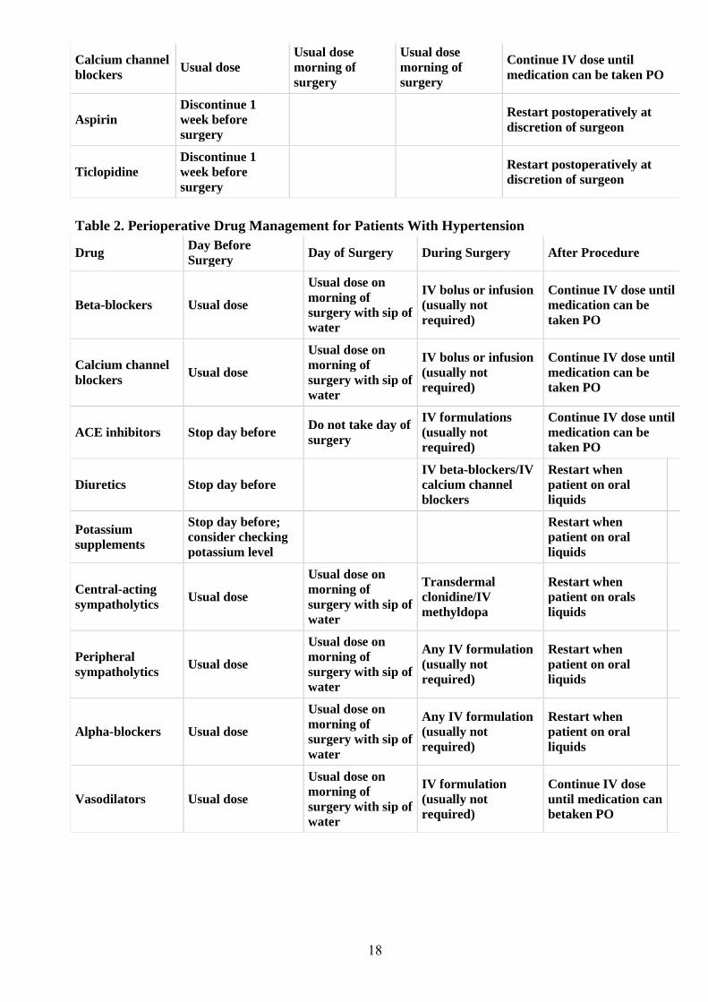
Table 2. Perioperative Drug Management for Patients With Hypertension
Drug Day Before
Surgery Day of Surgery During Surgery After Procedure
Beta-blockers Usual dose
Usual dose on
morning of
surgery with sip of
water
IV bolus or infusion
(usually not
required)
Continue IV dose until
medication can be
taken PO
Calcium channel
blockers Usual dose
Usual dose on
morning of
surgery with sip of
water
IV bolus or infusion
(usually not
required)
Continue IV dose until
medication can be
taken PO
ACE inhibitors Stop day before Do not take day of
surgery
IV formulations
(usually not
required)
Continue IV dose until
medication can be
taken PO
Diuretics Stop day before
IV beta-blockers/IV
calcium channel
blockers
Restart when
patient on oral
liquids
Potassium
supplements
Stop day before;
consider checking
potassium level
Restart when
patient on oral
liquids
Central-acting
sympatholytics Usual dose
Usual dose on
morning of
surgery with sip of
water
Transdermal
clonidine/IV
methyldopa
Restart when
patient on orals
liquids
Peripheral
sympatholytics Usual dose
Usual dose on
morning of
surgery with sip of
water
Any IV formulation
(usually not
required)
Restart when
patient on oral
liquids
Alpha-blockers Usual dose
Usual dose on
morning of
surgery with sip of
water
Any IV formulation
(usually not
required)
Restart when
patient on oral
liquids
Vasodilators Usual dose
Usual dose on
morning of
surgery with sip of
water
IV formulation
(usually not
required)
Continue IV dose
until medication can
betaken PO

19
8-Anticoagulant therapy Although anaesthesia and surgery are not contraindicated in patients taking anticoagulants, major
surgery poses an increased risk of haemorrhagic complications. There is good evidence that surgery
increases the risk of venous thromboembolisms (VTE) and so, for most patients (especially those at
high-risk of thromboembolism), some form of anticoagulant therapy should continue for most of the
peri-operative period.
Pre-operative management
The key principles of peri-operative anticoagulant management are summarised in table-1:
table-1 Principles of peri-operative anticoagulant therapy management 1-Discontinue oral anticoagulant
2-Start unfractionated heparin or low molecular weight heparin (LMWH)
3-Ensure that the international normalised ratio (INR) falls to the desired level before surgery
4-Discontinue unfractionated heparin or LMWH just before surgery
5-Restart unfractionated heparin or LMWH after surgery
6-Restart oral anticoagulant
7-Discontinue heparin when INR returns to within the desired range .
1-Warfarin is usually discontinued three to four days before surgery to allow the international
normalised ratio (INR) to fall below 1.5 " a level considered safe for most types of surgery to be
performed ".
Vitamin K can be used to reverse the anticoagulant effect if there is insufficient time to allow the INR to
fall to a desired level, but it should be noted that this can interfere with the effect of warfarin for many
days. In an emergency, administration of clotting factors or fresh frozen plasma may be warranted .
2-As the INR falls, intravenous unfractionated heparin (UH), or low molecular weight heparin (LMWH)
is started. The dose used depends on the risk of thromboembolism. All patients considered as high-risk
for VTE must be considered for a "treatment" dose of UH
3-UH or LMWH are discontinued for a few hours pre-operatively to provide the surgical team with
a short period when the patient has little systemic anticoagulation and it is safest to operate. The short
half-life of heparin allows surgery to proceed within four to six hours of its discontinuation, hence
minimising the period of "non-anticoagulation". Due to their longer duration of action, LMWHs
must be stopped at least 12 hours before surgery.
In an emergency, the effect of UH may be cautiously reversed using protamine sulphate. The
disadvantage of using an LMWH is that it is not possible to reverse anticoagulation rapidly if bleeding
occurs.
It should be noted that intramuscular injections administered to patients receiving full anticoagulant
doses of heparin or warfarin, may cause painful haematoma and abscess formation
4-Post-operative management If full anticoagulation is required post-operatively, UH can be restarted about 12 hours after surgery
(when haemorrhage risk is reduced) with close monitoring of activated partial prothrombin time, usually
six-hourly.
Warfarin can be restarted as soon the patient is able to tolerate oral medication and the risk of bleeding
has passed (eg, when all drains have been removed). Heparin treatment is continued until the desired
INR is reached once more (usually two or three days after recommencing warfarin). Different hospitals
adopt different warfarin loading dose regimens depending on for how long the warfarin has been
discontinued. References : Nafisa K Kuwajerwala, MD; Chief Editor: William A Schwer, MD. Perioperative Medication Management. Nov 11, 2015

20
2-3: Surgery in those on steroids (`1)
Patients on steroid therapy need extra cover to cope with the stress of surgery- their endogenous adrenal
hormone levels will be suppressed, even for a period after cessation of a course of treatment. The
amount of extra cover needed depends on the extent of the surgery and the pre-op dose of steroids. For
routine surgery, aim to reduce the dose of steroid as much as possible. Consider steroid cover for anyone
who has had high-dose glucocorticoid therapy in the last year (2).
A-Major surgery: Typically give hydrocortisone 50-100mg IV with the pre-med and then every 6-8h IV/IM for 3d, then
wean to previous medication (1).
B-Minor surgery: Prepare as for major surgery except that hydrocortisone is given for 24h only.
The major risk with adrenal insufficiency is hypotension, so if this is encountered without an obvious
cause, it may be worthwhile giving a STAT dose of 50mg hydrocortisone IV. See BNF section 6.3 for
steroid dose equivalences (2).
References:
1- Movig KL, Janssen MW, de Waal Malefijt J, et al. Relationship of Serotonergic
Antidepressants and Need for Blood Transfusion in Orthopedic Surgical Patients. Arch Intern
Med. 2003; 163: 2354‐2358.
2- Longmore, Murray; Wilkinson, Ian B; Turmezei, Tom; Cheung, Chee Kay. Oxford Handbook of
Clinical Medicine, 7th Edition . Copyright ©2007 Oxford University Press.
2-4 : Surgery and the contraceptive pill (1)
Oestrogen-containing contraceptive pills increase the risk of thromboembolic disease in women taking
them prior to surgery. Progesterone-only contraceptives appear to pose little or no additional risk and
may be continued during surgery. The increase in risk is related to the size of the operative procedure
and the existing co-morbidity; the advice is adjusted accordingly.
1-Low risk procedures: dental, day case, minor laparoscopic. Oestrogen-containing contraceptive
pills may be continued.
2-Medium risk: abdominal, orthopaedic, major breast surgery.
A- Oestrogen-containing contraceptive pills should be discontinued at least 1 month
prior to elective surgery.
B-Urgent or emergency surgery should be conducted with full thromboprophylaxis
3-High risk: pelvic, lower limb orthopaedic surgery, cancer.
A- Oestrogen-containing contraceptive pills should be discontinued at least 1 month prior to
elective surgery.
B-Urgent or emergency surgery should be conducted with extended thromboprophylaxis
References :
1- McLatchie, Greg; Borley, Neil; Chikwe, Joanna . Oxford
Handbook of Clinical Surgery, 3rd Edition .Copyright 2007©
Oxford University Press
3-1: Acute appendicitis
This is the most common surgical emergency. in which
Gut organisms invade the appendix wall (1).

21
Clinical features A-Symptoms (2).
1-malaise, anorexia, and fever;
2-diarrhoea common and may be mistaken for acute (gastro)enteritis.
3-abdominal pain starts centrally and localizes to the right iliac fossa.
4-abdominal pain caused by coughing and moving.
B-Signs (2).
1-fever, tachycardia; 2-abdominal tenderness .
C-Investigations may be normal and none are diagnostic or exclusive (2).
Establish a diagnosis The diagnosis is a clinical one in all but exceptional cases and investigations are usually unnecessary (2).
Complications (1)
1-Perforation .
2-Appendix mass May result when an inflamed appendix becomes covered with omentum.
3-Appendix abscess May result if an appendix mass fails to resolve.
Management
A-Acute appendicitis 1- Appendicectomy (2).
2-IV antibiotics are only indicated for perforation.
B-Appendix mass or appendix abscess (2).
1-IV antibiotics (e.g. cefuroxime 750mg tds + metronidazole 500mg tds),
2-If symptoms settle: delayed (interval) appendicectomy after 6 weeks,
3-If symptoms fail to settle: may need acute appendicectomy.
4-Appendix abscess may be amenable to drainage.
References
1- Longmore, Murray; Wilkinson, Ian B; Turmezei, Tom; Cheung, Chee Kay. Oxford Handbook of
Clinical Medicine, 7th Edition . Copyright 2007©آ Oxford University Press.
2-McLatchie, Greg; Borley, Neil;
Chikwe, Joanna . Oxford Handbook of
Clinical Surgery, 3rd Edition .Copyright
2007©آ Oxford University Press
3-2 : Gallstones: Pathological features(1)
Bile has three major constituents:
1-bile salts (primary: cholic and
chenodeoxycholic acids; secondary:
deoxycholic and lithocholic acids).
2-Phospholipids (90% lecithin).
3-cholesterol.
Bile containing excess cholesterol
relative to bile salts and lecithin is
predisposed to gallstone formation.
Types of gallstones (1)
1-Pure cholesterol (10%). Often solitary, large (> 2.5cm), round.
22
2-Pure pigment (bile salts; 10%). Pigment stones are of two types:
A-black (associated with haemolytic disease);
B-brown (associated with chronic cholangitis and biliary parasites).
3-Mixed (80%). Most common; usually multiple.
Predisposing conditions (1)
1-Increasing age.
2-Female (pregnancy and use of the oral contraceptive).
3-Obesity.
4-Multiparity.
5-Chronic haemolytic disorders (only for pigment stones).
Clinical features (common presentations)(1)
A-Biliary colic Intermittent severe epigastric and right upper quadrant; usually associated with nausea and vomiting.
Resolves after a few hours .
Acute cholecystitis(1)
Severe continuous right upper quadrant pain; often radiates to right flank and back; associated with
anorexia and pyrexia.
Complications of acute cholecystitis include (1)
1-formation of an empyema or abscess of the gallbladder (rare)
2-perforation with biliary peritonitis (very rare);
3-jaundice due to compression of the adjacent common bile duct by swelling .
Chronic cholecystitis (1)
A mucocele of the gallbladder or infection producing an empyema.
Diagnosis and investigations(1,2,3)
1-WCC 2-ultrasound (Ultrasonography is the method of choice for diagnosing gallstones )
3-Abdominal x-ray ( AXR ) only shows ~10% of gallstones.
Treatment: Asymptomatic gallstones found incidentally are not usually treated because the majority will never give
symptoms. Symptomatic gallstones are best treated surgically (3 ).
A-Surgical treatment Cholecystectomy
This is the treatment of choice for all patients fit for GA (general anesthesia )(1). If delayed, relapse
occurs in 18% and may be associated with more complications, so early surgery is generally
recommended (2).
B-Non-surgical treatments (1,3)
1-Dissolution therapy (chenodeoxycholic or ursodeoxycholic ):
a-Rarely used. Requires a functioning gallbladder, small stones.
b-Problems: requires prolonged treatment, less than 70% response, high rate of recurrence of stones,
toxicity of medication.
2-Extracorporeal shock wave lithotripsy (ESWL) (1).
Hardly ever used. Risk of visceral injury and high risk of stone recurrence

23
References :
1-McLatchie, Greg; Borley, Neil; Chikwe, Joanna . Oxford Handbook of Clinical Surgery, 3rd Edition .Copyright
2007©آ Oxford University Press
2- Longmore, Murray; Wilkinson, Ian B; Turmezei, Tom; Cheung, Chee Kay. Oxford Handbook of Clinical
Medicine, 7th Edition . Copyright 2007©آ Oxford University Press.
3- Nicholas A. Boon, Nicki R. Colledge and Brian R. Walker. Davidson's Principles and Pracrtice of Medicines .
23 th Edition 2006.
3-3 : Common bile duct stones Key facts Types of stones as per gallbladder stones.
Common bile duct (CBD) stones about 10% of patients with gallstones. Most pass from the gallbladder
into the CBD (secondary duct stones).Rarely form within the CBD (primary duct stones); almost always
associated with partial duct obstruction.
Clinicopathological features 1-May be Asymptomatic :
Usually found incidentally on ultrasound for
gallbladder stones.
2-Obstructive jaundice:
Usually due to CBD stone causing
obstruction; rarely due to stone-induced CBD
stricture.
Anorexia, nausea, itching.
Dark urine and pale stools.
Epigastric pain and fever due to bile
infection.
3- Cholangitis (bile duct infection )
4-Acute pancreatitis
Diagnosis and investigations 1- ( WCC in cholangitis and pancreatitis),
LFTs (conjugated bilirubin and
alkaline phosphatase), serum amylase
( in pancreatitis).
2-The most convenient method of demonstrating obstruction to the common bile duct is by
ultrasonography (2).
Management Cholangitis requires analgesia, intravenous fluids and broad-spectrum antibiotics such as cefuroxime
and metronidazole. Patients require urgent stone removal. Endoscopic stone extraction is the treatment
of choice, particularly in patients over the age of 60, and is successful in about 90% of patients. Less
commonly used techniques include extracorporeal lithotripsy (2).
Surgical treatment of choledocholithiasis is performed less frequently than ERCP because it carries
higher morbidity and mortality (2).
References :
1- McLatchie, Greg; Borley, Neil; Chikwe, Joanna . Oxford Handbook of Clinical Surgery, 3rd Edition
.Copyright ©2007 Oxford University Press

24
2- Nicholas A. Boon, Nicki R. Colledge and Brian R. Walker. Davidson's Principles and Pracrtice of Medicines .
23 th Edition 2006.
3-7: Thyroidectomy The surgical removal of part or all of the thyroid
gland, thyroidectomy allows treatment of
hyperthyroidism, respiratory obstruction from
goiter, and thyroid cancer.
Subtotal thyroidectomy, used to correct
hyperthyroidism when drug therapy fails or
radiation therapy is contraindicated, reduces
secretion of thyroid hormone. It also effectively
treats diffuse goiter. After surgery, the remaining
thyroid tissue usually supplies enough thyroid
hormone
for normal function .
Total thyroidectomy may be performed for certain
types of thyroid cancers, such as
papillary, follicular, medullary, or anaplastic
neoplasms. After this surgery, the patient requires
lifelong thyroid hormone replacement therapy.
Indications: Pressure symptoms, hyperthyroidism, carcinoma, cosmetic reasons (1).
1-Thyroid surgery for hyperthyroidism A-If severe, give carbimazole until euthyroid . Arrange operation date and 10-14d before this, start
aqueous iodine oral solution (Lugol's solution), 0.1-0.3mL/8h PO well diluted with milk or water.
Continue until surgery (1) .
B-Mild hyperthyroidism
Start propranolol 80mg/8h PO and Lugol's solution as above at the 1st consultation. Stop Lugol's
solution on the day of surgery but continue propranolol for 5d post-op (1) .
2-Non-thyroid surgery Thyroxine has a long tآ½ (~7d) so omitting a dose while nil by mouth will not have any major effects (1) .
Complications (1) .
1-Early:
Hoarseness , haemorrhage و hypoparathyroidism; thyroid storm (symptoms of severe hyperthyroidism -
treat by propranolol PO or IV, antithyroid drugs, and iodine, ).
2-Late:
Hypothyroidism; recurrent hyperthyroidis .
References :
1- Longmore, Murray; Wilkinson, Ian B; Turmezei, Tom; Cheung, Chee Kay. Oxford Handbook of Clinical
Medicine, 7th Edition . Copyright ©2007 Oxford University Press.

25
3-8 Bowel Obstruction: *A blockage prevents the contents of the intestines from passing normally through the digestive tract.
The problem causing the blockage can be inside or outside the intestine. Inside the intestine, a tumor or
swelling can fill and block the inside passageway of the intestine. Outside the intestine, it is possible for
an adjacent organ or area of tissue to pinch, compress or twist a segment of bowel.
* A bowel obstruction can occur in the small bowel (small intestine) or large bowel (large intestine or
colon). Also, a bowel obstruction can be total or partial, depending on whether any intestinal contents
can pass through the obstructed area.
* In the small intestine, the most common causes of bowel obstruction are:
1- Adhesions —Adhesions develop on the outside of injured intestine or pelvic organs as they heal
after surgery or infection. Gynecological surgeries and surgery involving the appendix or colon
are particularly likely to result in adhesions.
2- Hernia
3- Tumors – Cancerous tumors
*In the large intestine, the most common causes of bowel obstruction are:
1-Colorectal cancer
2-Volvulus – Volvulus is an abnormal twisting of a segment of bowel around itself. This twisting
motion typically produces a closed loop of bowel with a pinched base, leading to intestinal
obstruction. In Western countries, volvulus is most common among people over age 65, and these
patients often have a history of chronic (long-lasting) constipation.
3-Diverticular disease – In the large bowel, diverticula are small, balloon-shaped pouches that
protrude from the wall of the intestine. If diverticula become infected this is called diverticulitis.
During healing from infection, scars may form in the wall of the colon as it.
Symptoms
Symptoms of small-bowel obstruction can include:
1-Cramping abdominal pain, generally coming in intense waves that strike at intervals of five to 15
minutes and sometimes center either on the navel or between the navel and rib cage (Pain that becomes
constant may be a symptom of bowel strangulation)
2-Nausea and vomiting
3-No gas passing through the rectum
4-A bloated abdomen, sometimes with abdominal tenderness
5-Rapid pulse and rapid breathing during episodes of cramps
Symptoms of large-bowel obstruction can include:
1-A bloated abdomen
2-Abdominal pain, which can be either vague and mild, or sharp and severe, depending on the cause of
the obstruction

26
3-Constipation at the time of obstruction, and possibly intermittent bouts of constipation for several
months beforehand
Exams and Tests
Tests that show obstruction include:
Abdominal CT scan
Abdominal x-ray
Ultrasound
Treatment
Treatment involves placing a tube through the nose into the stomach or intestine to help relieve
abdominal swelling (distention) and vomiting. Volvulus of the large bowel may be treated by passing a
tube into the rectum.Surgery may be needed to relieve the obstruction if the tube does not relieve the
symptoms, or if there are signs of tissue death.
References
1- McKenzie S, Evers BM. Small intestine. In: Townsend CM, Beauchamp RD, Evers BM, Mattox
KL, eds. Sabiston Textbook of Surgery . 19th ed. St. Louis, Mo: WB Saunders; 2012:chap 50.
2- Fry RD, Mahmoud N, Maron DJ, Bleier JIS. Colon and rectum. In: Townsend CM, Beauchamp RD,
Evers BM, Mattox KL, eds. Sabiston Textbook of Surgery . 19th ed. St. Louis, Mo: WB Saunders;
2012: chap 52.
3-9 Pancreatitis
Pancreatitis is an inflammation of the pancreas. It
has several causes and symptoms and requires
immediate medical attention. It occurs when
pancreatic enzymes (especially trypsin) that digest
food are activated in the pancreas instead of the
small intestine. It may be acute—beginning
suddenly and lasting a few days, or chronic—
occurring over many years. Chronic pancreatitis
can lead to diabetes or pancreatic cancer. (1)
Causes: It can be initiated by several factors, including gallstones, alcohol, trauma, infections and
hereditary factors. About 75% of pancreatitis caused by gallstones or alcohol. (1, 2)
Symptoms
The most common symptoms of pancreatitis (2)
1-Severe upper abdominal burning pain radiating to the back,
2- Nausea, and vomiting that is worsened with eating.
3-Blood pressure may be elevated by pain or decreased by dehydration or bleeding.
4- Heart and respiratory rates are often elevated.

27
5-The abdomen is usually tender but to a lesser degree than the pain itself. The abdomen may be
distended with intraperitoneal fluid
6-Fever or jaundice may be present.
7- Unexplained weight loss may occur from a lack of pancreatic enzymes hindering digestion.
Diagnosis:
1-Characteristic abdominal pain since acute pancreatitis typically presents with severe upper abdominal
pain which may radiate through to the back and be associated with nausea and vomiting. On physical
examination, the patient may show tachycardia, tachypnea, hypotension, *note:-Cholecystitis, perforated
peptic ulcer, bowel infarction, and diabetic ketoacidosis can mimic pancreatitis by causing similar
abdominal pain and elevated enzymes. The diagnosis can be confirmed by ultrasound and/or CT.
2-Blood amylase or lipase will be 4-6 times higher than the normal variations, but this will be dependent
on the laboratory that is testing the blood.
3-Computed Tomography Scan (CT): Currently the best method to stage the acute pancreatitis is CT. A
CT allows identification of pancreatic edema, fluid or cysts, and the severity of pancreatitis
4-Ultrasound: Abdominal ultrasound (US) examination is the best way to confirm the presence of
gallstones in suspected biliary pancreatitis (3,4)
Treatment
The treatment of pancreatitis is supportive and depends on severity. Gallstones are the most common
cause of acute pancreatitis worldwide.
According to the physical examination, radiological findings and laboratory results the etiology of the
acute pancreatitis is diagnosed as biliary or non-biliary.
The most important initial treatment of biliary pancreatitis is conservative intensive care with the goals
of control of pain and oral food and fluid restriction, replacement of fluids and electrolytes parenterally
as assessed by central venous pressure and urinary excretion.
After stabilizing the patient, specific treatment and timing of the intervention have to be planned. The
issue of when to intervene for clearance of gallstones is controversial. General consensus is either urgent
intervention (cholecystectomy) within the first 48 to 72 hours of admission, or briefly delayed
intervention (after 72 hours, but during the initial hospitalization) to give an inflamed pancreas time to
recover. Cholecystectomy and common duct clearance is the best treatment of biliary acute pancreatitis.
(4)
Reference 1- "Pancreatitis". A.D.A.M., Inc. Retrieved 2013-01-05
2-NIDDK (July 2008). "Pancreatitis". National Digestive Diseases Information Clearinghouse. U.S. National Institute of
Diabetes and Digestive and Kidney Diseases. 08–1596.
3-Yadav D., Hawes R. H., Brand R. E., et al. (June 2009). "Alcohol consumption, cigarette smoking, and the risk of recurrent
acute and chronic pancreatitis". Arch. Intern. Med. 169 (11): 1035–45.
4-Luis Rodrigo. ACUTE PANCREATITIS. Sebastian Kaulitzki, 2011. Used under license from Shutterstock.com

28
3.10 Hernia A hernia occurs when an organ or fatty tissue squeezes through a weak spot in a surrounding muscle or
connective tissue called fascia. The most common types of hernia are inguinal (inner groin), incisional
(resulting from an incision), femoral (outer groin), umbilical (belly button), and hiatal (upper stomach).
In an inguinal hernia, the intestine or the bladder protrudes through the abdominal wall or into the
inguinal canal in the groin. About 96% of all groin hernias are inguinal, and most occur in men because
of a natural weakness in this area.
In an incisional hernia, the intestine pushes through the abdominal wall at the site of previous
abdominal surgery. This type is most common in elderly or overweight people who are inactive after
abdominal surgery.
A femoral hernia occurs when the intestine enters the canal carrying the femoral artery into the upper
thigh. Femoral hernias are most common in women, especially those who are pregnant or obese.

29
In an umbilical hernia, part of the small intestine passes through the abdominal wall near the navel.
Common in newborns, it also commonly afflicts obese women or those who have had many children.
A hiatal hernia happens when the upper stomach squeezes through the hiatus, an opening in the
diaphragm through which the esophagus passes.
Causes of Hernias:
Ultimately, all hernias are caused by a combination of pressure and an opening or weakness of muscle or
fascia; the pressure pushes an organ or tissue through the opening or weak spot. Sometimes the muscle
weakness is present at birth; more often, it occurs later in life.
Anything that causes an increase in pressure in the abdomen can cause a hernia, including:
Lifting heavy objects without stabilizing the abdominal muscles
Diarrhea or constipation
Persistent coughing or sneezing
In addition, obesity, poor nutrition, and smoking, can all weaken muscles and make hernias more likely.
3-11 Guidelines on Parenteral Nutrition in Surgery (1)
Parenteral nutrition is a way of delivering, in the form of intravenous infusion, the nourishments
necessary for the maintenance of life, such as amino acids—a source of proteins, glucose, and lipids—a
supply of energy; and water, electrolytes, microelements, and vitamins .
*Central Parenteral Nutrition: often called Total Parenteral
Nutrition (TPN); delivered into a central vein
*Peripheral Parenteral Nutrition (PPN): delivered into a
smaller or peripheral vein
*Inadequate oral intake for more than 14 days is associated
with a higher mortality.
*Compounding Methods
A- Total nutrient admixture (TNA) or 3-in-1 Dextrose, amino acids, lipid, additives are mixed together in one
container
Lipid is provided as part of the PN mixture on a daily basis and becomes an important energy substrate

30
B- 2-in-1 solution of dextrose, amino acids, additives Typically compounded in 1-liter bags
Lipid is delivered as piggyback daily or intermittently as a source of EFA
Uses: Parenteral nutrition is used primarily in therapies of gastrointestinal patients after stomach
resection, with short bowel syndrome, intestinal fistula, bowel obstruction, and absorption disorders
(Crohn’s disease, acute pancreatitis) and as perioperative treatment in malnourished or depleted patients
with extensive burns, and those in shock and during chemo- and radiotherapy
The role of the pharmacist should ensure the therapeutic safety of parenteral nutrition in all its
aspects including parenteral nutrition mixture preparation, choice of an appropriate administration route
and drug form for the ongoing medication, implementation of alternative treatment methods, monitoring
therapeutic and toxic effects, and instructing the medical and nursing staff about possible interactions of
drugs with parenteral nutrition.
Type of formula
The commonly used formula of 25 kcal/kg ideal body weight furnishes an approximate estimate of daily
energy expenditure and requirements. Under conditions of severe stress requirements may approach 30
kcal/kg ideal body weight.
*The Protein: Fat: Glucose caloric ratio should approximate to 20:30:50%

31
nourished patients who recover oral or enteral nutrition by postoperative day 5 there is a -well In Note:little evidence that intravenous supplementation of vitamins and trace elements is required
After surgery, in those patients who are unable to be fed via the enteral route, and in whom total or near
total parenteral nutrition is required, a full range of vitamins and trace elements should be supplemented
on a daily basis.
Complications
1-infections
TPN requires a chronic IV access for the solution to run through, and the most common complication is
infection of this catheter. Infection is a common cause of death in these patients, with a mortality rate of
approximately 15% per infection, and death usually results from septic shock.
2-Blood clots
Chronic IV access leaves a foreign body in the vascular system, and blood clots on this IV line are
common.[6] Death can result from pulmonary embolism wherein a clot that starts on the IV line but
breaks off and goes into the lungs.
Patients under long-term TPN will typically receive periodic heparin flush to dissolve such clots before
they become dangerous.
liver failureFatty liver and -3
Fatty liver is usually a more long term complication of TPN. The pathogenesis is due to using linoleic
acid (an omega-6 fatty acid component of soybean oil) as a major source of calories
Reference:
1-M. Stawny,1 R. Olijarczyk,1 E. Jaroszkiewicz,2 and A. Jelińska. Pharmaceutical Point of View on
Parenteral Nutrition. The Scientific World Journal Volume 2013 (2013),
(Review of Antibacterial Agents)
Antibiotic Overview Questions to ask before selecting an antibiotic:
Host factors:
1. Normal or abnormal immune status?
2. Underlying disease that will affect selection &/or dosing? (e.g. renal failure)
3. Seriousness of the infection?
Pathogen factors:
4. What are the most likely bugs based on the infection site?
5. Where was the infection acquired? (community or hospital setting?)
6. Local susceptibility patterns?
Drug factors:
7. Bioavailability at infected site? (e.g. blood-brain barrier)
8. Broad or narrow spectrum?
9. Bacteriocidal or bacteriostatic?
10. Side effect profile?
General Principles: 1. Be elegant. Use the antibiotic with the narrowest spectrum that covers the pathogen.
2. Be smart. If a patient is very sick or immunocompromised, it’s OK to cover broadly for the first 1-3
days while you identify the pathogen as long as you narrow your choice as soon as possible.

32
3. Follow the 3 day rule: Broad spectrum antibiotics markedly alter the normal host flora about 3 days
into therapy AND cultures should be back in 3 days so always reassess your antibiotic choices and
narrow it when possible.
4. New isn’t always better. When several antibiotics have similar coverage, select the least expensive.
Antibiotic Classes by Coverage:
Gram positive coverage: 1. Penicillins (ampicillin, amoxicillin) penicillinase resistant (Dicloxacillin, Oxacillin)
2. Cephalosporins (1st and 2nd generation)
3. Macrolides (Erythromycin, Clarithromycin, Azithromycin)
4. Quinolones (gatifloxacin, moxifloxacin, and less so levofloxacin)
5. Vancomycin (MRSA)
6. Sulfonamide/trimethoprim*(Increasing resistance limits use, very inexpensive)
7. Clindamycin
8. Tetracyclines
9. Chloramphenicol (§causes aplastic anemia so rarely used)
10. Other: Linezolid, Synercid (VRE)
Pseudomonas coverage Ciprofloxacin
Aminoglycosides
Some 3rd generation cephalosporins
4th generation cephalosporins
Broad spectrum penicillins
Gram negative coverage: 1. Broad spectrum penicillins (Ticarcillin-clavulanate, piperacillin-tazobactam)*
2. Cephalosporins (2nd, 3rd, and 4th generation)*
3. Aminoglycosides* (renal and ototoxicity)
4. Macrolides (Azithromycin)*
5. Quinolones (Ciprofloxacin)*
6. Monobactams (Azetreonam)*
7. Sulfonamide/trimethoprim*
8. Carbapenems (Imipenem)
9. Chloramphenicol
Anaerobic coverage: 1. Metronidazole
2. Clindamycin
3. Broad spectrum penicillins
4. Quinolones (Gatifloxacin, Moxifloxacin)
5. Carbapenems
6. Chloramphenicol
Atypical coverage: 1. Macrolides (Legionella, Mycoplasma, chlamydiae)
2. Tetracyclines (rickettsiae, chlamydiae)
3. Quinolones (Legionella, Mycoplasma, Chlamydia)
4. Chloramphenicol§ (rickettsiae, chlamydiae, mycoplasma)
5. Ampicillin (Listeria)
33
1- Cephalosporins
- 4 generations based on coverage with improving gram negative coverage as generation number
increases
- 1st generation (Cefazolin and Cephalexin): Good gram positive coverage, inexpensive, and used
primarily to treat skin and soft tissue infections.
- 2nd generation (Cefuroxime): Some gram positive and gram negative coverage, expensive, and
rarely used as 1st line therapy.
- 3rd generation (Ceftriaxone ,cefotaxime, , cefoperazone, cefpodoxime): Good gram negative
coverage except pseudomonas, long half-life (q24 hr dosing), crosses blood-brain barrier, biliary
and renal clearance.
- 4th generation (Cefipime): Good gram positive (except MRSA) and gram negative coverage,
including pseudomonas, crosses blood-brain barrier, good for nosocomial infections.
The Carbapenems
A. imipenem antimicrobial activity – broad spectrum
a) Works well against anaerobes
b) Works well against Pseudomonas
B. meropenem and ertapenem - same as imipenem but do not require cilastatin
C. aztrenam (a monobactam)
antimicrobial activity for B& C
a) Only active against G- bacteria, including Pseudomonas
b) Not effective against anaerobes
2-Tetracyclines
Tetracyclines are bacteriostatic antibiotics . They have unique roles in the treatment of Rickettsia,
Chlamydia and Mycoplasma infections.Their general use is limited because of widespread
resistance among more common bacterial pathogens (2).
Tetracycline (250-500 mg PO q6h) (2).
Doxycycline (100 mg PO/IV q12h) is the most commonly used tetracycline (2).
4-Aminoglycosides
These include amikacin, gentamicin, neomycin, netilmicin, streptomycin, and tobramycin. All are
bactericidal and active against some Gram-positive and many Gram-negative organisms.
Amikacin, gentamicin, and tobramycin are also active against Pseudomonas aeruginosa;
streptomycin is active against Mycobacterium tuberculosis and is now almost entirely reserved for
tuberculosis (1) .
Dosing and Administration
Traditional dosing of aminoglycosides involves standard divided daily dosing of Aminoglycosides (2).
Extended-interval dosing of Aminoglycosides (once daily dosing ) is an alternative method of
administration and is more convenient than traditional dosing for most indications (2).
Specific agents
A-Gentamicin By IM or by slow I.V injection over at least 3 minutes or by intravenous infusion,
3–5 mg/kg daily (in divided doses every 8 hours) (1) .
B-Tobramycin Tobramycin has similar activity to gentamicin. It is slightly more active against Ps.
aeruginosa but shows less activity against certain other Gram-negative bacteria (1) .
Dose : By IM or by slow I.V injection injection or by intravenous infusion, 3 mg/kg daily in
divided doses every 8 hours, in severe infections up to 5 mg/kg daily in divided doses every 6–8
hours (reduced to 3 mg/kg as soon as clinically indicated) (1) .
C-Amikacin
Amikacin is more stable than gentamicin to enzyme inactivation. Amikacin is used in the
treatment of serious infections caused by gentamicin-resistant Gram-negative bacilli (1) .
Dose : By IM or by slow I.V injection injection or by infusion, 15 mg/kg daily in 2 divided doses,
increased to 22.5 mg/kg daily in 3 divided doses in severe infections (1) .
D-Streptomycin is most commonly used for treating drug-resistant tuberculosis and enterococcal
endocarditis. Other indication for streptomycin is brucellosis (2) .

34
Adverse effects.
Nephrotoxicity is the major adverse effect of aminoglycosides. Nephrotoxicity is reversible when
detected early but can be permanent. Aminoglycosides should be used cautiously or avoided, if
possible, in patients with decompensated kidney disease (2) .
Ototoxicity (vestibular or cochlear) is also possible and requires weekly hearing tests with
extended therapy (>14 days). Streptomycin is unique in that it causes more ototoxicity with a
lower risk of nephrotoxicity. Concomitant administration of aminoglycosides with other known
nephrotoxic agents should be avoided if possible (2) .
5-Macrolide Antibiotics
A-Erythromycin Erythromycin (250-500 mg PO qid or 0.5-1.0 g IV q6h) has an antibacterial
spectrum that is similar but not identical to that of penicillin; it is thus an alternative in penicillin-
allergic patients. Erythromycin is also active against chlamydia and mycoplasmas (1) .
B-Clarithromycin it has slightly greater activity than the parent compound. It is given twice
daily(1) (250-500 mg PO bid or 1,000 mg XL PO once daily). It is an important component of
regimens used to eradicate Helicobacter pylori (2) . clarithromycin causes fewer gastro-intestinal
side-effects than erythromycin (1) .
Azithromycin is a macrolide with slightly less activity than erythromycin against Gram-positive
bacteria but enhanced activity against some Gram-negative organisms including H. influenzae. It
has a long tissue half-life and once daily dosage is recommended . Azithromycin causes fewer
gastro-intestinal side-effects than erythromycin (1) .
6-Quinolones
Nalidixic acid and norfloxacin (400 mg PO q12h) are effective in uncomplicated urinary-tract
infections (1).
12h) -400 mg IV q8-[Cipro XR] or 200 750 mg PO q12h or 500 mg PO daily-( 250 Ciprofloxacin
negative bacteria. It is particularly active -positive and Gram-is active against both Gram ., (2)
.(1)negative bacteria, including salmonella, shigella, and pseudomonas -against Gram
are newer fluoroquinolones with improved emifloxacing, and moxifloxacin Levofloxacin ,
coverage of aerobic Gram-positive bacteria (streptococci, staphylococci) and atypical respiratory
pathogens (Chlamydia pneumoniae, Mycoplasma, Legionella) but less Gram-negative activity
. (2)nst P. aeruginosa) than ciprofloxacin (especially agai
8-Others :
A-Sulfamethoxazole, and Trimethoprim .
Sulfamethoxazole (sulphamethoxazole) and trimethoprim are used in combination (as co-
trimoxazole) because of their synergistic activity. However, co-trimoxazole is associated with rare
but serious side-effects (e.g. Stevens-Johnson syndrome and blood dyscrasias, notably bone
marrow depression and agranulocytosis) (1) . The combination has a broad spectrum of activity
but typically does not inhibit P. aeruginosa, anaerobes, or group A streptococci (2) .
B-Chloramphenicol
Chloramphenicol Chloramphenicol is a potent broad-spectrum antibiotic; however, it is
associated with serious haematological side-effects when given systemically and should therefore
be reserved for the treatment of life-threatening infections, particularly those caused by
Haemophilus influenzae (1) .
C-Metronidazole and Tinidazole :
Metronidazole (250-750 mg PO/IV q6-12h) is only active against anaerobic bacteria and some
protozoa. Protozoal infections that are routinely treated with metronidazole include Giardia,
Entamoeba histolytica, and Trichomonas vaginalis (2).
Tinidazole is similar to metronidazole but has a longer duration of action (1) .
D-Clindamycin
Clindamycin(150-450 mg PO tid-qid or 600-900 mg IV q8h) (2) is active against Gram-positive
cocci, including streptococci and penicillin-resistant staphylococci, and also against many
anaerobes, especially Bacteroides fragilis. It is well concentrated in bone (1).Clindamycin has been
associated with antibiotic-associated colitis which may be fatal (1) .
E-Vancomycin and teicoplanin
35
The glycopeptide antibiotics vancomycin and teicoplanin have bactericidal activity against aerobic
and anaerobic Gram-positive bacteria including multi-resistant staphylococci (1). Vancomycin has
a relatively long duration of action and can therefore be given every 12 hours (2).
is very similar to vancomycin but has a significantly longer duration of action Teicoplanin
allowing once-daily administration. Unlike vancomycin, teicoplanin can be given by intramuscular
.(1)ous injection; it is not given by mouth as well as by intraven
F-Fusidic acid
The only indication for their use is in infections caused by penicillin-resistant staphylococci,
especially osteomyelitis, as they are well concentrated in bone; they are also used for
staphylococcal endocarditis (1) .
G-Nitrofurantoin
Nitrofurantoin is a bactericidal oral antibiotic that is useful for uncomplicated UTIs except those
caused by Proteus, P. aeruginosa, or Serratia (2).
Although it was commonly used in the past for UTI prophylaxis, this practice should be avoided,
as prolonged therapy is associated with chronic pulmonary syndromes that can be fatal (2).
Nitrofurantoin should not be used for pyelonephritis or any other systemic infections (2).
Adverse effects. Nausea is the most common adverse effect, and the drug should be taken with
food to minimize this problem. Patients should be warned that their urine may become brown
secondary to the medication. Furthermore, it should not be used in patients with an elevated
serum creatinine, as the risk for development of treatment-associated adverse effects is increased (2).
References
Southwick FS, Infectious Diseases in 30 Days. New York: McGraw Hill, 2003.
The Sanford Guide to Antimicrobial Therapy.