microdiscectomy for lumbar disc using caspar …
TRANSCRIPT

www.ejpmr.com
Rahman et al. European Journal of Pharmaceutical and Medical Research
45
MICRODISCECTOMY FOR LUMBAR DISC USING CASPAR RETRACTOR
1*Dr. Md. Moshiur Rahman,
2Dr. S.I.M. Khairun Nabi Khan,
3Dr. Robert Ahmed Khan,
4Dr. Md Rokibul
Islam, 5Dr. K. M. Ziaur Rahman and
6Prof. Mainul Haque Sarker
1Assistant Professor (CC), Neurosurgery Department, Holy Family Red Crescent Medical College.
2Assistant Professor, Neurosurgery Department, BSMMU. 3,4
Medical Officer, Neurosurgery Department BSMMU. 5Medical Officer, Neurosurgery Department Holy Family Red Crescent Medical College Hospital.
6Ex- Professor and Head, Neurosurgery Department, Dhaka Medical College.
Article Received on 05/12/2019 Article Revised on 25/12/2019 Article Accepted on 15/01/2020
INTRODUCTION
Lumbar discectomy is most widely used to relieve pain
and strengthen a neurological disorder for a herniated
lumbar disk. Throughout their lives, nearly 70-85% of
patients experience at least one episode of lower back
pain with or without leg pain.[1]
Different methods and
types of lumbar disc disease treatment have been
implemented and many changes have been made to the
lumbar disc herniation treatment modalities. Spengler,[2]
introduced a limited discectomy that removes fragments
of the extruded disk and any loose pieces in the space of
the disk. Spengler's method has been popularized as a
conventional microdiscectomy for lumbar
microdiscectomy. The Caspar Retractor is a specialized
lightweight retractor used in the operation of the lumbar
disk prolapse. Microdiscectomy and minimally invasive
discectomy reduce surgical exposure and trauma, with
success rates of around 90%. In intervertebral disk
surgery, there are two primary surgical modalities. One
is standard open discectomy with partial laminectomy
and disc removal, first recorded by Mixter and Barr in
1934.[3]
The other is minimally invasive discectomy with
percutaneous endoscopic lumbar discectomy and
microendoscopic discectomy (MED), first proposed by
Yasargil and Caspar in 1977.[4-5]
Lumbar
microdiscectomy is the gold standard for treatment when
conservative treatment of symptomatic lumbar disc
herniation with radiculopathy fails. Cochrane's lumbar
disc surgery study has shown substantial evidence of
discectomy efficacy in patients who have failed
conservative management.[6]
Microdiscectomy was
compared with standard open discectomy in three
studies.[7-9]
There are also restrictions on minimally
invasive spine surgery. Recurring herniation of the disc
is another issue related to limited exposure. Patients
whose symptoms do not improve with conservative
SJIF Impact Factor 6.222
Research Article
ISSN 2394-3211
EJPMR
EUROPEAN JOURNAL OF PHARMACEUTICAL
AND MEDICAL RESEARCH
www.ejpmr.com
ejpmr, 2020,7(2), 45-51
ABSTRACT
Background: Caspar retractor is being used by many neurosurgeons over the years for microdiscectomy in lumbar
disc prolapse surgery. Microdiscectomy and minimally invasive discectomy decrease surgical exposure and trauma
and have success rates of approximately 90%. Minimal access spinal technologies aim primarily at minimizing the
trauma associated with surgical exposure of the spine. This technique offers a small incision, excellent
magnification, gentle handling of the nerve root, and good exposure. The outcome of surgery depends on the
correct level diagnosis and patient selection. Objective: The main aim of the study is to assess the surgical
outcome of microdiscectomy for lumbar disc prolapse using Caspar retractor. Method: This is a retrospective
study. A total 650 cases were observed in a private hospital, Dhaka, Bangladesh. Male was 433 and female 217.
Study period was 2009 to 2017. Minimum follow up period was 2 years. More than one level surgery was in 24
cases. Inclusion criteria was back pain with sciatica which was not relieved by conservative treatment for 8 weeks.
Patients having cauda equina syndrome was excluded from the study. Results: Immediately after surgery all
patients were pain free. 32 patients needed revision surgery. 14 patients had iatrogenic dural tears. 6 patients had
discitis. Wrong level exploration was in 16 patients in whom the nerve root was not tight, and the next level found
pathological intra-operatively. There was no direct nerve root injury though 3 patients had weak extensors of toes
after surgery which was recovered over 2-3 months probably due to traction injury. Conclusion: Microdiscectomy
in lumbar disc surgery using Casper retractor through a paramedian incision has many advantages including short
hospital stay, less tissue trauma and early recovery. Surgical outcome of this procedure depends on clinical
correlation and the correct level surgery.
KEYWORDS: Lumbar, Microdiscectomy, disc, Surgery, Caspar.
*Corresponding Author: Dr. Md. Moshiur Rahman
Assistant Professor (CC), Neurosurgery Department, Holy Family Red Crescent Medical College.
DOI: 10.20959/ejpmr20202-7790

www.ejpmr.com
Rahman et al. European Journal of Pharmaceutical and Medical Research
46
treatment require surgical intervention.[10]
Minimally
invasive surgery should have a comparable or better
results than traditional surgery, but the access route
should be less painful and the natural anatomy should be
maintained as far as possible.[11]
Aging of the lumbar
spine results in some degenerative changes and also may
lead to lumbar spine stenosis. Both surgical and
conservative treatments are used for the treatment of
this.[12]
Minimum access to spinal equipment is primarily
intended to reduce injuries associated with spinal surgery
exposure. The outcome of the operation depends on the
correct diagnosis and selection of the patient. Developing
the percutaneous techniques for lumbar disc disease is an
attempt to improve operating efficiency, reduce post-
operative pain, limit the length of hospitalization of the
patient, reduce perineural fibrosis, and minimize spinal
instability. Reduced tissue damage enables early
ambulation, accelerated daily activity resumption and
less hospitalization.
OBJECTIVE Aim of the study is to assess the surgical outcome of
microdiscectomy for lumbar disc prolapse using caspar
retractor.
METHODS
Type of study: Retrospective Study.
Place of study: Private hospitals, Dhaka, Bangladesh.
Sample size: Total 650 cases were included in the study.
Study period: 2009 to 2017.
Follow up: Minimum follow up was 2 years
Among 650 cases male was 433 (67%) and female 217
(33%).
Figure 1: Male and female ratio.
More than one level surgery was in 24 cases. Inclusion
criteria was back pain with sciatica which was not
relieved by conservative treatment for 8 weeks. Patients
having cauda equina syndrome was excluded from the
study.
Figure 2: Levels of surgery in all cases.
Figure 3: The positioning of the patient.
www.ejpmr.com
Rahman et al. European Journal of Pharmaceutical and Medical Research
47
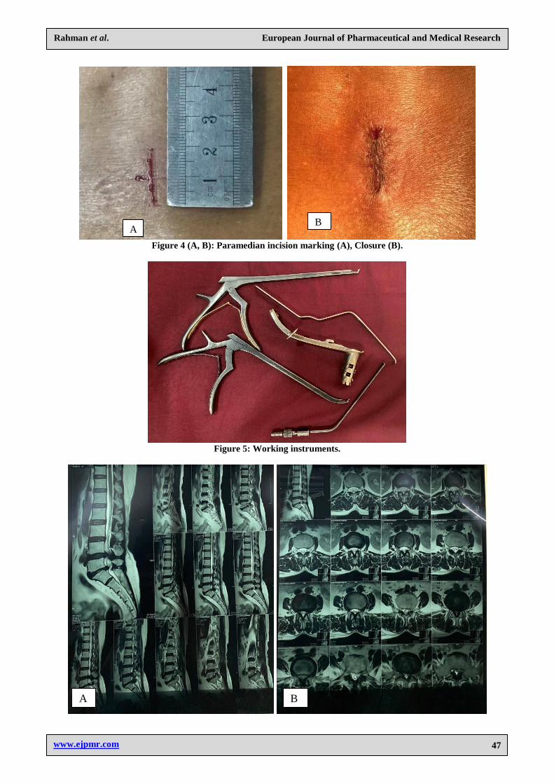
Figure 4 (A, B): Paramedian incision marking (A), Closure (B).
Figure 5: Working instruments.
A
B
A B
www.ejpmr.com
Rahman et al. European Journal of Pharmaceutical and Medical Research
48
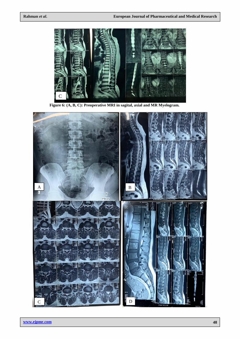
Figure 6: (A, B, C): Preoperative MRI in sagital, axial and MR Myelogram.
C
A B
C D

www.ejpmr.com
Rahman et al. European Journal of Pharmaceutical and Medical Research
49
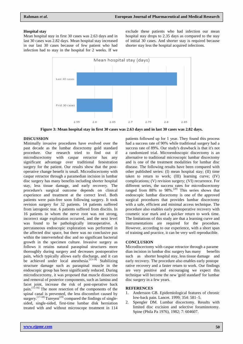
Figure 7: (A, B, C, D, E, F, G): Preoperative MRI and marking X Ray.
RESULTS
Immediately after surgery all patients were pain free. 32
patients needed revision surgery. 14 patients had
iatrogenic dural tears. 6 patients had discitis. Wrong
level exploration was in 16 patients in whom the nerve
root was not tight, and the next level found pathological
intra-operatively. There was no direct nerve root injury
though 3 patients had weak extensors of toes after
surgery which was recovered over 2-3 months probably
due to traction injury.
Table 1: Patient conditions after surgery.
Findings after surgery Number of patients
Revision surgery 32
Iatrogenic dural tear 14
Distics 6
Wrong level exploration 16
Traction injury (nerve) 3
So the final result shows that 95% had satisfactory
outcome. Because the revision surgery was done in 32
patients out of 650.
E F
F
G
www.ejpmr.com
Rahman et al. European Journal of Pharmaceutical and Medical Research
50
Hospital stay
Mean hospital stay in first 30 cases was 2.63 days and in
last 30 cases was 2.82 days. Mean hospital stay increased
in our last 30 cases because of few patient who had
infection had to stay in the hospital for 2 weeks. If we
exclude these patients who had infection our mean
hospital stay drops to 2.35 days as compared to the stay
of initial 30 cases. And shorter stay is required because
shorter stay less the hospital acquired infections.
Figure 3: Mean hospital stay in first 30 cases was 2.63 days and in last 30 cases was 2.82 days.
DISCUSSION
Minimally invasive procedures have evolved over the
past decade as the lumbar discectomy gold standard
procedure. Our research tried to find out if
microdiscectomy with caspar retractor has any
significant advantage over traditional fenestration
surgery for the patient. Our results show that the post-
operative change benefit is small. Microdiscectomy with
caspar retractor through a paramedian incision in lumbar
disc surgery has many benefits including shorter hospital
stay, less tissue damage, and early recovery. The
procedure's surgical outcome depends on clinical
experience and treatment at the correct level. Both
patients were pain-free soon following surgery. It took
revision surgery for 32 patients. 14 patients suffered
from iatrogenic tear. 6 patients suffered from discitis. In
16 patients in whom the nerve root was not strong,
incorrect stage exploration occurred, and the next level
was found to be pathologically intraoperative. A
percutaneous endoscopic exploration was performed in
the affected disc space, but there was no conclusive pus
within the intervertebral disc and no significant bacterial
growth in the specimen culture. Invasive surgery as
follows it retains natural paraspinal structures more
thoroughly during surgery and decreases postoperative
pain, which typically allows early discharge, and it can
be achieved under local anesthesia.[13-14]
Stabilizing
structure damage such as paraspinal muscle in the
endoscopic group has been significantly reduced. During
microdiscectomy, it was proposed that muscle dissection
and removal of posterior components, such as lamina and
facet joint, increase the risk of post-operative back
pain.[15-16]
The more resection of the components of the
spinal canal is prevented, the less discomfort caused by
surgery.[17-18]
Tureyen[19]
compared the findings of single-
sided, single-sided, first-time lumbar disk herniation
treated with and without microscope treatment in 114
patients followed up for 1 year. They found this process
had a success rate of 90% while traditional surgery had a
success rate of 89%. Our study's drawback is that it's not
a randomized trial. Microendoscopic discectomy is an
alternative to traditional microscopic lumbar discectomy
and is one of the treatment modalities for lumbar disc
disease. The following results have been compared with
other published series: (I) mean hospital stay; (II) time
taken to return to work; (III) learning curve; (IV)
complications; (V) revision surgery; (VI) recurrence. For
different series, the success rates for microdiscectomy
ranged from 88% to 98%.[20]
This series shows that
endoscopic lumbar discectomy is one of the approved
surgical procedures that provides lumbar discectomy
with a safe, efficient and minimal access technique. The
procedure also enables early postoperative recovery with
cosmetic scar mark and a quicker return to work time.
The limitations of this study are that a learning curve and
instrumentations are required for the procedure.
However, according to our experience, with a short span
of training and practice, it can be very well reproducible.
CONCLUSION
Microdiscectomy with caspar retractor through a parame
dian incision in lumbar disc surgery has many benefits
such as shorter hospital stay, less tissue damage and
early recovery. The procedure also enables early postope
rative recovery and a faster return to work. Our findings
are very positive and encouraging we expect this
technique will become the new 'gold standard' for lumbar
disc surgery in a few years.
REFERENCES
1. Andersson GB. Epidemiological features of chronic
low-back pain. Lancet. 1999; 354: 581–5.
2. Spengler DM. Lumbar discectomy. Results with
limited disc excision and selective foraminotomy.
Spine (Phila Pa 1976), 1982; 7: 604607.

www.ejpmr.com
Rahman et al. European Journal of Pharmaceutical and Medical Research
51
3. Mixter WJ, Barr JS: Rupture of the intervertebral
disc with involvement of the spinal canal. N Engl J
Med., 210-215.
4. Yasargil MG: Microsurgical operation for herniated
disc. Adv Neurosurg, 1977; 81.
5. Caspar W: A new surgical procedure for lumbar disk
herniation causing less tissue damage through a
microsurgical approach. Adv Neurosurg, 1977; 4:
74-77.
6. Gibson JN, Grant IC, Waddell G: The Cochrane
review of surgery for lumbar disc prolapse and
degenerative lumbar spondylosis. Spine, 1999; 24:
1820-1832.
7. Henriksen L, Schmidt K, Eskesen V, Jantzen E: A
controlled study of microsurgical versus standard
lumbar discectomy. Br J Neurosurg, 1996; 10:
289-293.
8. Lagarrigue J, Chaynes P: Comparative study of disk
surgery with or without microscopy. A prospective
study of 80 cases. Neurochirurgie, 1994; 40:
116-120.
9. Tullberg T, Isacson J, Weidenhielm L: Does
microscopic removal of lumbar disc herniation lead
to better results than the standard procedure? Results
of a one-year randomized study. Spine, 1993; 18:
24-27.
10. Wu X, Zhuang S, Mao Z, Chen H. Microendoscopic
discectomy for lumbar disc herniation: Surgical
technique and outcome in 873 consecutive
cases. Spine (Phila Pa 1976), 2006; 31: 2689–94.
[PubMed] [Google Scholar]
11. Evaniew N, Khan M, Drew B, Kwok D, Bhandari
M, Ghert M, et al. Minimally invasive versus open
surgery for cervical and lumbar discectomy: A
systematic review and meta-analysis. CMAJ Open,
2014; 2: E295–305. [PMC free article] [PubMed]
[Google Scholar]
12. Rahman M M, Surgical outcome of unilateral
approach in Lumbar Spinal Stenosis; Analysis of 62
cases, Jan, 2017; 12(2): 2-4. [Google Scholar]
13. Lee DY, Shim CS, Ahn Y, Choi YG, Kim HJ, Lee
SH: Compa- rison of percutaneous endoscopic
lumbar discectomy and open lumbar
microdiscectomy for recurrent disc herniation. J
Korean Neurosurg Soc., 2009; 46: 515-521.
14. Lee DY, Ahn Y, Lee SH: Percutaneous endoscopic
lumbar dis- cectomy for adolescent lumbar disc
herniation: surgical outcomes in 46 consecutive
patients. Mt Sinai J Med., 2006; 73: 864-870.
15. Ahn Y, Lee SH, Park WM, Lee HY, Shin SW, Kang
HY: Per- cutaneous endoscopic lumbar discectomy
for recurrent disc herniation: surgical technique,
outcome, and prognostic factors of 43 consecutive
cases. Spine (Phila Pa 1976), 2004; 29: E326-332.
16. Ozgen S, Naderi S, Ozek MM, Pamir MN: Findings
and outcome of revision lumbar disc surgery. J
Spinal Disord, 1999; 12: 287-292.
17. Kotilainen E, Valtonen S: Clinical instability of the
lumbar spine after microdiscectomy. Acta Neurochir
(Wien), 1993; 125: 120-126.
18. Schoeggl A, Maier H, Saringer W, Reddy M, Matula
C: Out- come after chronic sciatica as the only
reason for lumbar micro- discectomy. J Spinal
Disord Tech., 2002; 15: 415-419.
19. Tureyen K: One-level one-sided lumbar disc surgery
with and without microscopic assistance: 1-year
outcome in 114 consecutive patients. JNS Spine,
2003; 99(3): 247–250.
20. Anjan A Lath Microendoscopic discectomy for
prolapsed lumbar intervertebral disc Neurol India,
2006; 54: 1904.