micrornaについて...micrornaについて •蛋白をコードしないrna(non-coding rna...
TRANSCRIPT

microRNAについて
• 蛋白をコードしないRNA(non-coding RNA 約23,000個)の一種 (蛋白をコードするRNA:20,000個)
• 18-25塩基の最終的には1本鎖RNA
• 1/3の遺伝子はマイクロRNAにより発現が調整されている
• マイクロRNAの多くは番号が後ろにつく形で表現され、現在1800番台の番号がついている。 miR-122, miR-200など 例外 let-7
• ほ乳類の発生、器官形成、癌・感染症、生活習慣病などの病態に関与
• 診断や治療への応用が期待されている

RNA干渉 (RNA interference)
Fire A, Mello C ら 1998 Nature 2006年にノーベル生理学賞受賞
Potent and specific genetic interference by double-stranded RNA in Caenorhabditis elegans.
腺虫の一種(C elegans)を用いてunc-22(筋細胞)含むその他の蛋白発現を抑制する実験 unc-22の機能が低下すると筋細胞が収縮する
長さ 表現型 センス鎖: 742塩基 異常なし アンチセンス: 742塩基 異常なし センス、アンチセンス混合(二本鎖RNA): 742塩基 筋収縮
→ dsRNAのアンチセンスRNAは単純に1:1でmRNAに結合しているのではない
その後、細胞質内のRNaseであるDicerによりRNAが短く切断され短い2本鎖siRNA(small intefering RNA)ができる siRNAと蛋白質からなるRISC複合体(RNA-induced silencing complex)が再利用されながら 相補的なmRNAを分解する
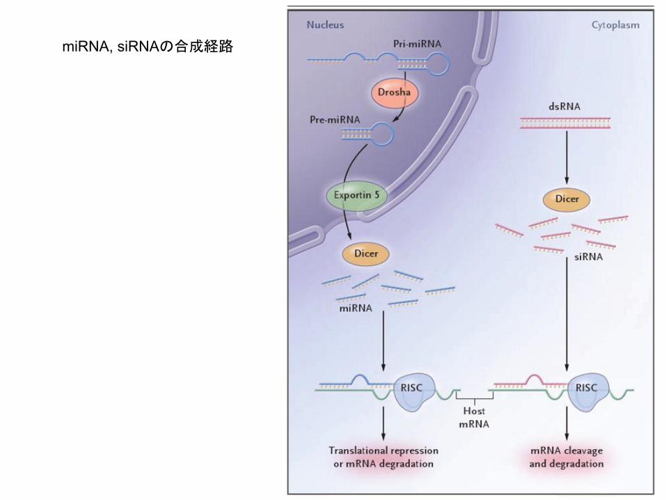
miRNA, siRNAの合成経路

siRNAとmiRNAの違い(miRNAの利点)
• miRNAは6-8塩基が相補的であれば結合し、蛋白合成を停止させる
• 体内でより安定であり、効果が長時間持続する
• 毒性が少ない
• 一つのmiRNAは100-1000個の蛋白発現を抑制することが考えられている (Target Scanというサイトで検索可能) 相乗効果が期待される。

Dicer, Drosha, and Outcomes in Patients with Ovarian Cancer Merritt. WM, Sood AK, N Eng J Med 2008
Dicer, DroshaのmRNAレベルと卵巣癌の予後因子について検討
DicerのmRNAが低い患者の予後は不良 RNA干渉を利用した治療をする上では不利

miRNAが脳神経の発生において重要な役割をはたしている1例
miR-124a is required for hippocampal axogenesis and retinal cone survival through Lhx2 suppression.
Sanuki R, Furukawa T Nat Neurosci 2011
miR-124aは中枢神経系で最も多く存在しており、脳に発現する全miRNA量の25-48%に達する。 中枢神経系での重要な機能が想定されていたが、いくつかのグループから相反する 結果が示されていた そこでmiR124a欠損マウスを作成し、脳の発達に影響があるかどうかを検討した
背景

図1 miR‐124a欠損による海馬の神経回路異常 海馬の歯状回の顆粒細胞(赤)に入った情報は苔状線維を介し
てCA3の錐体細胞(青)へと伝達され、さらにCA1へと伝わる(上図)。miR‐124a欠損マウス(KO)では、苔状線維とCA3錐体細胞の回路形成が正しい位置で形成されず、苔状線維の
CA3領域への異常侵入が認められた(下図、橙矢印)

図2 miR‐124a欠損による錐体視細胞の脱落 視覚情報の入り口である視細胞によって光信号は受容され、電
気信号へと変換される。その信号は双極細胞を介して神経節細胞へと伝達され、脳に情報が伝わる。この視覚情報入力系のうち錐体視細胞は視力と色覚にとって重要な働きをする(上図)。
miR‐124a欠損マウス(KO)では錐体視細胞が細胞死によって脱落し、その結果、野生型に比べて錐体視細胞が減少する。
錐体視細胞を蛍光色素で標識し、網膜を上から観察した(下図)

図3 miR‐124aの機能メカニズム miR‐124aは分化した神経細胞から発現し始め、成熟が進むにつれてその発現量が多くなる。この時、miR‐124aは標的遺伝子であるLhx2の発現を抑えることによって、神経細胞の発達・成熟と維持を制御して正常な神経機能を発揮する。miR‐124a欠損マ
ウスは神経細胞の成熟不良により、神経回路形成の異常や神経細胞死が起こる。

C型肝炎の新たな治療のターゲットにmiRNAが利用されている
• miR-122は肝臓において高い発現
• HCV RNAの2カ所とmiR-122は結合する
• miR-122の結合が肝細胞におけるHCVのRNAを増加させる。
• SPC3649はmiR-122の5’に相補的に結合し、miR-122の機能を抑制させる
背景
チンパンジーにHCVを感染させ、C型肝炎を作成 SPC3649 : high dose (5mg/kg), low dose(1mg/kg)の2群で治療 週1回 12週にわたり治療を継続
Therapeutic silencing of microRNA-122 in patients with chronic hepatitis C virus infection
Robert E, Henrik O Science 2010
方法

Silencing of miR-122 by SPC3649 in chimpanzees with chronic hepatitis C virus infection. (A) Analysis of HCV RNA levels in HCV-infected chimpanzees during the study. The HCV titers are shown as genomic equivalents (GE) for the high-dose animals (4x0513, blue triangles; 4x0514, magenta diamonds) and low-dose animals (4x0267, orange squares; 4x0358, red dots) in serum (GE/ml, solid lines) and liver (GE/μg liver RNA, dashed lines). The placebo and active treatment periods are indicated below. (B) Northern blot analysis of RNA from chimpanzee liver biopsies using LNA-modified probes detecting free and sequestered miR-122 (upper panel) and SPC3649 (lower panel). The first two lanes are positive controls for free miR-122 and preformed miR-122:SPC3649 heteroduplexes, respectively. (C) Detection of sequence variants in the miR-122 seed sites (boxed) by deep sequencing of the HCV 5′ NCR from the high-dose animals at four time points: baseline, end of treatment, viral rebound, and end of the follow-up period. (D) The two miR-122 seed sites (boxed) in the HCV 5′ NCR are conserved in all HCV genotypes and subtypes (see Supporting Online Material for details).
HCV RBA量 →治療に伴い低下 治療終了増加 肝細胞におけるmiR-122量 →治療に伴い発現低下

Treatment of HCV-infected chimpanzees with SPC3649 was well tolerated. (A) Plasma trough levels of SPC3649. (B and C) Alanine aminotransferase (ALT) levels (B) and creatinine levels (C) in HCV-infected chimpanzees during the study. (D to G) Photomicrographs of hematoxylin and eosin–stained sections from biopsies of a normal chimpanzee liver (D) and animal 4x0513 at week –4 (E), week 19 (F), and week 25 (G), respectively.
A 血清SPC3649濃度→治療終了後8週でほぼ検出されず B 血清ALT(GPT)濃度→治療に伴い低下、治療終了後、肝炎の再燃に伴いやや上昇 C 血清クレアチニン値→治療中、大きな変化なし 肝細胞は治療にて正常の構造に近づく
現在ヒトにおいて第1相臨床試験が始まっている

EMT (epithelial mesenchymal transition)
上皮性マーカーの欠失 (E-cadherinの発現低下) 間葉系性格の獲得(vimentin,fibronectin, ZEB1(E-cadherinの転写を抑える),ZEB2のは発現亢進

miR-200c expression induces an EMT phenotype in UMUC3 bladder cancer cell line. A, phase-contrast microscopy of UMUC3 cells transduced with a miR-200c containing retrovirus (UMUC3/200c) or with an empty, control, retroviral construct (UMUC3-E). Bar, 100 µm. B, measurement of in vitro cell migration by "wound-healing" assay. Representative pictures for same single spot. The experiment was done twice in triplicate experiments. Bar, 400 µm. C, quantification of miR-200c in empty virus–transduced (UMUC3-E) and miR-200c-transduced (UMUC3/200c) cells. Levels of miR-200c in UMUC5 cells are plotted for comparison. D, measurement by real-time RT-PCR of the epithelial and mesenchymal markers E-cadherin, ZEB1, and ZEB2 in empty virus–transduced cells and miR-200c-transduced UMUC3 cells. E, confocal microscopy analysis of the UMUC3 series costained for ZEB1 or ZEB2 (red pixels) and nuclear DNA (green pixels). Note the down-regulation of nuclear ZEB1 and ZEB2 in UMUC3 clones expressing the miR-200c. F, measurement of ERRFI-1 and E-cadherin protein levels by Western blot. Actin reprobing served as internal (loading) control. Bottom, absorbance relative values expressed as ratios between actin (internal control) and ERRFI-1. Note the up-regulation of E-cadherin and down-regulation of ERRFI-1 protein levels on miR-200c expression.
膀胱癌細胞株にretrovirusでmiR-200cを導入

miR-200c expression reverses EGFR resistance in UMUC3 cells. A, confocal microscopy analysis of the UMUC3 series costained for ERRFI-1 (red pixels) and EGFR (green pixels) and a DNA dye, showing the nucleus (blue pixels). Note the yellow pixels (left) as a result of red and green pixels colocalization. B, immunoblot of autophosphorylated EGFR, total EGFR, and total ERRFI-1 in cells transduced with miR-200c from the experiment above. In this experiment, actin served as the internal control. C, top, immunoblot of pMAPK, total MAPK, and total EGFR of the UMUC3 series. Cells grown in 2% serum-supplemented MEM were left untreated or treated with increased concentrations of cetuximab (C225) for 3 h. Bottom, absorbance relative values expressed as ratios between EGFR (internal control) and pMAPK. D, cell proliferation measurement of the UMUC3 series using radioactive thymidine incorporation. Each experiment was done in at least two different triplicates.
miR200cを導入すると ERRFI-1(ErbB receptor inhibitor-1)が減少し EGFRの発現が回復し C225(Cetuximab)の濃度依存的にp-MAPKが低下 (細胞の増殖が遅くなっている)

癌の進展におけるmiR-200の役割
miR200 低下
ZEB1 亢進
E-cadherin 低下
浸潤しやすく
ErbB receptor inhibitor-1 亢進
抗EGFR薬が効かなくなる
β-catenin 亢進
Cyclin D1 亢進
細胞増殖が早くなる
Tublin(微小管)の増加
タキサン系薬剤に耐性
