molecular cloning and sequence analysis of a …. 265, no. 12, issue of april 25, pp. 6556-6561,...
TRANSCRIPT

Vol. 265, No. 12, Issue of April 25, pp. 6556-6561, 1990 Printed in U.S A.
Molecular Cloning and Sequence Analysis of a cDNA Encoding a Porcine Kidney Renin-binding Protein*
(Received for publication, October 10, 1989)
Hiroyasu Inoue, Kiyoshi Fukui, Saori Takahashi, and Yoshihiro Miyake$ From the Department of Biochemistr.y, National Cardiovascular Center Research Institute, 7-1, 5.chome, Fujishiro-dai, Suita, Osaka 565, Japan
A complementary DNA encoding a renin-binding protein (RnBP) has been isolated from a porcine kidney cDNA library by immunological screening of in uitro translation products from the cDNAs. Analysis of the nucleotide sequence of the clone revealed a 1,342- nucleotide sequence with a 5’-terminal untranslated region of 52 nucleotides, an open reading frame of 1,206 nucleotides that encodes 402 amino acids, and a 3’-terminal untranslated region of 84 nucleotides that contains the polyadenylation signal sequence, AA- TAAA. The predicted amino acid sequence contains no hydrophobic amino-terminal sequence and does not show significant homology to those of other identified proteins. The irz uitro translated RnBP was found to have the same molecular weight, 42,000, as that of the purified RnBP from porcine kidney on sodium dodecyl sulfate-polyacrylamide gel electrophoresis and it formed a complex with renin purified from porcine kidney, which indicates that the cDNA encodes a func- tional RnBP without a propeptide sequence. The RnBP cDNA probe hybridized to a 1.5-kilobase mRNA in kidney, liver, adrenal, and pituitary glands, the amount being much greater in kidney than in the other tissues. Southern blot analysis showed the presence of a unique gene for RnBP in the porcine genome.
Renin-binding protein (RnBP)’ is a protein that binds to renin forming a protein complex called high molecular weight (HMW) renin. Therefore, this protein has been investigated in relation to renin and HMW renin. Renin (EC 3.4.23.15), an aspartyl proteinase that catalyzes the release of angioten- sin 1 from angiotensinogen is now known to exist in several forms with different molecular weights. In contrast to renin (iVfr 40,000), HMW renin is one of the forms of renin with a molecular weight of about 60,000 (1, Z), and another form of renin is prorenin, an inactive renin precursor which consists of a single polypeptide chain of Mr 44,000 (3,4). The molecular
* This work was supported in part by Research Grant 62A-1 for Cardiovascular Diseases from the Ministry of Health and Welfare of Japan, and Grants-in-Aid for Scien& Research (63570137 and 01770170) from the Mini&v of Education, Science and Culture of Japan. The costs of publication of this article were defrayed in part by the payment of page charges. This article must therefore be hereby marked “aduertisement” in accordance with 18 U.S.C. Section 1734 solely to indicate this fact
The nucleotide sequence(s) reported in this paper has been submitted to the GenBankrM/EMBL Data Bank with accession number(s) JO5399.
i To whom correspondence should be addressed. ’ The abbreviatiois used are: RnBP, renin-binding protein; HMW,
high molecular weight; bp, base pairs; SDS, sodium dodecyl sulfate; NEM, N-ethylmaleimide; DTNB, 5,5’-dithiobis-(2-nitrobenzoic acid); PMSF, phenylmethanesulfonyl fluoride; kbp, kilobase pairs.
properties of renin and prorenin have been well characterized by purification from animal tissues (3-8) and analysis of the gene structure (9, 10). However, the results were somewhat conflicting as to the properties of HMW renin in crude tissue extracts (11,12). On the other hand, it has been reported that sulfhydryl oxidizing- and alkylating-reagents induced the for- mation of HMW renin in a kidney extract and dithiothreitol reconverted it to renin and RnBP (13). In addition, a purified preparation of HMW renin from porcine kidney showed par- tial renin activity (1).
Recently, we purified HMW renin from porcine kidney and showed that the purified preparation consisted of two protein species (2). One was identified as renin and the other as a protein with a molecular weight of 42,000. The latter was denatured by acid treatment, which resulted in the release of renin and an increase in the level of renin activity to that of free renin. In a separate experiment, we also purified the jVp 42,000 protein from porcine kidney (14). The molecular weight of the purified protein was similar to that of the protein in HMW renin. The protein formed a complex with renin in the neutral pH region, although it is acid labile and easily dena- tured on acidification. Moreover, on complex formation the protein strongly inhibited the activity of renin, and analysis of the inhibition indicated that the complex is completely inactive, provided that it is composed of one molecule each of renin and the protein. The Mr 42,000 protein was thus iden- tified as RnBP. We also reported that the purified RnBP is a dimer which dissociates into the monomer on treatment with NEM and DTNB, and the DTNB-treated monomer is recon- verted to the dimer in the presence of dithiothreitol (15). As the monomerization occurred with sulfhydryl-alkylating and -oxidizing reagents, the RnBP is not disulfide-linked but is a noncovalently bound dimer. Despite evidence that RnBP forms a complex with renin, the structural characteristics of this protein as well as its physiological role remain to be elucidated.
In the present study, we isolated a cDNA encoding RnBP from a porcine kidney cDNA library using a newly developed cloning strategy. We determined the complete nucleotide se- quence of the cDNA and predict the primary structure of RnBP from the nucleotide sequence. In addition, the expres- sion of RnBP in tissues, the genomic organization of the RnBP gene and complex formation of in. uitro synthesized RnBP with the purified renin are also described.
EXPERIMENTAL PROCEDURES’
RESULTS
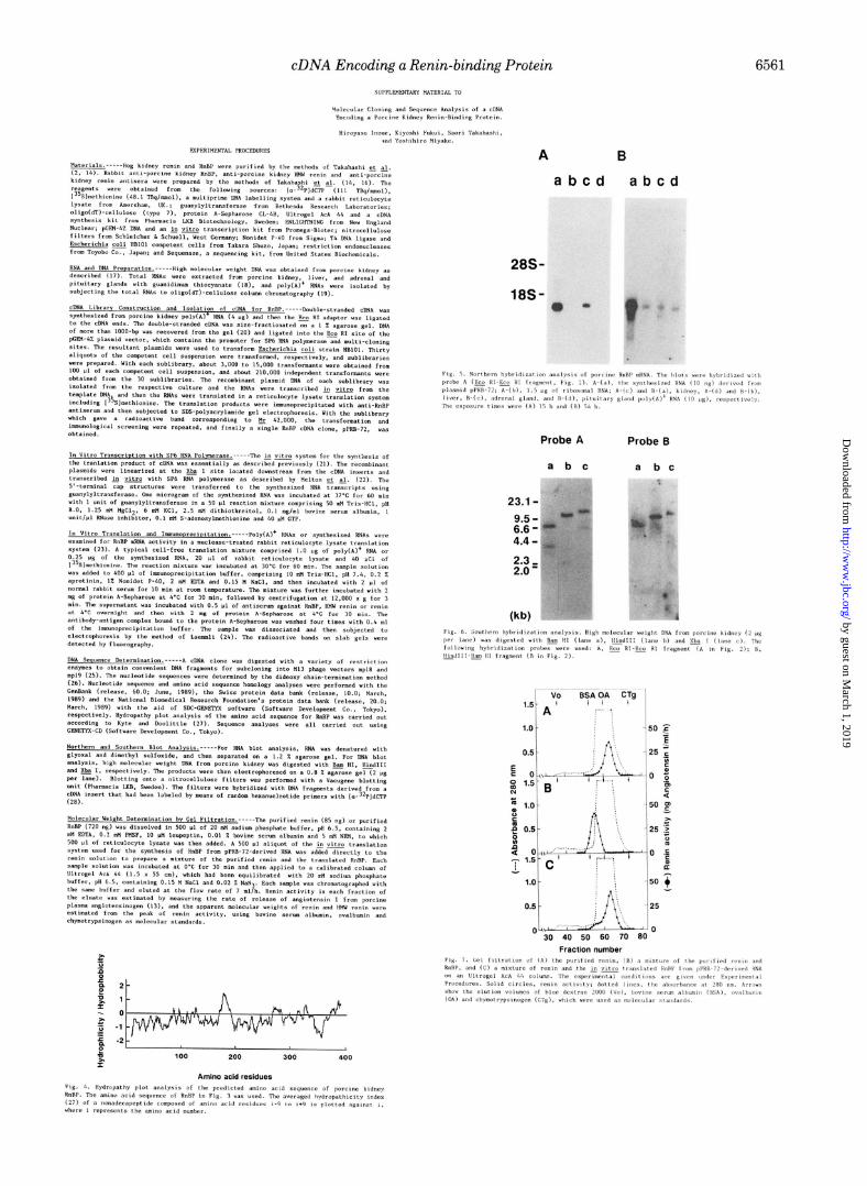
Isolation of a cDNA Clone for RnBP-To isolate RnBP cDNA, a newly developed cloning strategy was employed. The
’ Portions of this paper (including “Experimental Procedures” and Figs. 4-7) are presented in miniprint at the end of this paper. Miniprint is easily read with the aid of a standard magnifying glass. Full size photocopies are included in the microfilm edition of the Journal that is available from Waverly Press.
6556
by guest on March 1, 2019
http://ww
w.jbc.org/
Dow
nloaded from

cDNA Encoding a Renin-binding Protein 6557
method involves the fractionation of transformants from a porcine kidney cDNA library into sublibraries and the detec- tion of the in uitro translated products of the transformants in each sublibrary by immunoprecipitation. RnBP cDNA could be isolated through tertiary transformation and immu- nological screening. In fact, a single radioactive band was obtained for the in uitro translation products from porcine kidney poly(A)+ RNA (Fig. 1, lune b) and RNA synthesized with the single clone pPRB-72 (Fig. 1, lune c). The band in lane c disappeared on the addition of the purified RnBP (lane d). Moreover, the protein immunoreactive toward the anti- RnBP antiserum gave a single band on immunoprecipitation with the anti-HMW renin antiserum (lune e), but not with the anti-renin antiserum (lune f ). The immunoreactivities of the in uitro translation product from pPRB-72-derived RNA toward the antisera were fairly consistent with those of RnBP reported previously (14), and the bands in Fig. 1 corresponded to a molecular weight of 42,000, which was also identical to that of RnBP purified from porcine kidney (14).
Nucleotide Sequence Analysis-The restriction endonucle- ase map and an outline of the strategy used to determine the whole nucleotide sequence of the cDNA insert in clone pPRB- 72 are presented in Fig. 2. Fig. 3 shows the 1342-nucleotide sequence encoding the porcine RnBP in clone pPRB-72. The first ATG codon is located at nucleotides 53-55, i.e. down-
94 - 67 -
43- -- -
30 -
Fm. 1. Autoradiogram of the translation products for iden- tification by immunological screening of a cDNA encoding porcine kidney RnBP. The proteins were obtained by translation in a rabbit reticulocyte lysate translation system without added RNA (Lane a), with 1.0 Kg of kidney poly(A)+ RNA (lune b), and with the synthesized RNA (0.25 pg) derived from plasmid pPRB-72 (Lanes c- f). Proteins labeled with [%]methionine were immunoprecipitated and then analyzed by gel electrophoresis and autoradiography. Im- munoprecipitation with anti-RnBP antiserum in the absence (lanes a-c) or presence (lane ~0 of 5 pg of unlabeled RnBP, with anti-HMW renin antiserum (Lune e), and with anti-renin antiserum (Lune f). The exposure time for lanes a and b was 16 h, and that for lunes c-f 2 h.
pPRB-72
FIG. 2. Restriction endonuclease map and sequencing strat- egy for the cDNA insert. The map displays only the relevant restriction endonuclease sites. &oRI* denotes the ligated &oRI adaptor sites. The XbaI site, which was used for in uitro transcription, was not. nresent in the cDNA insert of DF’RB-72. A so&d box represents the cod& region for RnBP. Open &es represent the 5’--and 3’. untranslated regions. The cDNA insert is shown by a so/id bar. The direction and extent of the sequence determination are indicated by the horizontal orrow under the insert. Fragment A is the probe used for Northern and Southern hybridization analyses in Figs. 5 and 6, and fragment B for Southern hybridization analysis in Fig. 6.
stream from the in-frame terminator TGA (nucleotides 23- 25). The nucleotide sequence surrounding the ATG triplet encoding the initiating methionine agrees with the favored sequence that flanks functional initiation codons in eukary- otic mRNAs (29). In particular, a purine nucleotide is present at position -3 from the first nucleotide of the initiation codon and a guanine nucleotide is present at position +4. An open reading frame is followed by the TAG termination codon at nucleotides 1259-1261 and the 1206-nucleotide reading frame codes for 402 amino acids. The 3’-noncoding region of the cDNA is 84 nucleotides long, including a poly(A) tail of 9 nucleotides. The polyadenylation signal, AATAAA (30), is present 17 nucleotides upstream from the poly(A) tail.
The amino acid composition of the sequence predicted from the cDNA and that of RnBP purified from porcine kidney (15) were very close to each other (Table I). Moreover, the amino acid sequences of two fragments from lysyl endopep- tidase- and cyanogen bromide-treated RnBP3 agreed com- pletely with those predicted from the cDNA sequences at amino acid positions 4-12 and 326-357. The molecular weight of the protein was thus calculated to be 46,510 from the predicted amino acid sequence.
The nucleotide and predicted amino acid sequences of por- cine RnBP do not appear to be related to those of other proteins entered in nucleotide and protein sequence data banks. There is a potential asparagine-linked glycosylation site conforming to the consensus sequence of Asn-X-Ser/Thr at amino acid positions 228-230 (Fig. 3). Fig. 4 shows the results of hydropathy plot analysis of the predicted amino acid sequence of RnBP. The RnBP does not possess a hydro- phobic amino-terminal sequence indicative of a signal se- quence, whereas it has a hydrophobic segment at amino acid positions 164-189.
Expression of RnBP mRNA in Porcine Tissues-Northern blot analysis was carried out with the cDNA insert of pPRB- 72 (EcoRI-EcoRI; Fig. 2, probe A) as a hybridization probe (Fig. 5). The size of the synthesized RNA derived from clone pPRB-72 was 1.4 kilobases, which is in good agreement with that calculated from the transcribed sequences of the cDNA insert and the plasmid vector (Fig. 5A, lune u). A single 1.5- kilobase mRNA was identified in porcine kidney (Fig. 5A, lune c), and it was also detected in liver and adrenal and pituitary glands on long exposure (Fig. 5B, lunes b-d). There- fore, RnBP mRNA is much more abundant in kidney than in other tissues.
Genomic Organization of the RnBP Gene-Southern blot analysis of genomic DNA was performed to determine the copy number of the RnBP gene in the porcine genome. When the cDNA insert of pPRB-72 (EcoRI-EcoRI; Fig. 2, probe A) was used as a hybridization probe, three (6.6, 3.6, and 3.0 kbp), two (11 and 2.3 kbp), and two (14 and 6.5 kbp) bands were observed for high molecular weight DNA digested with BarnHI, HindIII, and XbaI, respectively (Fig. 6A). On the other hand, when a 0.42-kbp fragment (HindIII-BumHI; Fig. 2, probe B) was used as a hybridization probe, only a single band (6.6 kbp, BarnHI; 11 kbp, HindIII; 14 kbp, XbaI) was detected (Fig. 6B). These results suggest that the RnBP gene exists as a single copy in the porcine genome.
Complex Formation of Purified Renin with RnBP Synthe- sized in Vitro-The interaction of the in vitro translated RnBP from pPRB-72-derived RNA with renin purified from porcine kidney was investigated by gel filtration on an Ultro- gel AcA 44 column. Fig. 7A shows the elution profile of the purified renin, of which the elution volume corresponded to a molecular weight of 40,000. When an excess amount of RnBP
” S. Takahashi and Y. Miyake, unpublished results.
by guest on March 1, 2019
http://ww
w.jbc.org/
Dow
nloaded from

6558 cDNA Encoding a Renin-binding Protein
FIG. 3. The nucleotide and corre- sponding amino acid sequences of porcine kidney RnBP. Nucleotides are numbered in the 5’ to 3’ direction, beginning with the first nucleotide of the cDNA insert preceded by an EcoRI adap- tor site. The deduced amino acid residues are indicated below the nucleotide tri- plets. An upstream in-frame stop codon at nucleotides 23-25 is indicated by a &t&e ande&e. The amino acid se- quences that agree with those deter- mined directly by Edman degradation3 are underhed. The potential aspara- gine-linked glycosylation site is indi- cated by a broken-line and the polyade- nylation signal, AATAAA, is boxed.
purified from porcine kidney was added to the renin solution, the peak of renin activity in Fig. 7A shifted completely to a position corresponding to a molecular weight of 60,000 (Fig. 7B). The results in Fig. 7, A and B, show the complete conversion of renin to HMW renin, being quite consistent with the results reported previously (14). When the in vitro translated RnBP was added to the renin solution, a shoulder appeared in the elution profile (Fig. 7C), the elution volume being the same as that observed in Fig. 7B. However, the major peak of renin activity remained at a position corre- sponding to the elution volume of the peak in Fig. 7A, and the complete shift of the renin peak to a position correspond- ing to the high molecular species was not observed. The amount of the Lz uitro translated RnBP that was applied to the column was thus estimated from the height of the shoul- der, with the assumption that the complex is composed of one molecule each of renin and RnBP, and it was roughly esti- mated to be in the order of 10 ng. Accordingly, the amount of RnBP in the sample solution used for the experiment in Fig. 7C was much less than that of renin. Therefore, the results in Fig. 7C indicate that the shoulder and the large peak corre- spond to the in vitro translated RnBP bound to renin and to free renin, respectively, that is, the in vitro translated RnBP also binds to renin and forms HMW renin. In the experiment in Fig. 7C, the measurement of renin activity in the region of the shoulder became difficult when the amount of renin in the sample solution was reduced, and also it was difficult to increase the synthesis of RnBP under the present conditions.
DISCUSSION
We have obtained a cDNA clone encoding RnBP from a porcine kidney cDNA library using a newly developed cloning strategy. This strategy comprises the highly efficient in uitro transcription of RNAs from the cDNAs and specific immu- nological detection of in uitro translation products from the synthesized RNAs on SDS-polyacrylamide gel electrophore- sis. Several advantages of this approach deserve comment. First, the strategy can exclude cross-reactive undesired clones which cause considerable problems in conventional immuno- logical screening, because characterization of immunoreactive proteins can be made simultaneously. Second, the strategy permits rapid characterization of expressed protein even in primary screening of sublibraries, and purification of the positive cDNA clone can be achieved through stepwise frac- tionations of the cDNA clone mixture. In fact, essentially the same result as shown in Fig. 1 was obtained in the initial step of the screening before isolation of the single clone pPRB-72 (data not shown). Third, the positive clone obtained by this strategy should cover the entire coding region, since the cDNA was selected on the basis of the synthesis of an immunoreac- tive protein with the same molecular weight as that of the primary translation product of the mRNA. Northern blot analysis (Fig. 5) indicated that the cDNA insert in clone pPRB-72 covered almost the full length of the mRNA se- quence, excluding poly(A) tail. Fourth, functional assays can be carried out directly with the use of the translation product as shown in Fig. 7. The validity of this screening procedure
by guest on March 1, 2019
http://ww
w.jbc.org/
Dow
nloaded from
cDNA Encoding a Renin-binding Protein 6559
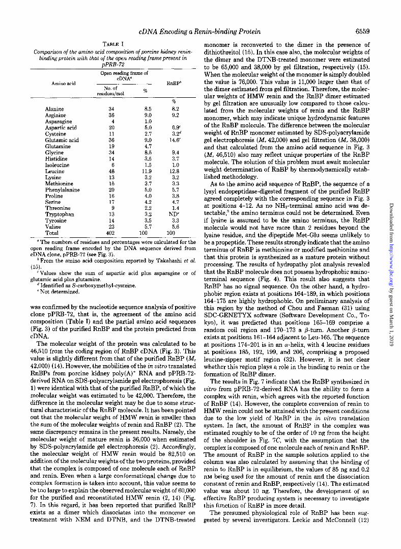
TABLE I Comparison of the amino acid composition of porcine kidney renin-
binding protein with that of the open reading frame present in pPRB-72
Open reading frame of C-DNA0
Amino acid ---..-
No. of residueslmol %
RnBPb
Alanine 34 Arginine 36 Asparagine 4 Aspartic acid 20 Cysteine 11 Glutamic acid 36 Glutamine 19 Glycine 34 Histidine 14 Isoleucine 6 Leucine 48 Lysine 13 Gethionine 15 Phenvlalanine 20 Proline 16 Serine 17 Threonine 9 Tryptophan 13 Tvrosine Valine
14 23
Total 402 I
% 8.5 8.2 9.0 9.2 1.0 5.0 6.9’ 2.7 3.2d 9.0 14.6 4.7 8.5 9.4 3.5 3.7 1.5 1.0
11.9 12.8 3.2 3.2 3.7 3.3 5.0 5.7 4.0 3.8 4.2 4.7
2.2 3.2 p&t
3.5 3.3 5.7 5.6
LOO 100
’ The numbers of residues and percentages were calculated for the open reading frame encoded by the DNA sequence derived from cDNA clone, pPRB-72 (see Fig. 3).
* From the amino acid composition reported by Takahashi et al. (15).
‘Values show the sum of aspartic acid plus asparagine or of glutamic acid plus glutamine.
’ Identified as S-carboxymethyl-cysteine. ’ Not determined.
was confirmed by the nucleotide sequence analysis of positive clone pPRB-72, that is, the agreement of the amino acid composition (Table I) and the partial amino acid sequences (Fig. 3) of the purified RnBP and the protein predicted from cDNA.
The molecular weight of the protein was calculated to be 46,510 from the coding region of RnBP cDNA (Fig. 3). This value is slightly different from that of the purified RnBP (Mr 42,000) (14). However, the mobilities of the in uitro translated RnBPs from porcine kidney poly(A)+ RNA and pPRB-72- derived RNA on SDS-polyacrylamide gel electrophoresis (Fig. 1) were identical with that of the purified RnBP, of which the molecular weight was estimated to be 42,000. Therefore, the difference in the molecular weight may be due to some struc- tural characteristic of the RnBP molecule. It has been pointed out that the molecular weight of HMW renin is smaller than the sum of the molecular weights of renin and RnBP (2). The same discrepancy remains in the present results. Namely, the molecular weight of mature renin is 36,000 when estimated by SDS-polyacrylamide gel electrophoresis (2). Accordingly, the molecular weight of HMW renin would be 82,510 on addition of the molecular weights of the two proteins, provided that the complex is composed of one molecule each of RnBP and renin. Even when a large conformational change due to complex formation is taken into account, this value seems to be too large to explain the observed molecular weight of 60,000 for the purified and reconstituted HMW renin (2, 14) (Fig. 7). In this regard, it has been reported that purified RnBP exists as a dimer which dissociates into the monomer on treatment with NEM and DTNB, and the DTNB-treated
monomer is reconverted to the dimer in the presence of dithiothreitol (1.5). In this case also, the molecular weights of the dimer and the DTNB-treated monomer were estimated to be 65,000 and 38,000 by gel filtration, respectively (15). When the molecular weight of the monomer is simply doubled the value is 76,000. This value is 11,000 larger than that of the dimer estimated from gel filtration. Therefore, the molec- ular weights of HMW renin and the RnBP dimer estimated by gel filtration are unusually low compared to those calcu- lated from the molecular weights of renin and the RnBP monomer, which may indicate unique hydrodynamic features of the RnBP molecule. The difference between the molecular weight of RnBP monomer estimated by SDS-polyacrylamide gel electrophoresis (Mr 42,000) and gel filtration (&fr 38,000) and that calculated from the amino acid sequence in Fig. 3 (iVr 46,510) also may reflect unique properties of the RnBP molecule. The solution of this problem must await molecular weight determination of RnBP by thermodynamically estab- lished methodology.
As to the amino acid sequence of RnBP, the sequence of a lysyl endopeptidase-digested fragment of the purified RnBP agreed completely with the corresponding sequence in Fig. 3 at positions 4-12. As no NH*-terminal amino acid was de- tectable,3 the amino terminus could not be determined. Even if lysine is assumed to be the amino terminus, the RnBP molecule would not have more than 2 residues beyond the lysine residue, and the dipeptide Met-Glu seems unlikely to be a propeptide. These results strongly indicate that the amino terminus of RnBP is methionine or modified methionine and that this protein is synthesized as a mature protein without processing. The results of hydropathy plot analysis revealed that the RnBP molecule does not possess hydrophobic amino- terminal sequence (Fig. 4). This result also suggests that RnBP has no signal sequence. On the other hand, a hydro- phobic region exists at positions 164-189, in which positions 164-175 are highly hydrophobic. On preliminary analysis of this region by the method of Chou and Fasman (31) using SDC-GENETYX software (Software Development Co., To- kyo), it was predicted that positions 165-169 comprise a random coil region and 170-173 a B-turn. Another p-turn exists at positions 161-164 adjacent to Leu-165. The sequence at positions 174-201 is in an a-helix, with 4 leucine residues at positions 185, 192, 199, and 206, comprising a proposed leucine-zipper motif region (32). However, it is not clear whether this region plays a role in the binding to renin or the formation of RnBP dimer.
The results in Fig. 7 indicate that the RnBP synthesized in vitro from pPRB-72-derived RNA has the ability to form a complex with renin, which agrees with the reported function of RnBP (14). However, the complete conversion of renin to HMW renin could not be attained with the present conditions due to the low yield of RnBP in the in uitro translation system. In fact, the amount of RnBP in the complex was estimated roughly to be of the order of 10 ng from the height of the shoulder in Fig. 7C, with the assumption that the complex is composed of one molecule each of renin and RnBP. The amount of RnBP in the sample solution applied to the column was also calculated by assuming that the binding of renin to RnBP is in equilibrium, the values of 85 ng and 0.2 nM being used for the amount of renin and the dissociation constant of renin and RnBP, respectively (14). The estimated value was about 10 ng. Therefore, the development of an effective RnBP producing system is necessary to investigate this function of RnBP in more detail.
The presumed physiological role of RnBP has been sug- gested by several investigators. Leckie and McConnell (12)
by guest on March 1, 2019
http://ww
w.jbc.org/
Dow
nloaded from
6560 cDNA Encoding a Renin-binding Protein
suggested that HMW renin is a complex of renin with a renin inhibitor of molecular weight 13,000 and that the inhibitor is decomposed on acid treatment. With respect to this presump- tion, we have shown, through kinetic analysis of the inhibi- tion, that purified RnBP strongly inhibits renin activity and that the complex is completely inactive (14). Moreover, we have also demonstrated that the dissociation constant of renin and RnBP is 0.2 nM by kinetic analysis. The renin activity in Fig. 7 (I3 and C) may thus be due to dissociation of renin from the complex under the extremely dilute conditions in the described radioimmunoassay (14). Murakami et ul. (33) showed that RnBP is present only in the kidney and pituitary gland and that the kidney RnBP reacts with kidney renin but not with renin in pituitary and submandibular glands. In addition, they showed that RnBP does not interact with other acid proteases in the kidney. These results suggest that kidney RnBP is highly specific for kidney renin. In this connection the present results show that RnBP mRNA exists not only in the kidney but also in extrarenal tissues, such as the liver, the pituitary, and adrenal glands. We also found the presence of RnBP in these tissues on in uitro translation of poly(A)+ RNA from each tissue (34). Boyd (11) proposed that RnBP is a renin carrier. It is noteworthy that RnBP has no propeptide, as indicated by the present results and our recent results (34), while the primary structure of human kidney renin predicted from renin cDNA includes both signal and propeptide se- quences (9). If RnBP is not excreted from kidney cells in ho, complex formation with extracellular renin would not occur. However, the HMW species of renin has been detected in plasma and amniotic fluid (35). Even if RnBP is not excreted and functions intracellularly, complex formation with renin would not occur unless it is synthesized in renin-producing juxtaglomerular cells. Further studies are necessary to solve these problems and to facilitate elucidation of the physiolog- ical significance of RnBP.
REFERENCES
I. Inagami, T., and Murakami, K. (1977) Circ. Res. 41, 11-11-11-16 2. Takahashi. S.. Ohsawa, T.. Miura. R.. and Mivake, Y. (1983) J.
Biochem: (Tokyo) 93; 2&274 - 3. Takahashi, S., Miura, R., and Miyake, Y. (1982) J. Biochem.
(Tokyo) 92,559-567 4. Takii, Y., and Inagami, T. (1982) Biochem, Biophys. Res. Com-
mu. 104,133-140 5. Cohen, S., Taylor, J. M., Murakami, K., Michelakis, A. M., and
Inagmi, T. (1972) Biochemistry 11, 4286-4293 6. Misono, K. S., Chang, J-J., and Inagami, T. (1982) Proc. Natl.
Acad. Sci. U. S. A. 79, 4858-4862
7.
8.
9.
10.
11. 12. 13.
14.
15.
16.
17.
18.
19.
20.
21.
22.
23.
24. 25.
26.
27. 28.
29. 30.
31.
32.
33.
34.
35.
Inagami, T., and Murakami, K. (1977) J. Biol. Chem. 252,2978- 2983
Takahashi, S., Miura, R., Yutani, C., and Miyake, Y. (1986) J. Biochem. (Z’okyo) 99,307-310
Imai, T., Miyazaki, H., Hirose, S., Hori, H., Hayashi, T., Kagey- ama, R., Ohkubo, H., Nakanishi, S., and Murakami. K. (1983) Proc. Natl. Acad. Sci, U. S. A. 80,7405-7409
Miyazaki, H., Fukamizu, A., Hirose, S., Hayashi, T., Hori, H., Ohkubo, H., Nakanishi, S., and Murakami, K. (1984) Proc. Natl. Acad. Sci. U. S. A. 81, 5999-6003
Boyd, G. W. (1974) Circ. Res. 35, 426-438 Leckie, B. J., and McConnell, A. (1975) Circ. Res. 36, 513-519 Murakami, K., Takahashi, S., Suzuki, F., Hirose, S., and Inagami,
T. (1980) Biomed. Res. 1, 392-399 Takahashi. S.. Ohsawa, T., Miura, R.. and Mivake, Y. (1983) J.
Biochem: (Tokyo) 93; 1583-1594 - Takahashi. S.. Miura. R.. and Mivake. Y. (1988) Biochem. Znt.
l6,105i-ii60 ' ' " ' ' Takahashi, S., Miura, R., and Miyake, Y. (1985) J. Biochem.
(Z’okyo) 97,671-677 Fukui, K., Watanabe, F., Shibata, T., and Miyake, Y. (1987)
Biochemistry 26,3612-3618 Chirgwin, J. M., Przybyla, A. E., MacDonald, R. J., and Rutter,
W. J. (1979) Biochemistry l&5294-5299 Aviv, H., and Leder, P. (1972) Proc. Natl. Acad. Sci. U. S. A. 69,
14081412 Dretzen, G., Bellard, M., Sassone-Corsi, P., and Chambon, P.
(1981) Anal. Biochem. 112, 295-298 Fukui, K., Momoi, K., Watanabe, F., and Miyake, Y. (1988)
Biochemi&-y 27,6693-6697 Melton, D. A., Krieg, P. A., Rebagliati, M. R., Maniatis, T., Zinn,
K., and Green, M. R. (1984) Nucleic Acids Res. 12, 7035-7056 Pelham, H. R. B., and Jackson, R. J. (1976) Eur. J. Biochem. 67,
247-256 Laemmli, U. K. (1970) Nature 227, 680-685 Yanisch-Perron, C., Vieira, J., and Messing, J. (1985) Gene
(Am&.) 33, 103-119 Sanger, F., Nicklen, S., and Coulson, A. R. (1977) Proc. Natl.
Acad. Sci. U. S. A. 74. 5463-5467 Kyte, J., and Doolittle, k. F. (1982) J. Mol. Biol. 157, 105-132 Feinberg, A. P., and Vogelstein, B. (1983) Anal. Biochem. 132,
6-13 Kozak, M. (1984) Nucleic Acids Res. 12,857-872 Breathnach. R.. and Chambon. P. (1981) Arznu. Reu. Biochem.
50,349~i83 Chou, P. Y., and Fasman, G. D. (1978) Adu. Enzymol. 47, 45-
148 Landschulz, W. H., Johnson, P. F., and McKnight, S. L. (1988)
Science 240,1759-1764 Murakami, K., Chino, S., Hirose, S., and Higaki, J. (1980)
Biomed. Res. 1, 476-481 Fukui, K., Inoue, H., Takahashi, S., and Miyake, Y. (1989)
Biochem. Biopkys. Res. Commun. 164,265-270 Day, R. P., Luetscher, J. A., and Gonzales, C. M. (1975) J. C&n.
Endocrinol. Metab. 40, 1078-1084
by guest on March 1, 2019
http://ww
w.jbc.org/
Dow
nloaded from
cDNA Encoding a Renin-binding Protein 6561
A B
abed abed
28S-
Probe A Probe 6
a b c a bc
23.1-
by guest on March 1, 2019
http://ww
w.jbc.org/
Dow
nloaded from

H Inoue, K Fukui, S Takahashi and Y Miyakerenin-binding protein.
Molecular cloning and sequence analysis of a cDNA encoding a porcine kidney
1990, 265:6556-6561.J. Biol. Chem.
http://www.jbc.org/content/265/12/6556Access the most updated version of this article at
Alerts:
When a correction for this article is posted•
When this article is cited•
to choose from all of JBC's e-mail alertsClick here
http://www.jbc.org/content/265/12/6556.full.html#ref-list-1
This article cites 0 references, 0 of which can be accessed free at
by guest on March 1, 2019
http://ww
w.jbc.org/
Dow
nloaded from