(multidrug resistant...
TRANSCRIPT

다제내성균 감시검사(MULTIDRUG RESISTANT ORGANISMS)
조지현원광의대 진단검사의학과
감염관리를 위한 진단검사

1. MDRO 정의 (multidrug resistant organisms)2. MDRO 중요성과 국내 MDRO 현황3. MDRO 검사 (screen, confirm)
1) MRSA, VRSA/VISA, CNS2) VRE3) ESBL4) CRE, MRPA, MRAB
4. 보균자 검사 (전파예방 및 관리)5. 요약
참고자료1) CDC. HAIs; Laboratory Resources
(http://www.cdc.gov/HAI/settings/lab/lab_settings.html)2) CLSI 2014 M100‐S24 Performance Standards for AST3) 질병관리본부. 다제내성균 감염 관리지침 2012.10.4) 질병관리본부. 카바페넴내성 장내균속 감염 관리지침 2012.5) 질병관리본부. 의료관련감염증 표준진단법(송원근 2013).6) 원광대병원 2013년 감수성 통계
다제내성균 감시검사

No consensus But, Microorganisms, predominantly bacteria, that are
resistant to one or more classes of antimicrobial agents
G(+) Staphylococcus ‐MRSA, VISA, VRSA Enterococcus ‐ VRE Streptococcus ‐ S. pneumoniae
G(‐) Enterobacteriaceae
‐ K. pneumoniae, E. coli (ESBL)‐ Enterobacter, Serratia, Citrobacter, Morganella, etc
Non‐fermenter‐ A. baumannii, P. aeruginosa, ‐ S. maltophilia, B. cepacia, R. picketti
Clostridium difficileMycobacterium – MTB
MDRO 정의 : No consensus But, Microorganisms, predominantly bacteria, that are
resistant to one or more classes of antimicrobial agents
G(+) Staphylococcus ‐MRSA, VISA, VRSA Enterococcus ‐ VRE Streptococcus ‐ S. pneumoniae
G(‐) Enterobacteriaceae
‐ K. pneumoniae, E. coli (ESBL)‐ Enterobacter, Serratia, Citrobacter, Morganella, etc
Non‐fermenter‐ A. baumannii, P. aeruginosa, ‐ S. maltophilia, B. cepacia, R. picketti
Clostridium difficileMycobacterium – MTB
MDRO 정의 :

감염병 원인1) serious, difficult‐to‐treat 2) morbidity, mortality3) length of stay 4) cost
예방 가능1) Judicious use of antimicrobials
decrease incidence of microorgnisms developing antibiotic resistance
2) Acquired via transmission by 근무자 ‐ 환자 환경 ‐ 환자 환자 ‐ 환자
중요성 :

손위생 (Hand hygiene) – 가장 중요 격리주의 (Isolation precautions)
Follow standard precautions for all patients‐ Personal protection equipment (gloves, gown, mask, goggle, face shield)
Develop & utilize systems ‐ to identify patients with MDROs & notify infection preventionists, physicians & direct caregivers
Place patients with a confirmed MDRO or history of an MDRO in singe‐patient rooms
Group patients with the same MDRO in designated areas if single‐patient room unavailable
Implement contact precautions immediately for all patients infected with MDROs, previously identified MDRO or have a history of being colonized with target MDROs.
Notify infection prevention and control after placing the patient in isolation
접촉주의 (Contact precautions)
환경관리 (Environmental measures)
전파방지 :
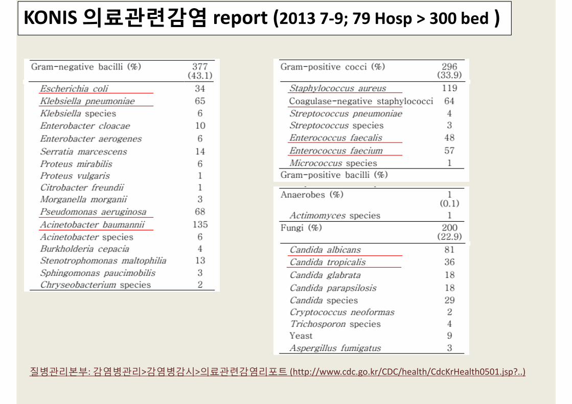
KONIS 의료관련감염 report (2013 7‐9; 79 Hosp > 300 bed )
질병관리본부: 감염병관리>감염병감시>의료관련감염리포트 (http://www.cdc.go.kr/CDC/health/CdcKrHealth0501.jsp?..)
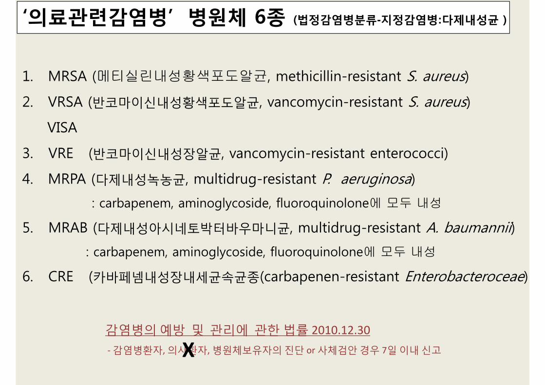
‘의료관련감염병’ 병원체 6종 (법정감염병분류-지정감염병:다제내성균 )
1. MRSA (메티실린내성황색포도알균, methicillin-resistant S. aureus)
2. VRSA (반코마이신내성황색포도알균, vancomycin-resistant S. aureus)
VISA
3. VRE (반코마이신내성장알균, vancomycin-resistant enterococci)
4. MRPA (다제내성녹농균, multidrug-resistant P. aeruginosa)
: carbapenem, aminoglycoside, fluoroquinolone에 모두 내성
5. MRAB (다제내성아시네토박터바우마니균, multidrug-resistant A. baumannii)
: carbapenem, aminoglycoside, fluoroquinolone에 모두 내성
6. CRE (카바페넴내성장내세균속균종(carbapenen-resistant Enterobacteroceae)
감염병의 예방 및 관리에 관한 법률 2010.12.30‐ 감염병환자, 의사환자, 병원체보유자의 진단 or 사체검안 경우 7일 이내 신고X

‘의료관련감염병’ 병원체 6종 신고현황
2012 감염병 감시연보. 보건복지부, 질병관리본부

KONIS 의료관련감염 report (2013, 모든 검체)
보고시스템>감염병웹통계시스템>표본감시통계http://is.cdc.go.kr/nstat/index.jsp
http://is.cdc.go.kr/nstat/index.jsp
Gram(+) Cocci
MRSA VRSA/VISA, CNS VRE

MRSA (ORSA): methicillin, oxacillin 병원분리 S. aureus 특성
PBP2a encoded by the mecA gene 변형된 penicillin‐binding protein, lower avidity to beta‐lactams
MRSA 중요성 Pathogenicity Transmissible
(모든 VRSA는 MRSA임) Limited treatment options: All b‐lactam R (예외…..)
SAU (%)
1,085 주 R
Penicillin 96.8
Oxacillin 78.2

B‐Lactam Families SIEMENS, CLSI AST Update for 2100, by Lisa Walker
Cefoxitin
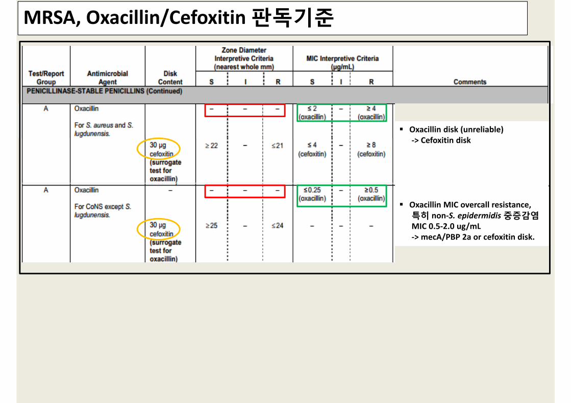
MRSA, Oxacillin/Cefoxitin 판독기준
Oxacillin disk (unreliable) ‐> Cefoxitin disk
Oxacillin MIC overcall resistance, 특히 non‐S. epidermidis 중증감염MIC 0.5‐2.0 ug/mL‐> mecA/PBP 2a or cefoxitin disk.

Screening Tests for MRSA in Staphylococcus species
Transmitted light. > 1 colony . light film of growth=> MRSA
≤ 21mm = mecA(+)≥ 21mm = mecA(‐)
≤ 24mm = mecA(+)≥ 25mm = mecA(‐)
≥ 4ug/mL = mecA(+)≤ 4ug/mL = mecA(‐)
Cefoxitin > Oxacillin > MethicillinOxacillin > Methicillin: storage, heteroresistant strains. Cefoxitin > Oxacillin : mecA inducer, easy reading (clear endpoints)
33 to 35℃, ambient air.
(Testing at temperature above 35℃ may not detect MRSA.)
≤ 2 ≥ 4 ≤ 4 ≥ 8

Tests for MRSA Detection Cefoxitin/Oxacillin test
Broth‐ & Agar‐based Tests Heteroresistance: 내성유전자(+), 일부만 in vitro 내성표현
Grow more slowly Missed at Tm above 35C.
at 33‐35° C (maximum of 35°C), ambient air for a full 24 hours NaCl (4% w/v; 0.68 mol/L)
Latex agglutination test for PBP2a
Nucleic acid amplification tests (PCR) for mecA gene mecA‐mediated oxacillin resistance의 surrogate mecA(‐) & oxacillin R ‐> ‘oxacillin R’, not cefoxitin R
: all beta‐lactam ‘R’ or ‘No’ report except anti‐MRSA activity
VISA/VRSA
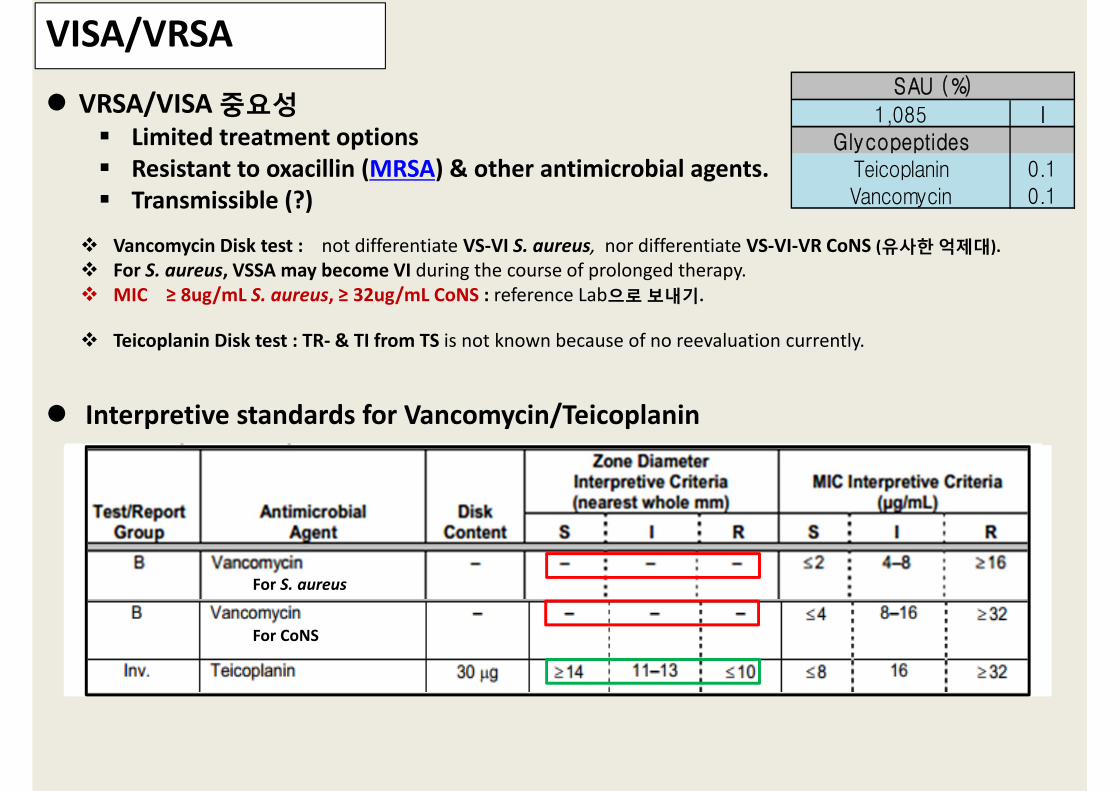
VRSA/VISA 중요성 Limited treatment options Resistant to oxacillin (MRSA) & other antimicrobial agents. Transmissible (?)
1,085 IGlycopeptides
Teicoplanin 0.1Vancomycin 0.1
SAU (%)
Vancomycin Disk test : not differentiate VS‐VI S. aureus, nor differentiate VS‐VI‐VR CoNS (유사한 억제대). For S. aureus, VSSA may become VI during the course of prolonged therapy. MIC ≥ 8ug/mL S. aureus, ≥ 32ug/mL CoNS : reference Lab으로 보내기.
Teicoplanin Disk test : TR‐ & TI from TS is not known because of no reevaluation currently.
For CoNS
For S. aureus
Interpretive standards for Vancomycin/Teicoplanin
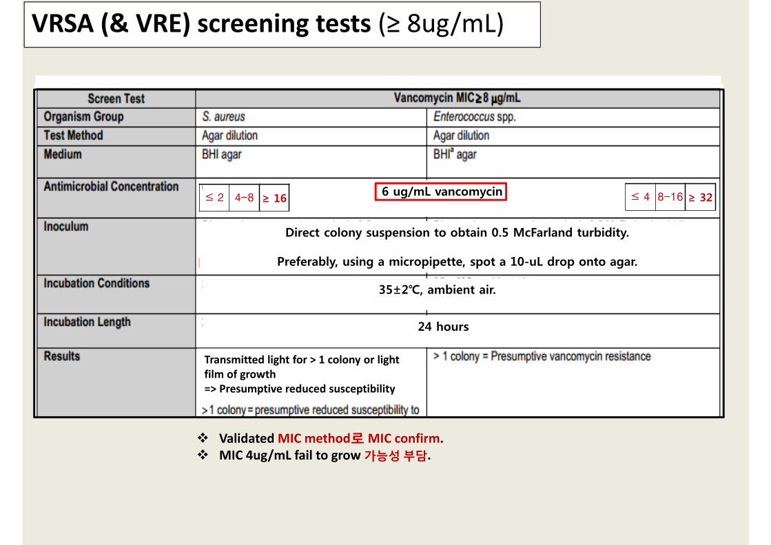
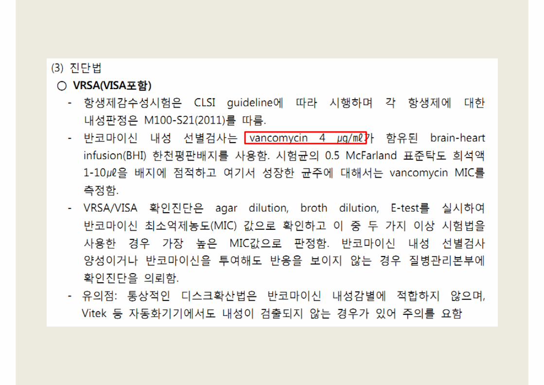
VRSA (& VRE) screening tests (≥ 8ug/mL)
Validated MIC method로 MIC confirm. MIC 4ug/mL fail to grow 가능성 부담.
Transmitted light for > 1 colony or light film of growth => Presumptive reduced susceptibility
35±2℃, ambient air.
6 ug/mL vancomycin
Direct colony suspension to obtain 0.5 McFarland turbidity.
Preferably, using a micropipette, spot a 10-uL drop onto agar.
24 hours
≤ 2 4-8 ≥ 16 ≤ 4 8-16 ≥ 32
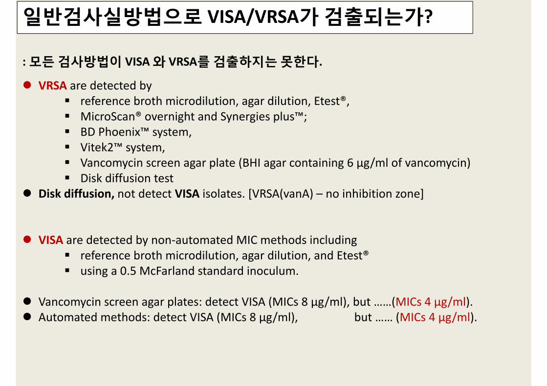
일반검사실방법으로 VISA/VRSA가 검출되는가?
: 모든 검사방법이 VISA 와 VRSA를 검출하지는 못한다.
VRSA are detected by reference broth microdilution, agar dilution, Etest®, MicroScan® overnight and Synergies plus™; BD Phoenix™ system, Vitek2™ system, Vancomycin screen agar plate (BHI agar containing 6 µg/ml of vancomycin) Disk diffusion test
Disk diffusion, not detect VISA isolates. [VRSA(vanA) – no inhibition zone]
VISA are detected by non‐automated MIC methods including reference broth microdilution, agar dilution, and Etest® using a 0.5 McFarland standard inoculum.
Vancomycin screen agar plates: detect VISA (MICs 8 µg/ml), but ……(MICs 4 µg/ml). Automated methods: detect VISA (MICs 8 µg/ml), but …… (MICs 4 µg/ml).

After VRSA/VISA (+) screening Verified by repeating a validated MIC method, & the organism identification.
Additional antimicrobial agents:: Clindamycin, daptomycin, linezolid, quinupristin/dalfopristin, rifampin, trimetho‐sulfa.
To stock: Minimum repeated subcultures
All VRSA = MRSA Most VISA = MRSA
가능하면, Vancomycin agar screen plate를 적용
all MRSA useful for detecting VISA (MIC = 8 µg/ml)

CNS with Decreased Susceptibility to the Glycopeptides
CoNS 중요성 difficult to determine the significance, increasing numbers of healthcare‐associated infections
invasive devices(catheters…)impaired host defenses
CoNS 의 Decreased Susceptibility to Glycopeptides 중요성 Limited treatment options. Detection can be difficult.
heteroresistance.
Identifying CoNS to species level about 20 CoNS species (S. haemolyticus and S. epidermidis) some species are more resistant outbreak recognition & tracking resistance trends
Can routine susceptibility tests detect these isolates? Not all methods. Should be reported to infection control? Yes!

VRE : Enterococci 에서 2 가지 Vancomycin 내성양상
Intrinsic resistance Enterococcus gallinarum, E. casseliflavus/E. flavescens low‐level resistance vanC (intrinsic) low vancomycin MICs of 2 to 16 µg/ml, S to teicoplanin
Acquired resistance. E. faecium, E. faecalis vanA, vanB, vanC, vanD, vanE그외 E. raffinosus, E. avium, E. durans, 기타 enterococcal species
vanA high vancomycin (>128 µg/ml) & teicoplanin (≥16 µg/ml) vanB lower vancomycin (MICs 16 to 64 µg/ml), S to teicoplanin (MICs ≤1 µg/ml). vanD, vanE moderate vancomycin (MICs 64 to 128) & teicoplanin (MICs 4‐8 µg/ml)
EFA (%) EFM (%)587 428
GlycopeptidesTeicoplanin 1.4 23.4Vancomycin 1.7 23.4

VRE: Enterococcus species 수준까지 동정?
Aids in confirming intrinsic (vanC) or acquired resistance (vanA or vanB).
감염관리에서 중요성 vanA and vanB : transferable, spread from organism to organism. vanC genes : not transferable, less serious infections, no outbreaks.
감별: Motility, Yellow pigment, Susceptibility profile E. faecium, E. faecalis : non‐motile, no pigment, E. casseliflavus/E. flavescens : motile, yellow E. Gallinarum : motile, no pigment

For VRE detection, a full 24hr incubation needed. Transmitted light: a haze or any growth in inhibition zone => confirm MIC MIC 8‐16ug/mL: species identification
Interpretive Standards for Vancomycin for Enterococci
Test/Report Group
Antimicrobial Agent
Disk Content
Zone diameter Interpretive Criteria (nearest whole mm)
MIC Interpretive Criteria
(ug/mL)
S I R S I R
B Vancomycin 30ug ≥ 17 15-16 ≤ 14 ≤ 4 8-16 ≥ 32
Inv. Teicoplanin 30ug ≥ 14 11-13 ≤ 10 ≤ 8 16 ≥ 32
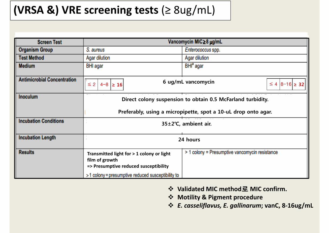
Validated MIC method로 MIC confirm. Motility & Pigment procedure E. casseliflavus, E. gallinarum; vanC, 8‐16ug/mL
(VRSA &) VRE screening tests (≥ 8ug/mL)
Transmitted light for > 1 colony or light film of growth => Presumptive reduced susceptibility
35±2℃, ambient air.
6 ug/mL vancomycin
Direct colony suspension to obtain 0.5 McFarland turbidity.
Preferably, using a micropipette, spot a 10-uL drop onto agar.
24 hours
≤ 2 4-8 ≥ 16 ≤ 4 8-16 ≥ 32

Gram(-) bacilli
ESBL CRE MRPA, MRAB

ESBLs (extended spectrum b‐lactamase)
ESBLs (extended‐spectrum β‐lactamases) destroy extended‐spectrum cephalosporins (G3: ceftazidime, cefotaxime, ceftriaxone),
Narrow spectrum/broad spectrum b‐lactams (Penicillin, Ampicillin, 1st G cephalosporins)monobactams (aztreonam) not cephamycins (cefoxitin, cefotetan) not carbapenems (meropenem,imipenem)
Isolates other than K. pneumoniae, K. oxytoca, E. coli or P. mirabilis produce ESBLs? Yes. Other isolates of Enterobacteriaceae (Enterobacter spp, Morganella morganii, Serratia marcescens, Salmonella spp), P. aeruginosa
ECO (%) KPN (%)1,326 657
R R3rd Cephalosporins
Cefotaxime 29 34.4Ceftazidime 28.3 35.9Cefepime 26 32.2Aztreonam 27 34.3
E.C KL.P
No. 1,326 657
ESBL(+) 26.3 31.2
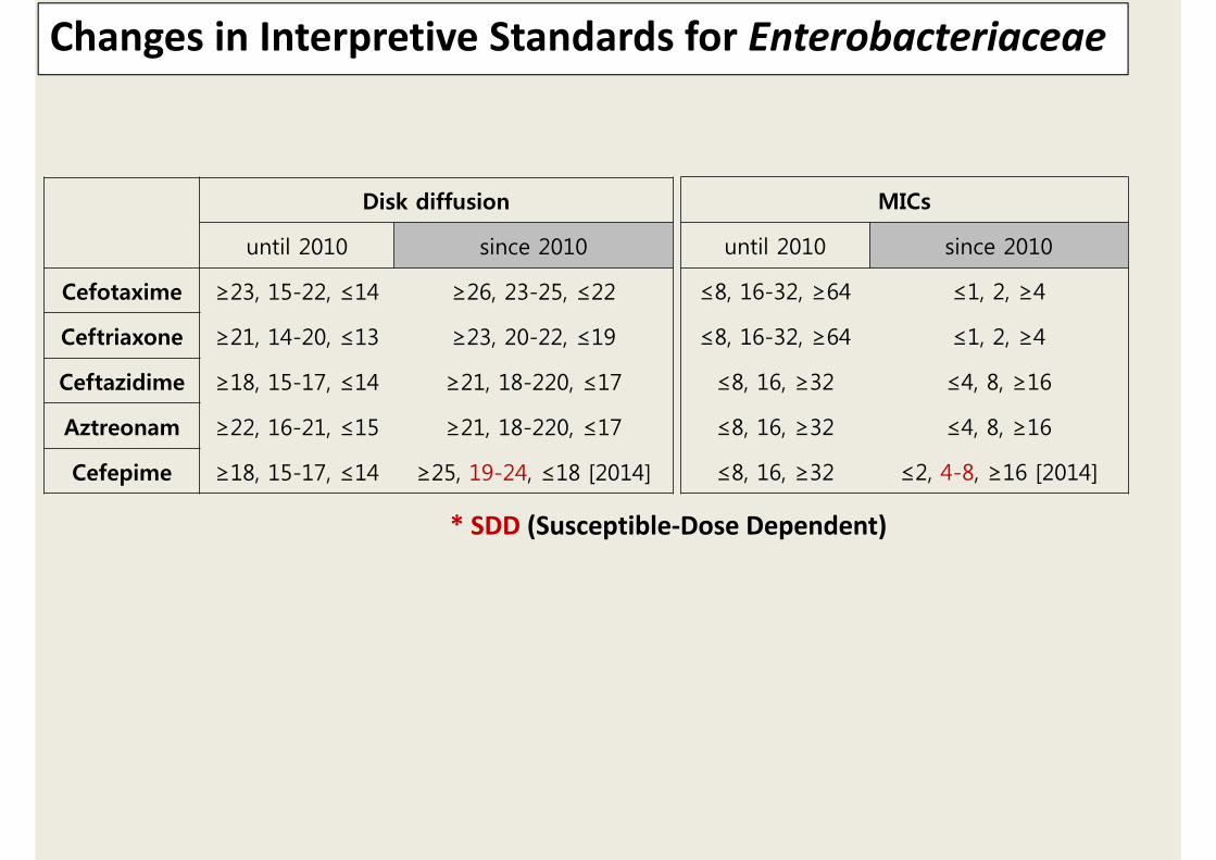
Changes in Interpretive Standards for Enterobacteriaceae
Disk diffusion
until 2010 since 2010
Cefotaxime ≥23, 15-22, ≤14 ≥26, 23-25, ≤22
Ceftriaxone ≥21, 14-20, ≤13 ≥23, 20-22, ≤19
Ceftazidime ≥18, 15-17, ≤14 ≥21, 18-220, ≤17
Aztreonam ≥22, 16-21, ≤15 ≥21, 18-220, ≤17
Cefepime ≥18, 15-17, ≤14 ≥25, 19-24, ≤18 [2014]
MICs
until 2010 since 2010
≤8, 16-32, ≥64 ≤1, 2, ≥4
≤8, 16-32, ≥64 ≤1, 2, ≥4
≤8, 16, ≥32 ≤4, 8, ≥16
≤8, 16, ≥32 ≤4, 8, ≥16
≤8, 16, ≥32 ≤2, 4-8, ≥16 [2014]
* SDD (Susceptible‐Dose Dependent)

ESBLs: Standard Breakpoints Did Not Detect Resistance‐ SIEMENS, CLSI AST Update for 2100, by Lisa Walker

Screening & Confirmatory Tests for ESBLs in E. coli, K. pneumoniae, K. oxytoca, P. mirabilis
Confirmatory testing requires use of both cefotaxime & ceftazidime, alone & in combination with clavulate.
Using > 1 antimicrobials for screening improves the sensitivity of ESBL detection.
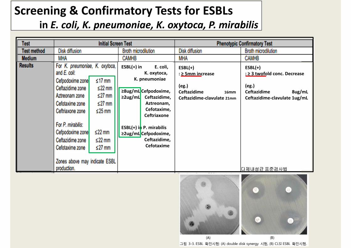
Screening & Confirmatory Tests for ESBLs in E. coli, K. pneumoniae, K. oxytoca, P. mirabilis
ESBL(+) in E. coli, K. oxytoca,
K. pneumoniae
≥8ug/mL Cefpodoxime,≥2ug/mL Ceftazidime,
Aztreonam,Cefotaxime,Ceftriaxone
ESBL(+) in P. mirabilis≥2ug/mL Cefpodoxime,
Ceftazidime, Cefotaxime
ESBL(+): ≥ 5mm increase
(eg.)Ceftazidime 16mmCeftazidime‐clavulate 21mm
ESBL(+): ≥ 3 twofold conc. Decrease
(eg.)Ceftazidime 8ug/mL Ceftazidime‐clavulate 1ug/mL
다제내성균표준검사법

중요성 Treat a variety of serious infections by highly resistant organisms Treat nosocomial and mixed bacterial infections. R Limits therapeutic options, Transmissible
Activity : species dependent. Ertapenem ‐ EnterobacteriaceaeMeropenem ‐ slightly more active than imipenem against G(‐)
Carbapenem: imipenem, meropenem, ertapenem
Drug Entero-bacteriaeae
Non-fermentors Anaerobes
MSSA & Strep spp.
Imipenem + + + +
Meropenem + + + +
Ertapenem + Limited + +
Doripenem + + + +
내성(+) 세균Intrinsic R : Stenotrophomonas maltophilia,
Morganella morgagnii, P. mirabilis, Providentia, Serratia marcescensProteus species, Serratia marcescens, Enterobacter species, Klebsiella pneumoniae. Pseudomonas aeruginosa, Burkholderia cepacia, Acinetobacter species,

Carbapenem 내성기전
Carbapenemase Enzyme Most Common Bacteria
Class A KPC, SME, IMI, NMC, GESEnterobacteriaceae
(rare reports in P. aeruginosa) Class B
IMP, VIM, GIM, SPM, NDMP. aeruginosa
(metallo‐b‐lactamse) EnterobacteriaceaAcinetobacter spp.
Class D OXA Acinetobacter spp.
Enterobacteriaceae Cephalosporinase + porin lossCarbapenemase
P. aeruginosa Porin lossUp‐regulated effluxCarbapenemase
Acinetobacter Cephalosporinase + porin lossCarbapenemase
> 1 enzyme & > 1 porin modification : high MIC (>16 µg/ml). porin changes alone : lower MICs (2‐8 µg/ml). AmpC‐type enzyme or an ESBL, combined with porin loss. Carbapenem resistant vs Carbpenemase producing
Carbapenemase (outbreak, transmissible) vs non‐carbapenemase produsing
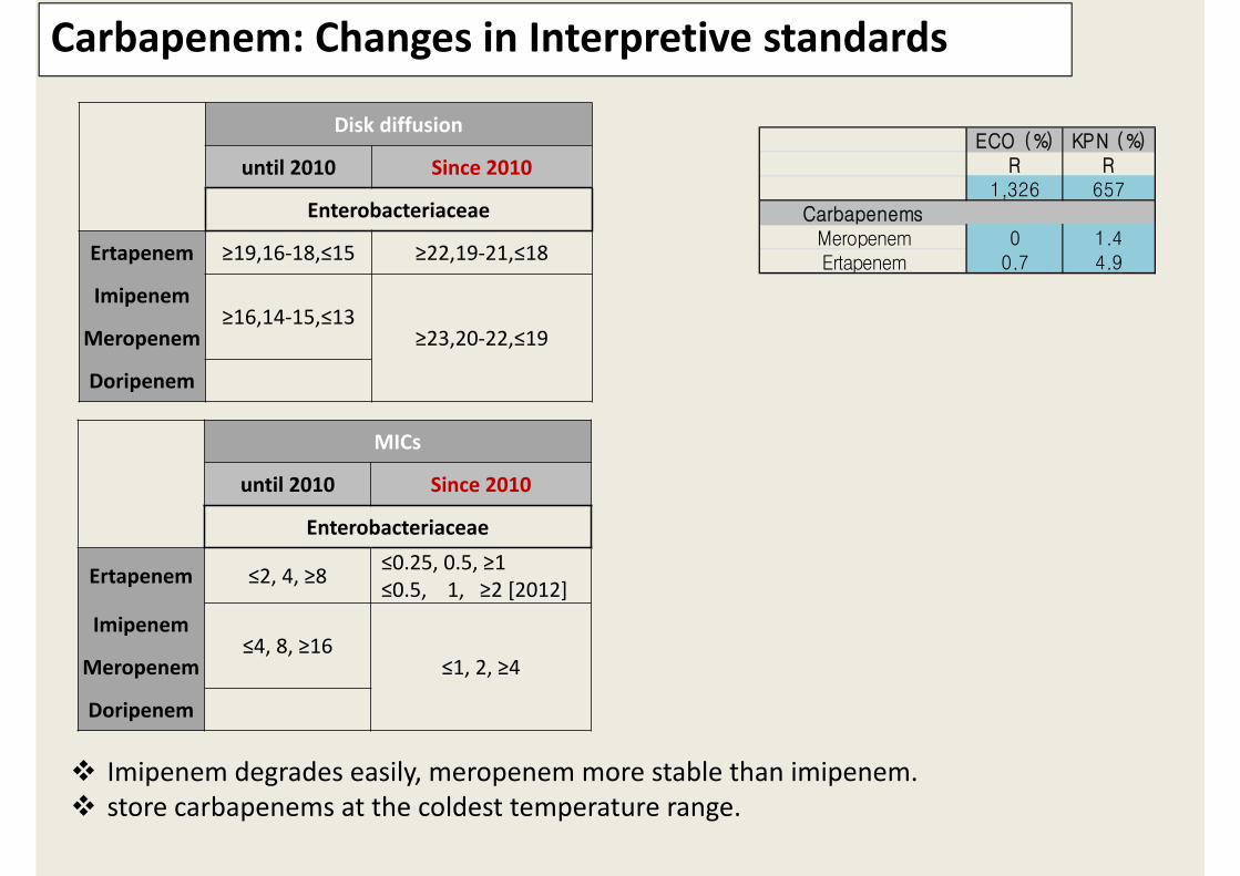
Carbapenem: Changes in Interpretive standards
Disk diffusion
until 2010 Since 2010
Enterobacteriaceae
Ertapenem ≥19,16‐18,≤15 ≥22,19‐21,≤18
Imipenem≥16,14‐15,≤13
≥23,20‐22,≤19Meropenem
Doripenem
MICs
until 2010 Since 2010
Enterobacteriaceae
Ertapenem ≤2, 4, ≥8 ≤0.25, 0.5, ≥1≤0.5, 1, ≥2 [2012]
Imipenem≤4, 8, ≥16
≤1, 2, ≥4Meropenem
Doripenem
ECO (%) KPN (%)R R
1,326 657Carbapenems
Meropenem 0 1.4Ertapenem 0.7 4.9
Imipenem degrades easily, meropenem more stable than imipenem. store carbapenems at the coldest temperature range.
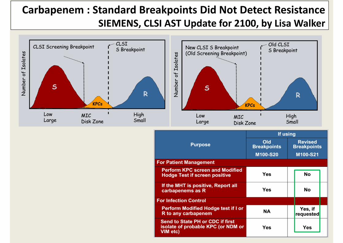
Carbapenem : Standard Breakpoints Did Not Detect ResistanceSIEMENS, CLSI AST Update for 2100, by Lisa Walker
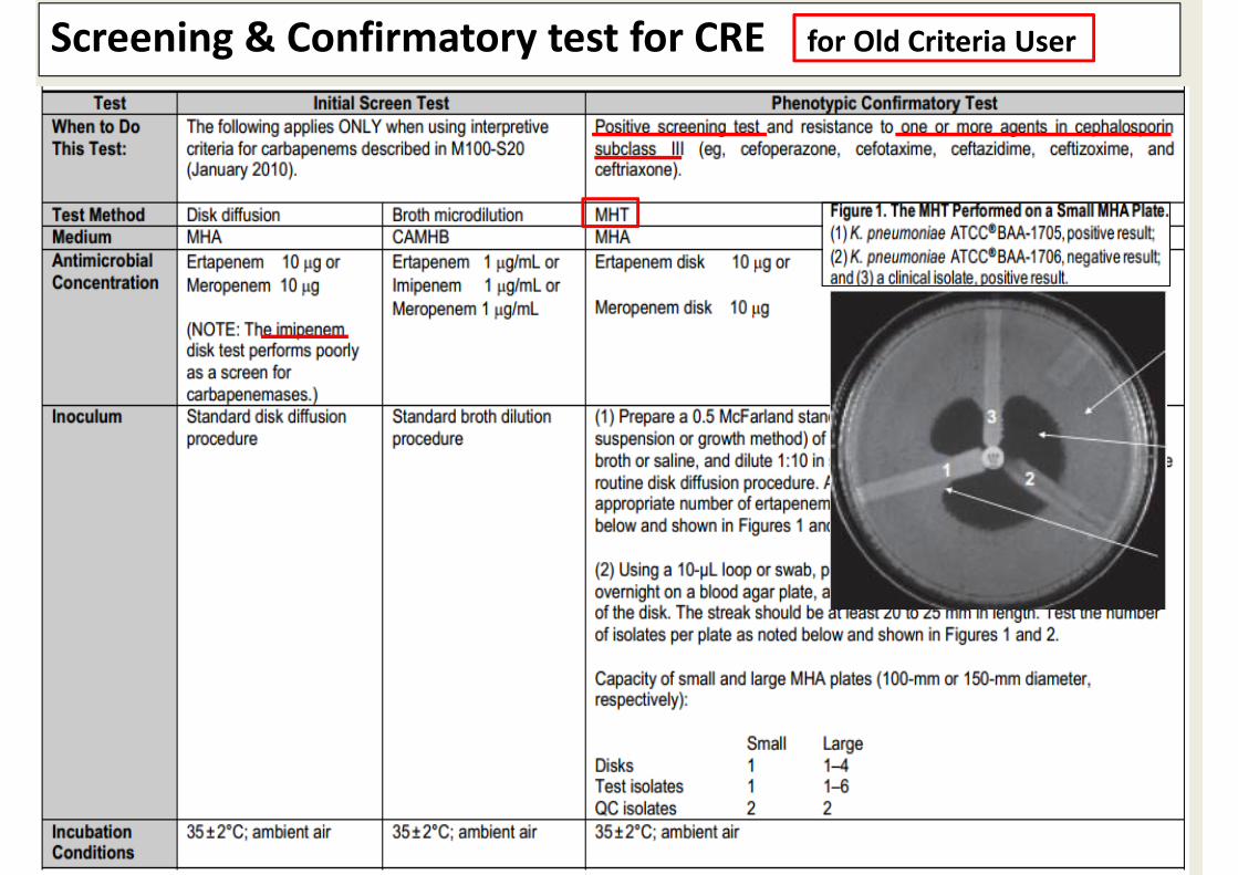
Screening & Confirmatory test for CRE for Old Criteria User

Screening & Confirmatory test for CRE ‐ continued
Not all carbapenemase‐producing Enterobacteriaceae are MHT(+) – other resistance mechanisms.
Old criteria user‐MHT(+), all carbapenem R; MHT(‐), according to criteria.
New criteria user‐ Rt confirmatory test만 infection control & epidemiological investigation 목적으로 사용.‐ 역학조사대상 Carbapenemase producing isolates: 1 or more carbapenems에 I or R,
1 or more cephalosporin subclass III에 I or R.

Carbapenemase 검출시험
Modified Hodge test
Carbapenemase inhibition test : 내성기전 감별 ROSCO confirmative tabletsMBL E‐test (imipenem or meropenem ± EDTA)
Carba NP test: Patrice N et al, 2012 Emerg Inf Ds Rapid detection of …. 37°C, 2hr sensitivity, specificity (100%)
PCR (real‐time PCR), Sequencing
2012 JCM Diana D et al JCM Lab detection of Enterobacteriaceae that produce carbapenemase

P. Aeruginosa (MRPA), Acinetobacter baumannii(MRAB)
PAE (%) ABA (%)585 541R R
CarbapenemsImipenem 31.7 56.0Meropenem 23.8 53.6
AminoglycosidesGentamicin 20.3 51.1Tobramycin 51.6Amikacin 11.8 41.2
QuinolonesCiprofloxacin 34.4 59.0Levofloxacin 35.6 51.7
Disk diffusion
until 2010 Since 2010
PAE,ABA PAE,ABA
Ertapenem
Imipenem≥16,14‐15,≤13
≥19,16‐18,≤15 PAE 2012Im ≥22,19‐21,≤18 AB 2014Me ≥18,15‐17,≤14 AB 2014Do ≥18,15‐17,≤14 AB 2014
Meropenem
Doripenem
MICs
until 2010 Since 2010
PAE,ABA PAE,ABA
Ertapenem
Imipenem≤4, 8, ≥16
≤2, 4, ≥8Meropenem
Doripenem
Changes in Carbapenem Interpretive standards

실제 어떤 방법으로 내성을 검사할 것인가?


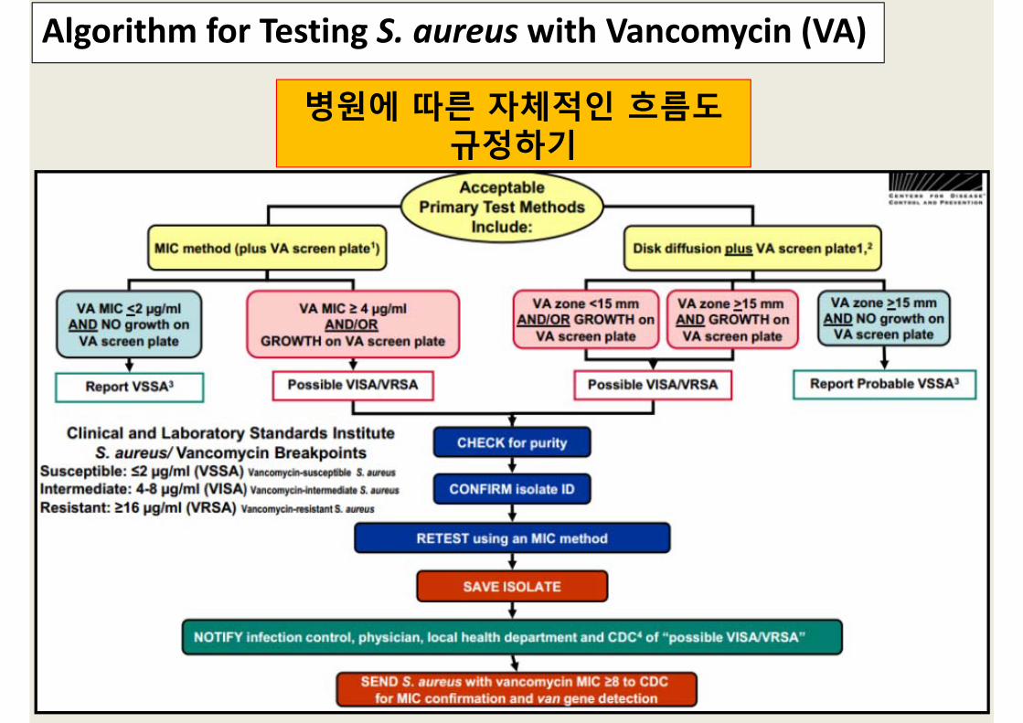
Algorithm for Testing S. aureus with Vancomycin (VA)
병원에 따른 자체적인 흐름도규정하기

실제 Outbreak에 어떻게 대처할 것인가?
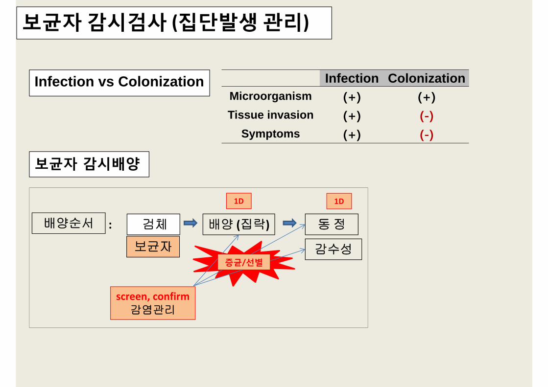
보균자 감시검사 (집단발생 관리)
Infection vs Colonization Infection ColonizationMicroorganism (+) (+)Tissue invasion (+) (-)
Symptoms (+) (-)
보균자 감시배양
배양 (집락) 동정
감수성
배양순서 검체
screen, confirm 감염관리
:
1D 1D
증균/선별
보균자

질병발생 (Concept of Disease Occurrence)

다제내성균 선별검사의 시행과 격리에 대한 알고리즘–질병관리본부 2012.10.
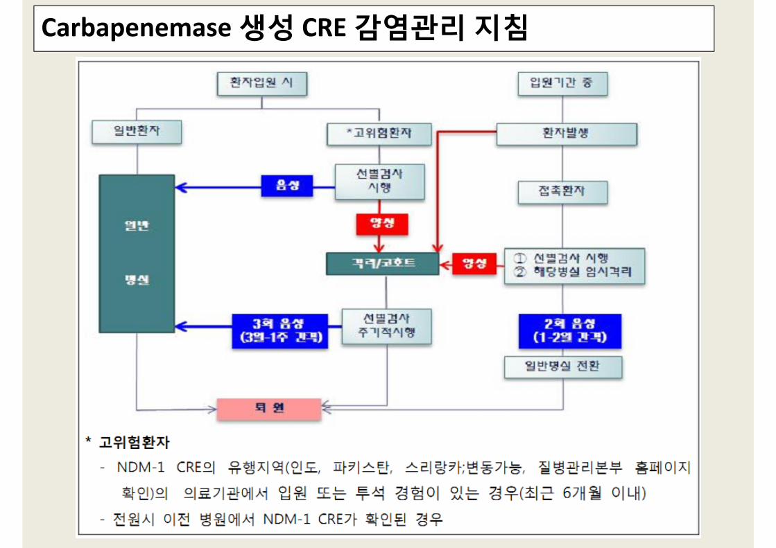
Carbapenemase 생성 CRE 감염관리 지침

Carbapenemase 생성 CRE 감염관리 지침
1. 과거 입원(3개월 이내) 당시 균이 분리되었던 사실이 확인되는 경우에는
즉시 격리조치를 취하고 선별검사(1‐2일 간격)를 실시
vs 가능한 격리조치를 취하고
2. 능동감시대상 (선별검사 대상) 지정
3. 집단감염발생 시 관리

Carbapenemase 생성 CRE에 대한 검사실내 감시지침

다제내성균별 감염관리
과거 입원(3개월 이내)에서 균이 분리되어 선제 격리된 환자는 감시배양에서 2-3회 음성(1-2일 간격)이면 격리 해제
환자의 퇴원여부에 대해서는 임상판단에 따르며, 다제내성균의 보균상태로 인해 퇴원을 연기할 근거는 없음. 다만 퇴원 시 접촉주의지침에 대한 교육을 시행하고, 타 의료시설로 전원할 경우 전원 대상시설에 다제내성균에 관한정보 제공.
CRE

CRE/카바페넴분해효소 생성 CRE 감시배양 질병관리본부
CDC CRE screening test 2008 Dec.

Bile‐esculin broth
(VAN. 6µg/ml)
ChromID‐VREE. FaecalisE. faecium
ModificationVancomycin R Enterococcus
Rectal swabStool
배양
Multiplex PCR vanA, vanB,
vanC
Any Specimen
동정, 감수성
보균자환 자

정의: Culture media for the simple and fast detection of bacteria The chromogenic mixture contains chromogenic substrates Certain enzymes, produced by some bacteria, cleave substrates ‐> coloration of colonies.
Chromogenic agar
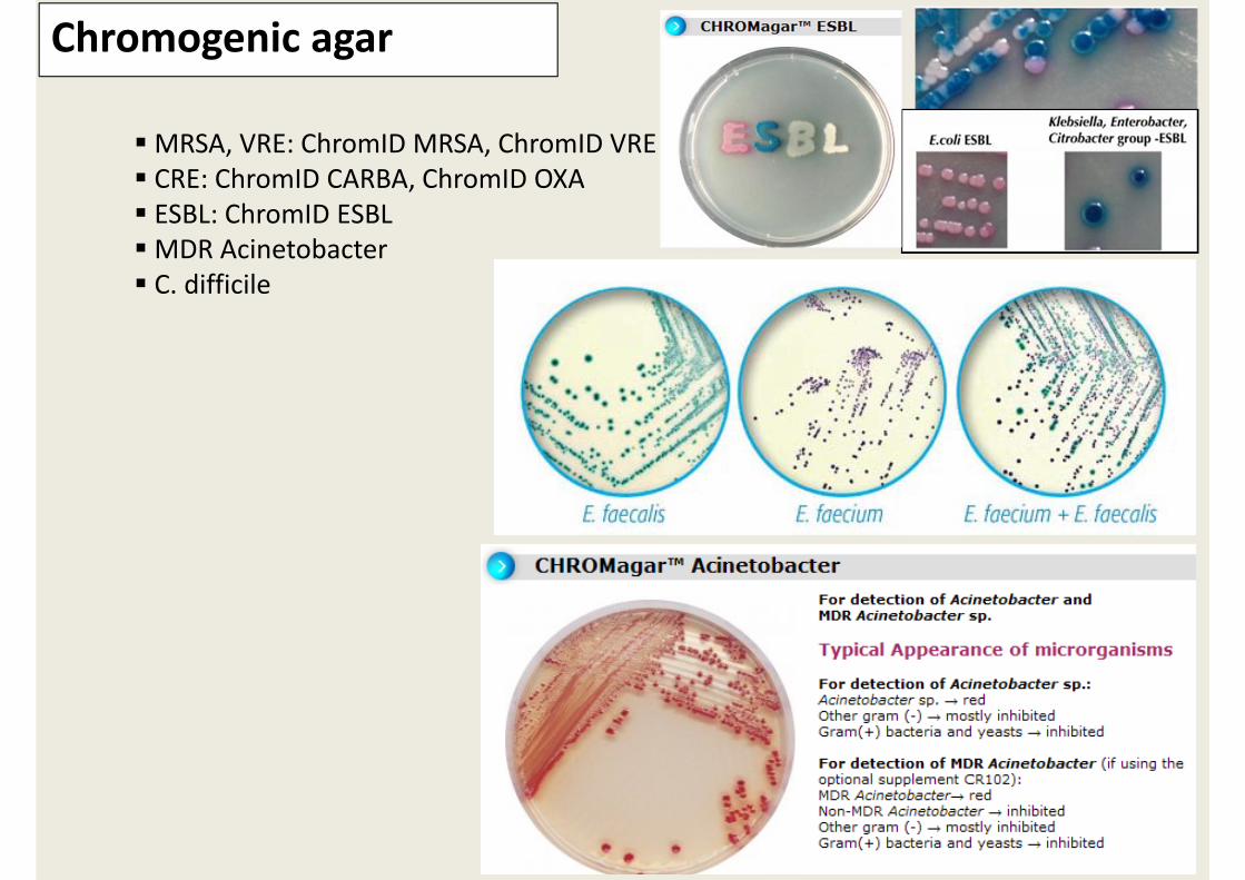
MRSA, VRE: ChromID MRSA, ChromID VRE CRE: ChromID CARBA, ChromID OXA ESBL: ChromID ESBL MDR Acinetobacter C. difficile
Chromogenic agar

마무리?

세균 동정과 내성 검사방법
각 검사실에서 사용하는 방법 표현형검사: 생화학적방법, 항원검사 정확한 검사방법과 한계 숙지
내성(+) 세균검출순수 집락분리재검 ‐ 표준방법의 준수검사실에서 가능한 방법으로 내성 확인균주보관 및 질병관리본부로 이송 준비
감염관리위원회에 통보병원차원에서의 준비 – 검사방법에 대한 제안격리 및 주의 환기전파방지를 위한 조치감시배양과 격리해제를 위한 배양

• MDRO 정의 및 감수성 기준설정 이해
• 감수성 screen & confirm 방법 이해
• 목표설정
• 병원환경과 현실적인 기대 수준 설정
• 실현가능한 형태로 수정 및 보완
요 약