mycobacterium tuberculosis
TRANSCRIPT

MYCOBACTERIUM TUBERCULOSIS
Deepshikha ChhetriMSc. FSN

INTRODUCTION: Mycobacteria is an obligate aerobe growing most
successfully in tissues with a high oxygen content.(the upper lobe of the lung and the kidney).
Its cell wall contains several complex lipids ( long-chain fatty acids called mycolic acids).
It is relatively resistant to acids and alkalis. NaOH is used to concentrate clinical specimens; it destroys unwanted bacteria, human cells, and mucus but not the organism.
They are Facultative intracellular pathogens usually infecting mononuclear phagocytes (e.g. macrophages).

MORPHOLOGY: SHAPE - long, slender, straight or slightly curved rod. SIZE - 3 x 0.3 µm Intracellular. Mycolic acid, waxes & lipids are present in
cell wall The high lipid content (approximately 60%)
of their cell wall makes mycobacteria acid-fast and hydrophobic.
Because the cells are hydrophobic and tend to clump together, they are impermeable to the usual stains, e.g. Gram's stain
Neither gram-positive nor gram-negative.
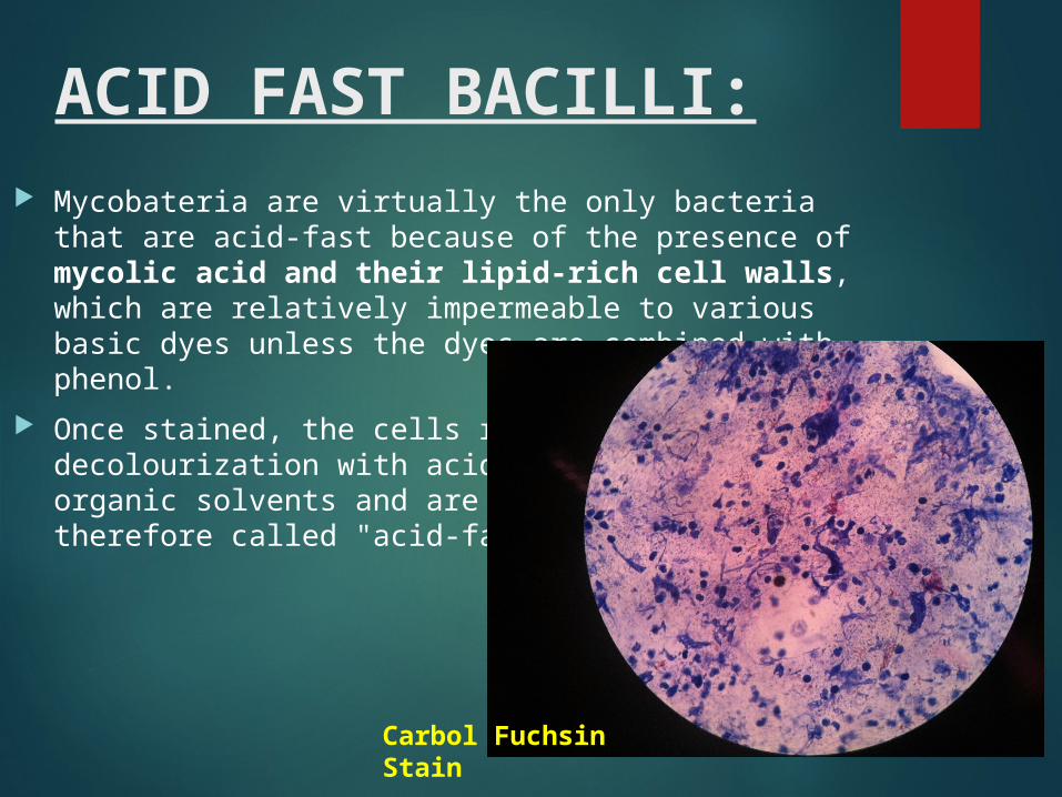
ACID FAST BACILLI: Mycobateria are virtually the only bacteria that are
acid-fast because of the presence of mycolic acid and their lipid-rich cell walls, which are relatively impermeable to various basic dyes unless the dyes are combined with phenol.
Once stained, the cells resist decolourization with acidified organic solvents and are therefore called "acid-fast".
Carbol Fuchsin Stain
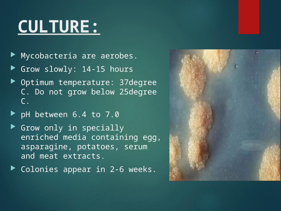
CULTURE: Mycobacteria are aerobes. Grow slowly: 14-15 hours Optimum temperature: 37degree C.
Do not grow below 25degree C. pH between 6.4 to 7.0 Grow only in specially enriched
media containing egg, asparagine, potatoes, serum and meat extracts.
Colonies appear in 2-6 weeks.
RESISTANCE: Mycobateria can survive in dust for several months in
sputum for 20-30 hours. Killed at temperature of 60degree C for 20 minutes
and instantly at 100degree C. Sensitive to ultraviolet ray and sunlight.

TAXONOMY

Species Growth on Bacteriologic Media
Preferred Temperature in Vivo (˚C)
Source or Mode of Transmission
M. tuberculosis Slow (weeks) 37 Respiratory droplets
M. bovis Slow (weeks) 37 Milk from infected animals
M. leprae None 32 Prolonged close contact
Atypical mycobacteria (ex. M. kansasii)
Slow (weeks) 37 Soil and water
M. marinum Slow (weeks) 32 Water
M. avium-intracellulare complex
Slow (weeks) 37 Soil and water
M. fortuitum-chelonei complex Rapid (days) 37 Soil and water

Diseases caused by Mycobacterium species: M. leprae - leprosy M. avium - lung and skin infections in immunocompromised
hosts - lymphadenopathy in children - catheter-related infections
M. bovis - Primarily infection among the cattle . It infects Tonsils, Cervical nodes. Enter through Intestines – infects the ileocecal
region.

MYCOBACTERIUM TUBERULOSIS It causes TUBERCULOSIS which is the most common cause
of death due to bacterial infection worldwide. It is closely related to M. bovis. and it is believed to have
evolved from M. bovis after domestication of cattle (8,000-4,000 BC)
Archeologists have shown that tuberculosis of the bones seems to have preceded the disease of the lungs.
M. tuberculosis is resistant to dehydration and so survives in dried expectorated sputum; this property may be important in its transmission by aerosol.

Mycobacterium tuberculosis under Electron Micrograph:
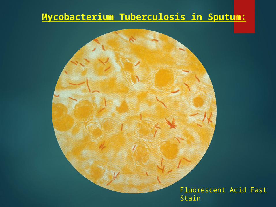
Mycobacterium Tuberculosis in Sputum:
Fluorescent Acid Fast Stain
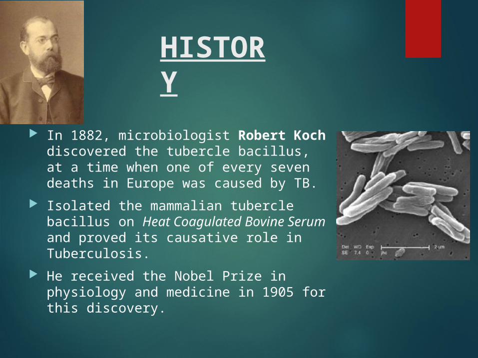
HISTORY
In 1882, microbiologist Robert Koch discovered the tubercle bacillus, at a time when one of every seven deaths in Europe was caused by TB.
Isolated the mammalian tubercle bacillus on Heat Coagulated Bovine Serum and proved its causative role in Tuberculosis.
He received the Nobel Prize in physiology and medicine in 1905 for this discovery.
TRANSMISSION AND EPIDEMIOLOGY
M. tuberculosis is transmitted from person to person by respiratory aerosol, and its initial site of infection is the lung. In the body, it resides within macrophages.
In developed countries, tuberculosis is almost exclusively a human disease, and most tuberculosis is due to reactivation in elderly, malnourished men.
In developing countries, M. bovis is found in cow's milk, which, unless pasteurized, can cause gastrointestinal tuberculosis in humans.
The risk of infection and disease is highest among socio-economically disadvantaged people, who have poor housing and poor nutrition.
Coughing projects droplet nuclei into the air that contain tubercle bacilli. One cough can release 3,000 droplet nuclei. One sneeze can release tens of thousands of droplet nuclei.

Pre-Disposing Factors: Genetic basis, Age Stress, Nutrition, Co existing infections Eg. HIV. HIV association will lead to rapid
spread of tuberculosis. HIV kills CD4+ T Helper cells which normally inhibit M. tuberculosis.
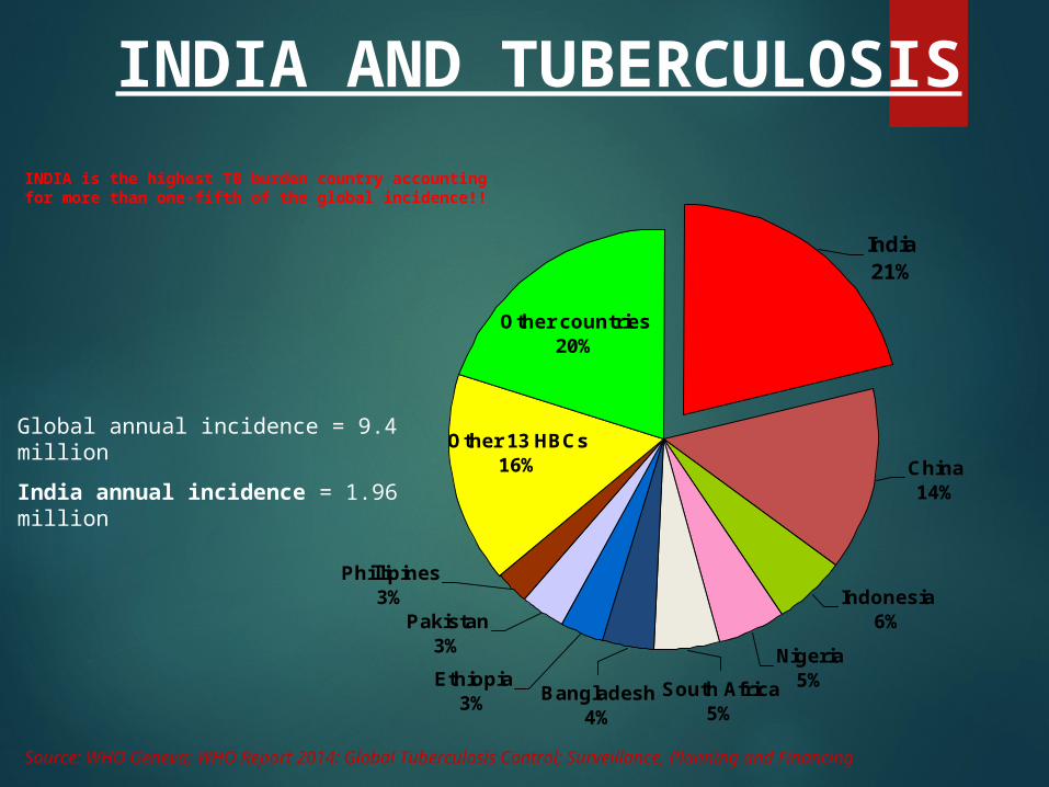
INDIA is the highest TB burden country accounting for more than one-fifth of the global incidence!!
Indonesia6%
Nigeria5%
Other countries20%
Other 13 HBCs16% China
14%
South Africa5%
Bangladesh4%
Ethiopia3%
Pakistan3%
Phillipines3%
India21%
Source: WHO Geneva; WHO Report 2014: Global Tuberculosis Control; Surveillance, Planning and Financing
Global annual incidence = 9.4 millionIndia annual incidence = 1.96 million
INDIA AND TUBERCULOSIS
Estimated incidence 1.96 million new cases annually
Estimated mortality 330,000 deaths due to TB each year Over 1000 deaths a day
2 deaths every 3 minutes Someone in the world is newly infected with TB bacilli every
second. Overall, one-third of the world's population is currently infected
with the TB bacillus. 5-10% of people who are infected with TB bacilli (but who are
not infected with HIV) become sick or infectious at some time during their life.
PATHOGENESIS
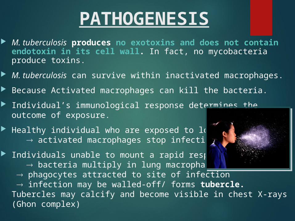
M. tuberculosis produces no exotoxins and does not contain endotoxin in its cell wall. In fact, no mycobacteria produce toxins.
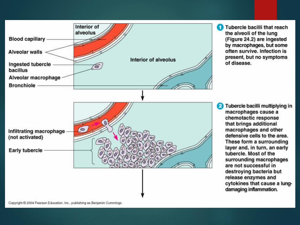
M. tuberculosis can survive within inactivated macrophages. Because Activated macrophages can kill the bacteria. Individual’s immunological response determines the outcome of
exposure. Healthy individual who are exposed to low dose
activated macrophages stop infection. Individuals unable to mount a rapid response
bacteria multiply in lung macrophages. phagocytes attracted to site of infection infection may be walled-off/ forms tubercle.Tubercles may calcify and become visible in chest X-rays(Ghon complex)
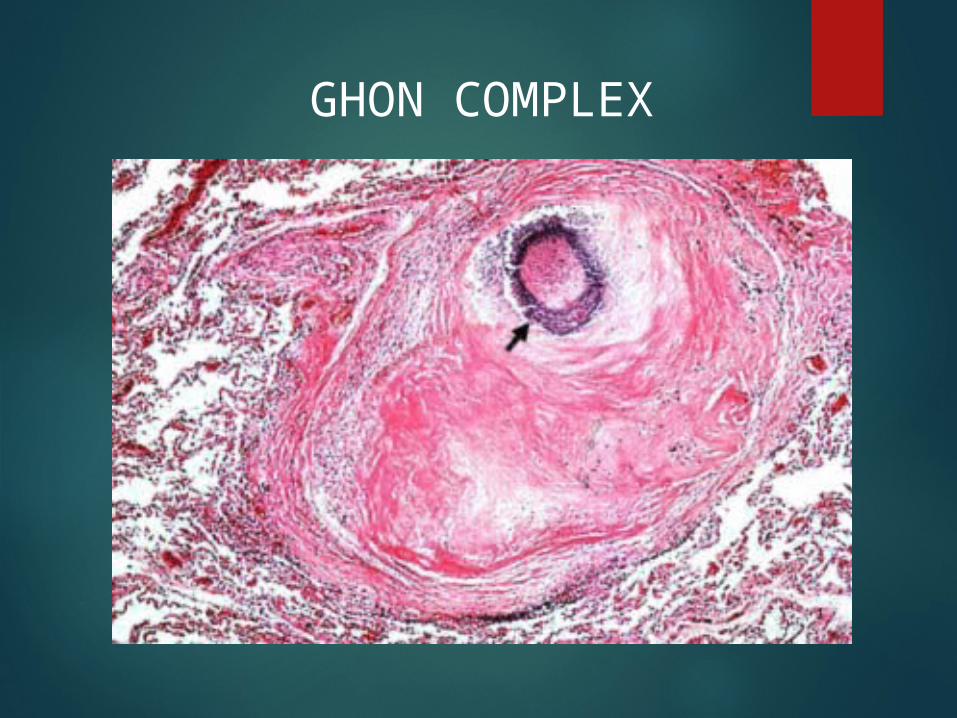
GHON COMPLEX
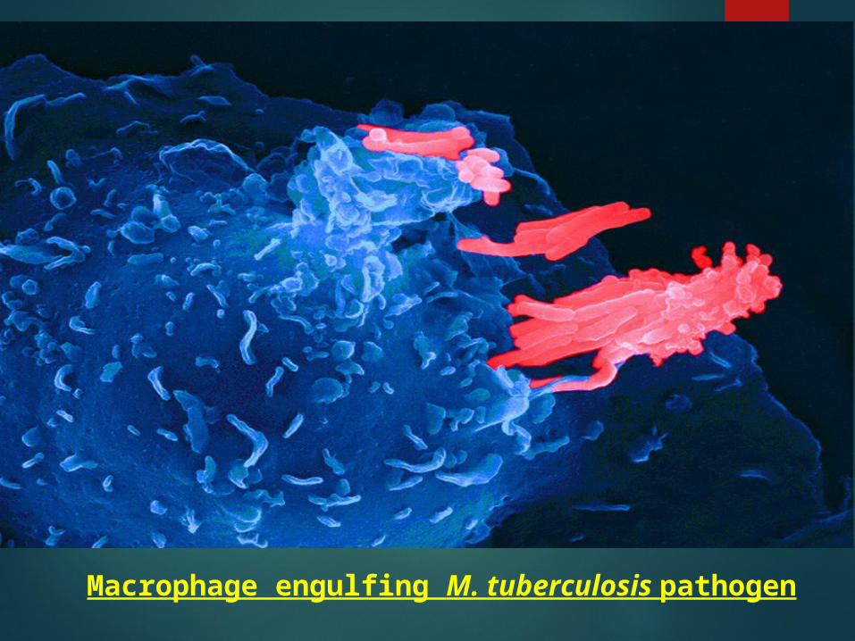
Macrophage engulfing M. tuberculosis pathogen
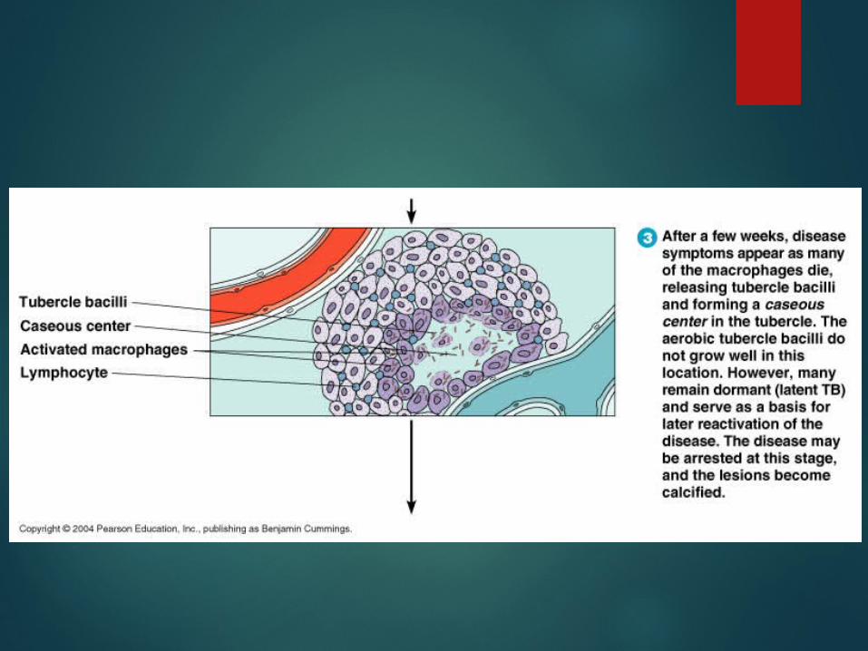


Bacteria in tubercles may survive for decades (latency).
Suppression of immune system may allow bacteria to break out of lesions and multiply (reactivation).
Old age, cancer, immunosuppressive drugs and HIV infection can lead to reactivation.
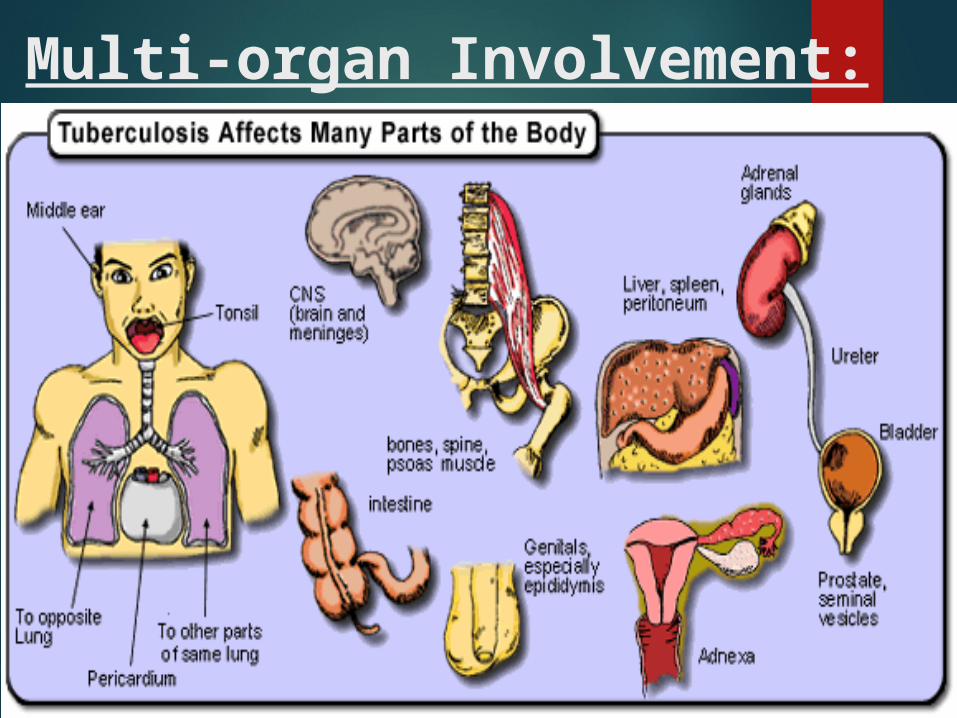
Multi-organ Involvement:
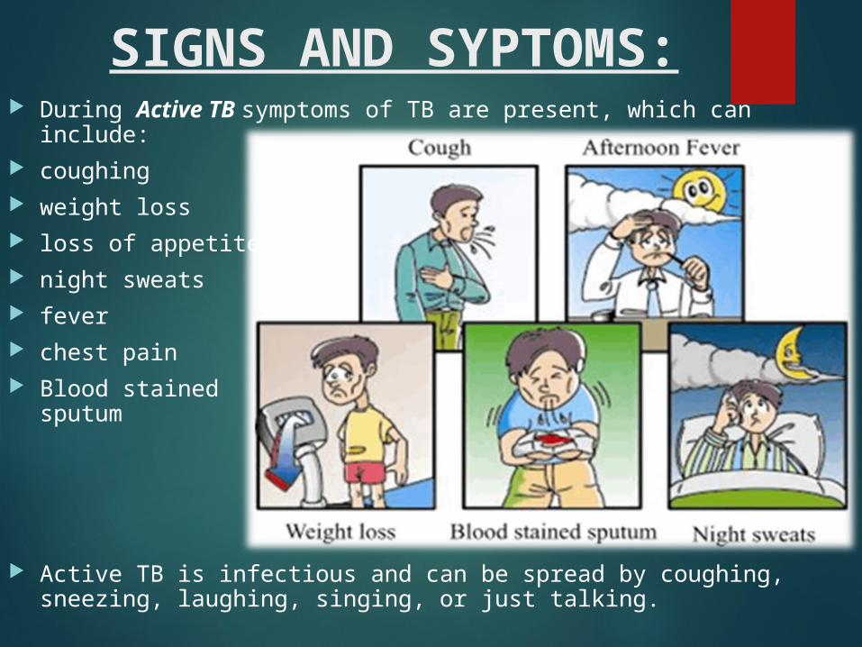
SIGNS AND SYPTOMS: During Active TB symptoms of TB are present, which can
include: coughing weight loss loss of appetite night sweats fever chest pain Blood stained
sputum
Active TB is infectious and can be spread by coughing, sneezing, laughing, singing, or just talking.

M. tuberculosis does not spread by:
Sharing dishes and utensils
Using towels and linens
Handling food
Sharing cell phones
Touching computer keyboard
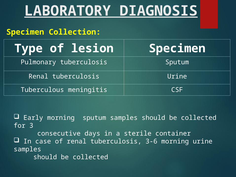
Early morning sputum samples should be collected for 3 consecutive days in a sterile container In case of renal tuberculosis, 3-6 morning urine samples should be collected
Type of lesion SpecimenPulmonary tuberculosis Sputum
Renal tuberculosis UrineTuberculous meningitis CSF
LABORATORY DIAGNOSISSpecimen Collection:

DIAGNOSTIC METHODS
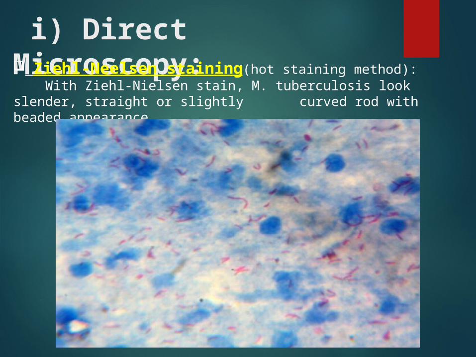
i) Direct Microscopy: Ziehl-Neelsen staining(hot staining method): With Ziehl-Nielsen stain, M. tuberculosis look slender, straight or slightly curved rod with beaded appearance.
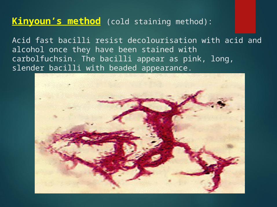
Kinyoun’s method (cold staining method):
Acid fast bacilli resist decolourisation with acid and alcohol once they have been stained with carbolfuchsin. The bacilli appear as pink, long, slender bacilli with beaded appearance.

Fluorescent staining by Auramine O or auramine rhodamine: Mycobacterium spp. will fluoresce yellow against dark
background under fluorescent microscope.

ii) Culture: Concentrated specimen is generally inoculated on Lowenstein – Jensen’s
medium (solid medium) and incubated at 370C for 2 – 8 weeks. Lowenstein – Jensen’s medium contains coagulated egg, Mineral salt
solution, Asparagine's, Malachite green, Agar Colonies appear as buff coloured, dry, irregular colonies with wrinkled
surface and not easily emulsifiable (Buff, rough and tough colonies). Colonies are creamy white to yellow colour with smooth surface.
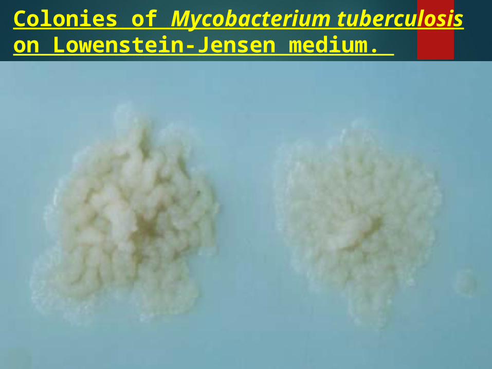
Colonies of Mycobacterium tuberculosis on Lowenstein-Jensen medium.
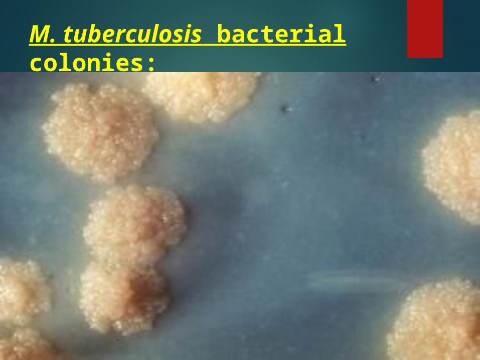
M. tuberculosis bacterial colonies:
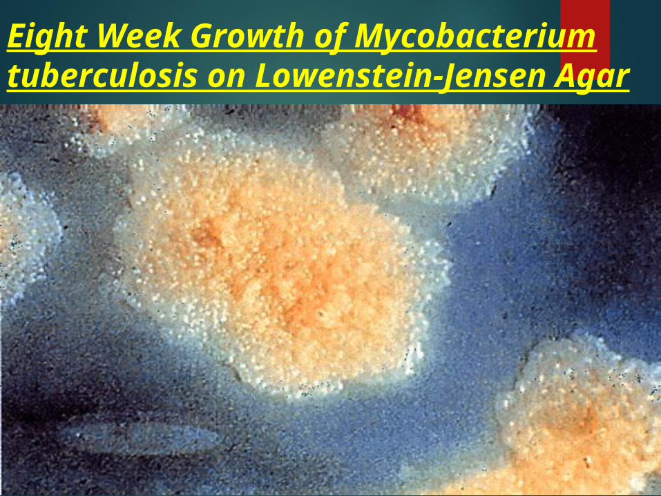
Eight Week Growth of Mycobacterium tuberculosis on Lowenstein-Jensen Agar
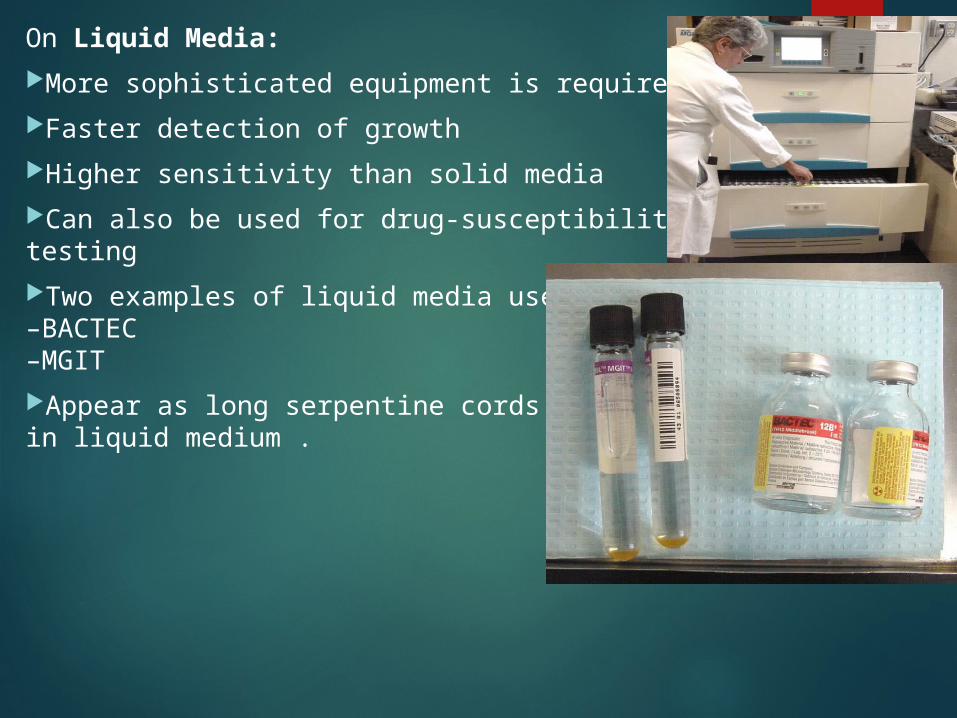
On Liquid Media:More sophisticated equipment is required Faster detection of growth Higher sensitivity than solid media Can also be used for drug-susceptibility testing Two examples of liquid media used: –BACTEC –MGIT Appear as long serpentine cordsin liquid medium .
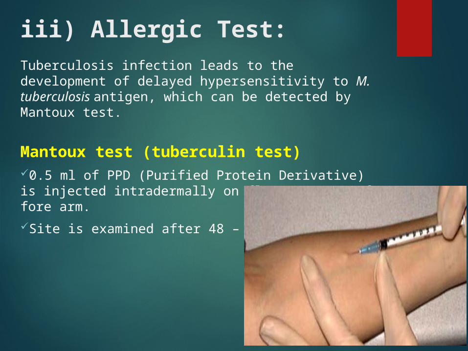
iii) Allergic Test:Tuberculosis infection leads to the development of delayed hypersensitivity to M. tuberculosis antigen, which can be detected by Mantoux test.
Mantoux test (tuberculin test)0.5 ml of PPD (Purified Protein Derivative) is injected intradermally on flexor aspect of fore arm.Site is examined after 48 – 72 hrs.

Induration of 10 mm or more is considered positive. positive test leads to red area at injection site
iv) Detection of Antibodies: Various methods such as enzyme linked immunosorbent
assay (ELISA), radio immunoassay (RIA), latex agglutination assay have been employed for detection of antibodies in patient serum.
However, diagnostic utility of these methods is doubtful.
WHO has recommended that these tests should not be used for diagnosis of active tuberculosis.

v) Biochemical Reactions: Niacin test: M. tuberculosis lacks the enzyme that converts Niacin
to Niacin ribonucleotide due to this large amount of Niacin accumulates in the culture medium.
Niacin is detected by addition of 10% cyanogen bromide and 4% aniline in 96% ethanol.
Positive reaction – canary yellow M. tuberculosis – Positive M. bovis - Negative
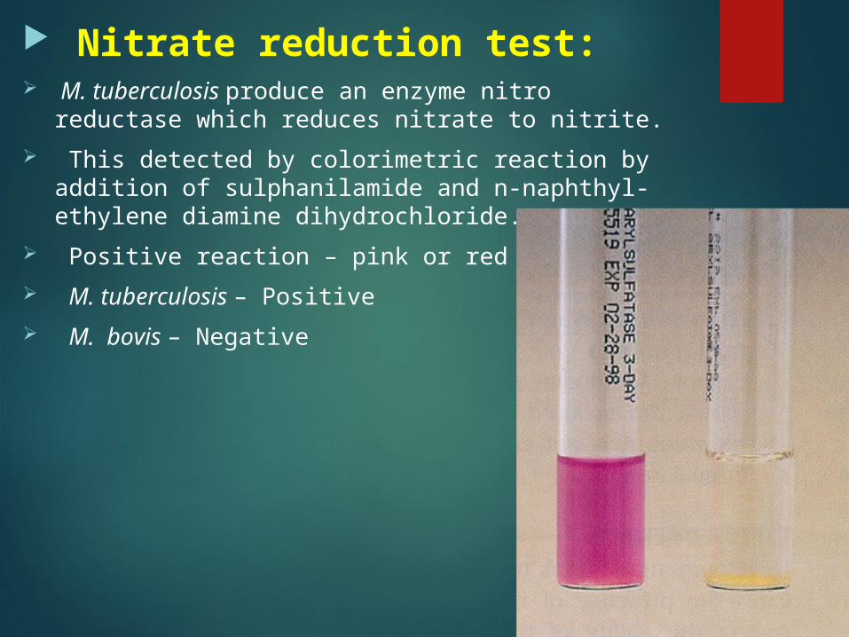
Nitrate reduction test: M. tuberculosis produce an enzyme nitro reductase
which reduces nitrate to nitrite. This detected by colorimetric reaction by addition of
sulphanilamide and n-naphthyl- ethylene diamine dihydrochloride.
Positive reaction – pink or red colour M. tuberculosis – Positive M. bovis – Negative

M. tuberculosis is resistant to TCH (Thiophene - 2 - carboxylic acid hydrazide); hence, growth occurs.
M. bovis is susceptible; therefore, does not grow. Growth in presence of TCH
M. tuberculosis
M. bovis
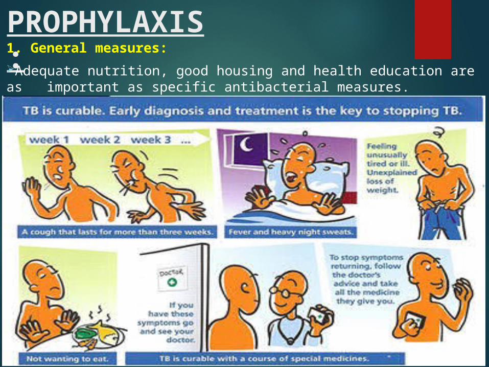
PROPHYLAXIS:1. General measures:Adequate nutrition, good housing and health education are as important as specific antibacterial measures.

2. Immunoprophylaxis:The BCG (Bacille Calmette-Guerin) vaccine (0.1 ml), administered soon after birth by intradermal Injection which may be given at any time during the first year of life.BCG is live attenuated strain derived from M. bovis → stimulates development of hypersensitivity to M. tuberculosis.
Albert Calmette Camille Guérin
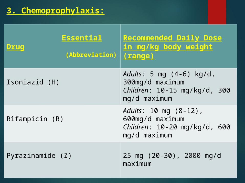
3. Chemoprophylaxis:
Essential Drug (Abbreviation)
Recommended Daily Dose in mg/kg body weight (range)
Isoniazid (H) Adults: 5 mg (4-6) kg/d, 300mg/d maximum Children: 10-15 mg/kg/d, 300 mg/d maximum
Rifampicin (R) Adults: 10 mg (8-12), 600mg/d maximum Children: 10-20 mg/kg/d, 600 mg/d maximum
Pyrazinamide (Z) 25 mg (20-30), 2000 mg/d maximum
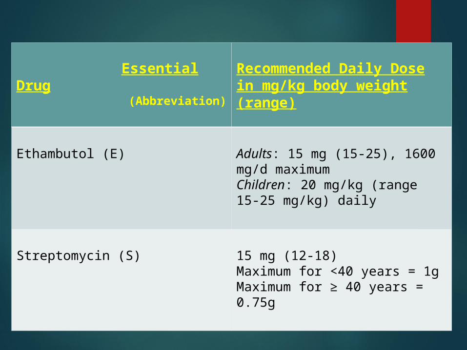
Essential Drug (Abbreviation)
Recommended Daily Dose in mg/kg body weight (range)
Ethambutol (E) Adults: 15 mg (15-25), 1600 mg/d maximum Children: 20 mg/kg (range 15-25 mg/kg) daily
Streptomycin (S) 15 mg (12-18) Maximum for <40 years = 1g Maximum for ≥ 40 years = 0.75g

DOTS – Directly Observed Therapy Short Course DOTS is the name given to the World Health
Organization-recommended tuberculosis control strategy. DOTS has five main components:
1. Government commitment (including political) at all levels, and establishment of a centralized and prioritized system of TB monitoring, recording and training).
2. Case detection by sputum smear microscopy.3. Standardized treatment regimen directly of six to nine months observed by a
healthcare worker or community health worker for at least the first two months.
4. A drug supply.5. A standardized recording and reporting system that allows assessment of
treatment results. MDR TB (Drug Resistant TB)
MDR-TB refers to strains of the bacterium which are proven in a laboratory to be resistant to the two most active anti-TB drugs, isoniazid and rifampicin. Treatment of MDRTB is extremely expensive, toxic, arduous, and often unsuccessful.
DOTS has been proven to prevent the emergence of MDR-TB, and also to reverse the incidence of MDR-TB where it has emerged.


THANK YOU