naosite: nagasaki university's academic output...
TRANSCRIPT

This document is downloaded at: 2018-07-14T22:00:49Z
Title Reduced FOXO1 Expression Accelerates Skin Wound Healing andAttenuates Scarring
Author(s)
Mori, Ryoichi; Tanaka, Katsuya; de Kerckhove, Maiko; Okamoto,Momoko; Kashiyama, Kazuya; Tanaka, Katsumi; Kim, Sangeun; Kawata,Takuya; Komatsu, Toshimitsu; Park, Seongjoon; Ikematsu, Kazuya;Hirano, Akiyoshi; Martin, Paul; Shimokawa, Isao
Citation The American Journal of Pathology, 184(9), pp.2465-2479; 2014
Issue Date 2014-09
URL http://hdl.handle.net/10069/34774
Right
© 2014 American Society for Investigative Pathology. Published byElsevier Inc.; NOTICE: this is the author’s version of a work that wasaccepted for publication in American Journal of Pathology. Changesresulting from the publishing process, such as peer review, editing,corrections, structural formatting, and other quality control mechanismsmay not be reflected in this document. Changes may have been made tothis work since it was submitted for publication. A definitive version wassubsequently published in American Journal of Pathology, 184, 9, (2014)
NAOSITE: Nagasaki University's Academic Output SITE
http://naosite.lb.nagasaki-u.ac.jp

Reduced FOXO1 accelerates skin wound healing and attenuates scarring
Ryoichi Mori1*, Katsuya Tanaka1, 2, Maiko de Kerckhove1, 4, Momoko Okamoto1,
Kazuya Kashiyama2, Katsumi Tanaka2, Sang Eun Kim1, Takuya Kawata1,
Toshimitsu Komatsu1, Seongjoon Park1, Kazuya Ikematsu3, Akiyoshi Hirano2,
Paul Martin5 and Isao Shimokawa1 1Departments of Pathology, 2Plastic and Reconstructive Surgery, and 3Forensic
Pathology and Science, School of Medicine, Graduate School of Biomedical Sciences,
Nagasaki University, 1-12-4 Sakamoto, Nagasaki 852-8523, Japan 4Center for Medical Education and Research, Nagasaki Municipal Hospital, 6-39
Shinchi-machi, Nagasaki 850-8555, Japan 5Schools of Biochemistry and Physiology & Pharmacology, Faculty of Medical and
Veterinary Sciences, University of Bristol, University Walk, Bristol BS8 1TD, UK
*To whom correspondence should be addressed: Ryoichi Mori, Ph.D. Department of Pathology, Graduate School of Biomedical Sciences, Nagasaki
University, 1-12-4 Sakamoto, Nagasaki 852-8523, Japan
Tel: +81-95-819-7051 Fax: +81-95-819-7052 E-mail: [email protected]
Short title: Foxo1 in skin wound healing and scarring
Grants: This work was supported in part by the Ministry of Education, Culture, Sports,
Science and Technology of Japan (Grants-in-Aid for Young Scientists [A] 21689049 and
24689069, Challenging Exploratory Research 23650484 and 25560055 to R.M.), Takeda
Science Foundation (R. M.), Uehara Memorial Foundation (R. M.), and Nakatomi
Foundation (R. M.), The Wellcome Trust (Senior Investigator Award 097791MA P. M.),
and The Royal Society (International Joint Project, R. M. and P. M.).
1

Number of text pages, 49; Figures, 6; Tables; 3
2

Summary (219 words) 1
The FOXO family has been extensively investigated in aging and metabolism, but its 2
role in tissue-repair processes remains largely unknown. In the present study, we aimed 3
to clarify the molecular aspect of the FOXO family in skin wound healing. We 4
demonstrated that Foxo1 and Foxo3a were both upregulated during murine skin wound 5
healing. Partial full body knockout of Foxo1 in Foxo1+/- mice led to accelerated skin 6
wound healing with enhanced keratinocyte migration, reduced granulation tissue 7
formation, and collagen density, accompanied by an attenuated inflammatory response, 8
but we observed no wound phenotype in Foxo3a-/- mice. Fibroblast growth factor2, 9
adiponectin, and notch1 genes were significantly increased at wound sites in Foxo1+/- 10
mice, along with markedly altered ERK1/2 and AKT phosphorylation. Similarly, 11
transient knockdown of Foxo1 at the wound site by local delivery of antisense 12
oligodeoxynucleotides enhanced skin wound healing. The link between FOXO1 and 13
scarring extends to clinical patients, in particular keloid scars where we see FOXO1 14
expression markedly increased in fibroblasts and inflammatory cells within the 15
otherwise normal dermis in the immediate vicinity of the keloid by comparison to the 16
center of the mature keloid, indicating that FOXO1 is associated with the overgrowth of 17
this fibrotic response into adjacent normal skin. Overall, our data indicate that molecular 18
targeting of FOXO1 may improve the quality of healing and reduce pathological 19
scarring.20
3

Introduction 1
The skin is our surface organ and, as such plays a major role in protecting us against all 2
extrinsic traumatic factors (i.e. microbes, ultraviolet radiation, heat, and chemicals). 3
Damage to the skin immediately triggers tissue repair mechanisms alongside a robust 4
inflammatory response for host defense.1 Skin wound healing is generally considered to 5
consist of 3 phases: inflammation, proliferation/migration, and maturation. During an 6
acute wound inflammatory response, large numbers of neutrophils rapidly migrate into 7
damaged tissues to protect against microbes, followed by macrophages that contribute 8
to formation of an associated granulation tissue, including the wound angiogenic 9
response; unfortunately, this wound inflammatory response also contributes to the final 10
fibrotic outcome of adult tissue repair.2 In parallel with connective tissue repair, 11
epithelial cells migrate over the newly forming granulation tissue to cover the wound 12
site in a process known as re-epithelialisation.3 Finally, the wound tissues are partially 13
remodeled, including some removal of excess extracellular matrix at the scar site by 14
proteolytic degradation.4 15
Tissue repair speed and quality is dependent on aging, and metabolic status at a 16
whole-body level, in addition to local immunity and cellular responses at the wound 17
site.5, 6 The skin is one of the clearest indicators of aging, and skin healing is highly 18
associated with the aging process. Skin repair occurs perfectly, without scarring, until 19
fairly late in gestation (embryonic 14 days (d) in the mouse and the end of the second 20
trimester in humans).7 By contrast the elderly are known to exhibit impaired healing. To 21
date, several studies have therefore focused on the molecular mechanisms linking tissue 22
4

repair and skin biology with age- and/or metabolic-related genes.8-12 A worldwide 1
increase in patients with delayed skin wound healing due to an abnormal healing 2
process is linked with aging, diabetes, malnutrition, chemotherapy, and hereditary 3
diseases. 4
The mammalian forkhead box O (FOXO) is a family of transcription factors 5
consisting of FOXO1, FOXO3A, FOXO4, and FOXO6. These proteins remain 6
transcriptionally active in the nucleus in the absence of environmental and growth 7
factors.13 Modification of FOXO leads to its translocation to the cytoplasm and/or its 8
degradation, resulting in the suppression of transcriptional activity. Foxo1 deficiency 9
(Foxo1-/-) in embryonic mice has been shown to be lethal, causing abnormal vascular 10
development.14, 15 We have previously reported that FOXO1 plays a key role in aging 11
and in caloric restriction and exhibits anti-neoplastic characteristics.16 Several lines of 12
evidence suggest that FOXO proteins may play several key roles during tissue repair. 13
Sarcopenia is an age-associated degenerative condition resulting in the loss of skeletal 14
muscle mass and muscle tissue repair.17 Activation of the FOXO family is implicated in 15
skeletal muscle regeneration.18 In human skin wounds, Foxo1 and Foxo4 are overly 16
expressed in the transcription factor binding sites of promoters from many differentially 17
expressed genes in the epidermis.19 Moreover, in diabetic mice, impaired skin wound 18
healing is associated with enhanced activation of FOXO1.20 Other studies have recently 19
reported that re-epithelialization during scalp wound healing is impaired in 20
keratinocyte-specific Foxo1-/- mice.21 However, the full involvement of FOXO family 21
members and their mechanism of action in all cell lineages involved in skin wound 22
5

healing and scarring in vivo remain largely unknown. 1
In the present study, we aimed to investigate the molecular functions for FOXO family 2
gene members in skin wound healing, and their potential application in a clinical setting. 3
We find that heterozygous Foxo1-deficient (Foxo1+/-) mice exhibit accelerated skin 4
wound healing, decreased scarring, and enhanced keratinocyte migration. Acute 5
knockdown of the FOXO1 protein at wound sites (using Foxo1 antisense 6
oligodeoxynucleotides [AS ODN]) improved the quality of healing. And we suggest that 7
FOXO1 may be implicated in the development of human keloids, since the altered 8
expression of FOXO1 during this process is race-dependent. Our findings suggest that 9
modulating expression of FOXO1 may regulate wound healing and scar formation and 10
thus are potential therapeutic targets for improving wound healing in the clinic.11
6

Materials and methods 1
Wound model 2
All experiments were conducted according to the Ethics Review Committee for Animal 3
Experimentation at Nagasaki University (No. 1108010940-7 and 1311121101). The 4
generation of Foxo1+/- and Foxo3a-/- mice has been described previously.16, 22 The 5
wound model was performed as previously described23. In brief, four full-thickness 6
excisional (4-mm biopsy punch; Kai Industries, Tokyo, Japan) wounds or 2 7
full-thickness incisional wounds (1-cm) in the dorsal skin (after shaving under 8
anesthesia) were performed in mice (7–12 wks old). Wounds were then harvested using 9
a 6-mm biopsy punch (Kai Industries) and recorded using a digital camera. Areas were 10
calculated using PhotoshopCS4 (Adobe systems, San Jose, CA). 11
12
Human samples 13
Human keloid tissue samples were harvested from Japanese and African American 14
patients at the time of surgery, and diagnosis was confirmed by routine pathological 15
examination (Supplemental Information Table S1). Normal skin tissue samples were 16
harvested from the immediate vicinity of the keloid site. All experiments were conducted 17
with the approval of the ethics committee of Nagasaki University Hospital 18
(No.09062523-2), and in accordance with the Declaration of Helsinki principles. 19
Written informed consent was obtained from each individual. 20
21
Histology 22
7

Tissue was fixed in 4% PFA for paraffin embedding. Sections (6 μm thick) were used 1
for various staining techniques: H&E, Masson’s Trichrome, Picrosirius red staining, 2
and immunohistochemistry (IHC) for FOXO1, neutrophils, F4/80, proliferating cell 3
nuclear antigen (PCNA), and phospho–extracellular signal-regulated kinase (pERK). 4
Observations were made via microscopy (polarized light, epifluorescence, or confocal 5
[C2+ system; Nikon Corporation, Tokyo, Japan]). NIS-Elements C or AR software 6
(Nikon Corporation) was used for data analysis. Immunostaining procedures and 7
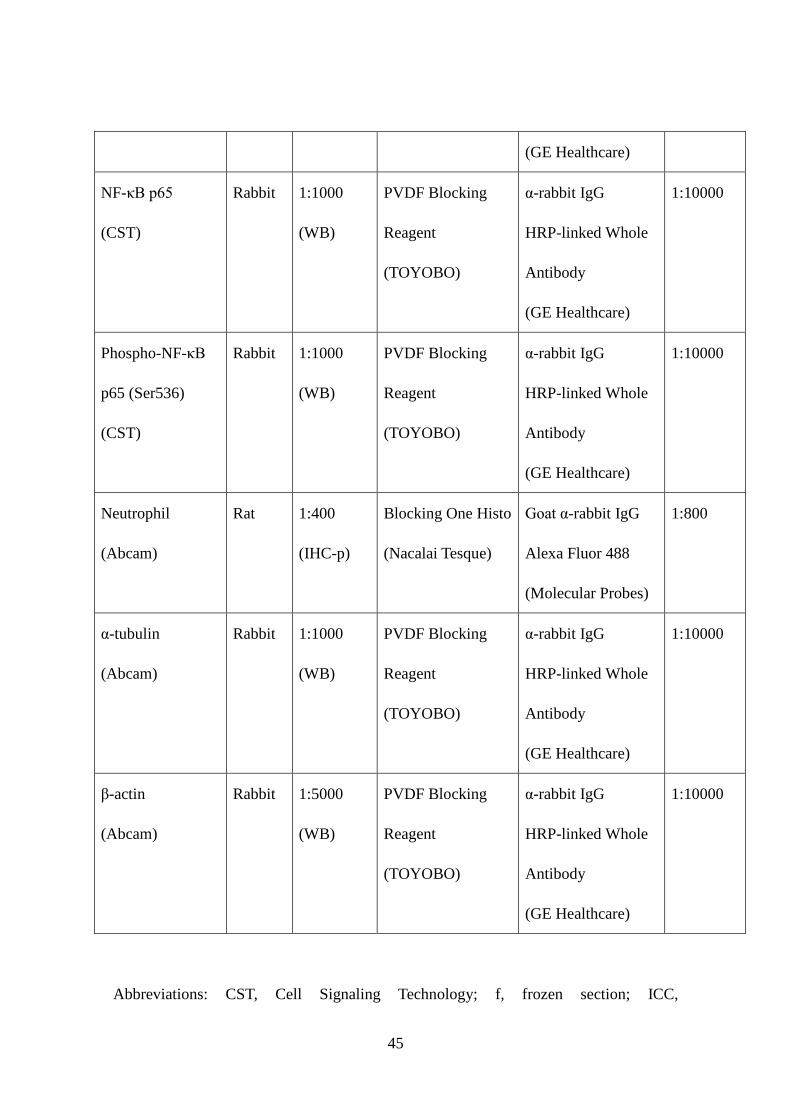
antibody information are listed in Table 1. 8
9
Analysis of epithelial tongue and area of granulation tissue 10
Measurement of epithelial tongue and area of granulation tissue were performed as 11
previously described.23 In brief, the epithelial tongue on H&E-stained wound sections 12
and areas of granulation tissue on Masson’s Trichrome staining sections were measured 13
using NIS-Elements AR software. 14
15
Analysis of angiogenesis 16
Wounded skin was fixed in 4% PFA for 16 hours (h), then exposed to 10%, 20%, and 17
30% sucrose (each percentage for 16 h), and frozen in OCT compound. Sections (50 μm 18
thick) were permeabilized with histology blocking reagent (Blocking One Histo, 19
Nacalai Tesque, Kyoto, Japan) and 0.3% Triton X-100 for 2 h. IHC for CD31, including 20
antibody information are listed in Table 1. Evaluation of 3-dimensional imaging for 21
blood vessels and vascular density were obtained by confocal microscopy, 22
8

NIS-Elements C software, and IMARIS software (BITPLANE, Zurich, Switzerland). 1
2
Transmission electron microscopy (TEM) and morphological analysis of collagen 3
TEM and morphological analysis of collagen were performed as previously described.23, 4
24 5
6
Measurement of macrophages and FOXO1-positive cells at human intact skin and 7
keloid sites 8
F4/80-positive cells (indicative of macrophages) and FOXO1-positive cells in the wound 9
bed (defined as the area surrounded by unwounded skin, fascia, regenerated epidermis, 10
and eschar), keloids, or intact skin were counted from 3 random fields (0.14 mm2). 11
12
RNA isolation and quantitative polymerase chain reaction (qPCR) 13
RNA isolation and qPCR were performed as previously described.24 The gene-specific 14
primers and probes for qPCR analysis were obtained from TaqMan gene expression 15
assays (Applied Biosystems, Foster City, CA) and gene-specific primer sets (Takara Bio, 16
Shiga, Japan). 17
18
Extraction of nuclear protein and measurement of FOXO1 activity 19
Nuclear proteins were extracted by the Nuclear Extract kit (Active Motif Japan, Tokyo, 20
Japan), according to the manufacturer’s instructions. Harvested skin wound sites were 21
homogenized using TissueLyzer II (Qiagen, Hilden, Germany). 22
9

FOXO activity was measured with the TransAM FKHR (FOXO1/4) (Active motif 1
Japan) according to the manufacturer’s instructions. FOXO1 consensus 2
oligonucleotide-treated extracts were used as the negative control. Absorbance was read 3
by the spectrophotometer (model; LS-PLATEmanager 2004, Wako Pure Chemical 4
Industries, Osaka, Japan). The degree of FOXO1 binding activity was calculated as 5
follows: FOXO1 binding activity (arbitrary units) = optical density/μg of nuclear 6
protein. 7
8
Total protein extraction and western immunoblotting 9
Harvested skin wound sites were homogenized using TissueLyzer II (Qiagen) and were 10
added to T-PER Reagent (Thermo Fisher Scientific, Waltham, MA) consisting of 11
proteinase and dephosphorylation inhibitor (Thermo Fisher Scientific). Supernatant 12
debris was eliminated using Ultrafree-MC 0.45 μm filter (Millipore, Bedford, MA). 13
Filtered protein samples were separated on a 4–12% NuPAGE Novex Bis-Tris gel (Life 14
Technology, Carlsbad, CA), transferred to PVDF, and blotted according to standard 15
protocols (antibody details are listed in Table 1). Protein bands were visualized by 16
chemiluminescence (Thermo Fisher Scientific), and band intensity was calculated using 17
Image J 1.47a software (National Institutes of Health). 18
19
Cell culture, Foxo1 siRNA treatment, wound scratch assay, and measurement of 20
pERK activity 21
Mouse primary keratinocytes (PKs) (Cell Lines Service, Eppelheim, Germany) were 22
10

transfected with 100 nM Stealth RNAi Negative Control Medium GC Duplex #2 or 1
Stealth Foxo1 siRNA (Life Technologies, Carlsbad, CA) using the Neon Transfection 2
System (1400 V, 20 ms, 2 pulses) (Life Technologies). The wound scratched assay was 3
performed as previously described.25 Recombinant mouse fibroblast growth factor 2 4
(FGF2) (100 ng/mL) (Cell Signaling Technology, Danvers, MA) was used. The 5
intensity of pERK fluorescence was measured by NIS-Elements AR software (Nikon 6
Corporation) 7
8
Lipopolysaccharide (LPS) challenge 9
LPS (from E.coli serotype O55, phenol extraction, was obtained from Wako Pure 10
Chemical Industries) was reconstituted in saline. Mice (8-12 wks, body weight 30 g) 11
were intraperitoneally injected with 1.0 mg of LPS, and their survival was monitored. 12
13
ELISA 14
Extracted proteins were measured by the myeloperoxidase (MPO) mouse ELISA kit 15
(abcam, Cambridge, UK), according to the manufacturer’s instructions. 16
17
Microarray analysis 18
Cyanine3-labeled complementary RNA (Cy3-cRNA) was generated from 200 ng of 19
total RNA using the Low input quick amp labeling kit, one color (Agilent Technologies, 20
Santa Clara, CA), and was purified by the RNeasy mini kit (Qiagen), according to the 21
manufacturer’s instructions. Fragmented Cy3-cRNA (600 ng) was hybridized to 22
11

SurePrint G3 mouse GE microarray, 8 × 60 K (Agilent Technologies) at 65°C for 17 h. 1
The microarray was then washed, and scanned using the Agilent DNA microarray 2
scanner. 3
Analysis of microarray data was performed using Ingenuity iReport (Ingenuity 4
System, Redwood City, CA). Probeset intensities were summarized and normalized 5
using Robust Multi-Array Average. Significant differential expression was determined 6
by a moderated t-test (Limma) using a p value cutoff of 0.05 and a fold-change cutoff of 7
1.5. All raw data are available in the GEO database (GSE48473, 8
http://www.ncbi.nlm.nih.gov/geo/query/acc.cgi?acc=GSE48473). 9
10
Screening AS ODNs candidates for knockdown studies 11
AS ODNs and the in vitro AS ODN cleavage experiments were both designed as 12
previously described.23 BLAST searches for AS ODNs sequences (Table 2) were 13
conducted to exclude any sequences that were nonspecific for Foxo1 mRNA (GenBank; 14
NM_019739). Foxo1 mRNA was transcribed from the Riken FANTOM FLS Clone 15
(Clone ID: E430027H20; DNAFORM, Kanagawa, Japan) 16
For in vivo experiments involving ODN delivery, ODNs (1 or 10 μM in 50 μL 30% 17
Pluronic F-127 gel [Sigma-Aldrich, St. Louis, MO], which acts as a slow release 18
vehicle23) were topically applied immediately after wounding (50 μL; 1 or 10 μM of 19
ODNs). 20
21
Statistical analysis 22
12

All data are expressed as the mean ± SEM. Statistical significance was assessed by 1
analysis of variance, followed by: (1) Tukey’s post hoc test for multiple comparisons; 2
(2) Dunnett’s post hoc test for comparisons of all columns vs. control; or (3) paired or 3
unpaired Student’s t-test. Survival curve were analyzed using Kaplan-Meier survival 4
analysis and were compared with the log-rank test. Statistical analysis was performed 5
using GraphPad Prism software (GraphPad Software, La Jolla, CA). Significance was 6
reached at values of p<0.05. 7
8
Results 9
Skin wound healing was accelerated in Foxo1+/- mice 10
We investigated the expression of Foxo in wild type (WT) mice after dorsal aseptic skin 11
wounding by qPCR. Gene expression of Foxo1 was significantly increased 3 and 7 d 12
after injury and Foxo3a, 7 d after injury by comparison with unwounded skin (Figure 13
1A). Gene expression of Foxo4 was markedly low, and Foxo6 was not significantly 14
induced. These results indicated that Foxo1 and Foxo3a genes are the Foxo family 15
members predominantly expressed during the skin repair process. 16
We then explored the role of FOXO1 and FOXO3A in skin wound healing in 17
Foxo1+/- and Foxo3a-/- male mice, respectively. Foxo1+/- male mice are viable despite 18
expressing less than 50% of both FOXO1 in intact skin (data not shown), and Foxo1 19
mRNA in liver, spleen, muscle, adipose tissue, and hippocampus.16 After 3 d of injury, 20
Foxo1+/- mice exhibited a significantly smaller wound area (57 ± 3.2%) by comparison 21
to time-matched WT mice (72 ± 4.0%) (Figure 1, B and C). By contrast, wound closure 22
13

in Foxo3a-/- mice was not altered compared with WT mice (data not shown). These 1
results prompted us to further analyze the function of FOXO1 in skin wound healing. 2
To determine which cells express FOXO1 protein during skin wound healing, we 3
performed IHC analysis. By only 1 d post injury, FOXO1 was markedly present in the 4
leading edge and basal layer of keratinocytes, and hair follicles, and in recruited 5
neutrophils (Figure 1D and Supplementary Figure S1). Seven d after injury, FOXO1 6
was present in macrophages, fibroblasts, and endothelial cells at the wound site (Figure 7
1, E and F). Histological analysis allowed us to quantify both the extent of 8
re-epithelialization and the area of granulation tissue at various time points during repair. 9
The length of epithelial wound tongues in Foxo1+/- mice 3 d after injury was markedly 10
higher (813 ± 94 μm) than for WT mice (513 ± 52 μm) (Figure 1, G and H). IHC for the 11
proliferation marker, PCNA, showed that the percentage of proliferating cells in the 12
epithelial tongue of Foxo1+/- mice 3 d after injury was markedly increased compared 13
with WT mice (55% versus 40%). Masson's Trichrome staining of sections of excisional 14
wounds revealed that the area of granulation tissue in the mid-wound region of Foxo1+/- 15
mice was significantly reduced (0.15 ± 0.014 mm2) compared with WT mice (0.26 ± 16
0.021 mm2) (Figure 1, I and J). 17
Since angiogenesis is crucial for granulation tissue formation, and FOXO1 is known 18
to be involved in vasculogenesis in embryonic and fetal development 14 we investigated 19
vessel outgrowth during the repair process in Foxo1+/- versus WT mice. The 20
3-dimensional blood vessel network of the wound was reconstructed via confocal 21
microscopy of sections stained for for PECAM/CD31 which is a marker for endothelial 22
14

cells.26 These studies revealed no difference in the vessel network in intact skin of 1
Foxo1+/- and WT mice (0.033 ± 0.0024 μm3/μm3 and 0.026 ± 0.0057 μm3/μm3, 2
respectively) and the same was true at both 7 and 14 d wound sites (Supplemental 3
Figure S2). 4
Our analyses suggest that attenuated expression of FOXO1 protein leads to 5
accelerated repair and improved quality of skin wound healing, owing to enhanced 6
migration of keratinocytes in the early stage of wound healing, followed by decreased 7
area of granulation tissue formation, but this is not due to differences in wound 8
angiogenesis. 9
10
The inflammatory response was attenuated at wound sites of Foxo1+/- mice 11
Several leukocyte lineages infiltrate wound sites at various time points during the skin 12
repair process.2 Our IHC staining showed that wound-infiltrated neutrophils and 13
macrophages expressed FOXO1 protein (Figure 1, D and E). Neutrophils, revealed 14
either by neutrophil IHC or measurement of MPO were reduced in Foxo1+/- wounds 15
versus WT controls (Figure 2, A and B). F4/80 IHC for macrophages27 confirms a 16
similar reduction (by 40%) in macrophage numbers at wound sites of Foxo1+/- mice 17
(Figure 2, C and D). 18
Since NF-κB plays a pivotal role in the inflammatory response28 we performed 19
western immunoblot analysis and showed that phosphorylation levels of NF-κB p65 20
(Ser536) at wound sites of Foxo1+/- mice 3 d after injury were markedly reduced (by 21
38%) compared with WT mice (Figure 2, E and F). In addition, we found that Foxo1+/- 22
15

mice exhibited significant resistance to high dose LPS-induced endotoxin shock that 1
leads to activation of NF-κB signaling via TLR4 in vivo (Supplemental Figure S3).29 2
Collectively, these data suggest that FOXO1 may regulate inflammatory cell 3
recruitment to wound sites, and the reduced FOXO1 in Foxo1+/- mice dampens down 4
this inflammatory response. 5
6
Expression and phosphorylation of the FOXO family at wound sites 7
Our findings provide clear evidence that Foxo1+/- mice exhibit accelerated skin wound 8
healing and enhanced re-epithelialization at the early stage of skin wound healing and a 9
diminished inflammatory response. We next performed a comprehensive gene 10
expression profile at wound sites of Foxo1+/- and WT mice, after first confirming 11
reduced FOXO1 DNA binding activity. ELISA studies indicate reduced binding of 12
FOXO1 in cells from wound sites of Foxo1+/- mice 3 and 7 d after injury (66 ± 13% and 13
56 ± 15%, respectively) compared with WT mice (Figure 3A). Similar results were 14
found for both gene (Table 3) and protein (Figure 3B) expression of FOXO1. 15
In human fibroblasts, FOXO1 and FOXO3A have been shown to impact on Foxo1 16
gene expression,30 and so we examined the protein expression and phosphorylation 17
levels of FOXO1, FOXO3A, and FOXO4 at wound sites 3 d after injury Foxo1+/- mice 18
(Figure 3B). We find that FOXO1 protein expression and its phosphorylation (pFOXO1 19
[Thr24]) level at wound sites were markedly decreased in Foxo1+/- mice, but expression 20
of FOXO3A, pFOXO3A (Ser318/321), and FOXO4 were not altered in either group. 21
These results, and the pattern of expression of Foxo family genes during skin repair 22
16

(Figure 1A) indicates that FOXOs at the wound site are predominantly regulated by 1
pFOXO1 (Thr24) and pFOXO3A (Ser318/321) in the early stage of skin wound 2
healing. 3
4
Activation of ERK1/2 was enhanced in wound sites of Foxo1+/- mice 5
To determine the molecular mechanisms underlying enhanced skin wound healing when 6
FOXO1 protein expression was reduced, we performed microarray analysis on 3 d skin 7
wound samples from Foxo1+/- versus WT mice. Using a fold-change cutoff of 1.5 (with 8
a p-value cutoff of 0.05), we identify 387 and 269 genes differentially regulated genes 9
(DRGs) that are up- and down-regulated in Foxo1+/- mice, respectively (Supplementary 10
Table S2). We next screened for those molecules/pathways that might be most likely to 11
be promoting healing in Foxo1+/- mice by analyzing the molecular interactions between 12
DRGs, such as gene expression, activation, post-translational modification, and physical 13
interactions (Supplementary Table S3). Previous in vivo studies have revealed skin 14
wound healing-related genes: fibroblast growth factor2 (Fgf2),31-33 Adiponectin 15
(Adipoq),8, 9 and Notch1.34 Our results showed that Fgf2, Adipoq, and Notch1 were 16
significantly (p < 0.05) increased 1.65-fold, 1.82-fold, and 1.94-fold, respectively in 3 d 17
wound sites of Foxo1+/- mice (Table 3). 18
Two key signaling pathways may contribute to the FOXO1 phenotype we observe. 19
The ERK1/2 signaling pathway is involved in cell migration and proliferation,25 and the 20
AKT signaling pathway is associated with skin wound healing functions upstream of 21
FOXO1.35 The ERK1/2 and AKT pathways are activated by FGF2 and ADIPOQ, 22
17

contributing to epithelial and fibroblast cell proliferation.8, 36 Therefore, we investigated 1
whether activation of ERK and AKT pathways in wound sites of Foxo1+/- mice were 2
altered. The phosphorylation levels of ERK1/2 (Thr202/Tyr204) in wound sites of 3
Foxo1+/- mice 3 d after injury was markedly increased compared with WT (1.5 ± 0.13 4
and 1.0 ± 0.11, respectively) (Figure 3C). In contrast, the phosphorylation levels of 5
AKT (Ser473) in wound sites of Foxo1+/- mice 7 d after injury was markedly decreased 6
compared with WT (0.56 ± 0.054 and 1.0 ± 0.20, respectively) (Figure 3D). 7
Our microarray analysis showed that several cell migration- and proliferation-related 8
signals were significantly up-regulated, including myosin heavy chain 10 (Myh10) 9
(1.64-fold). MYH10 generates non-muscle Myosin IIb isoform,37 which is downstream 10
of the ERK1/2 pathway.38 Myosin IIb is expressed in both epidermis and wound 11
fibroblasts,39 and Myh10 is markedly induced at wound sites in Foxo1+/- mice (Table 3) 12
in the current study. We also see that protein levels of the Myosin IIb isoform are 13
markedly increased in wound sites of Foxo1+/- mice compared with WT (1.5 ± 0.21 and 14
1.0 ± 0.067, respectively) (Figure 3, C and D). Collectively, these results suggest that 15
expression of Myosin IIb may be enhanced via the ERK pathway at early stage of 16
wound repair in Foxo1+/- mice. 17
We next tested whether FOXO1 was involved in wound FGF2 signaling, which 18
contributes to the enhancement of cell proliferation and migration in PKs. The in vitro 19
wound scratch assay demonstrated that wound closure in Foxo1 siRNA-treated PKs was 20
not altered compared with control PKs (1.23 ± 0.15 and 1.0 ± 0.04, respectively). 21
Interestingly, the migration of mFGF2-treated Foxo1 siRNA-treated PKs was 22
18

significantly enhanced from the wound edge to the center of the wound (Figure 3, E and 1
F). ICC was then used to investigate the localization of pERK. In PKs exposed to 2
mFGF2-treated Foxo1 siRNA, pERK activation was mainly observed at the wound 3
edge rather than away from the wound site 24 h after scratching. This response was 4
significantly higher compared with control wounds (4.95 ± 0. 63 and 2.82 ± 0.41, 5
respectively) (Figure 3, G and H). Taken together, these findings indicate that FOXO1 6
may play a role in re-epithelialization and migration of keratinocytes at wound sites via 7
the ERK and FGF2 pathway. 8
9
Collagen organization was altered at wound sites of Foxo1+/- mice 10
Scarring is the final consequence of the wound healing process, and is a measure of 11
wound healing quality.1 To investigate whether altered FOXO1 expression influences 12
the development of scarring at wound sites, we monitored scarring 21 d after making 13
1-cm incisional wounds to Foxo1+/- and WT mice (Figure 4A). Picrosirius red staining 14
showed type I collagen (red and yellow) and type III collagen (green) bundle 15
organization40 to be markedly reduced in Foxo1+/- mice 21 d after injury (Figure 4B). 16
To further analyze development of scar, we performed TEM to reveal gross collagen 17
bundling patterns, individual collagen fibril diameter, and the density of fibrils at wound 18
site. Morphology of collagen within intact skin of Foxo1+/- and WT mice was 19
indistinguishable (Supplemental Figure S4). Interestingly, the fibril diameter at the 20
mid-region wound sites of Foxo1+/- mice were markedly (p < 0.001) reduced (61.5 ± 21
0.49 nm) compared with WT mice (63.3 ± 0.46 nm) (Figure 4, C and D). Furthermore, 22
19

intra collagen bundle spaces at wound sites of Foxo1+/- mice were significantly (p < 1
0.05) increased (0.62 ± 0.094 μm2/μm2) compared with WT mice (0.38 ± 0.023 2
μm2/μm2) (Figure 4E), more closely resembling that of unwounded skin. Gene 3
expression of type I collagen α1 (Col1α1), was significantly decreased at wound sites of 4
Foxo1+/- mice 7 d after injury compared with WT mice (0.58 ± 0.073 and 0.82 ± 0.063, 5
respectively) (Figure 4F). We suggest that these differences of collagen assembly at 6
wound sites play a key role in reducing scar formation during the maturation phase of 7
healing in Foxo1+/- mice. 8
9
Acute knock down of FOXO1 using AS ODNs improved skin wound healing 10
Our Foxo1+/- mouse data provides experimental evidence that attenuation of FOXO1 11
expression may improve skin wound healing. To further address our hypothesis and test 12
whether reducing levels of this transcription factor during the repair process is a 13
potential therapeutic strategy for improving healing, we designed and optimized Foxo1 14
specific AS ODNs in vitro (Figure 5A). We applied Foxo1 AS ODN (1717) (10 μM in 15
30% Pluronic gel for 6 h at the wound site23) versus control ODN, (with sequence 16
predicted to be non-binding to other mRNAs), to 4-mm diameter adult skin wound sites 17
(Figure 5, B and C). Macroscopic analysis indicated that wound closure in Foxo1 AS 18
ODN-treated wound sites 3 d after injury was markedly accelerated (67 ± 2.4%) at early 19
time points during the repair process, compared with the control ODN-treated wound 20
(76 ± 3.0%) (Figure 5, D and E). Next, we made a 1-cm incisional wound in the dorsal 21
skin and analyzed scarring (via TEM) with a 21-d treatment of control ODN or Foxo1 22
20

AS ODN at the wound sites. The fibril diameter at the mid-region was not altered (66.6 1
± 0.65 nm, n = 1336 from 3 mice) at Foxo1 AS ODN-treated wound sites compared 2
with control ODN-treated wound site (65.1 ± 0.55 nm, n = 1534 from 3 mice). Neither 3
was vacant extracellular space at Foxo1 AS ODN-treated wound sites altered (0.55 ± 4
0.038 μm2/μm2, n = 3) compared with control ODN-treated wound sites (0.44 ± 5
0.038μm2/μm2, n = 3). These results indicated that acute down-regulation of FOXO1 6
protein at the wound site using Foxo1 AS ODN accelerated skin wound healing, but did 7
not significantly alter scar quality. 8
9
Increased FOXO1 is associated with human keloid scars 10
Because of the altered level of scarring in our whole body Foxo1+/- mouse studies, we 11
next chose to investigate whether the expression pattern of FOXO1 was altered in 12
human keloid scars, which are an extreme instance of human skin fibrotic disease, 13
typified by a hypertrophic epidermis, and overgrowth of granulation tissue which 14
expands in a claw-like way to invade adjacent normal skin.41, 42 IHC showed that 15
FOXO1 was prominently present in suprabasal keratinocytes of the hypertrophic 16
epidermal layer of keloids (Figure 6A), in addition to some fibroblasts and 17
inflammatory cells (Figure 6B). Although FOXO1 in fibroblasts was not strongly 18
expressed at deep keloid tissue sites (Figure 6C), it was notably present in numerous 19
fibroblasts and inflammatory cells in the immediate vicinity of keloid sites of the 20
normal dermal layer (Figure 6, D, E and G). These results suggested that FOXO1 was 21
involved in expanding the growth of keloid into adjacent normal skin sites and might be 22
21
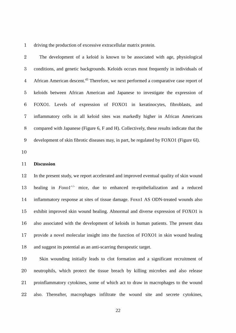
driving the production of excessive extracellular matrix protein. 1
The development of a keloid is known to be associated with age, physiological 2
conditions, and genetic backgrounds. Keloids occurs most frequently in individuals of 3
African American descent.43 Therefore, we next performed a comparative case report of 4
keloids between African American and Japanese to investigate the expression of 5
FOXO1. Levels of expression of FOXO1 in keratinocytes, fibroblasts, and 6
inflammatory cells in all keloid sites was markedly higher in African Americans 7
compared with Japanese (Figure 6, F and H). Collectively, these results indicate that the 8
development of skin fibrotic diseases may, in part, be regulated by FOXO1 (Figure 6I). 9
10
Discussion 11
In the present study, we report accelerated and improved eventual quality of skin wound 12
healing in Foxo1+/- mice, due to enhanced re-epithelialization and a reduced 13
inflammatory response at sites of tissue damage. Foxo1 AS ODN-treated wounds also 14
exhibit improved skin wound healing. Abnormal and diverse expression of FOXO1 is 15
also associated with the development of keloids in human patients. The present data 16
provide a novel molecular insight into the function of FOXO1 in skin wound healing 17
and suggest its potential as an anti-scarring therapeutic target. 18
Skin wounding initially leads to clot formation and a significant recruitment of 19
neutrophils, which protect the tissue breach by killing microbes and also release 20
proinflammatory cytokines, some of which act to draw in macrophages to the wound 21
also. Thereafter, macrophages infiltrate the wound site and secrete cytokines, 22
22

chemokines, and growth factors according to the extent of tissue damage and infection 1
state, thus reflecting the degree of the inflammatory phase.1 The FOXO family has been 2
previously shown to regulate the homeostasis of the immune system and the 3
inflammatory response.44 Conditional Foxo1-/- mice exhibit an altered phenotype, 4
including T-cell homeostasis and tolerance.45-47 FOXO1 may play a role during the 5
infection recognition and clearance process 48 since it is known that the bacterial 6
product N-formylmethionyl-leucyl-phenylalanine triggers neutrophils to upregulate 7
myeloid leukemia cell differentiation protein MCL1, which can form a complex with 8
FOXO1. Moreover, Chip-sequencing analysis using next generation sequencing has 9
revealed that FOXO1 significantly enhances TLR4 signaling in macrophages.49 10
Activation of NF-κB affects the AKT-FOXO1 signaling pathway.50 In the present study, 11
the infiltration of neutrophils and macrophages into wound sites and the 12
phosphorylation of NF-κB and AKT were attenuated in Foxo1+/- mice. Overall, the 13
FOXO1-mediated inflammatory response may link in to leukocyte recruitment and 14
activation in the skin wound healing process. 15
Re-epithelialization, involving keratinocyte migration and proliferation, commences 16
soon after skin damage and is regulated by various factors including keratinocyte 17
growth factor and others released by infiltrating inflammatory cells, and fibroblasts.2, 3 18
Previous studies suggest that the effect of FOXO1 in guiding cell 19
migration/proliferation may be cell-type specific. For example, knockdown of Foxo1 in 20
PDGF-treated fibroblasts has been shown to enhance proliferation, indicating that 21
attenuation of the expression of FOXO1 is sufficient for the enhancement of cell 22
23

growth.30 In contrast, keratinocyte specific Foxo1-/- in mice impairs scalp wound 1
healing due to reduced expression of Tgfβ1.21 In our current study, enhancement of 2
re-epithelialization in Foxo1+/- mice accelerated skin wound healing, which 3
corresponded with an increase in the expression of Fgf2, Adipoq, Notch1, and Myo10 4
(Table 3), and each of these, in turn, is known to play key roles in various aspects of the 5
repair process: FGF2 is crucial for re-epithelialization in skin wound healing,33 whilst 6
genetic and pharmacological inhibition of Notch1 in mice markedly impairs skin wound 7
healing,34 and calmodulin-like protein-mediated expression of MYO10 contributes to 8
keratinocyte motility and migration in humans and mice.51 ADIPOQ promotes 9
keratinocyte proliferation and migration via the ERK pathway in vivo.8, 9 We are 10
currently exploring the mechanism underlying FOXO-mediated regulation of cell 11
proliferation and migration in the presence of FGF2 at wound sites. 12
As well as enhanced rate of wound repair, we observe reduced scarring in Foxo1+/- 13
mice. Scarring appears at the final stage of the skin wound healing process, and the 14
phenotype of scarring is dependent on diverse factors, including inflammation, delayed 15
healing, physiological condition, age, and race.6, 43, 52 The regulation of collagen 16
expression via FOXO1 may depend on the tissue and cell type, and this may be a 17
consequence of both direct and/or indirect effects. Knockdown of Foxo1 in 18
UV-irradiated human dermal fibroblasts was shown to significantly decrease the 19
expression of Col1α1.53 In contrast, expression of liver Col1α1 was increased in the bile 20
duct ligation-induced experimental liver fibrosis model of Foxo1+/-, resulting in liver 21
fibrosis.54 Collagen organization is controlled by several enzymes and extracellular 22
24
matrix proteins,55 and considerably altered by normal aging.56, 57 Activation of AKT is 1
one of the main signaling pathways for type I procollagen synthesis.58 In the present 2
study, expression of Col1α1, collagen density, and AKT phosphorylation were all 3
markedly decreased in wound sites of Foxo1+/- mice 7 d after injury. We also found that 4
knockdown of FOXO1 in PKs significantly enhanced the ERK pathway after mFGF2 5
application. Local administration of FGF2 to the human incisional wound reduces 6
scarring.59 We speculate that the attenuation of FOXO1 in wound fibroblasts contributes 7
to reduced scarring through the FGF2 pathway. We are presently exploring how 8
FOXO1 regulates scarring at wound sites in the presence of several wound growth 9
factors, including FGF2. Further studies using other models, such as 10
keratinocyte-specific Foxo1-/- mice,21 are required to better elucidate the 11
pathophysiological significance of FOXO1 function in skin fibrosis. 12
In patients, the most extreme scarring phenotype is that of keloid scarring where scar 13
tissue spills out from the initial site of tissue damage. Our keloid studies showed 14
FOXO1 is highly expressed in fibroblasts and inflammatory cells at the margin of a 15
keloid compared with those fibroblasts in mature keloid sites, and that FOXO1 16
expression level was altered between African Americans and Japanese. These results 17
indicate that FOXO1 may regulate the expression of collagen, and thus, its expression 18
level may play a key role in scarring and keloids. Current investigations are focusing on 19
the implications of FOXO1 polymorphisms on fibrotic diseases. Disequilibrium of 20
FOXO1 is believed to affect human longevity,60, 61 and thus polymorphism of FOXO1 21
may also affect homeostasis at the cellular and individual level as well. Therefore, a 22
25

functional analysis of FOXO1 polymorphisms may further elucidate the differences in 1
keloid morbidity and repair phenotypes by age and race. Determining how the FOXO1 2
signaling pathway regulates keloid progression for the homeostatic maintenance between 3
proliferation and differentiation will thus be important to explore in future studies. 4
In conclusion, the age-related gene, FOXO1, plays a central role for tissue repair and 5
remodeling and, may be considered a potential therapeutic target for enhancing tissue 6
repair and remodeling as well as for dampening inflammatory diseases and fibrosis. 7
8
Acknowledgments 9
The authors are grateful to Takashi Suematsu (Department of Electron Microscopy, 10
Nagasaki University) for assistance with the TEM analysis, and Kazutaka Hayashida and 11
Shin-ichi Yokota (Nikon Instech, Japan) for assistance with microscopic and imaging 12
analysis. 13
14
Conflict of interest 15
The authors have no financial conflicts of interest.16
26

References 1
1. Shaw TJ, Martin P: Wound repair at a glance, J Cell Sci 2009, 122:3209-3213 2
2. Eming SA, Krieg T, Davidson JM: Inflammation in wound repair: molecular 3
and cellular mechanisms, J Invest Dermatol 2007, 127:514-525 4
3. Werner S, Krieg T, Smola H: Keratinocyte-fibroblast interactions in wound 5
healing, J Invest Dermatol 2007, 127:998-1008 6
4. Martins VL, Caley M, O'Toole EA: Matrix metalloproteinases and epidermal 7
wound repair, Cell Tissue Res 2013, 351:255-268 8
5. Gosain A, DiPietro LA: Aging and wound healing, World J Surg 2004, 9
28:321-326 10
6. Guo S, Dipietro LA: Factors affecting wound healing, J Dent Res 2010, 11
89:219-229 12
7. Ferguson MW, O'Kane S: Scar-free healing: from embryonic mechanisms to 13
adult therapeutic intervention, Philos Trans R Soc Lond B Biol Sci 2004, 359:839-850 14
8. Shibata S, Tada Y, Asano Y, Hau CS, Kato T, Saeki H, Yamauchi T, Kubota N, 15
Kadowaki T, Sato S: Adiponectin regulates cutaneous wound healing by promoting 16
keratinocyte proliferation and migration via the ERK signaling pathway, J Immunol 17
2012, 189:3231-3241 18
9. Salathia NS, Shi J, Zhang J, Glynne RJ: An in vivo screen of secreted proteins 19
identifies adiponectin as a regulator of murine cutaneous wound healing, J Invest 20
Dermatol 2013, 133:812-821 21
10. Goren I, Kampfer H, Podda M, Pfeilschifter J, Frank S: Leptin and wound 22
27

inflammation in diabetic ob/ob mice: differential regulation of neutrophil and 1
macrophage influx and a potential role for the scab as a sink for inflammatory cells and 2
mediators, Diabetes 2003, 52:2821-2832 3
11. Squarize CH, Castilho RM, Bugge TH, Gutkind JS: Accelerated wound 4
healing by mTOR activation in genetically defined mouse models, PLoS One 2010, 5
5:e10643 6
12. Serravallo M, Jagdeo J, Glick SA, Siegel DM, Brody NI: Sirtuins in 7
dermatology: applications for future research and therapeutics, Arch Dermatol Res 2013, 8
305:269-282 9
13. Huang H, Tindall DJ: Dynamic FoxO transcription factors, J Cell Sci 2007, 10
120:2479-2487 11
14. Furuyama T, Kitayama K, Shimoda Y, Ogawa M, Sone K, Yoshida-Araki K, 12
Hisatsune H, Nishikawa S, Nakayama K, Ikeda K, Motoyama N, Mori N: Abnormal 13
angiogenesis in Foxo1 (Fkhr)-deficient mice, J Biol Chem 2004, 279:34741-34749 14
15. Hosaka T, Biggs WH, 3rd, Tieu D, Boyer AD, Varki NM, Cavenee WK, 15
Arden KC: Disruption of forkhead transcription factor (FOXO) family members in mice 16
reveals their functional diversification, Proc Natl Acad Sci U S A 2004, 101:2975-2980 17
16. Yamaza H, Komatsu T, Wakita S, Kijogi C, Park S, Hayashi H, Chiba T, Mori 18
R, Furuyama T, Mori N, Shimokawa I: FoxO1 is involved in the antineoplastic effect of 19
calorie restriction, Aging Cell 2010, 9:372-382 20
17. Snijders T, Verdijk LB, van Loon LJ: The impact of sarcopenia and exercise 21
training on skeletal muscle satellite cells, Ageing Res Rev 2009, 8:328-338 22
28

18. Lara-Pezzi E, Winn N, Paul A, McCullagh K, Slominsky E, Santini MP, 1
Mourkioti F, Sarathchandra P, Fukushima S, Suzuki K, Rosenthal N: A naturally 2
occurring calcineurin variant inhibits FoxO activity and enhances skeletal muscle 3
regeneration, J Cell Biol 2007, 179:1205-1218 4
19. Roupe KM, Alberius P, Schmidtchen A, Sorensen OE: Gene expression 5
demonstrates increased resilience toward harmful inflammatory stimuli in the 6
proliferating epidermis of human skin wounds, Exp Dermatol 2010, 19:e329-332 7
20. Siqueira MF, Li J, Chehab L, Desta T, Chino T, Krothpali N, Behl Y, Alikhani 8
M, Yang J, Braasch C, Graves DT: Impaired wound healing in mouse models of 9
diabetes is mediated by TNF-alpha dysregulation and associated with enhanced 10
activation of forkhead box O1 (FOXO1), Diabetologia 2010, 53:378-388 11
21. Ponugoti B, Xu F, Zhang C, Tian C, Pacios S, Graves DT: FOXO1 promotes 12
wound healing through the up-regulation of TGF-beta1 and prevention of oxidative 13
stress, J Cell Biol 2013, 203:327-343 14
22. Miyamoto K, Araki KY, Naka K, Arai F, Takubo K, Yamazaki S, Matsuoka S, 15
Miyamoto T, Ito K, Ohmura M, Chen C, Hosokawa K, Nakauchi H, Nakayama K, 16
Nakayama KI, Harada M, Motoyama N, Suda T, Hirao A: Foxo3a is essential for 17
maintenance of the hematopoietic stem cell pool, Cell Stem Cell 2007, 1:101-112 18
23. Mori R, Shaw TJ, Martin P: Molecular mechanisms linking wound 19
inflammation and fibrosis: knockdown of osteopontin leads to rapid repair and reduced 20
scarring, J Exp Med 2008, 205:43-51 21
24. Mori R, Ikematsu K, Kitaguchi T, Kim SE, Okamoto M, Chiba T, Miyawaki 22
29

A, Shimokawa I, Tsuboi T: Release of TNF-alpha from macrophages is mediated by 1
small GTPase Rab37, Eur J Immunol 2011, 41:3230-3239 2
25. Matsubayashi Y, Ebisuya M, Honjoh S, Nishida E: ERK activation propagates 3
in epithelial cell sheets and regulates their migration during wound healing, Curr Biol 4
2004, 14:731-735 5
26. Newman PJ: The biology of PECAM-1, J Clin Invest 1997, 99:3-8 6
27. Austyn JM, Gordon S: F4/80, a monoclonal antibody directed specifically 7
against the mouse macrophage, Eur J Immunol 1981, 11:805-815 8
28. DiDonato JA, Mercurio F, Karin M: NF-kappaB and the link between 9
inflammation and cancer, Immunol Rev 2012, 246:379-400 10
29. Kawai T, Akira S: The role of pattern-recognition receptors in innate 11
immunity: update on Toll-like receptors, Nat Immunol 2010, 11:373-384 12
30. Essaghir A, Dif N, Marbehant CY, Coffer PJ, Demoulin JB: The transcription 13
of FOXO genes is stimulated by FOXO3 and repressed by growth factors, J Biol Chem 14
2009, 284:10334-10342 15
31. Meyer M, Muller AK, Yang J, Moik D, Ponzio G, Ornitz DM, Grose R, 16
Werner S: FGF receptors 1 and 2 are key regulators of keratinocyte migration in vitro 17
and in wounded skin, J Cell Sci 2012, 125:5690-5701 18
32. Miller DL, Ortega S, Bashayan O, Basch R, Basilico C: Compensation by 19
fibroblast growth factor 1 (FGF1) does not account for the mild phenotypic defects 20
observed in FGF2 null mice, Mol Cell Biol 2000, 20:2260-2268 21
33. Ortega S, Ittmann M, Tsang SH, Ehrlich M, Basilico C: Neuronal defects and 22
30
delayed wound healing in mice lacking fibroblast growth factor 2, Proc Natl Acad Sci U 1
S A 1998, 95:5672-5677 2
34. Chigurupati S, Arumugam TV, Son TG, Lathia JD, Jameel S, Mughal MR, 3
Tang SC, Jo DG, Camandola S, Giunta M, Rakova I, McDonnell N, Miele L, Mattson 4
MP, Poosala S: Involvement of notch signaling in wound healing, PLoS One 2007, 5
2:e1167 6
35. Somanath PR, Chen J, Byzova TV: Akt1 is necessary for the vascular 7
maturation and angiogenesis during cutaneous wound healing, Angiogenesis 2008, 8
11:277-288 9
36. Chrissouli S, Pratsinis H, Velissariou V, Anastasiou A, Kletsas D: Human 10
amniotic fluid stimulates the proliferation of human fetal and adult skin fibroblasts: the 11
roles of bFGF and PDGF and of the ERK and Akt signaling pathways, Wound Repair 12
Regen 2010, 18:643-654 13
37. Conti MA, Adelstein RS: Nonmuscle myosin II moves in new directions, J 14
Cell Sci 2008, 121:11-18 15
38. Kim JH, Wang A, Conti MA, Adelstein RS: Nonmuscle myosin II is required 16
for internalization of the epidermal growth factor receptor and modulation of 17
downstream signaling, J Biol Chem 2012, 287:27345-27358 18
39. Bond JE, Ho TQ, Selim MA, Hunter CL, Bowers EV, Levinson H: Temporal 19
spatial expression and function of non-muscle myosin II isoforms IIA and IIB in scar 20
remodeling, Lab Invest 2011, 91:499-508 21
40. Wang T, Feng Y, Sun H, Zhang L, Hao L, Shi C, Wang J, Li R, Ran X, Su Y, 22
31

Zou Z: miR-21 regulates skin wound healing by targeting multiple aspects of the 1
healing process, Am J Pathol 2012, 181:1911-1920 2
41. Hunasgi S, Koneru A, Vanishree M, Shamala R: Keloid: A case report and 3
review of pathophysiology and differences between keloid and hypertrophic scars, J 4
Oral Maxillofac Pathol 2013, 17:116-120 5
42. Robles DT, Moore E, Draznin M, Berg D: Keloids: pathophysiology and 6
management, Dermatol Online J 2007, 13:9 7
43. Shih B, Bayat A: Genetics of keloid scarring, Arch Dermatol Res 2010, 8
302:319-339 9
44. Peng SL: Foxo in the immune system, Oncogene 2008, 27:2337-2344 10
45. Gubbels Bupp MR, Edwards B, Guo C, Wei D, Chen G, Wong B, Masteller E, 11
Peng SL: T cells require Foxo1 to populate the peripheral lymphoid organs, Eur J 12
Immunol 2009, 39:2991-2999 13
46. Ouyang W, Beckett O, Flavell RA, Li MO: An essential role of the 14
Forkhead-box transcription factor Foxo1 in control of T cell homeostasis and tolerance, 15
Immunity 2009, 30:358-371 16
47. Ouyang W, Liao W, Luo CT, Yin N, Huse M, Kim MV, Peng M, Chan P, Ma 17
Q, Mo Y, Meijer D, Zhao K, Rudensky AY, Atwal G, Zhang MQ, Li MO: Novel 18
Foxo1-dependent transcriptional programs control T(reg) cell function, Nature 2012, 19
491:554-559 20
48. Crossley LJ: Neutrophil activation by fMLP regulates FOXO (forkhead) 21
transcription factors by multiple pathways, one of which includes the binding of FOXO 22
32

to the survival factor Mcl-1, J Leukoc Biol 2003, 74:583-592 1
49. Fan W, Morinaga H, Kim JJ, Bae E, Spann NJ, Heinz S, Glass CK, Olefsky 2
JM: FoxO1 regulates Tlr4 inflammatory pathway signalling in macrophages, EMBO J 3
2010, 29:4223-4236 4
50. Kim DH, Kim JY, Yu BP, Chung HY: The activation of NF-kappaB through 5
Akt-induced FOXO1 phosphorylation during aging and its modulation by calorie 6
restriction, Biogerontology 2008, 9:33-47 7
51. Bennett RD, Mauer AS, Pittelkow MR, Strehler EE: Calmodulin-like protein 8
upregulates myosin-10 in human keratinocytes and is regulated during epidermal wound 9
healing in vivo, J Invest Dermatol 2009, 129:765-769 10
52. Martin P, Leibovich SJ: Inflammatory cells during wound repair: the good, 11
the bad and the ugly, Trends Cell Biol 2005, 15:599-607 12
53. Tanaka H, Murakami Y, Ishii I, Nakata S: Involvement of a forkhead 13
transcription factor, FOXO1A, in UV-induced changes of collagen metabolism, J 14
Investig Dermatol Symp Proc 2009, 14:60-62 15
54. Adachi M, Osawa Y, Uchinami H, Kitamura T, Accili D, Brenner DA: The 16
forkhead transcription factor FoxO1 regulates proliferation and transdifferentiation of 17
hepatic stellate cells, Gastroenterology 2007, 132:1434-1446 18
55. Canty EG, Kadler KE: Procollagen trafficking, processing and fibrillogenesis, 19
J Cell Sci 2005, 118:1341-1353 20
56. Puschmann S, Rahn CD, Wenck H, Gallinat S, Fischer F: Approach to 21
quantify human dermal skin aging using multiphoton laser scanning microscopy, J 22
33

Biomed Opt 2012, 17:036005 1
57. Wu S, Li H, Yang H, Zhang X, Li Z, Xu S: Quantitative analysis on collagen 2
morphology in aging skin based on multiphoton microscopy, J Biomed Opt 2011, 3
16:040502 4
58. Park JH, Kim SR, An HJ, Kim WJ, Choe M, Han JA: Esculetin promotes type 5
I procollagen expression in human dermal fibroblasts through MAPK and PI3K/Akt 6
pathways, Mol Cell Biochem 2012, 368:61-67 7
59. Ono I, Akasaka Y, Kikuchi R, Sakemoto A, Kamiya T, Yamashita T, Jimbow 8
K: Basic fibroblast growth factor reduces scar formation in acute incisional wounds, 9
Wound Repair Regen 2007, 15:617-623 10
60. Kleindorp R, Flachsbart F, Puca AA, Malovini A, Schreiber S, Nebel A: 11
Candidate gene study of FOXO1, FOXO4, and FOXO6 reveals no association with 12
human longevity in Germans, Aging Cell 2011, 10:622-628 13
61. Li Y, Wang WJ, Cao H, Lu J, Wu C, Hu FY, Guo J, Zhao L, Yang F, Zhang 14
YX, Li W, Zheng GY, Cui H, Chen X, Zhu Z, He H, Dong B, Mo X, Zeng Y, Tian XL: 15
Genetic association of FOXO1A and FOXO3A with longevity trait in Han Chinese 16
populations, Hum Mol Genet 2009, 18:4897-4904 17
62. Poindexter NJ, Williams RR, Powis G, Jen E, Caudle AS, Chada S, Grimm 18
EA: IL-24 is expressed during wound repair and inhibits TGFalpha-induced migration 19
and proliferation of keratinocytes, Exp Dermatol 2010, 19:714-722 20
63. Gregorio J, Meller S, Conrad C, Di Nardo A, Homey B, Lauerma A, Arai N, 21
Gallo RL, Digiovanni J, Gilliet M: Plasmacytoid dendritic cells sense skin injury and 22
34

promote wound healing through type I interferons, J Exp Med 2010, 207:2921-2930 1
64. Gardner H, Broberg A, Pozzi A, Laato M, Heino J: Absence of integrin 2
alpha1beta1 in the mouse causes loss of feedback regulation of collagen synthesis in 3
normal and wounded dermis, J Cell Sci 1999, 112 ( Pt 3):263-272 4
65. Ekstrand AJ, Cao R, Bjorndahl M, Nystrom S, Jonsson-Rylander AC, Hassani 5
H, Hallberg B, Nordlander M, Cao Y: Deletion of neuropeptide Y (NPY) 2 receptor in 6
mice results in blockage of NPY-induced angiogenesis and delayed wound healing, Proc 7
Natl Acad Sci U S A 2003, 100:6033-6038 8
66. Akita S, Daian T, Ishihara H, Fujii T, Akino K: Leukemia inhibitory 9
factor-transfected embryonic fibroblasts and vascular endothelial growth factor 10
successfully improve the skin substitute wound healing by increasing angiogenesis and 11
matrix production, J Dermatol Sci 2004, 36:11-23 12
67. Meyer M, Muller AK, Yang J, Moik D, Ponzio G, Ornitz DM, Grose R, 13
Werner S: FGF receptors 1 and 2 are key regulators of keratinocyte migration in vitro 14
and in wounded skin, J Cell Sci 2012, 15
68. Sun LL, Xu LL, Nielsen TB, Rhee P, Burris D: Cyclopentyladenosine 16
improves cell proliferation, wound healing, and hair growth, J Surg Res 1999, 87:14-24 17
18
19
35

Figure legends 1
Figure 1. Skin wound healing is accelerated in Foxo1+/- mice. 2
(A) Gene expression of murine Foxo family in skin wound healing measured by qPCR 3
relative to 18S ribosomal RNA (n = 4-6). (B) Representative images for gross 4
appearances of excisional wound in WT and Foxo1+/- mice (C) The proportion of the 5
wound remaining open relative to the initial wound area at each time point (n = 12). (D) 6
IHC for FOXO1 and neutrophils showing neutrophils, epithelium and hair follicle 7
expressing FOXO1 1 d after injury in WT mice. Nuclei were counter stained with DAPI 8
(wound margin [closed arrowhead], leading edge of epithelia [arrow], and 9
FOXO1-expressing neutrophils [open arrowheads]). (E and F) IHC of FOXO1 and F4/80 10
or CD31 showing wound-infiltrated macrophages (E), fibroblasts (E, arrowhead), and 11
endothelial cells (F) at 7 d after injury in WT mice. (G) H&E staining of 12
re-epithelialization (wound margin [arrowheads] and the leading edge of epithelia 13
[arrows]). Upper panels correspond to the higher-power field of the lower panels. (H) 14
Measurement of epithelial tongue on 3 d after injury (n = 13). (I) Middle of wound tissue 15
at 14 d after injury stained with Masson's Trichrome, and the extent (cross-sectional area) 16
of granulation tissue visualized and quantified at the mid-point of the wound (indicated 17
by dotted line) (wound margin [arrowheads]). (J) Quantification of the area of 18
granulation tissue 14 d after injury (n = 6). Scale bars = 10 μm (E and F), 100 μm (D and 19
lower panels in G), 500 μm (low-power fields in G and I). All values represent mean ± 20
SEM. *P < 0.05, **P < 0.01, ***P < 0.001. 21
22
36

Figure 2. Attenuation of neutrophil and macrophage infiltration and inflammatory 1
responses at wound sites of Foxo1+/- mice. 2
(A) IHC for neutrophils (red) showing the number of wound-infiltrated neutrophils at 3
wound sites 1 d after injury, which is reduced in Foxo1+/- mice compared with WT mice 4
(nuclei were counter stained with DAPI). (B) MPO concentration using ELISA reveals 5
that MPO levels at wound sites of Foxo1+/- mice are significantly reduced compared 6
with WT mice (n = 8 per group). (C) IHC for macrophages using F4/80 at the middle 7
area of wound sites 7 d after injury. (D) Quantification of F4/80-positive macrophages at 8
wound sites of WT mice (n = 6 per group for 3 d and n = 5 per group for 7 d) and 9
Foxo1+/- mice (n = 6 per group for 3 and 7 d). (E) Western immunoblot of pNF-κB p65 10
and total NF-κB. Full scans for western immunoblot in Supplemental Figure S5A. (F) 11
Densitometric analysis for pNF-κB activity over total NF-κB (n = 4-5). Scale bars = 12
10 μm (A and C). All values represent mean ± SEM. *P < 0.05. 13
14
Figure 3. ERK1/2 activation and Myosin IIb expression is enhanced at wound sites 15
of Foxo1+/- mice. 16
(A) FOXO1 binding activity in the nuclear wound tissue extract was measured by 17
immobilized oligonucleotide ELISA. FOXO1 consensus oligonucleotide-treated 18
extracts were used as negative control (n = 2-4). (B) Western immunoblot of wound 19
tissue at 3 d shows weak expression of total FOXO1 and pFOXO1 (Thr24) at wound 20
sites of Foxo1+/- mice. Bands for total FOXO3A, pFOXO3A (Ser318/321), and FOXO4 21
remain unchanged in both groups. Full scans for western immunoblot in Supplemental 22
37

Figure S5B. (C) Western immunoblot of pERK1/2 (Thr202/Tyr204), total ERK1/2, 1
pAKT (Ser473), total AKT, and Myosin IIb. Full scans for western immunoblot in 2
Supplemental Figure S5C. (D) Densitometric analysis of pERK1/2 (Thr202/Tyr204) and 3
pAKT (Ser473) over total ERK1/2 and AKT, respectively, and Myosin IIb (n = 4-5). (E) 4
Wound scratch assay 24 h after treatment of mFGF2 in PKs with control (left) or Foxo1 5
siRNAs (right). (F) Wound closure ratio (n = 4). (G) Confocal images of pERK in 6
mFGF2-treated control PKs (left) and Foxo1 siRNA-treated PKs (right) 24 h after 7
scratching (n = 3). (H) Fluorescence intensity of pERK by mFGF2 24 h after scratching 8
in control PKs and Foxo1 siRNA-treated PKs (n = 3). Scale bar = 100 μm (E), and 50 9
μm (G). All values represent mean ± SEM. *P < 0.05. 10
11
Figure 4. Scarring at wound sites is attenuated in Foxo1+/- mice. 12
(A) Gross appearance of scarring at the incisional wound 21 d after injury (*wound 13
edge). Images shown are representative of six independent experiments. (B) Picrosirius 14
red-stained sections of incisional wound sites at 21 d after injury for analysis of 15
collagen fibers and alignments (type I collagen [red and yellow]; type III collagen 16
[green]; wound edge [arrowhead]). Granulation tissue was visualized at the mid-point of 17
the wound (indicated by dotted line). Images shown are representative of eight 18
independent experiments. Low magnifications were taken as non-polarized images. 19
High-magnification details from boxed areas indicated are differential interference 20
contract images using polarized light microscopy. (C) TEM images of connective tissue 21
from mid-wound sites 21 d after injury. High magnification insets illustrate differing 22
38

collagen fibril diameters in this tissue. (D) Histogram of total range of fibril diameters 1
in the wound site 21 d after injury (n = 1090 fibrils from 3 WT mice and n = 1354 2
fibrils from 3 Foxo1+/- mice). Fibril diameters at wound sites of Foxo1+/- mice tend to be 3
smaller than WT mice. (E) Quantification of vacant extracellular spaces in wound sites 4
at 21 d (n = 3-4). (F) Quantification of gene expression of Col1α1 7 d after injury at 5
wound sites (measured by qPCR), relative to 18S ribosomal RNA (n = 8). Scale bars = 6
50 μm (B), and 1 μm and 100 nm (inset box) (C). All values represent mean ± SEM. *P 7
< 0.05. 8
9
Figure 5. Skin wound healing is accelerated in Foxo1 AS ODN-treated wounds. 10
(A) Optimization of Foxo1 AS ODNs in vitro. Cleavage was visualized when 11
transcribed Foxo1 mRNA was incubated with control and AS ODNs in vitro 12
(representative of n = 2 independent experiments) (M1 and M2 = RNA loading marker). 13
Sequences are shown in Table 2. Foxo1 AS ODN (1717) is more efficient than other 14
Foxo1 AS ODNs. (B) Western immunoblot from wound sites (6 h after injury) to detect 15
the effective dose of Foxo1 AS ODN (1717) to decrease FOXO1 protein expression in 16
wounds in vivo. Full scans for western immunoblot in Supplemental Figure S5D. (C) 17
Quantification of FOXO1 protein expression in wound sites exposed to 10 μM Foxo1 18
AS ODN (1717) (n = 6 per group) reveals that FOXO1 is significantly reduced. Data 19
are presented as the mean ± SEM and analyzed by Tukey's multiple comparison tests. 20
(D) Representative photomicrographs for gross appearances of excisional control and 21
Foxo1 AS ODN-treated wounds at various time points after wounding (n = 12). (E) The 22
39

proportion of the wound remaining open relative to the initial wound area at each time 1
point after the injury in control versus Foxo1 AS ODN-treated wounds (n = 12 per group). 2
All values represent mean ± SEM. *P < 0.05, **P < 0.01. 3
4
Figure 6. Expression of FOXO1 at human keloids. 5
(A to C) IHC for FOXO1 shows that FOXO1 (brown) is highly present in basal lamina 6
keratinocytes at human keloid sites in the Japanese population (nuclei are stained with 7
hematoxylin [violet]) (FOXO1 expressing cells [arrowheads]). (D and E) FOXO1 is 8
present in normal epidermis and dermis in the immediate vicinity of keloid sites 9
compared with mature keloid sites. (F) IHC for FOXO1 shows highly prominent 10
FOXO1-positive cells at the surface and deep in keloid sites of African Americans 11
compared with Japanese (mature deep keloid sites are shown inset). Micrographs are 12
representative of 3 and 6 sections for D to F and A to C, respectively. (G) Percentage of 13
FOXO1-positive cells at the intact skin in the immediate vicinity of keloid sites (3 case) 14
and keloid sites (6 case). (H) Percentage of FOXO1-positive cells at mature keloid sites 15
of an African American (3 case) and two Japanese (6 case) patients. (I) A proposed 16
model of the exacerbation of keloids by FOXO1. Elevated expression of FOXO1 at the 17
epidermis may cause hyperplasia of keloids. In contrast, numbers of FOXO1-expressing 18
cells at deeply mature keloid sites are markedly reduced. Intact skin in the vicinity of 19
keloids appears normal. However, expression of FOXO1 is markedly increased 20
compared with mature keloid, suggesting that FOXO1 positive cells are associated with 21
keloid expansion. Collectively, many FOXO1-expressing cells produce collagen and 22
40

enhance the inflammatory response, leading to the exacerbation of keloid scarring. 1
Scale bars = 500 μm (A and D), and 100 μm (B, C, E, and F). All values represent mean 2
± SEM. *P < 0.05.3
41
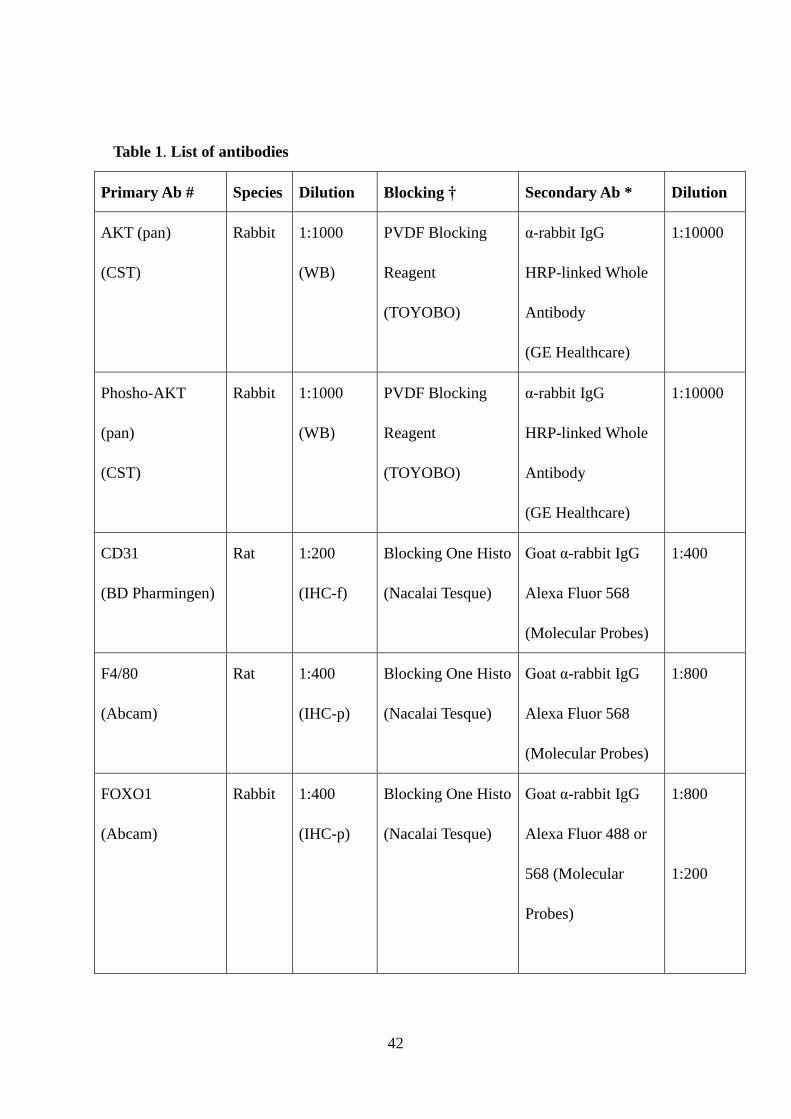
Table 1. List of antibodies
Primary Ab # Species Dilution Blocking † Secondary Ab * Dilution
AKT (pan)
(CST)
Rabbit 1:1000
(WB)
PVDF Blocking
Reagent
(TOYOBO)
α-rabbit IgG
HRP-linked Whole
Antibody
(GE Healthcare)
1:10000
Phosho-AKT
(pan)
(CST)
Rabbit 1:1000
(WB)
PVDF Blocking
Reagent
(TOYOBO)
α-rabbit IgG
HRP-linked Whole
Antibody
(GE Healthcare)
1:10000
CD31
(BD Pharmingen)
Rat 1:200
(IHC-f)
Blocking One Histo
(Nacalai Tesque)
Goat α-rabbit IgG
Alexa Fluor 568
(Molecular Probes)
1:400
F4/80
(Abcam)
Rat 1:400
(IHC-p)
Blocking One Histo
(Nacalai Tesque)
Goat α-rabbit IgG
Alexa Fluor 568
(Molecular Probes)
1:800
FOXO1
(Abcam)
Rabbit 1:400
(IHC-p)
Blocking One Histo
(Nacalai Tesque)
Goat α-rabbit IgG
Alexa Fluor 488 or
568 (Molecular
Probes)
1:800
1:200
42

Biotinylated
α-rabbit IgG (Vector
lab)
FOXO1
(CST)
Rabbit 1:1000
(WB)
PVDF Blocking
Reagent
(TOYOBO)
α-rabbit IgG
HRP-linked Whole
Antibody
(GE Healthcare)
1:10000
Phosho-FOXO1
(Thr24)
(CST)
Rabbit 1:1000
(WB)
PVDF Blocking
Reagent
(TOYOBO)
α-rabbit IgG
HRP-linked Whole
Antibody
(GE Healthcare)
1:10000
FOXO3A
(CST)
Rabbit 1:1000
(WB)
PVDF Blocking
Reagent
(TOYOBO)
α-rabbit IgG
HRP-linked Whole
Antibody
(GE Healthcare)
1:10000
Phosho-FOXO3A
(Ser318/321)
(CST)
Rabbit 1:1000
(WB)
PVDF Blocking
Reagent
(TOYOBO)
α-rabbit IgG
HRP-linked Whole
Antibody
(GE Healthcare)
1:10000
FOXO4
(CST)
Rabbit 1:1000
(WB)
PVDF Blocking
Reagent
α-rabbit IgG
HRP-linked Whole
1:10000
43

(TOYOBO) Antibody
(GE Healthcare)
p44/42 MAPK
(ERK1/2)
(CST)
Rabbit 1:1000
(WB)
PVDF Blocking
Reagent
(TOYOBO)
α-rabbit IgG
HRP-linked Whole
Antibody
(GE Healthcare)
1:10000
Phosho-p44/42
MAPK (ERK1/2)
(CST)
Rabbit 1:1000
(WB)
1:200
(ICC)
PVDF Blocking
Reagent
(TOYOBO)
Blocking One Histo
(Nacalai tesque)
α-rabbit IgG
HRP-linked Whole
Antibody
(GE Healthcare)
Goat α-rabbit IgG
Alexa Fluor 568
(Molecular Probes)
1:10000
1:1000
PCNA
(abcam)
mouse 1:400
(IHC-p
and -f)
Blocking One Histo
(Nacalai tesque)
Histofine
mousestain kit
(Nichirei
biosciences Inc)
Myosin IIb
(CST)
Rabbit 1:1000
(WB)
PVDF Blocking
Reagent
(TOYOBO)
α-rabbit IgG
HRP-linked Whole
Antibody
1:10000
44
(GE Healthcare)
NF-κB p65
(CST)
Rabbit 1:1000
(WB)
PVDF Blocking
Reagent
(TOYOBO)
α-rabbit IgG
HRP-linked Whole
Antibody
(GE Healthcare)
1:10000
Phospho-NF-κB
p65 (Ser536)
(CST)
Rabbit 1:1000
(WB)
PVDF Blocking
Reagent
(TOYOBO)
α-rabbit IgG
HRP-linked Whole
Antibody
(GE Healthcare)
1:10000
Neutrophil
(Abcam)
Rat 1:400
(IHC-p)
Blocking One Histo
(Nacalai Tesque)
Goat α-rabbit IgG
Alexa Fluor 488
(Molecular Probes)
1:800
α-tubulin
(Abcam)
Rabbit 1:1000
(WB)
PVDF Blocking
Reagent
(TOYOBO)
α-rabbit IgG
HRP-linked Whole
Antibody
(GE Healthcare)
1:10000
β-actin
(Abcam)
Rabbit 1:5000
(WB)
PVDF Blocking
Reagent
(TOYOBO)
α-rabbit IgG
HRP-linked Whole
Antibody
(GE Healthcare)
1:10000
Abbreviations: CST, Cell Signaling Technology; f, frozen section; ICC,
45

immunocytochemistry; p, paraffin section; WB, western immunoblotting.
* Overnight incubation at 4°C (IHC-p, HIC-f, ICC and WB).
† Blocking time: 2 h at room temperature.
‡ Incubation for 1 h at room temperature.
46
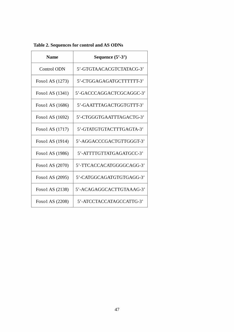
Table 2. Sequences for control and AS ODNs
Name Sequence (5’-3’)
Control ODN 5’-GTGTAACACGTCTATACG-3’
Foxo1 AS (1273) 5’-CTGGAGAGATGCTTTTTT-3’
Foxo1 AS (1341) 5’-GACCCAGGACTCGCAGGC-3’
Foxo1 AS (1686) 5’-GAATTTAGACTGGTGTTT-3’
Foxo1 AS (1692) 5’-CTGGGTGAATTTAGACTG-3’
Foxo1 AS (1717) 5’-GTATGTGTACTTTGAGTA-3’
Foxo1 AS (1914) 5’-AGGACCCGACTGTTGGGT-3’
Foxo1 AS (1986) 5’-ATTTTGTTATGAGATGCC-3’
Foxo1 AS (2070) 5’-TTCACCACATGGGGCAGG-3’
Foxo1 AS (2095) 5’-CATGGCAGATGTGTGAGG-3’
Foxo1 AS (2138) 5’-ACAGAGGCACTTGTAAAG-3’
Foxo1 AS (2208) 5’-ATCCTACCATAGCCATTG-3’
47
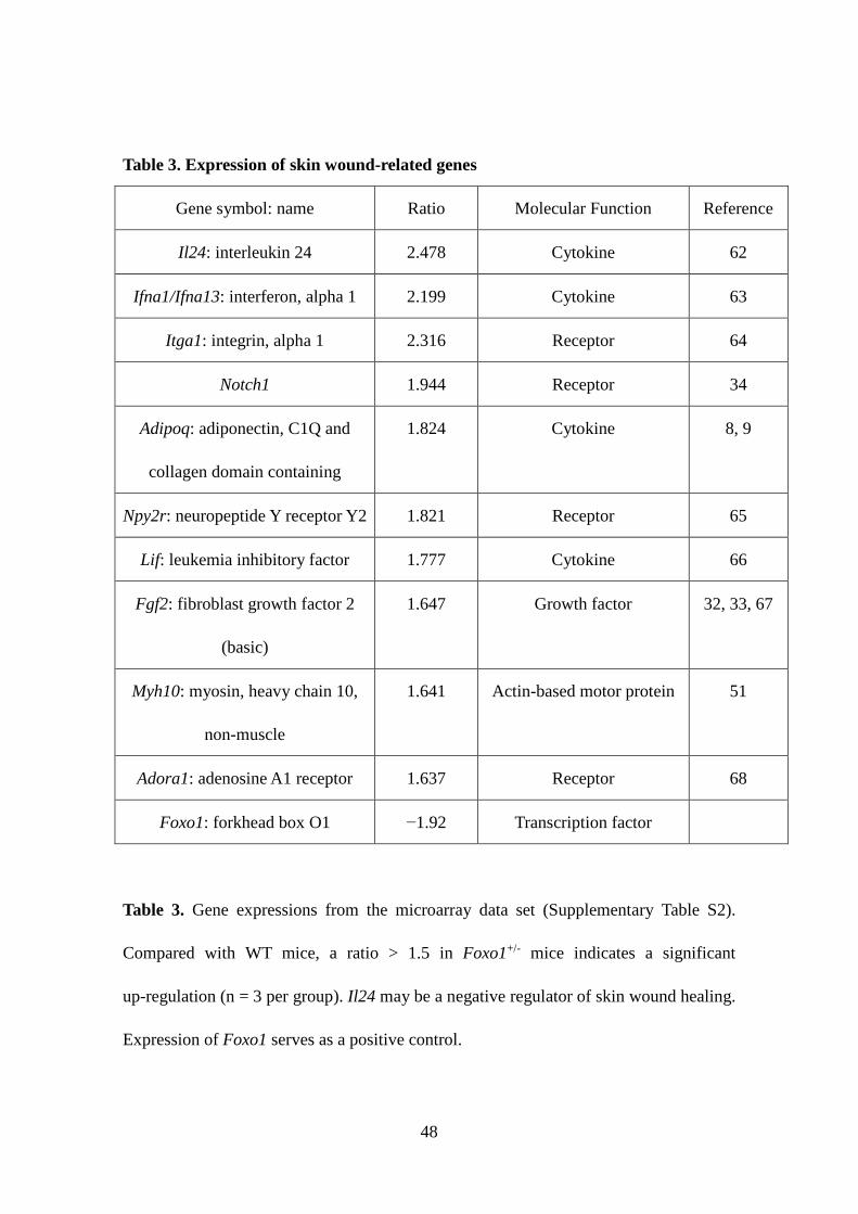
Table 3. Expression of skin wound-related genes
Gene symbol: name Ratio Molecular Function Reference
Il24: interleukin 24 2.478 Cytokine 62
Ifna1/Ifna13: interferon, alpha 1 2.199 Cytokine 63
Itga1: integrin, alpha 1 2.316 Receptor 64
Notch1 1.944 Receptor 34
Adipoq: adiponectin, C1Q and
collagen domain containing
1.824 Cytokine 8, 9
Npy2r: neuropeptide Y receptor Y2 1.821 Receptor 65
Lif: leukemia inhibitory factor 1.777 Cytokine 66
Fgf2: fibroblast growth factor 2
(basic)
1.647 Growth factor 32, 33, 67
Myh10: myosin, heavy chain 10,
non-muscle
1.641 Actin-based motor protein 51
Adora1: adenosine A1 receptor 1.637 Receptor 68
Foxo1: forkhead box O1 −1.92 Transcription factor
Table 3. Gene expressions from the microarray data set (Supplementary Table S2).
Compared with WT mice, a ratio > 1.5 in Foxo1+/- mice indicates a significant
up-regulation (n = 3 per group). Il24 may be a negative regulator of skin wound healing.
Expression of Foxo1 serves as a positive control.
48


