neuropathology - semmelweis egyetem
TRANSCRIPT
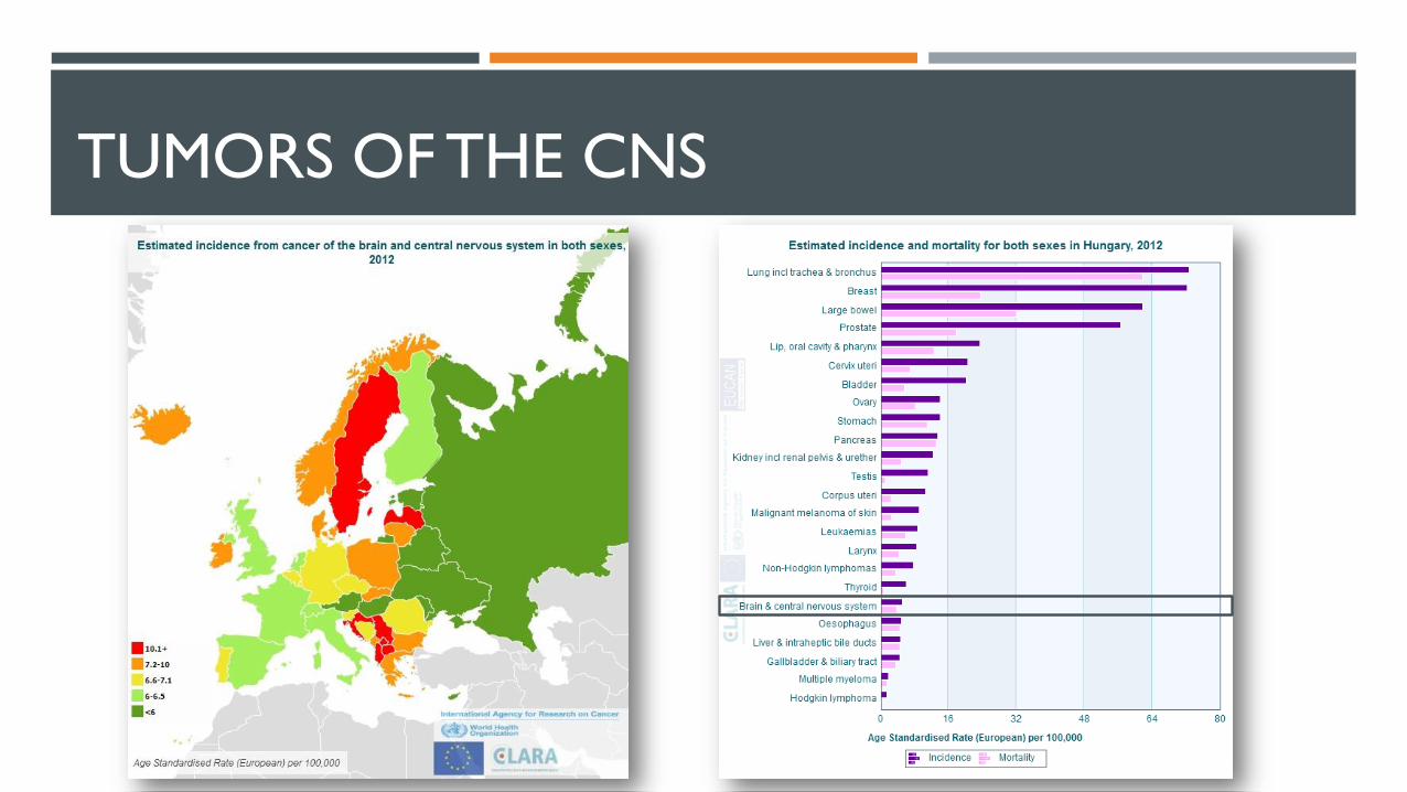
TUMORS OF THE CNS

32%
23%
10%
7%
6%
5%
4%
3%3%
7%
INCIDANCE OF CHILDHOOD NEOPLASMS
Leukemia
CNS
Lymphoma
Neuroblastoma
Kidney tumor
Bone tumor
Rhabdomyosarcoma
Retinoblastoma
Germ cell tumor
Egyéb

PRIMARY TUMORS OF THE CNS
1. Gliomas
2. Neuronal or mixed glioneural tumors
3. Choroid plexus neoplasms
4. Embryonal tumors
5. Meningial tumors
6. Other parenchymal tumors
• Haematologic malignancies
• Germ cell tumors
Glioblastoma
21%Astrocytoma
10%
Ependymoma
2%
Oligodendroglioma
4%
Embrional
tumors
2%
Meningioma
31%
Hypophysis
tumors
6%
Lymphoma
3%
Other
21%

CHARACTERISTICS
• Do not have premalignant or in situ stages
• Rarely spread outside of the CNS
• Symptoms• Epilepsy (focal or generalized)
• Focal neurologic deficits
• Signs of raised intracranial pressure
• Hydrocephalus
• Diagnosis• Age
• Sex
• Site of neoplasm
• Family history
Grade• Predicting the biological behaviour
• Grade 1
• Low proliferative potential
• Possibility of curative resection
• Grade II
• Infiltrative
• Often recur
• Progression
• Grade III
• Histological evidence of malignancy
• High mitotic activity, atypia
• Grade IV
• Cytologically malignant
• Rapid disease evolution
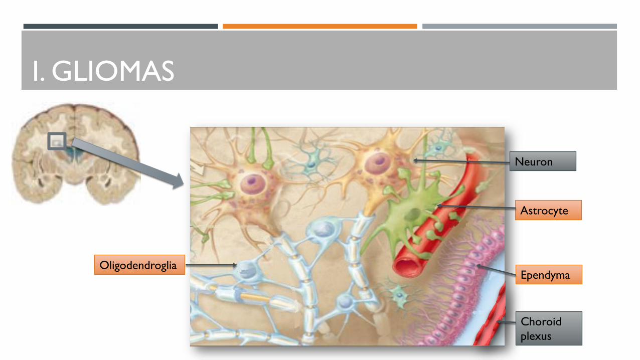
I. I. GLIOMAS
Oligodendroglia
Astrocyte
Ependyma
Neuron
Choroid
plexus

I. I. Astrocytoma
1. Pilocytic astrocytoma (Grade I)• Children and young adults
• Benign tumors
• Most commonly infratentorial – Cerebellum
Rosenthal fibers
Eosinophilic granular bodies
Astrocyte

1I. Diffuse astrocytoma (Grade II-IV)
Astrocyta
• Fourth through the sixth decades of life
• Cerebral hemispheres – Focal signs, headache
Well-differentiated astrocytoma Grade II
• Atypia+ High cellularity
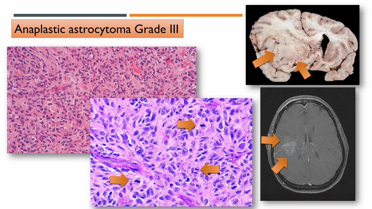
Anaplastic astrocytoma Grade III
• Atypia + High cellularity + High mitotic activity
Glioblastoma Grade IV
• Atypia + High cellularity + High mitotic activity +
Necrosis/Endothel proliferation

Well-differentiated astrocytoma Grade II
Anaplastic astrocytoma Grade III
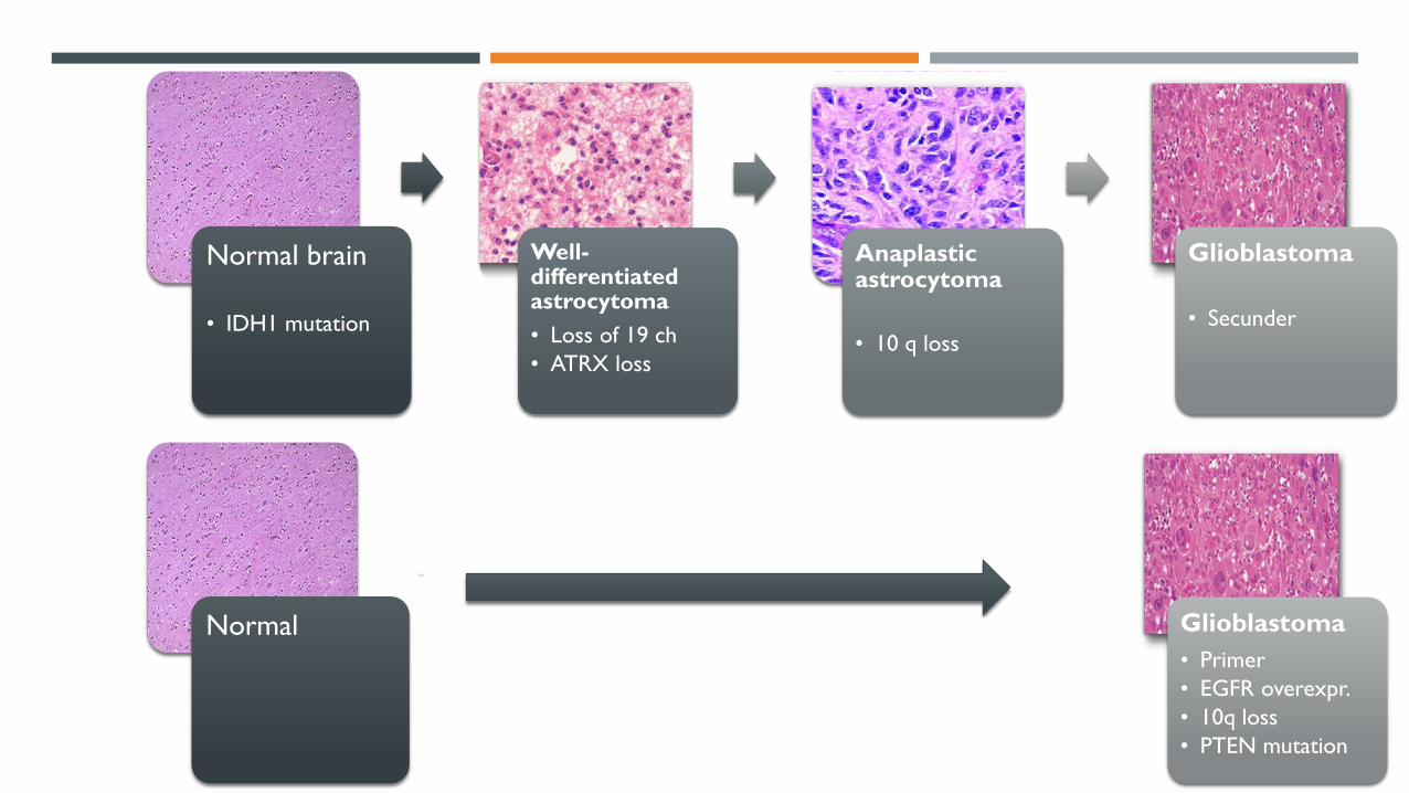
Normal brain
• IDH1 mutation
Well-differentiatedastrocytoma
• Loss of 19 ch
• ATRX loss
Anaplasticastrocytoma
• 10 q loss
Glioblastoma
• Secunder
Normal Jól differenciált diffúz astrocytoma
• Kr 19. vesztés
• ATRX vesztés
Anaplasztikusastrocytoma
• 10 q vesztés
Glioblastoma
• Primer
• EGFR overexpr.
• 10q loss
• PTEN mutation

Glioblastoma Grade IV
Vascular
proliferation
Necrosis
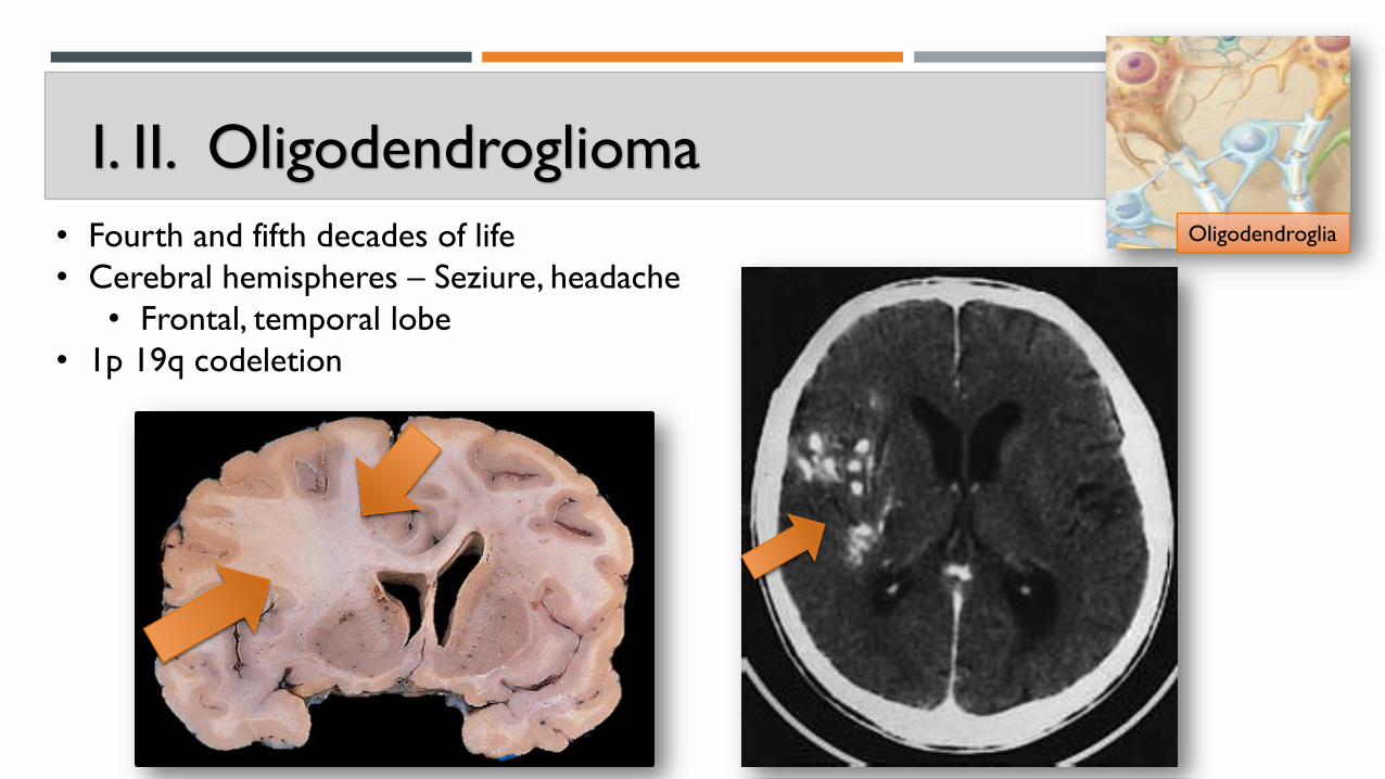
I. II. Oligodendroglioma
• Fourth and fifth decades of life
• Cerebral hemispheres – Seziure, headache
• Frontal, temporal lobe
• 1p 19q codeletion
Oligodendroglia

Well-differentiated oligodendroglioma
Grade II
Anaplastic oligodendroglioma
Grade III
Vascular
proliferation

I. III. Ependymoma
Ependyma• Intracranial – Childhood
• IV. ventricle, III. ventricle
• Spinal ependymoma – 20-40 years of age

Ependymoma Grade IIAnaplastic ependymoma
Grade III
Necrosis
Pseudorosettes

I. II. NEURONAL/GLIONEURONAL TUMORS
Oligodendroglia
Astrocyte
Ependyma
Neuron
Choroid
plexus

I. Central neurocytoma Grade II
• Intraventricular neoplasms located predominantly in
the vicinity of the septum pellucidum
• Young adultsNeuron

II. Gangliocytoma, ganglioglioma Grade I
• Mature appearing neurons ± Glial cells
• Glial compomonent – with time anaplasticNeuron
Ganglion cells
Ganglion cells
Glial cells
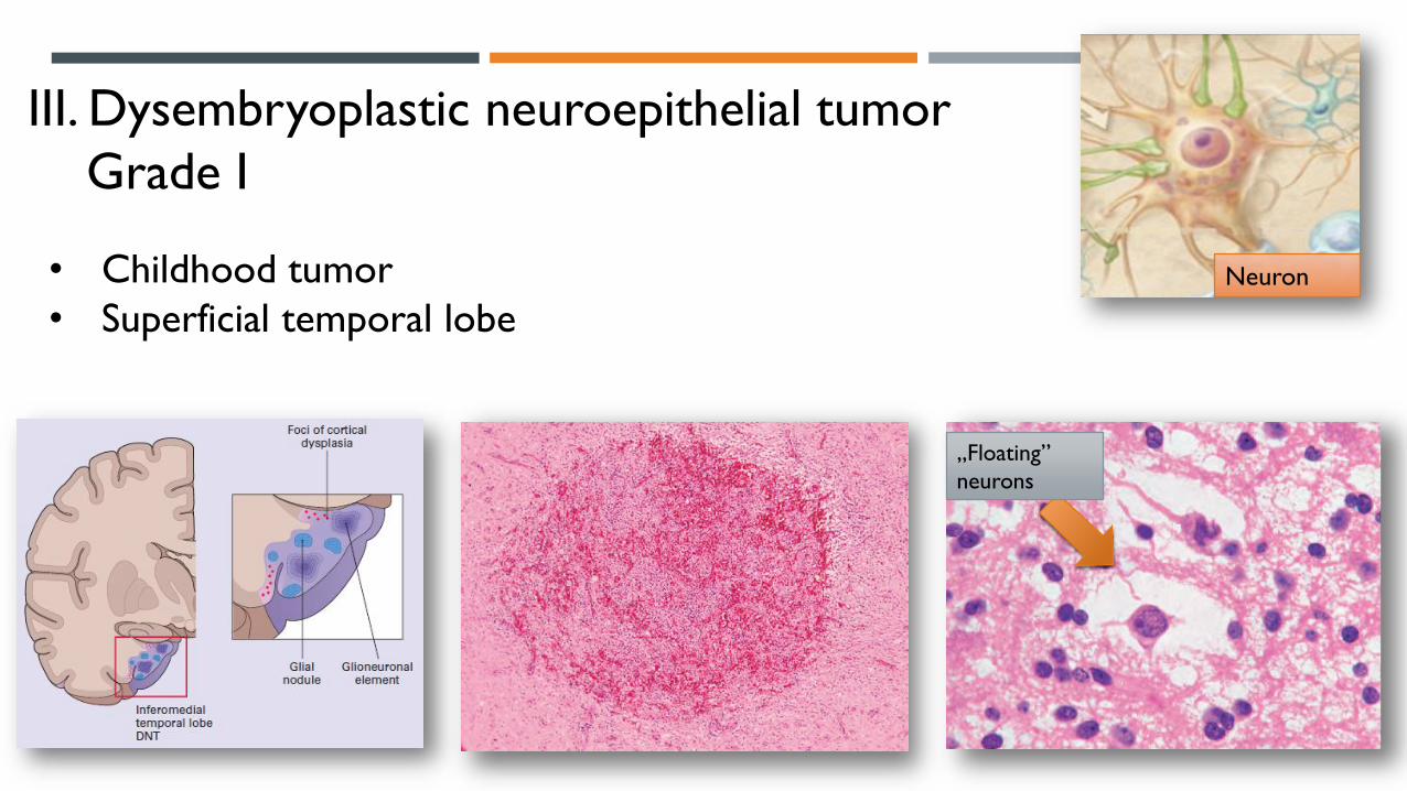
III. Dysembryoplastic neuroepithelial tumor
Grade I
• Childhood tumor
• Superficial temporal lobeNeuron
„Floating”
neurons

I. III. CHOROID PLEXUS NEOPLASMS
Oligodendroglia
Astrocyte
Ependyma
Neuron
Choroid
plexus

Choroideus plexus papilloma Plexus choroideus carcinoma
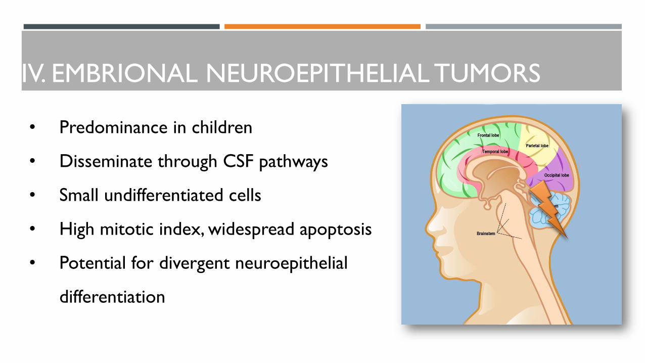
I. IV. EMBRIONAL NEUROEPITHELIAL TUMORS
• Predominance in children
• Disseminate through CSF pathways
• Small undifferentiated cells
• High mitotic index, widespread apoptosis
• Potential for divergent neuroepithelial
differentiation
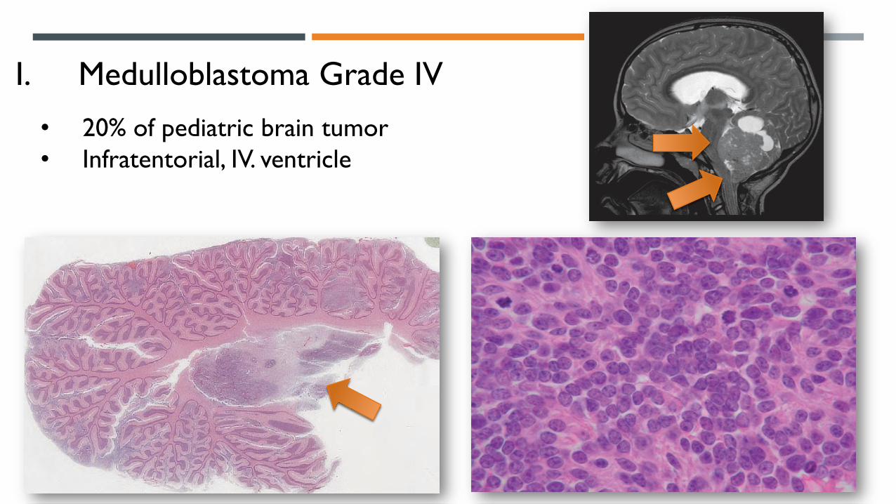
I. Medulloblastoma Grade IV
• 20% of pediatric brain tumor
• Infratentorial, IV. ventricle

II. Atypical teratoid/rhabdoid tumor (ATRT) Grade IV
• Most commonly <5 years
• Poor prognosis
• Anywhere in the CNS
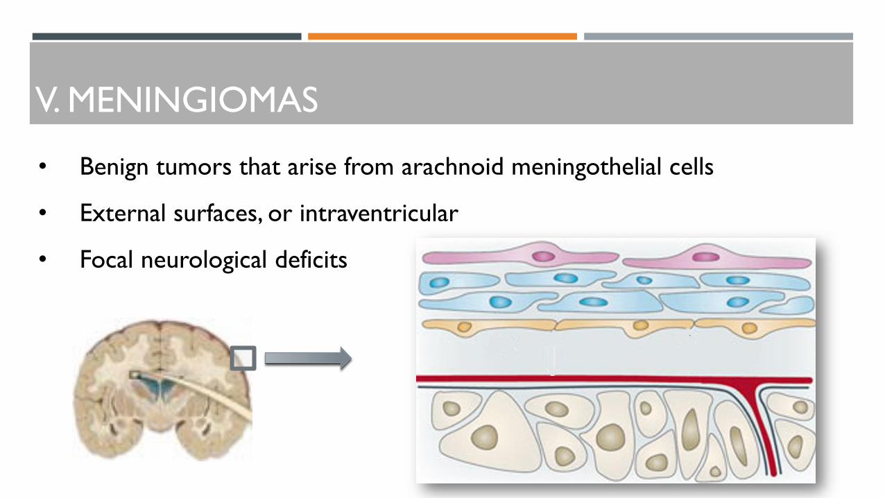
I. V. MENINGIOMAS
• Benign tumors that arise from arachnoid meningothelial cells
• External surfaces, or intraventricular
• Focal neurological deficits

I. Meningioma Grade 1.
• Incidence rises with age
• Primary CNS tumors ~30% meningioma
• Several histological variants
Psammoma
body

II. Atypical Meningioma
Grade 1I.
III. Anaplastic Meningioma
Grade I1I.
Ki67
Necrosis

I. VI. PRIMARY CNS LYMPHOMA
• DLBCL type
• Most common CNS neoplasm in
immunosuppressed persons
• nearly always positive for the EBV

I. VII. CNS GERM CELL TUMORS
• Germinoma - 50%
• Teratoma - 20%• Mature teratoma
• Immature teratoma
• Teratoma with malignant transformation
• Yolk sac tumor
• Embryonal carcinoma 5%
• Choriocarcinoma
• Mixed tumor - 25%
Germinoma
Teratoma
Choriocarcinoma

I. VIII. METASTATIC TUMORS
Lung tumor Breast tumor
Melanoma Gasrointestinal tumor
Kidney Other
• Gray-white junction
• Sharply demarcated masses
• Perifocal edema
Breast carcinomaSCLC

INFECTIONS OF THE NERVOUS SYSTEM
1. Parenchyma: encephalitis , myelitis, encephalomyelitis.
2. Meninges: meningitis, pachymeningitis.
3. Parenchyma and meninges: meningoencephalitis.
Localisation:

I. SPREAD
1. Hematogenous spread
2. Direct implantation – Trauma, Iatrogenic
3. Local extension – Otitis media, Congenital malformation
4. Peripheral nerves

I. INFECTIOUS AGENTS
1. Baterial infection• Bakterial meningitis
• Brain abcessus
• Tuberculosis
• Neurosyphilis
2. Virus infection• Viral meningitis
• Herpesvirus
• Cytomegalovirus
• Poliovirus
• Rabies
• HIV
• Progressive multifocal leukoencephalopathia
3. Fungal infection• Candida
• Mucormycosis
• Aspergillus
• Cryptococcus
4. Protozoal infection• Toxoplasma
5. Parazite infection• Cystercosis
• Echinococcus
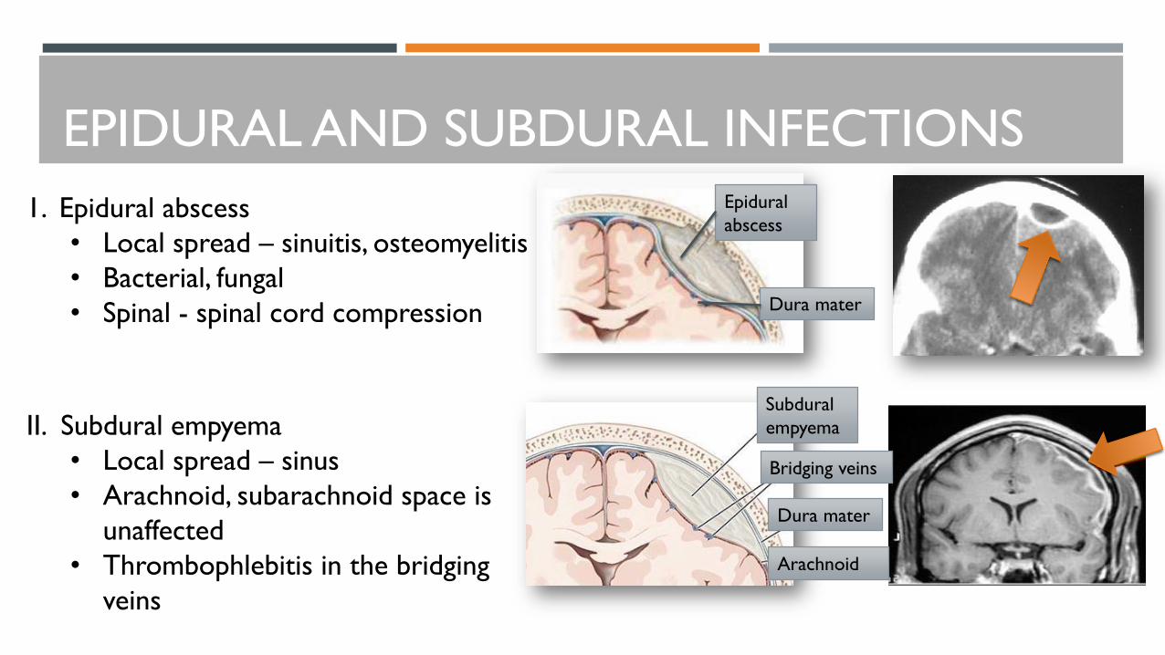
EPIDURAL AND SUBDURAL INFECTIONS
1. Epidural abscess
• Local spread – sinuitis, osteomyelitis
• Bacterial, fungal
• Spinal - spinal cord compression
II. Subdural empyema
• Local spread – sinus
• Arachnoid, subarachnoid space is
unaffected
• Thrombophlebitis in the bridging
veins
Epidural
abscess
Dura mater
Subdural
empyema
Dura mater
Bridging veins
Arachnoid
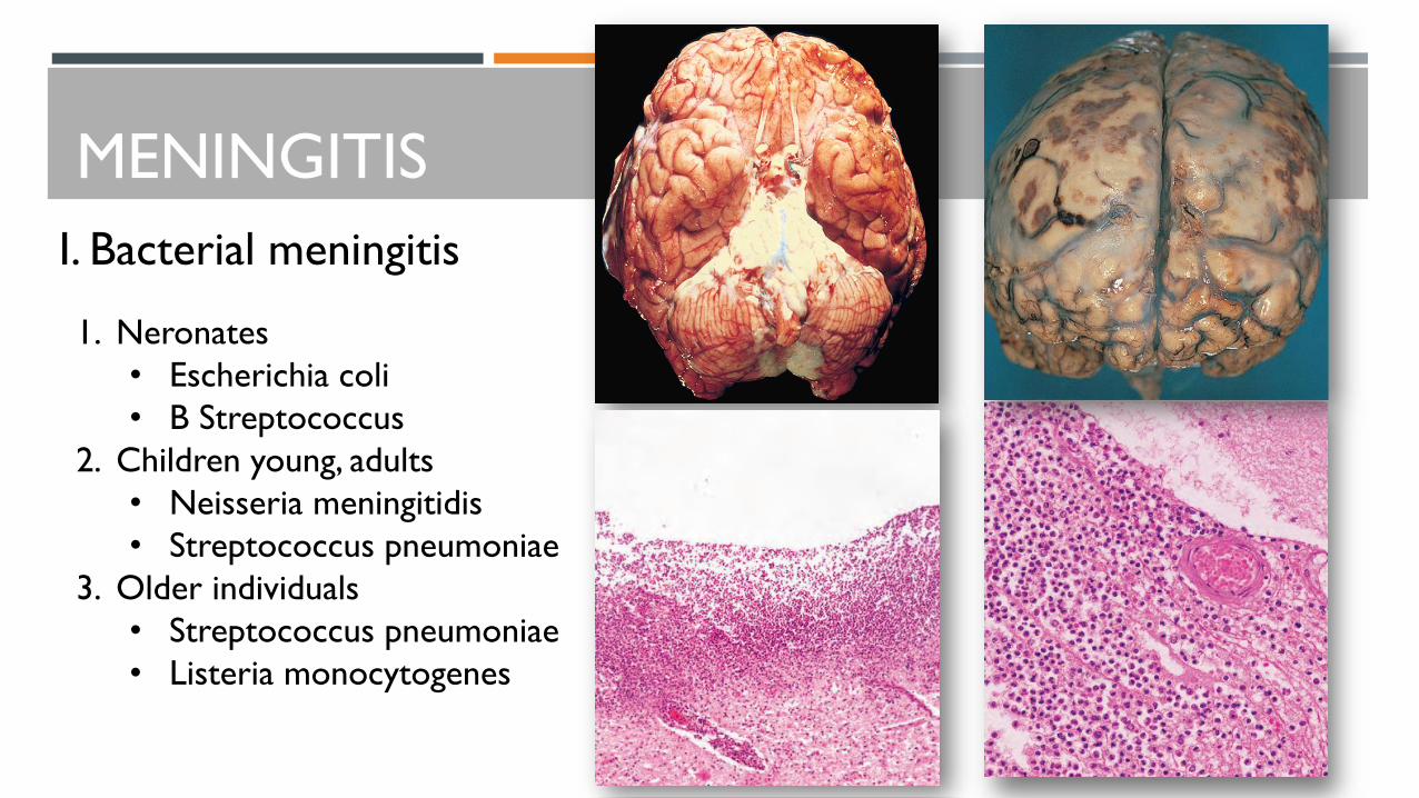
MENINGITIS
1. Neronates
• Escherichia coli
• B Streptococcus
2. Children young, adults
• Neisseria meningitidis
• Streptococcus pneumoniae
3. Older individuals
• Streptococcus pneumoniae
• Listeria monocytogenes
I. Bacterial meningitis

II. Aseptic/Viral meningitis
Echovirus
Coxsackie B
Coxsackie A
Herpes simplex virus (HSV)-2
Mumps
Human immunodeficiency virus (HIV)
Lymphochoriomeningitis virus
Arbovirus
Rubeola
Parainfluenza virus
Adenovirus

III. Chronic meningitis
III.I. Mycobacterium tuberculosis• Meningitis – Fibrinuos exudate
• Intraparenchymal mass (tuberculoma)
• Chronic tuberculotic infection - arachnoideal
fibrosis - hydrocephalus
III.II. Spirochaetal infections
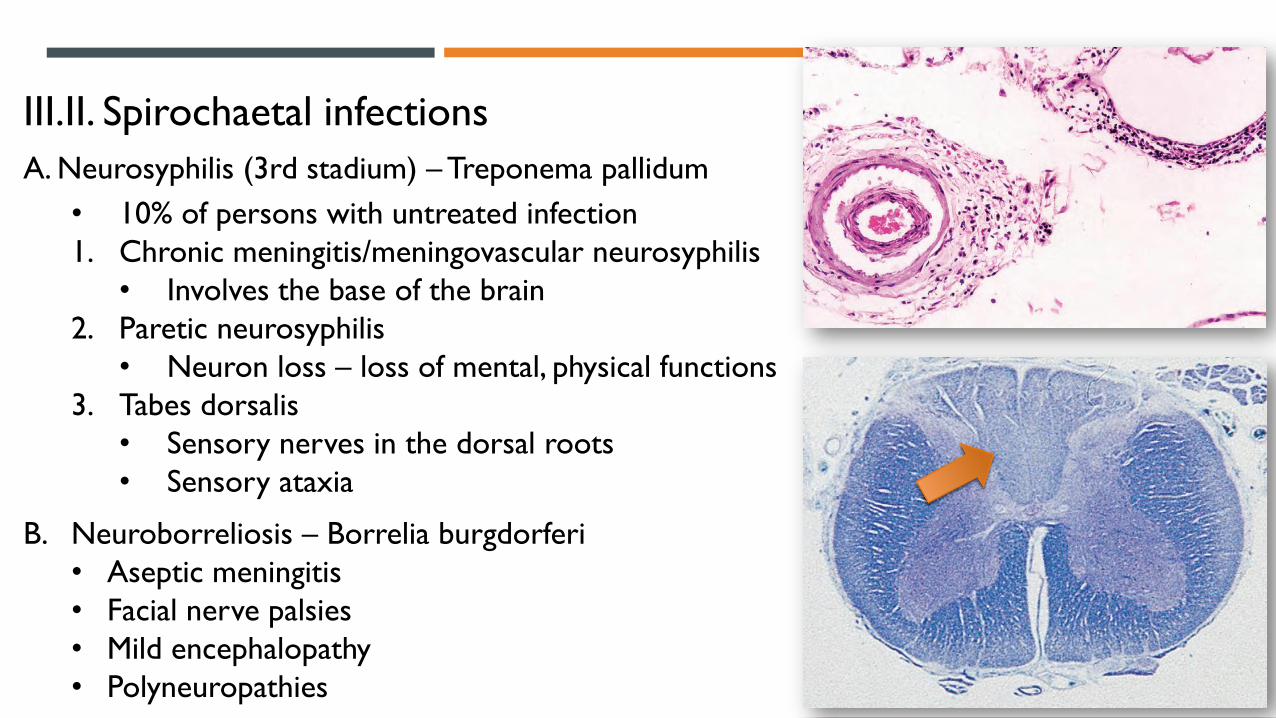
A. Neurosyphilis (3rd stadium) – Treponema pallidum
• 10% of persons with untreated infection
1. Chronic meningitis/meningovascular neurosyphilis
• Involves the base of the brain
2. Paretic neurosyphilis
• Neuron loss – loss of mental, physical functions
3. Tabes dorsalis
• Sensory nerves in the dorsal roots
• Sensory ataxia
B. Neuroborreliosis – Borrelia burgdorferi
• Aseptic meningitis
• Facial nerve palsies
• Mild encephalopathy
• Polyneuropathies
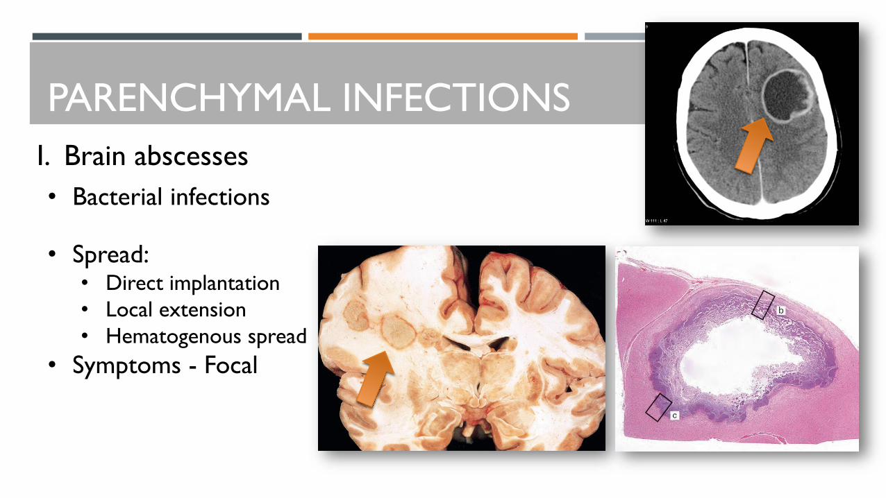
PARENCHYMAL INFECTIONS
I. Brain abscesses
• Bacterial infections
• Spread: • Direct implantation
• Local extension
• Hematogenous spread
• Symptoms - Focal
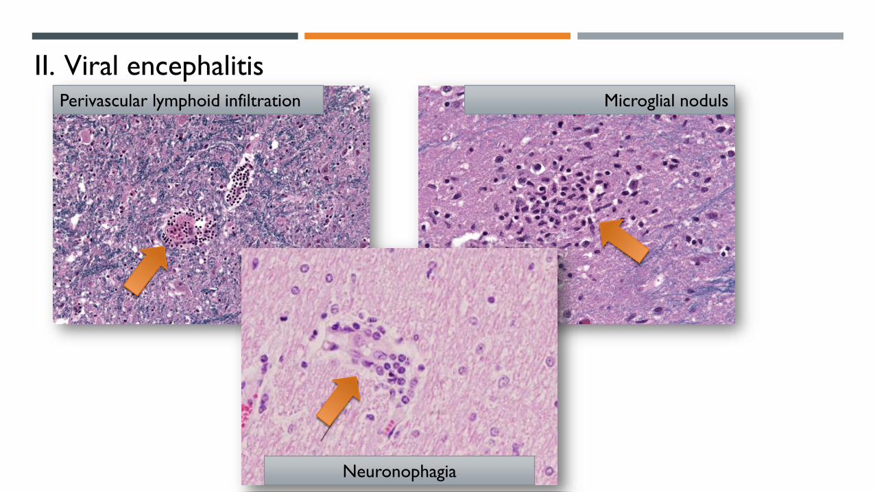
II. Viral encephalitisPerivascular lymphoid infiltration Microglial noduls
Neuronophagia

II. I. Herpes virus
A. Herpes simplex-1
• Children and young adults
• Frontal, temporal lobe involvement
• Necrotizing encephalitis
B. Herpes simplex-2
• Adults
• Viral meningitis
• Primary HSV genital inf - neonates
C. Varicella zoster
• Immunosuppressed patients
• HZV encephalitis
Necrotizing encephalitis
Cowdry A body

II. II. Cytomegalovirus
A. Fetus
• Periventricular necrosis
• Microcephalia
• Periventricular calcification
B. Adult
• Immunosuppressed persons
• Periventricular
• Subacut encephalitis
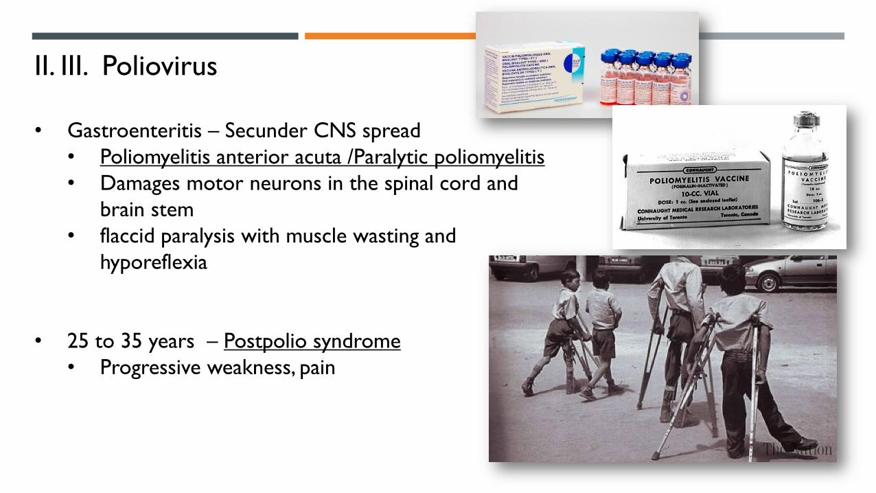
II. III. Poliovirus
• Gastroenteritis – Secunder CNS spread
• Poliomyelitis anterior acuta /Paralytic poliomyelitis
• Damages motor neurons in the spinal cord and
brain stem
• flaccid paralysis with muscle wasting and
hyporeflexia
• 25 to 35 years – Postpolio syndrome
• Progressive weakness, pain

II. IV. RabiesVirus
• Rabies
• Rabid animals, usually by a bite
• Ascending along the peripheral nerves
• the incubation period depends on the
distance between the wound and the brain
• Symptoms:
• Non specific
• Signs of CNS excitability
• Pain, hydrophobia
• Mania-coma
Retrograde trans-synaptic spread

II. V. Human Immundeficiency virus
A. Aseptic meningitis
• Within 1 to 2 weeks of onset of primary infection by
HIV in about 10% of patients
B. HIV Encephalitis (HIVE)
• Perivascular lymphoid infiltration
• Myelin loss in the hemispheres (Leukoencephalopathia)
• Microglial noduls
• Giant cells
C. Opportunistic infections
D. Primary CNS lymphoma

II. VI. JC virus / Progressive multifocal leukoencephalopathy
• Polyoma virus
• Infects oligodendroglial cells
• Demyelinisation
• White matter – Hemispheres, Cerebellum
• Progressive neurologic symptoms

III. Fungal infections
A. Candida Albicans• Multiplex microabscessusok
B. Mucormycosis• Nasal cavity, sinus infection
• Direct extension, Vascular invasion
C. Aspergillus fumigatus• Hemorrhagic infarctions
• Vascular invasion
D. Cryptococcus neoformans• Meningitis, Meningoencephalitis
• Fulminant

Candida albicans Mucormycosis
Aspergillus fumigatus Cryptococcus neoformans
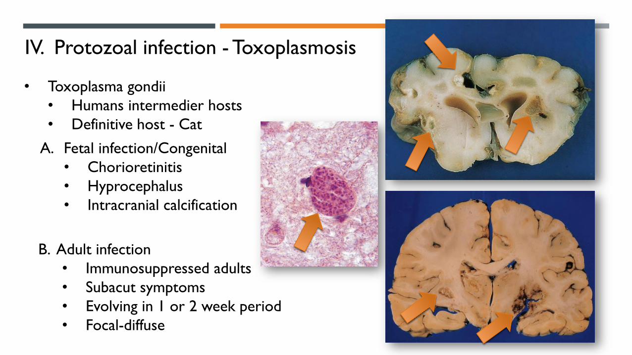
IV. Protozoal infection - Toxoplasmosis
• Toxoplasma gondii
• Humans intermedier hosts
• Definitive host - Cat
A. Fetal infection/Congenital
• Chorioretinitis
• Hyprocephalus
• Intracranial calcification
B. Adult infection
• Immunosuppressed adults
• Subacut symptoms
• Evolving in 1 or 2 week period
• Focal-diffuse

IV. Parazitic infection
I. Tenia solium - Cysticercosis
• End-stage infection
• Larval organisms leave the lumen of the
gastrointestinal tract
• Encyst – Brain – subarachnoid space
• Symptoms
• Focal symptoms
• Epilepsy

2. Echinococcus /Hydatidosis/
• Childhood
• Contact with dogs
• Encysts – Usually liver, lung rarely brain
• Symptoms
• Focal signs
• Epilepsy

Robbins Basic Pathology, 9th Edition
Neuropathology: A Reference Text of
CNS Pathology, 3rd Edition