stabilisation of pcr reagents for storing at room temperature · stabilisation of pcr reagents for...
TRANSCRIPT
Stabilisation of PCR reagents for storing at room
temperature
Hana Crlíková
Propojení výuky oborů Molekulární a buněčné biologie a Ochrany a tvorby životního prostředí OPVK
(CZ.1.07/2.2.00/28.0032)

Main aims
• Suitable mixtures
• Tablets
• Stabilisation of reagents
Substances used
• Pullulan - polysacharide
• Trehalose - disacharide
• Xanthan - polysacharide
procedure
• Dissolving of substances
• Dilution series
• RT PCR
• Drying of pullulan, xanthan, trehalose
• Combination of pullulan, xanthan,
trehalose
sources
• Zagon, Jutta, et al. (2012): "Preservation of
primer and probes on "ready-to-use" 96-well
microtiter plates: A step forward towards
enhancing throughput and harmonization of
real-time PCR applications in food and feed
control." Food Control 25.2: 709- 716.
• JahanshahiAnbuhi, Sana, et al. (2014):
"Pullulan Encapsulation of Labile
Biomolecules to Give Stable Bioassay
Tablets." Angewandte Chemie International
Edition 53(24):6155-8.
• Rombach, Markus, et al. (2014):
"Benchmarks. Real-time stability testing of
air-dried primer and fluorogenic hydrolysis
probes stabilized by trehalose and xathan"
BioTechniques 57: 151-155.

Thank you for your attention

Identification of protein factors
specifically binding to the p16
promoter region under replication
stress conditions
Martin Liptay
Propojení výuky oborů Molekulární a buněčné biologie a Ochrany a tvorby životního prostředí OPVK
(CZ.1.07/2.2.00/28.0032)
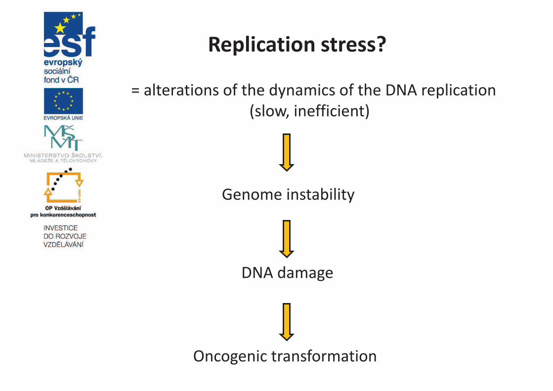
Replication stress?
= alterations of the dynamics of the DNA replication
(slow, inefficient)
Genome instability
DNA damage
Oncogenic transformation
Senescence?
- irreversible growth arrest, morphology of the cells is
changed (flat, enlarged shapes), cells become
resistant to apoptosis
- Program triggered by a variety of stresses, including
DNA damage, telomere erosion, oncogenic
activation…
- Triggered through the activation of expression of cdk
inhibitors like p16 and p21
- Normal physiological process, important tumor
supressor mechanism!
p16
- Cdk4/6 inhibitor, tumor supressor activity
- Cellular senescence
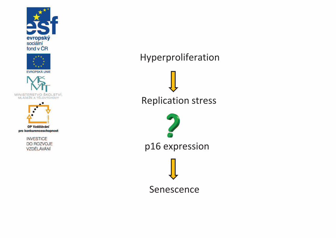
Hyperproliferation
Replication stress
p16 expression
Senescence
enCHIP
• ENgineered DNA-binding molecule-
mediated CHromatin
ImunoPrecipitation
• For purification of specific genomic
regions using engineered DNA-
binding molecule – FLAG tagged TALE
protein, catalytically inactive
CRISPR/Cas9; retains molecular
interactions in vivo

FLAG
FLAG
FLAG
https://www.mirusbio.com/applications/crispr-cas-transfection
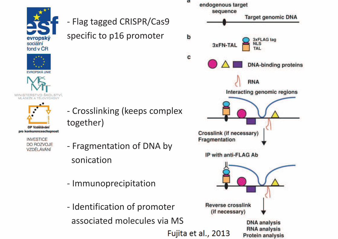
- Flag tagged CRISPR/Cas9
specific to p16 promoter
- Crosslinking (keeps complex
together)
- Fragmentation of DNA by
sonication
- Immunoprecipitation
- Identification of promoter
associated molecules via MS

Sources
• Kong Y, Cui H, Ramkumar C, Zhang H. Regulation of
Senescence in Cancer and Aging. Journal of Aging
Research. 2011;2011:963172.
doi:10.4061/2011/963172.
• Rayess H, Wang MB, Srivatsan ES. Cellular senescence
and tumor suppressor gene p16. International Journal
of Cancer. Journal International du
Cancer 2012;130(8):1715-1725. doi:10.1002/ijc.27316.
• Fujita T, Asano Y, Ohtsuka J, Takada Y, Saito K, Ohkiet R,
Fujii H. Identification of telomere-associated molecules
by engineered DNA-binding molecule-mediated
chromatin immunoprecipitation (enChIP). Sci. Rep. 3.
2013

Thank you for your attention!

The analysis of NOD2/CARD15 polymorphisms in patients with acute myocardial infarction
Department of Pathological Physiology,
Laboratory of cardiogenomic
Faculty of Medicine and Dentistry, Palacký University
and University Hospital Olomouc
Karolína Světlíková
Propojení výuky oborů Molekulární a buněčné biologie a Ochrany a tvorby životního prostředí OPVK
(CZ.1.07/2.2.00/28.0032)
What is it …?
• NOD2 (nucleotide-binding-oligomerization
domain-2) - structure responsible for
microbial sensing
• CARD15 (caspase activation recruitment
domain family-15) – the gene coding for
NOD2 protein
• NOD2/CARD15 - gene involved in the
innate immune response and inflammation

• Accute myocardial infarction (AMI) -
heart attack → the patient directly life-
threatening → rupture of an
atherosclerotic plaque
• Inflammation and immune response →
an important role in the pathogenesis of
atherosclerosis
Aims of the practical part of the thesis:
• Genotyping of SNPs (TNF-308, R702W,
G908R, 1007 Ins C) of NOD2/CARD15 gene
• Verify of genetic association of the SNPs
with increased risk of the AMI in the
patients
• Statistical evaluation of allelic and
genotypic frequencies in the patients +
analysis of the Hardy-Weinberg
equilibrium

Methods:
• Microisolation of DNA from peripheral
blood of patients
• PCR reactions
– Preamplification
– SAP reaction
– iPLEX reaction
• Preparation of the microarray
• Mass spectrometry (MassArray analysis)
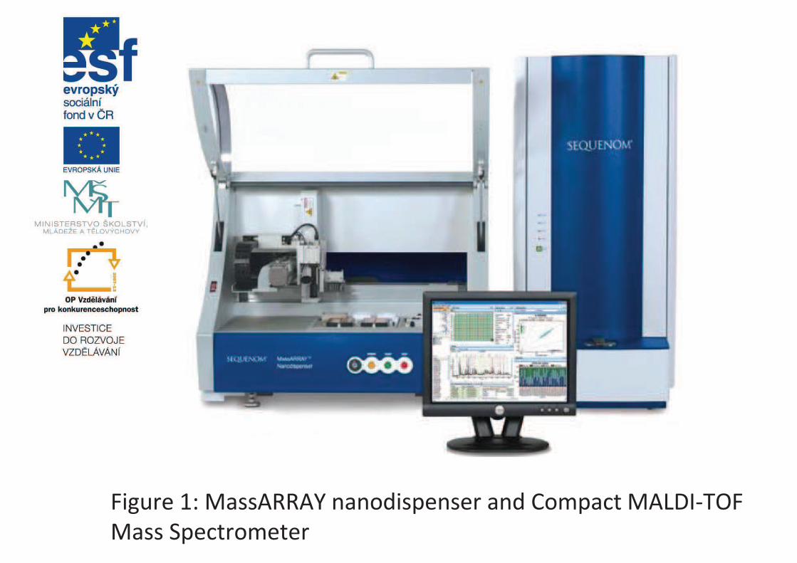
Figure 1: MassARRAY nanodispenser and Compact MALDI-TOF
Mass Spectrometer

Figure 2: Diagram of the genotypes for rs2066844

Sources
http://galleryhip.com/blood-test-tubes.html
www.sequenom.com
www.relax.lidovky.cz
Liu, H.Q., Zhang, X.Y., Edfeldt, K., Nijhuis, M.O., Idborg, H., Bäck,
M., Roy, J., Hedin, U., Jakobsson, P.J., Laman, J.D., de Kleijn, D.P.,
Pasterkamp, G., Hansson, G.K., Yan, Z.Q. (2013): NOD2-Mediated
Innate Immune Signaling Regulates the Eicosanoids in
Atherosclerosis. Arteriosclerosis, Thrombosis, and Vascular
Biology; 33: 2193-2201
Yazdanyar, S., Nordestgaard, B.G. (2010): NOD2/CARD15
genotype, cardiovascular disease and cancer in 43 600
individuals fromthe general population. Journal of Internal
Medicine; 268: 162-170

Thank you for your
attention!

Typing of systemic amyloidosis by
mass spectrometry-based proteomics
Lucie Broskevičová
Propojení výuky oborů Molekulární a buněčné biologie a Ochrany a tvorby životního prostředí OPVK
(CZ.1.07/2.2.00/28.0032)
Typing of systemic amyloidosis ..
• from paraffin embedded tissue using laser
microdissection (LMD) and mass spectrometric-
based proteomic analysis
• determination of the minimum amount of deposits
of amyloidogenic tissue
• from adipose tissue using MS-based proteomics

Amyloidosis
• a rare group of diseases characterized by
deposition of abnormal (misfolded) proteins in
various tissues of the body
• more than 28 types of amyloid
• localized and systemic
• systemic amyloidosis affect more than one organ
or tissue
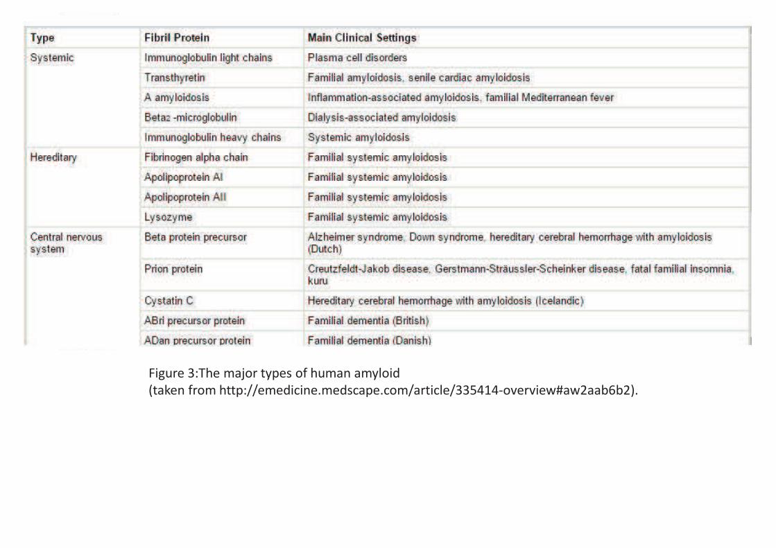
Figure 3:The major types of human amyloid
(taken from http://emedicine.medscape.com/article/335414-overview#aw2aab6b2).

• treatments are available for many types but they are
type specific
• typing of amyloid is essential before starting
treatment
Figure 2: Detection workflow (taken from Dasari et al, 2014).
Method
• Congo Red staining of paraffin-embedded tissue
biopsy
• Congo red-positive areas are dissected by laser
microdissection
• trypsin digestion
• LC-MS analysis
• identification proteins that form the amyloid
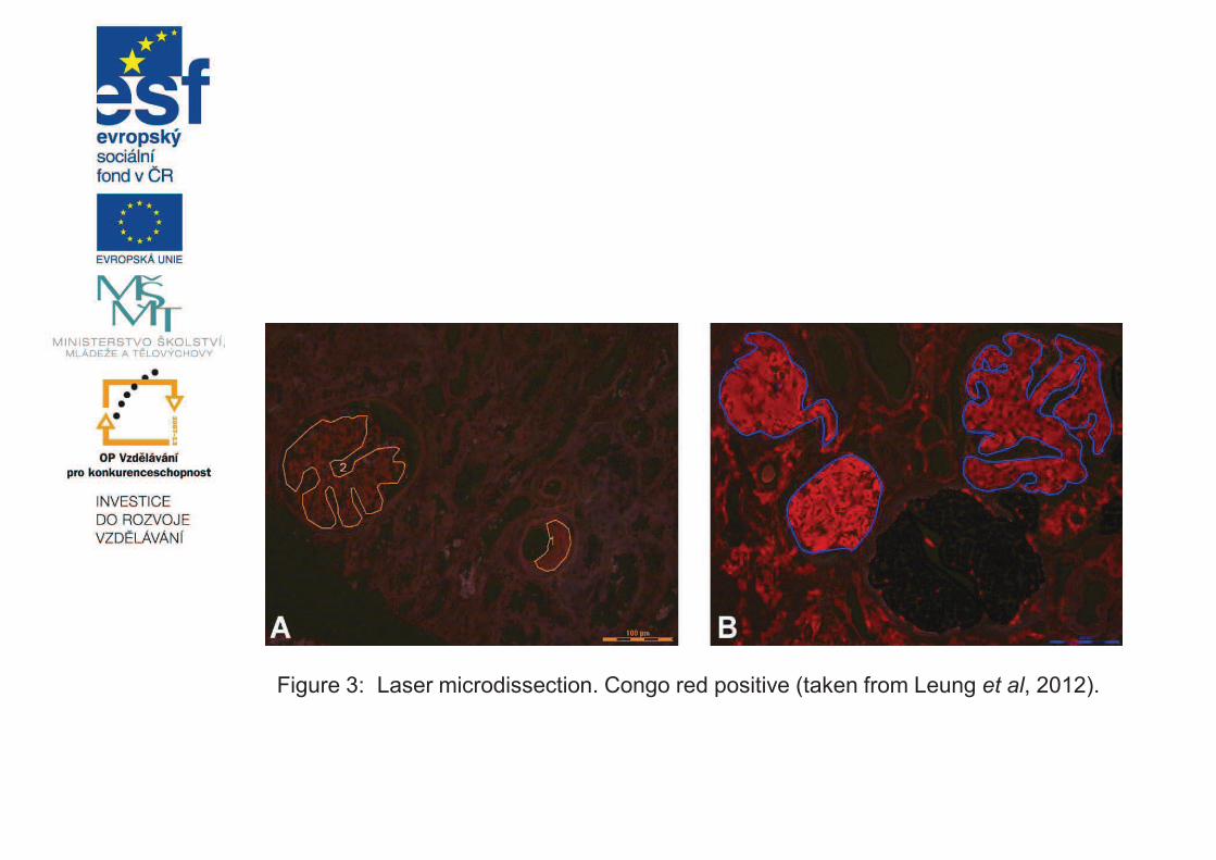
Figure 3: Laser microdissection. Congo red positive (taken from Leung et al, 2012).
Sources
Dasari, S., Theis, J. D., Vrana, J. A., Zenka, R. M., Zimmermann, M. T.,
Kocher, J. P. A., Dogan, A. (2014): Clinical proteome informatics
workbench detects pathogenic mutations in hereditary
amyloidoses. Journal of proteome research 13(5): 2352-2358.
Leung, N., Nasr, S. H., Sethi, S. (2012): How I treat amyloidosis: the
importance of accurate diagnosis and amyloid typing. Blood 120(16):
3206-3213.
www.medscape.com

Thank you for your attention