the cellular basis of life unit two
TRANSCRIPT

38
Unit Two:The Cellular Basis of Life UNC-CH Brain Explorers May be reproduced for non-profit educational use only. Please credit source.
THE CELLULAR BASIS OF LIFE
UNIT TWO

39
The CELLULAR BASIS of LIFESUMMARY KEY POINTS
UNIFYING CONCEPTS*
INTRODUCTION TO MICROSCOPY • • • • • • • • • • • • • • • • • • • • • • • • • • • • • •
Students use hand-held micro-scopes to explore and consider how this tool helps extend the senses. They then observe a photo using the scopes and discover that the pictures are made of dots. Like a picture is made of dots, our body is made of building blocks called cells.
• Big things (such as photos) are made up of little things (dots) that do not necessarily look like the big thing.• Complicated things are made up of simple building blocks. • Microscopes magnify things so that their smaller structural components are visible.
• Scientifi c instruments extend our Sensory experiences. Tools help scientists make better observa-tions. They help scientists see measure and do things that they could not otherwise see, measure and do. * • Students practice skills of observing, discovering, and describing.*
CELLS: THE BUILDING BLOCKS OF LIFE • • • • • • • • • • • • • • • • • • • • • • • •
Students use the Virtual Mi-croscope to explore cells. They become familiar with the parts and operation of a microscope, and view plant and animal cells. In part II, they are shown posters of elodea and human body cells, then draw these and label the parts.
• The cell is the basic building block of all living things. • There many different kids of cells in our bodies.• Every organ in our bodies is made up of cells.• The form of the cell relates to its function.
• Cells have different structures that serve distinct functions in the body.* • Specialized cells perform specialized functions in multicellular organisms. * • Simple instruments such as magnifiers provide more informtation than scientists obtain using only their senses.* •Describe the basic structure and function of human body sysrtems.**
NEURON STRUCTURE • • • • • • • • • • • • • • • • • • • • • • • • • • • • • • • • • • • • • • •
Students learn the parts of the neuron and their function by creating models of neurons out of clay or craft materials. This lesson is followed by a Jeopardy game or quiz to assess learning from the unit.
• Neurons have a very complicated structure compared to other cells.• Neurons are an integral part of a communication network throughout the body.• The shape of neurons of neurons relates to their function of sending and recieveing messages.
• All organisms are composed of cells - a fundamental unit of life. Cells carry on the many functions to sustain life. * • Specialized cells perform specialized functions in multicellular organisms. * • Explain how health is influenced by the interaction of body systems.**
* Source: National Science Standards** Source: National Health Standards
Unit Two:The Cellular Basis of Life UNC-CH Brain Explorers May be reproduced for non-profit educational use only. Please credit source.

40
Unit Two:The Cellular Basis of Life UNC-CH Brain Explorers May be reproduced for non-profit educational use only. Please credit source.
Lesson OverviewStudents explore the world of the classroom with hand-held microscopes. This ever-poplular activity engages them and helps introduce the concepts of extending the senses with tools and the idea that little things, or building blocks, makle up larger things. This is the core idea of the next lesson introducing cells.
Engage (5 minutes) • Using an LCD projector, introduce the Virtual Microscope. • Students explore the images in the miucroscope. Encourage questions.
Explore (10 minutes) • Pass out hand-held 30x microscopes. Review: location of the light switch, the magnifying glass and microscope features, and how to focus. Teach a few and have them show their peers. • Students explore the world around them: e.g. hair, clothing, skin.
Explain (20 minutes)• Students discuss how big things are made up of little things that don’t resemble the big things. What are some examples? (e.g. a brick wall) What tools do we need to use to see very little things? (Magnifying glasses and microscopes.)• Write magnify, magnifying glass and microscope on the board. What does magnify mean? (Discuss). What does the prefix “micro” mean? (Discuss). These are 30x microscopes. What does 30x mean? (Explain that 30 x means that objects look 30 times bigger with a 30x microscope.) Can we see things with a microscope that we can’t see with just our eyes?
Expand (10 minutes)• Distribute mounted color pictures to the students to observe with the 30X microscopes. What do you see? The pictures are made up of little dots that do not in any way resemble the pictures themselves. • This is an example of both big things made up of little things, and seeing something with a microscope that you can’t see with just your eyes.
LE
SS
ON
4
Supplies: Colored pencils, mounted magazine images, hand-held microscopes, and student handout.
41
Introduction to MIC
RO
SCO
PY
Unit Two:The Cellular Basis of Life UNC-CH Brain Explorers May be reproduced for non-profit educational use only. Please credit source.
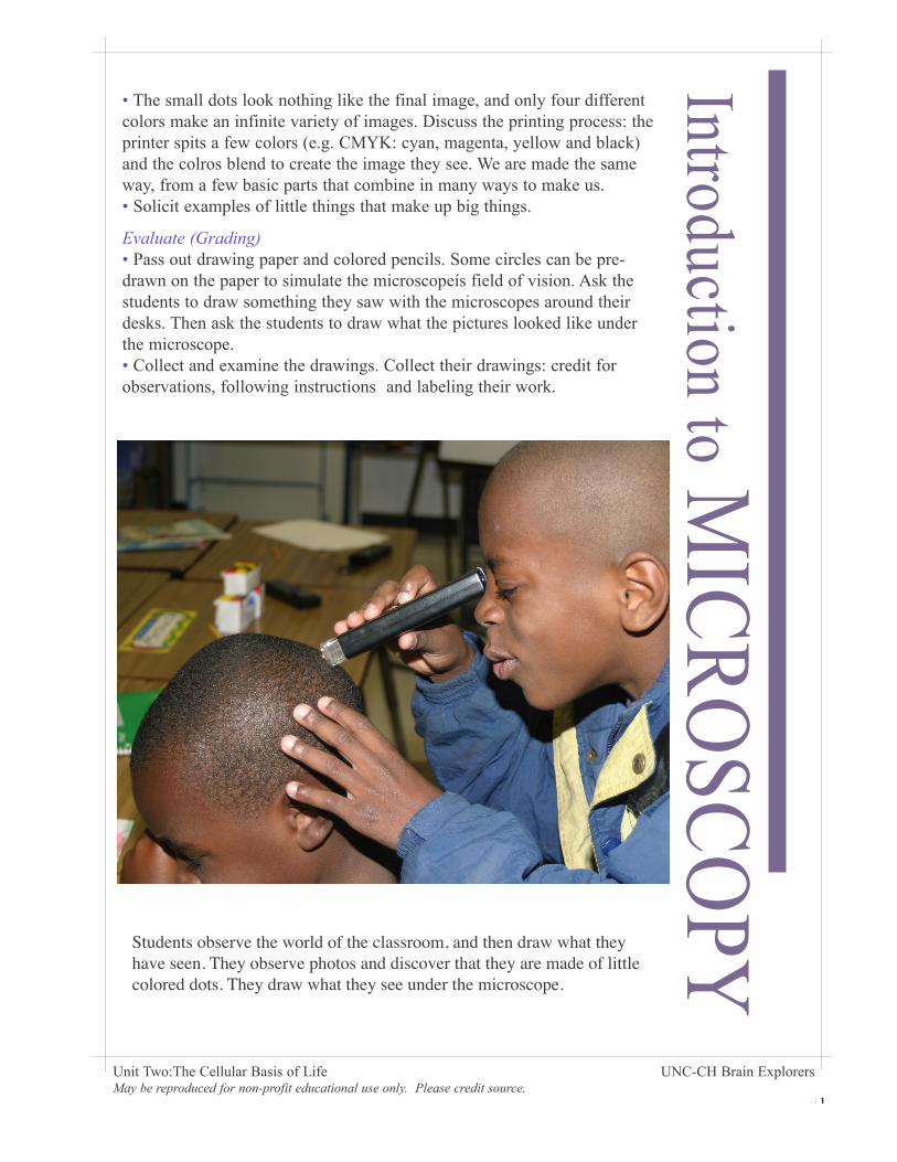
Students observe the world of the classroom, and then draw what they have seen. They observe photos and discover that they are made of little colored dots. They draw what they see under the microscope.
• The small dots look nothing like the final image, and only four different colors make an infinite variety of images. Discuss the printing process: the printer spits a few colors (e.g. CMYK: cyan, magenta, yellow and black) and the colros blend to create the image they see. We are made the same way, from a few basic parts that combine in many ways to make us. • Solicit examples of little things that make up big things.
Evaluate (Grading) • Pass out drawing paper and colored pencils. Some circles can be pre-drawn on the paper to simulate the microscopeís field of vision. Ask the students to draw something they saw with the microscopes around their desks. Then ask the students to draw what the pictures looked like under the microscope. • Collect and examine the drawings. Collect their drawings: credit for observations, following instructions and labeling their work.
42
Unit Two:The Cellular Basis of Life UNC-CH Brain Explorers May be reproduced for non-profit educational use only. Please credit source.
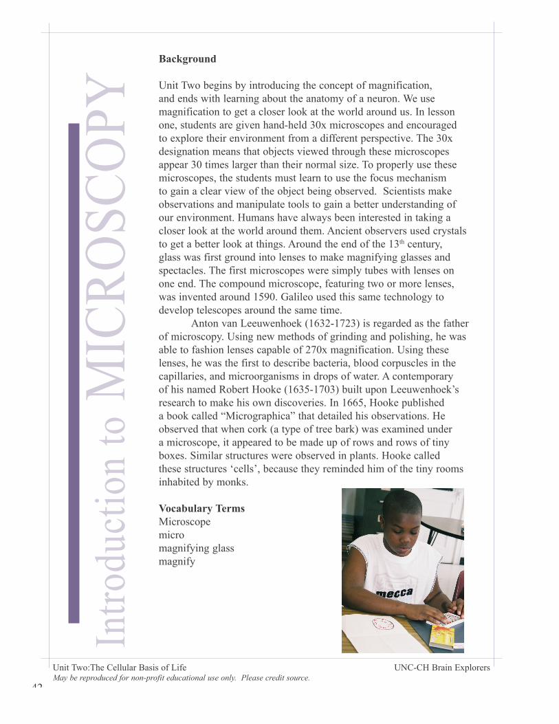
Background
Unit Two begins by introducing the concept of magnification, and ends with learning about the anatomy of a neuron. We use magnification to get a closer look at the world around us. In lesson one, students are given hand-held 30x microscopes and encouraged to explore their environment from a different perspective. The 30x designation means that objects viewed through these microscopes appear 30 times larger than their normal size. To properly use these microscopes, the students must learn to use the focus mechanism to gain a clear view of the object being observed. Scientists make observations and manipulate tools to gain a better understanding of our environment. Humans have always been interested in taking a closer look at the world around them. Ancient observers used crystals to get a better look at things. Around the end of the 13th century, glass was first ground into lenses to make magnifying glasses and spectacles. The first microscopes were simply tubes with lenses on one end. The compound microscope, featuring two or more lenses, was invented around 1590. Galileo used this same technology to develop telescopes around the same time. Anton van Leeuwenhoek (1632-1723) is regarded as the father of microscopy. Using new methods of grinding and polishing, he was able to fashion lenses capable of 270x magnification. Using these lenses, he was the first to describe bacteria, blood corpuscles in the capillaries, and microorganisms in drops of water. A contemporary of his named Robert Hooke (1635-1703) built upon Leeuwenhoek’s research to make his own discoveries. In 1665, Hooke published a book called “Micrographica” that detailed his observations. He observed that when cork (a type of tree bark) was examined under a microscope, it appeared to be made up of rows and rows of tiny boxes. Similar structures were observed in plants. Hooke called these structures ‘cells’, because they reminded him of the tiny rooms inhabited by monks.
Vocabulary TermsMicroscopemicromagnifying glassmagnify
Intro
duct
ion
to M
ICR
OSC
OPY
43
Name: __________________________
Draw and label something you saw with the hand held microscope.
Draw how a photograph appears when viewed through the hand-held microscope.
44
Unit Two:The Cellular Basis of Life UNC-CH Brain Explorers May be reproduced for non-profit educational use only. Please credit source.
Intro
duct
ion
to M
ICR
OSC
OPY
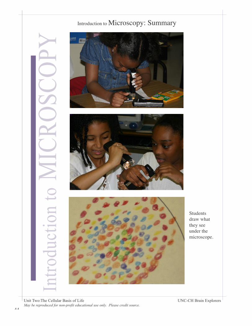
Introduction to Microscopy: Summary
Students draw what they see under the microscope.

45
46
Unit Two:The Cellular Basis of Life UNC-CH Brain Explorers May be reproduced for non-profit educational use only. Please credit source.
Lesson OverviewStudents learn the basic parts of a cell and explore different microscopic images. They use a Virtual Microscope and/or view large poster-sized images of cells.
Engage (10 minutes) • A cell is the basic building block of all living things. Are brick walls living? Trees? A rock? What are some of the differences between living and non-living things? Find examples from the room of objects with cells.. Are you made out of cells? We are living things; we are made out of cells. What are some types of cells in humans? Are plants and animals made out of the same kinds of cells? Why or why not?
Explore:• Display image of the plant cell (elodea or aquatic wisteria, at either 100x or 400 x.) Display posters of red blood and white blood cells, striated muscle cells and neuron cells. Ask the class to guess what kind of jobs each type of cell does. See “guide to Posters” for more information.
Explain (25 minutes)Discuss how form reflects function. Give hints by pointing out features of each cell.
Expand (25 minutes)• Pass out Venn Diagram handout and colored pencils. Ask the students to draw and labeltwo different kinds of cells. Write all the pertinent information on the board if it is not already there.
Evaluate (Grading)• Collect and grade their work for clarity of detail and comprehension.
LE
SS
ON
5
Supplies:LCD projector and computer access with the Virtual Microscope. Posters depicting red and white blood, striated muscle, and neuron cells. Handouts (1 per student) and colored pencils for groups of students.
47
Unit Two:The Cellular Basis of Life UNC-CH Brain Explorers May be reproduced for non-profit educational use only. Please credit source.
CELLS:B
UILD
ING
BLO
CK
S of LIFE
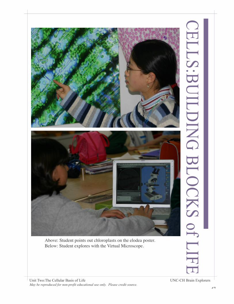
Above: Student points out chloroplasts on the elodea poster. Below: Student explores with the Virtual Microscope.
48
Unit Two:The Cellular Basis of Life UNC-CH Brain Explorers May be reproduced for non-profit educational use only. Please credit source.
CEL
LS:B
UIL
DIN
G B
LOC
KS
of L
IFE Background
The cell is now recognized as the basic building block of all living things. Plants and animals are both made up of cells, although there are a few basic differences between the two. Plant cells have cell walls that give the cells rigidity for structural support. Plant cells also contain organelles that produce food for the plant through the process of photosynthesis. Animal cells have less rigid membranes instead of cell walls.
The cell theory, first developed in the 19th century, states that all living things are made up of one or more cells. All cells come from preexisting cells and contain organelles that provide all the functions necessary for the cell to live. Finally, all cells contain the genetic information (DNA) needed to regulate cell function and pass all the needed genetic information to future generations of cells. The DNA is located in the nucleus, the control center of the cell. All living things are made up of cells, from single celled organisms like the amoeba up to humans whose bodies are made of hundreds of billions of cells.
The human body is made up of over 200 different kinds of cells. Each of these kinds of cells has a specific structure dictated by their function within the body. In the Brain Explorers lessons, we examine blood cells, muscle cells, and nerve cells (neurons).
Blood Cells: Whole blood is living tissue that circulates through the body carrying nourishment, electrolytes, hormones, vitamins, antibodies, heat, and oxygen. Whole blood contains red blood cells, white blood cells, and platelets suspended in yellowish liquid called plasma. Blood cells are made in the marrow of flat bones such as the skull, ribs, sternum, and pelvis.
Red blood cells carry oxygen throughout the body. They do this by mean of a protein called hemoglobin, which is also what makes red blood cells red. There are about 1 billion red blood cells in two or three drops of blood. For every 600 red blood cells, there are 40 platelets and one white blood cell. Red blood cells live for about 120 days and are eventually removed by the spleen. Unlike other cells in the body, mature red blood cells do not have a nucleus.
White blood cells protect the body from bacteria, fungi, and viruses. There are five different types of white blood cells. Some surround and destroy bacteria and viruses, while others help the immune system keep us healthy.
Platelets are very small cells that help in the clotting process. Without platelets, we would not stop bleeding when we got a cut. When you see a scab form on a cut, you are seeing platelets at work.
49
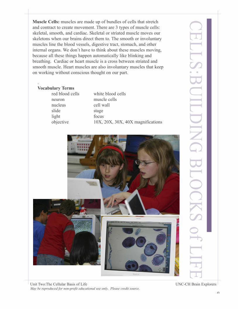
Muscle Cells: muscles are made up of bundles of cells that stretch and contract to create movement. There are 3 types of muscle cells: skeletal, smooth, and cardiac. Skeletal or striated muscle moves our skeletons when our brains direct them to. The smooth or involuntary muscles line the blood vessels, digestive tract, stomach, and other internal organs. We don’t have to think about these muscles moving, because all these things happen automatically like blinking and breathing. Cardiac or heart muscle is a cross between striated and smooth muscle. Heart muscles are also involuntary muscles that keep on working without conscious thought on our part.
CELLS:B
UILD
ING
BLO
CK
S of LIFE
Unit Two:The Cellular Basis of Life UNC-CH Brain Explorers May be reproduced for non-profit educational use only. Please credit source.
. Vocabulary Terms red blood cells white blood cells neuron muscle cells nucleus cell wall slide stage light focus objective 10X, 20X, 30X, 40X magnifications
50
CEL
LS:B
UIL
DIN
G B
LOC
KS
of L
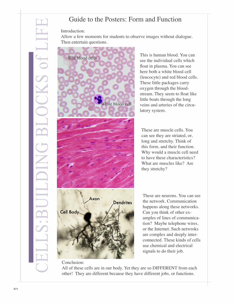
IFE Guide to the Posters: Form and Function
This is human blood. You can see the individual cells which fl oat in plasma. You can see here both a white blood cell (leucocyte) and red blood cells. These little packages carry oxygen through the blood-stream. They seem to fl oat like little boats through the long veins and arteries of the circu-latory system.
These are muscle cells. You can see they are striated, or, long and stretchy. Think of this form, and their function. Why would a muscle cell need to have these characteristics? What are muscles like? Are they stretchy?
These are neurons. You can see the network. Communication happens along these networks. Can you think of other ex-amples of lines of communica-tion? Maybe telephone wires, or the Internet. Such netwroks are complex and deeply inter-connected. These kinds of cells use chemical and electrical signals to do their job.
Conclusion:All of these cells are in our body. Yet they are so DIFFERENT from each other! They are different because they have different jobs, or functions.
Introduction: Allow a few moments for students to observe images without dialogue. Then entertain questions.
White blood cell
Red blood cells

51
Name
Draw a picture of Elodea Draw another cell and label it.
Unique to cell #1 Unique to cell #2
Common characteristics
Make a Venn Diagram: Label and describe each cell individually on the left and right sides, then write what they have in common in the center.

52
Unit Two:The Cellular Basis of Life UNC-CH Brain Explorers May be reproduced for non-profit educational use only. Please credit source.
Background
Neurons: Neurons or nerve cells are very specialized cells that carry messages throughout the brain and body. Neurons come in many different shapes and sizes, depending upon where they are in the body and what sort of messages they carry. These messages are transmitted through an electrochemical process called neurotransmission.
The human brain alone contains over 100 billion neurons. Neurons carry the motor and sensory messages that enable us to move and receive stimuli from the world around us. Neurons in our brains let us decipher all this information and make decisions accordingly. Unlike other cells in our bodies, neurons do not replace themselves when they die. We are born with all the neurons we will ever need, and for the most part they are never replaced. Recent studies have revealed that some new neurons are created in the hippocampus, the part of the brain responsible for long-term memory storage. Because neurons do not generally reproduce, it is important to avoid activities that can damage or destroy them such as abusing drugs or alcohol.
While there are many kinds of neurons, they all share the same basic anatomical structures. The nucleus and organelles of a neuron are located in the cell body. Radiating from the cell body are the dendrites. The term ‘dendrite� comes from the Greek word used to describe the branches of a tree. Receptors on the dendrites catch the chemical messages called neurotransmitters that are sent from other neurons.
Once the dendrites catch enough of these chemical messages, the cell body becomes excited and sends an electrical impulse called the action potential down a wire-like structure called the axon. When the electrical impulse reaches the end of the axon, called the axon terminal, more neurotransmitters are released to float across the gap between either the dendrites of another neuron or receptors on a muscle cell. This gap, about a millionth of an inch wide, is called the synapse.
PART
S an
d PI
ECES
: The
NEU
RO
N
53
PARTS and PIEC
ES: The NEU
RO
N
Unit Two:The Cellular Basis of Life UNC-CH Brain Explorers May be reproduced for non-profit educational use only. Please credit source.
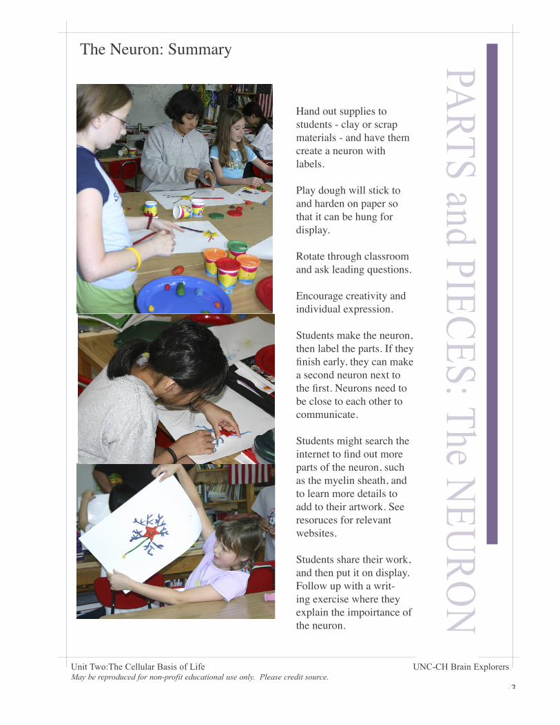
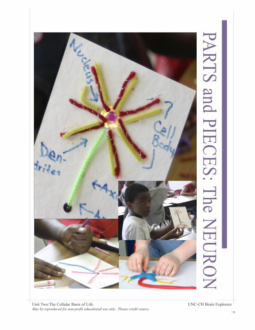
The Neuron: Summary
Hand out supplies to students - clay or scrap materials - and have them create a neuron with labels.
Play dough will stick to and harden on paper so that it can be hung for display.
Rotate through classroom and ask leading questions.
Encourage creativity and individual expression.
Students make the neuron, then label the parts. If they fi nish early, they can make a second neuron next to the fi rst. Neurons need to be close to each other to communicate.
Students might search the internet to fi nd out more parts of the neuron, such as the myelin sheath, and to learn more details to add to their artwork. See resoruces for relevant websites.
Students share their work, and then put it on display. Follow up with a writ-ing exercise where they explain the impoirtance of the neuron.
54
Unit Two:The Cellular Basis of Life UNC-CH Brain Explorers May be reproduced for non-profit educational use only. Please credit source.
PART
S an
d PI
ECES
: The
NEU
RO
N
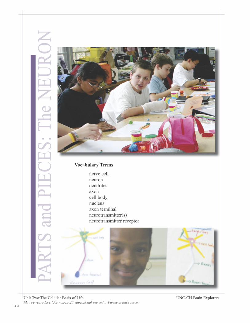
Vocabulary Terms
nerve cell neuron dendrites axon cell body nucleus axon terminal neurotransmitter(s) neurotransmitter receptor
55
Unit Two:The Cellular Basis of Life UNC-CH Brain Explorers May be reproduced for non-profit educational use only. Please credit source.
PARTS and PIEC
ES: The NEU
RO
N
56
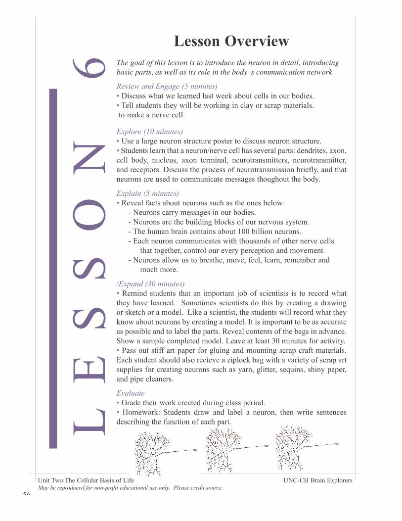
Lesson OverviewThe goal of this lesson is to introduce the neuron in detail, introducing basic parts, as well as its role in the body�s communication network
Review and Engage (5 minutes) • Discuss what we learned last week about cells in our bodies. • Tell students they will be working in clay or scrap materials. to make a nerve cell.
Explore (10 minutes)• Use a large neuron structure poster to discuss neuron structure. • Students learn that a neuron/nerve cell has several parts: dendrites, axon, cell body, nucleus, axon terminal, neurotransmitters, neurotransmitter, and receptors. Discuss the process of neurotransmission briefly, and that neurons are used to communicate messages thoughout the body.
Explain (5 minutes)• Reveal facts about neurons such as the ones below. - Neurons carry messages in our bodies. - Neurons are the building blocks of our nervous system. - The human brain contains about 100 billion neurons. - Each neuron communicates with thousands of other nerve cells that together, control our every perception and movement. - Neurons allow us to breathe, move, feel, learn, remember and much more.
/Expand (30 minutes)• Remind students that an important job of scientists is to record what they have learned. Sometimes scientists do this by creating a drawing or sketch or a model. Like a scientist, the students will record what they know about neurons by creating a model. It is important to be as accurate as possible and to label the parts. Reveal contents of the bags in advance. Show a sample completed model. Leave at least 30 minutes for activity. • Pass out stiff art paper for gluing and mounting scrap craft materials. Each student should also recieve a ziplock bag with a variety of scrap art supplies for creating neurons such as yarn, glitter, sequins, shiny paper, and pipe cleaners.
Evaluate• Grade their work created during class period.• Homework: Students draw and label a neuron, then write sentences describing the function of each part.LE
SS
ON
6
Unit Two:The Cellular Basis of Life UNC-CH Brain Explorers May be reproduced for non-profit educational use only. Please credit source.
57
Unit Two:The Cellular Basis of Life UNC-CH Brain Explorers May be reproduced for non-profit educational use only. Please credit source.
PARTS and PIEC
ES: The NEU
RO
N
58
Name __________________________
Teacher _________________________
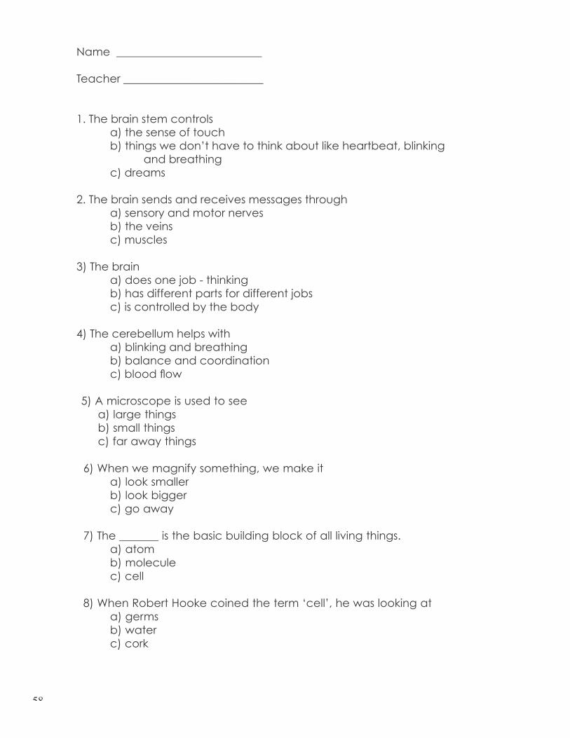
1. The brain stem controls a) the sense of touch b) things we don’t have to think about like heartbeat, blinking and breathing c) dreams
2. The brain sends and receives messages through a) sensory and motor nerves b) the veins c) muscles
3) The brain a) does one job - thinking b) has different parts for different jobs c) is controlled by the body
4) The cerebellum helps with a) blinking and breathing b) balance and coordination c) blood fl ow
5) A microscope is used to see a) large things b) small things c) far away things
6) When we magnify something, we make it a) look smaller b) look bigger c) go away
7) The _______ is the basic building block of all living things. a) atom b) molecule c) cell
8) When Robert Hooke coined the term ‘cell’, he was looking at a) germs b) water c) cork
59
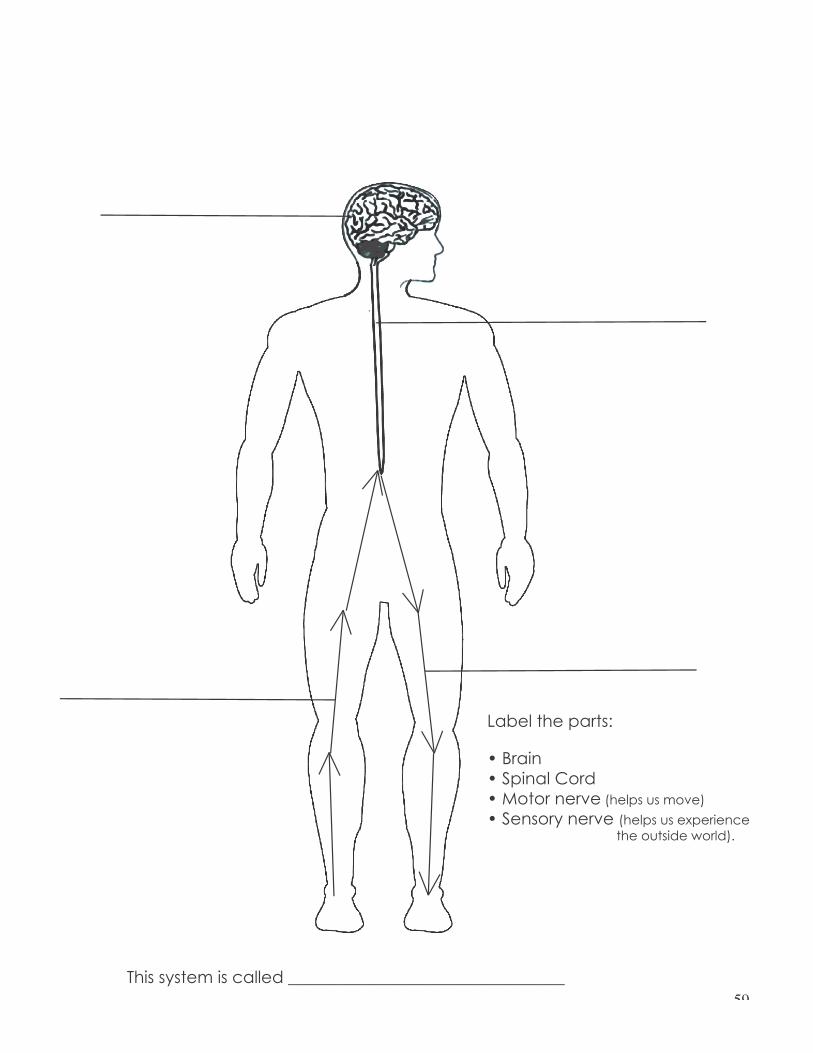
Label the parts:
• Brain• Spinal Cord• Motor nerve (helps us move)
• Sensory nerve (helps us experience the outside world).
This system is called _________________________________
60
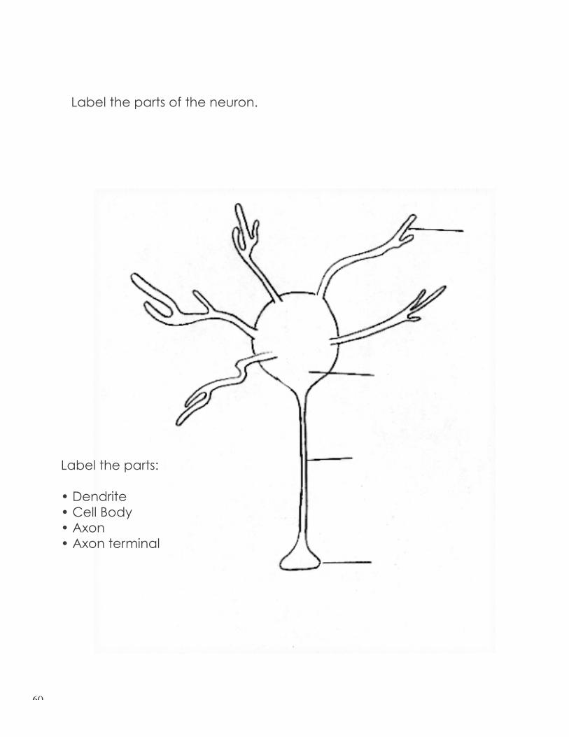
Label the parts of the neuron.
Label the parts:
• Dendrite• Cell Body• Axon• Axon terminal
61
In the space below, write a paragraph (or several sentences) about what you learned and what you enjoyed in Brain Explorers. The activities we did were:
a) Full Body Tracing: outine of body with yarn b) Magic Wand: Dr. Penfi eld’s experiments c) Clay Brains: making brains from play-dough d) Mid-point review: doctor/patient game e) Introduction to Microscopy: hand-held microscopes f) Cells: poster guessing game, drawing cells g) The Neuron: scratch lite drawing of a neuron or scrap materials

62

63