tracheoesophageal fistula after total resection of gastric ... · tracheoesophageal fistula after...
TRANSCRIPT
CASE REPORT Open Access
Tracheoesophageal fistula after totalresection of gastric conduit forgastro-aortic fistula due to gastric ulcerYayoi Sakatoku1, Masahide Fukaya1*, Hironori Fujieda1, Yuzuru Kamei2, Akihiro Hirata3, Keita Itatsu1
and Masato Nagino1
Abstract
Background: Tracheoesophageal fistula (TEF) is a rare but life-threatening complication after esophagectomy. It hasa high mortality rate and often leads to severe aspiration pneumonia. Various types of surgical repair procedureshave been reported, but the optimal management of TEF is challenging and controversial. Treatment should beindividualized to each patient.
Case presentation: A 66-year-old female underwent transthoracic esophagectomy with gastric tube reconstructionand an intrathoracic anastomosis for esophageal cancer. Three years later, she had hematemesis and was diagnosedwith a gastro-aortic fistula due to a gastric ulcer. She underwent endovascular aortic repair urgently at another hospital.Two days later, she underwent total resection of the gastric tube, during which time an injury to the trachea occurred;it was repaired by patching the stump of the esophagus to the injury site. Two months later, descending aorticreplacement was performed due to infection of the stent graft. Six months after the first operation, a TEF developed.The patient was referred to our hospital for further treatment. The fistula was ligated and divided via a cervicalapproach, and a pectoralis major muscle flap was used to cover the defect. Esophageal reconstruction with thepedunculated jejunum was performed via a subcutaneous route. The postoperative course was uneventful. The patientwas discharged after 6 months of physical and dysphagia rehabilitation.
Conclusion: A TEF located near the cervicothoracic border was successfully treated with a pectoralis major muscleflap through a cervical approach. Total resection of a gastric conduit in the posterior mediastinum carries a risk oftracheobronchial injury; however, if such an injury occurs, surgeons should be able to repair the injury using a suitableflap depending on the injury site.
Keywords: Tracheoesophageal fistula, Gastric conduit ulcer, Pectoralis major muscle flap
BackgroundTracheoesophageal fistula (TEF) is a rare but life-threatening complication after esophagectomy. It has ahigh mortality rate and often leads to severe aspirationpneumonia [1, 2]. Various types of surgical repair proce-dures have been reported, but the optimal managementof TEF is challenging and controversial. Treatmentshould be individualized to each patient.
Herein, we report a patient with a TEF after total re-section of a gastric conduit for gastro-aortic fistula dueto a gastric ulcer, successfully repaired with a pectoralismajor muscle flap through a cervical approach.
Case presentationA 66-year-old woman with esophageal cancer underwenttransthoracic esophagectomy with three-field lymph nodedissection via a muscle-sparing thoracotomy as previouslyreported [3], with gastric conduit reconstruction and anintrathoracic anastomosis. A high-dose proton pump in-hibitor (PPI) was administered postoperatively due to thepatient’s history of gastric ulcers; she discontinued the
* Correspondence: [email protected] of Surgical Oncology, Department of Surgery, Nagoya UniversityGraduate School of Medicine, 65 Tsurumai-cho, Showa-ku, Nagoya 466-8550,JapanFull list of author information is available at the end of the article
© The Author(s). 2017 Open Access This article is distributed under the terms of the Creative Commons Attribution 4.0International License (http://creativecommons.org/licenses/by/4.0/), which permits unrestricted use, distribution, andreproduction in any medium, provided you give appropriate credit to the original author(s) and the source, provide a link tothe Creative Commons license, and indicate if changes were made.
Sakatoku et al. Surgical Case Reports (2017) 3:90 DOI 10.1186/s40792-017-0371-6
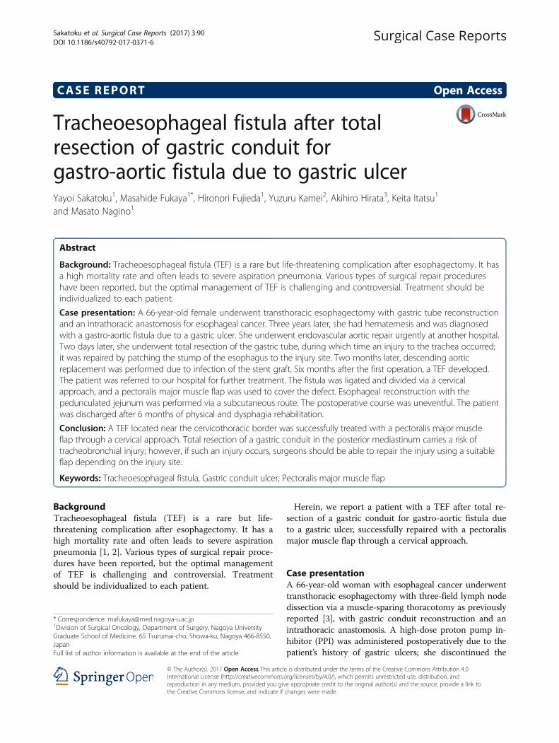
medication of her own volition. Three years after surgery,she was admitted to a local hospital for mediastinitis dueto a perforated gastric ulcer in the conduit (Fig. 1a). Shewas treated with antibiotics and fasting. Seventeen dayslater, she had hematemesis and was diagnosed with agastro-aortic fistula due to a gastric ulcer (Fig. 1b, c). Shewas transferred to a nearby university hospital with shockstatus and underwent endovascular aortic repair urgently,using two GORE® TAG® devices (W.L. Gore & Associates,Flagstaff, AZ). Two days later, total resection of the gastrictube was performed via a right posterolateral thoracot-omy. There was an abscess cavity between the gastric tubeand the descending aorta which consisted of necrotic tis-sue and old blood. A 2-cm wall defect was found on theright wall of the middle of gastric tube. The clot-filled gas-tric tube was resected. The perforation of the descendingaorta was left. During this operation, a tracheal injury oc-curred while the remnant esophagus was being separatedfrom the trachea; this injury was subsequently repaired bypatching the stump of the esophagus to the injury site.Tracheostomy and a feeding jejunostomy were also
performed. The operative time was 8 h and 40 min, andthe blood loss was 2380 ml. The remnant esophagus hadbeen decompressed by nasal tube since the total resectionof gastric conduit till the following reconstructive surgery.Two months later, a descending aortic replacement wasperformed due to infection of the stent graft. Stent graftwas removed, and the descending aorta between the fifthvertebra and the eleventh vertebra level was replaced withrifampicin-soaked 24-mm J graft (JUNKEN MEDICAL,Tokyo, Japan). Infected vascular intima of the aortaaround the gastro-aortic fistula was resected. Theoperative time was 8 h and 10 min. Purulent matter wasfound around the stent graft, and Candida albicans wasrecognized by the bacterial culture of the pus. Post-operative severe pneumonia due to methicillin-resistantStaphylococcus aureus (MRSA) occurred after aortic re-placement and required artificial respirator. Postoperativepneumonia was gradually improved by antibiotics. Threemonths later, she was transferred to previous local hos-pital. When a TEF developed 6 months after the first oper-ation, in spite of decompression of the remnant esophagus
a
b cFig. 1 a Gastrointestinal endoscopy showed an ulcerated lesion on the right wall of middle of gastric tube. b Horizontal and c sagittal enhancedcomputed tomography image showed an irregular ulceration on the anterior wall of the descending aorta, no extravasation, and absence of thedescending aortic wall and gastric wall, suggesting sealed rupture of the descending aorta (yellow arrow)
Sakatoku et al. Surgical Case Reports (2017) 3:90 Page 2 of 6
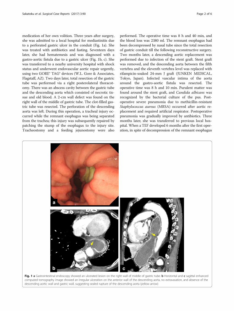
by nasal tube, she still had required respirator due to pro-longed postoperative pneumonia. Her poor general condi-tion could not allow reconstructive surgery. She couldwithdraw from respirator 5 months after the aortic re-placement (7 months after the first operation). Thoughshe suffered from repeated bouts of aspiration pneumonia,she could walk after rehabilitation for 5 months (1 yearafter the first operation), and transferred to our hospitalfor reconstructive surgery. This patient’s time course issummarized in Fig. 2.Bronchoscopy showed a fistula on the membranous
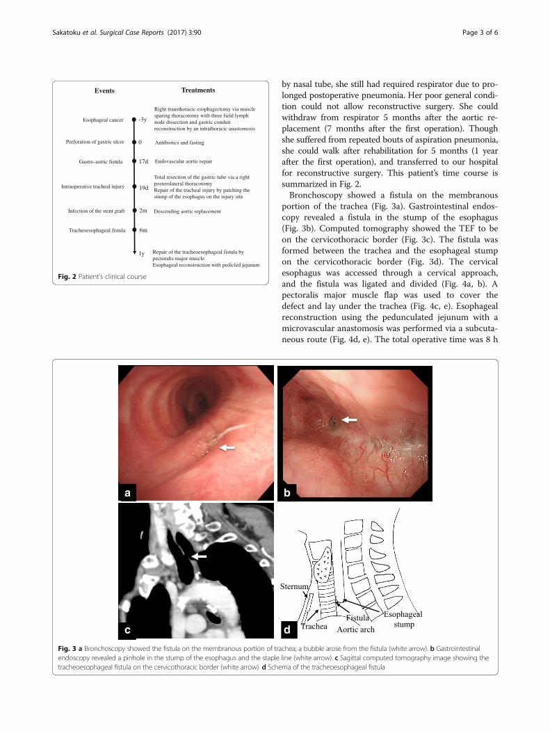
portion of the trachea (Fig. 3a). Gastrointestinal endos-copy revealed a fistula in the stump of the esophagus(Fig. 3b). Computed tomography showed the TEF to beon the cervicothoracic border (Fig. 3c). The fistula wasformed between the trachea and the esophageal stumpon the cervicothoracic border (Fig. 3d). The cervicalesophagus was accessed through a cervical approach,and the fistula was ligated and divided (Fig. 4a, b). Apectoralis major muscle flap was used to cover thedefect and lay under the trachea (Fig. 4c, e). Esophagealreconstruction using the pedunculated jejunum with amicrovascular anastomosis was performed via a subcuta-neous route (Fig. 4d, e). The total operative time was 8 h
Right transthoracic esophagectomy via muscle sparing thoracotomy with three field lymph node dissection and gastric conduit reconstruction by an intrathoracic anastomosis
Endovascular aortic repair
Total resection of the gastric tube via a right posterolateral thoracotomyRepair of the tracheal injury by patching the stump of the esophagus on the injury site
Descending aortic replacement
Repair of the tracheoesophageal fistula by pectoralis major muscle Esophageal reconstruction with pedicled jejunum
Esophageal cancer
Perforation of gastric ulcer
Gastro-aortic fistula
Infection of the stent graft
Tracheoesophageal fistula
-3y
0
17d
19d
2m
6m
1y
Intraoperative tracheal injury
Antibiotics and fasting
Events Treatments
Fig. 2 Patient’s clinical course
a b
c d
Fig. 3 a Bronchoscopy showed the fistula on the membranous portion of trachea; a bubble arose from the fistula (white arrow). b Gastrointestinalendoscopy revealed a pinhole in the stump of the esophagus and the staple line (white arrow). c Sagittal computed tomography image showing thetracheoesophageal fistula on the cervicothoracic border (white arrow). d Schema of the tracheoesophageal fistula
Sakatoku et al. Surgical Case Reports (2017) 3:90 Page 3 of 6
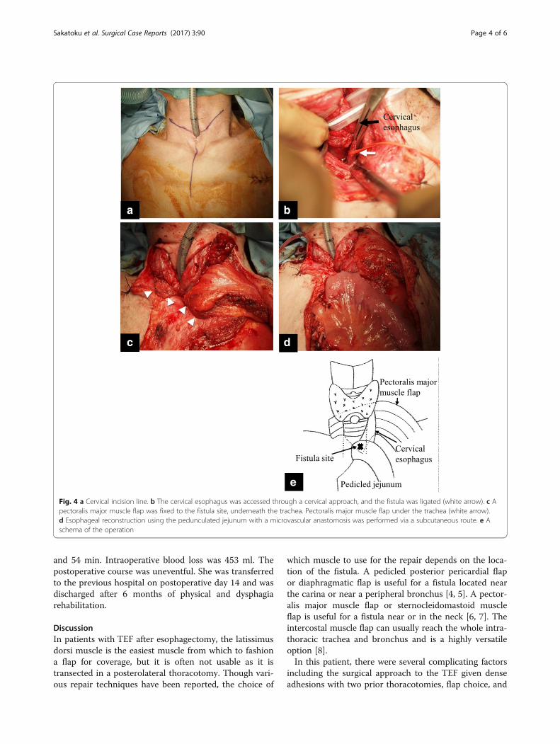
and 54 min. Intraoperative blood loss was 453 ml. Thepostoperative course was uneventful. She was transferredto the previous hospital on postoperative day 14 and wasdischarged after 6 months of physical and dysphagiarehabilitation.
DiscussionIn patients with TEF after esophagectomy, the latissimusdorsi muscle is the easiest muscle from which to fashiona flap for coverage, but it is often not usable as it istransected in a posterolateral thoracotomy. Though vari-ous repair techniques have been reported, the choice of
which muscle to use for the repair depends on the loca-tion of the fistula. A pedicled posterior pericardial flapor diaphragmatic flap is useful for a fistula located nearthe carina or near a peripheral bronchus [4, 5]. A pector-alis major muscle flap or sternocleidomastoid muscleflap is useful for a fistula near or in the neck [6, 7]. Theintercostal muscle flap can usually reach the whole intra-thoracic trachea and bronchus and is a highly versatileoption [8].In this patient, there were several complicating factors
including the surgical approach to the TEF given denseadhesions with two prior thoracotomies, flap choice, and
a b
c d
e
Fig. 4 a Cervical incision line. b The cervical esophagus was accessed through a cervical approach, and the fistula was ligated (white arrow). c Apectoralis major muscle flap was fixed to the fistula site, underneath the trachea. Pectoralis major muscle flap under the trachea (white arrow).d Esophageal reconstruction using the pedunculated jejunum with a microvascular anastomosis was performed via a subcutaneous route. e Aschema of the operation
Sakatoku et al. Surgical Case Reports (2017) 3:90 Page 4 of 6
conduit choice for esophageal reconstruction. Fortunately,the TEF was located on the cervicothoracic border, and acervical approach was therefore chosen. A pectoralismajor muscle or sternocleidomastoid muscle (SCM) flapcan be suitable for repairing this tracheoesophageal fistula.A pectoralis major muscle flap is larger and thicker, andits covering area is wider. A SCM flap can be created eas-ier in the same operative field. The larger pectoralis majormuscle flap is considered to be more suitable for this pa-tient to fill the dead space behind the membranous por-tion of the trachea. There was a problem about bloodsupply of SCM in this case. The upper third of SCM issupplied by branches of occipital artery. The middle thirdof SCM is supplied by branches of superior thyroid artery.The lower third of SCM is supplied by branches of thesuprascapular artery [9]. There are two types of SCM flap:the superiorly based flap which is the commonly used andsupplied from occipital artery and superior thyroid arteryand the inferiorly based flap which is suitable for the le-sion on the lower neck or upper mediastinum [10].Though the inferiorly based sternocleidomastoid muscleflap was required according to the fistula position, the ne-crosis of the flap might occur because of no blood supplyfrom branches of superior thyroid artery and suprascapu-lar artery by previous cervical lymph node dissection.Therefore, the pectoralis major muscle was chosen overthe SCM. With respect to esophageal reconstruction,small intestine reconstruction using the pedunculated je-junum with a microvascular anastomosis was chosen overa colonic conduit as there were dense adhesions of thetransverse colon to the upper abdominal organs due to aprevious total resection of the former gastric conduit.During gastric conduit resection, dense adhesions
around the conduit, particularly around the esophagogas-trostomy in the upper mediastinum [11], make dissectiondifficult and a tracheobronchial injury a possibility. In thiscase, a tracheal injury occurred while the remnant esopha-gus was being dissected from the trachea via a thoracot-omy. The injury was repaired with the remnant esophaguspatch and resulted in a delayed TEF. Because the patienthad previously undergone a muscle-sparing thoracotomyfor esophageal cancer, thus preserving the latissimus dorsimuscle, a posterolateral thoracotomy with a latissimusdorsi muscle flap or intercostal muscle flap would havebeen a good option for repair of the initial tracheobron-chial injury. When the tracheal injury occurred, the sur-geons should have created an intercostal muscle flap torepair the injury and then performed a cervical esopha-gostomy. A pectoralis major muscle flap or a sternocleido-mastoid muscle flap could also have been an option as theinjury was near the neck.The primary cause of this gastro-aortic fistula was
discontinuation of PPI medication of her own volition.The frequency of peptic ulcer in the reconstructed
gastric tube was reported to be 6.6–19.4% [12–15]. Themortality of patients with perforation of gastric tube ulcerwas 56.5% in the review of Japanese literature and 84.6%in the review of the English literature [16]. Once gastrictube ulcer develops to gastro-aortic fistula, it causes a fatalresult. We recommend PPI medication for patients withgastric tube reconstruction after esophagectomy.
ConclusionsA TEF located near the cervicothoracic border was suc-cessfully treated with a pectoralis major muscle flapthrough a cervical approach. Because total resection of agastric conduit in the posterior mediastinum carries arisk of tracheobronchial injury, thoracotomy with cre-ation of an intercostal muscle flap should be performedin preparation for a tracheobronchial injury in such situ-ations. If such an injury occurs, surgeons should be ableto repair the injury using a suitable flap depending onthe injury site.
AbbreviationsPPI: Proton pump inhibitor; SCM: Sternocleidomastoid muscle;TEF: Tracheoesophageal fistula
Authors’ contributionsMF, YS, HF, KI, and YK performed the surgery. YS and HF took charge ofpostoperative care in our hospital. AH took charge of conservative therapyfor mediastinitis due to a perforated gastric ulcer in the conduit andpostoperative physical and dysphagia rehabilitation in the local hospital. YSprepared the manuscript. MF and MN assisted in drafting the manuscriptand reviewed the article. All authors read and approved the final manuscript.
Ethics approval and consent to participateThis study was carried out in accordance with the principles of theDeclaration of Helsinki.
Consent for publicationWritten informed consent was obtained from the patient for publication ofthis case report and any accompanying images.
Competing interestsThe authors declare that they have no competing interests.
Publisher’s NoteSpringer Nature remains neutral with regard to jurisdictional claims inpublished maps and institutional affiliations.
Author details1Division of Surgical Oncology, Department of Surgery, Nagoya UniversityGraduate School of Medicine, 65 Tsurumai-cho, Showa-ku, Nagoya 466-8550,Japan. 2Department of Plastic and Reconstructive Surgery, Nagoya UniversityGraduate School of Medicine, 65 Tsurumai-cho, Showa-ku, Nagoya 466-8550,Japan. 3Department of Surgery, Shizuoka Kosei Hospital, 23 Kitaban-cho,Aoi-ku, Shizuoka 420-8623, Japan.
Received: 14 March 2017 Accepted: 17 August 2017
References1. Lambertz R, Holscher AH, Bludau M, Leers JM, Gutschow C, Schroder W.
Management of tracheo- or bronchoesophageal fistula after Ivor-Lewisesophagectomy. World J Surg. 2016;40(7):1680–7. PubMed PMID: 26913731.
2. Yasuda T, Sugimura K, Yamasaki M, Miyata H, Motoori M, Yano M, et al. Tencases of gastro-tracheobronchial fistula: a serious complication after
Sakatoku et al. Surgical Case Reports (2017) 3:90 Page 5 of 6
esophagectomy and reconstruction using posterior mediastinal gastric tube.Dis Esophagus. 2012;25(8):687–93. PubMed PMID: 22292530.
3. Miyata K, Fukaya M, Itatsu K, Abe T, Nagino M. Muscle sparing thoracotomyfor esophageal cancer: a comparison with posterolateral thoracotomy. SurgToday. 2016;46(7):807–14. PubMed PMID: 26311005.
4. Yasuda T, Makino T, Shiraishi O, Sogabe S. Pedicled posterior pericardial repairof tracheoesophageal fistula after chemoradiotherapy for esophageal cancer.J Thorac Cardiovasc Surg. 2016;151(6):e95–7. PubMed PMID: 26948167.
5. Mineo TC, Ambrogi V. The diaphragmatic flap: a multiuse material inthoracic surgery. J Thorac Cardiovasc Surg. 1999;118(6):1084–9. PubMedPMID: 10595982.
6. Shichinohe T, Okushiba S, Morikawa T, Kitashiro S, Manase H, Kawarada Y, et al.Salvage of a massive esophago-tracheal fistula resulting from a stentingtreatment. Dis Esophagus. 2006;19(4):299–304. PubMed PMID: 16866865.
7. Arimoto J, Hatada A, Kawago M, Nishimura O, Maebeya S, Okamura Y. Closureof esophagotracheal fistula after esophagectomy for esophageal cancer.Gen Thorac Cardiovasc Surg. 2015;63(11):636–9. PubMed PMID: 26189183.
8. Deshpande G, Samarasam I, Banerjee S, Gnanamuthu RB, Chandran S,Mathew G. Benign esophagorespiratory fistula: a case series and a noveltechnique of definitive management. Dis Esophagus. 2013;26(2):141–7.PubMed PMID: 22486830.
9. Kierner AC, Aigner M, Zelenka I, Riedl G, Burian M. The blood supply of thesternocleidomastoid muscle and its clinical implications. Arch Surg.1999;134(2):144–7. PubMed PMID: 10025452.
10. Yugueros P, Woods JE. The sternocleidomastoid myocutaneous flap: areappraisal. Br J Plast Surg. 1996;49(2):93–6. PubMed PMID: 8733347.
11. Akita H, Doki Y, Ishikawa O, Takachi K, Miyashiro I, Sasaki Y, et al. Total removalof the posterior mediastinal gastric conduit due to gastric cancer afteresophagectomy. J Surg Oncol. 2004;85(4):204–8. PubMed PMID: 14991878.
12. Yang L. Surgical treatment for carcinoma of gastric stump—a report on 13patients. Zhonghua Zhong Liu Za Zhi. 1992;14(2):123–6. PubMed PMID:1618080.
13. Koide N, Hiraguri M, Nishio A, Hanazaki K, Adachi W, Shikama N, et al. Ulcerin the gastric tube for esophageal replacement: a comparison of 12esophageal cancer patients with or without postoperative radiotherapy.J Gastroenterol Hepatol. 2001;16(2):137–41. PubMed PMID: 11207892.
14. Maier A, Tomaselli F, Sankin O, Anegg U, Fell B, Renner H, et al. Acid-relateddiseases following retrosternal stomach interposition. Hepato-Gastroenterology.2001;48(39):899–902. PubMed PMID: 11462952.
15. Motoyama S, Saito R, Kitamura M, Suzuki H, Nakamura M, Okuyama M, et al.Prospective endoscopic follow-up results of reconstructed gastric tube.Hepato-Gastroenterology. 2003;50(51):666–9. PubMed PMID: 12828056.
16. Ubukata H, Nakachi T, Tabuchi T, Nagata H, Takemura A, Shimazaki J, et al.Gastric tube perforation after esophagectomy for esophageal cancer.Surg Today. 2011;41(5):612–9. PubMed PMID: 21533931.
Sakatoku et al. Surgical Case Reports (2017) 3:90 Page 6 of 6