vanja opačić galić , violeta petrović , vukoman...
TRANSCRIPT

Address: 1 Kraljice Natalije Street, 11000 Belgrade, Serbia
+381 11 4092 776, Fax: +381 11 3348 653
E-mail: [email protected], Web address: www.srpskiarhiv.rs
Paper Accepted* ISSN Online 2406-0895
Original Article / Оригинални рад
Vanja Opačić-Galić1,†, Violeta Petrović1, Vukoman Jokanović2, Slavoljub Živković1
Histological Evaluation of Tissue Reactions to Newly Synthetized Calcium
Silicate- and Hydroxyapatite-Based Bioactive Materials: in vivo Study Хистолошкe реакције ткива на новосинтетисане биоактивне материјале на
бази калцијум силикатних система и хидроксиапатита – in vivo студија
1 Department for Restorative Dentistry and Endodontics, School of Dental Medicine, University of Belgrade; 2 Institute for Nuclear Sciences Vinča, University of Belgrade, Serbia
Received: July 19, 2016
Accepted: October 4, 2016
Online First: March 10, 2017
DOI: 10.2298/SARH160719063O
* Accepted papers are articles in press that have gone through due peer review process and have been
accepted for publication by the Editorial Board of the Serbian Archives of Medicine. They have not
yet been copy edited and/or formatted in the publication house style, and the text may be changed
before the final publication.
Although accepted papers do not yet have all the accompanying bibliographic details available, they
can already be cited using the year of online publication and the DOI, as follows: the author’s last
name and initial of the first name, article title, journal title, online first publication month and year,
and the DOI; e.g.: Petrović P, Jovanović J. The title of the article. Srp Arh Celok Lek. Online First,
February 2017.
When the final article is assigned to volumes/issues of the journal, the Article in Press version will be
removed and the final version will appear in the associated published volumes/issues of the journal.
The date the article was made available online first will be carried over. † Correspondence to:
Vanja Opačić Galić
School of Dental Medicine, Rankeova 4, 11000 Belgrade, Serbia
E-mail: [email protected]

Srp Arh Celok Lek 2017│Online First March 10, 2017│ DOI: 10.2298/SARH160719063O
DOI: 10.2298/SARH160719063O Copyright © Serbian Medical Society
2
Histological Evaluation of Tissue Reactions to Newly Synthetized Calcium
Silicate- and Hydroxyapatite-Based Bioactive Materials: in vivo Study
Хистолошкe реакције ткива на новосинтетисане биоактивне материјале на бази
калцијум силикатних система и хидроксиапатита – in vivo студија
SUMMARY
Introduction/Objective Developing of the materials,
which could be used as biological bone substitutes, is
one of the most valuable and active fields of
biomaterial research.
Objective The goal was to research the reaction of the
tissue on newly synthesized nano-materials with the
calcium silicate system (CS) and hydroxyapatite (CS-
HA) based, after being implanted into the
subcutaneous tissue of a rats and direct pulp capping
of the tooth of the rabbits.
Methods The tested materials were implanted in 40
Wistar male rats, sacrificed after 7, 15, 30, 60 days.
The direct pulp capping is realized on the teeth of the
rabbits. Cavities were prepared on the vestibular
surface of the incisors. The animals are sacrificed after
10 and 15 days. The control material was mineral
trioxide aggregate (MTA). Histological analysis
covered inflammatory reaction cellular components
tracking, presence of gigantic cells and necrosis of the
tissue.
Results Seven days after implantation the strongest
inflammatory response was given by the MTA
(3,30±0,48), while CS and CS-HA scored 3,00±0,71.
After 60 days the rate of inflammatory reactions
dropped, which was the least visible with CS-HA
(0,20±0,45). The least visible inflammatory reaction of
the rabbit’s pulp tissue was spotted with the CS
(1,83±0,75), than with the MTA and CS-HA
(2.67±1.53, 3.00±0.63).
Conclusion The newly synthesized materials caused a
slight reaction of the subcutaneous tissue. CS-HA
showed the best tissue tolerance. Nanostructural
biomaterials caused a slight to moderate inflammatory
reaction of the rabbit`s pulp tissue only in the
immediate vicinity of the implanted material.
Keywords: Biocompatibility; calcium silicate system;
hydroxyapatite; mineral trioxide aggregate
САЖЕТАК
Увод/Циљ Усавршавање материјала, који би могли да се користе као биолошке замене кости, једна је од најзначајнијих и најактивнијих области истраживања биоматеријала. Циљ овог рада је био да се испита одговор ткива на новосинтетисане наноматеријале на бази калцијум силикатних система(КС) и хидроксиапатита (КС-ХА) после имплантације у поткожно ткиво пацова и директног прекривања пулпе зуба кунића. Методе У поткожно ткиво 40 Вистар пацова су имплантирани тестирани материјали, а после 7, 15, 30 и 60 дана зивотиње су жртвоване. Директно прекривање пулпе је реализовано на зубима кунића. На вестибуларним површинама секутића прекривани су кавитети. Животиње су жртвоване после 10 и 15 дана. Контролни материјал у оба експеримента је био минерални триоксидни агрегат МТА). Хистолошка анализа је обухватила праћење ћелијске компоненте запаљења, присуства гигантских ћелија и некрозе ткива. Резултати Седам дана после субкутане имплантације најјачи запаљенски одговор дао је МТА (3,30±0,48), док је за КС и КС-ХА он оцењен са 3,00±0,71. После 60 дана дошло је до опадања знакова запаљења, које је било најмање изражено око КС-ХА (0,20±0,45). Најмање изражена запаљенска рекција пулпног ткива кунића уочена је код материјала КС (1,83±0,75), затим код МТА и КС-ХА (2.67±1.53, 3.00±0.63). Закључак Новосинтетисани материјали су изазвала благу запаљенску реакцију поткожног ткива пацова, а КС-ХА је показао најбољу ткивну толеранцију. Наноструктурни биоматеријали КС и КС-ХА су узроковали благу до умерену запаљенску реакцију пупног ткива кунића само у непосредној близини имплантираног материјала. Кључне речи: биокомпатибилност; калцијум силикатни систем; хидроксиапатит; минерал триоксид агрегат
INTRODUCTION
Biocompatibility of dental materials and the constraints imposed by their toxicity in contact
with dental and other oral tissues are an important segment in the research of the newly synthetized
materials. Cytotoxicity of the material can cause inflammatory reaction in contact with the
surrounding tissue, significantly affecting the therapy outcomes [1, 2]. Therefore, multiple testing of
the synthetized materials is required in order to ensure their reliable application in day-to-day clinical

Srp Arh Celok Lek 2017│Online First March 10, 2017│ DOI: 10.2298/SARH160719063O
DOI: 10.2298/SARH160719063O Copyright © Serbian Medical Society
3
practice. For many years now the scientific community has been focusing on the need to evaluate the
biological properties of new materials at the pre-clinical stage by using various in vitro and in vivo
methods, as these mostly remain in long-term contact with local cells and tissues [3]. In this
interaction, the onset of the foreign body reaction usually takes place immediately after the material is
implanted (going through the stages of inflammation and healing, and involving a number of different
cell types), making the in vivo biocompatibility testing one of the most important steps towards their
prospective clinical application.
While the evaluation made based on in vitro assays may be faster in rendering biological
interaction data, the reliability of such data remains questionable compared to the data obtained in
more complex in vivo conditions. In vivo assays are normally carried out on animal models
(implantation in subcutaneous, muscle, bone or other tissues), and they precede assays on target
animal tissues and human clinical trials [4].
Subcutaneous tissues are tissues of choice for the biocompatibility evaluation of the implanted
material. In the opinion of the authors they are found on highly accessible sites, enabling the
evaluation of the biological reactions to biomaterials or, in other words, facilitating detection of
inflammatory tissue reactions to the agents in the implanted material [3]. Histological examination is
the most frequently used method in the research of tissue compatibility and its capability to restrict the
inflammatory reaction to the implanted material [5-9].
The direct pulp capping (DPC) is a therapeutic procedure for preserving the dental pulp vitality
by covering the exposed pulp injury with materials that will foster reparative dentine formation [10].
The direct capping material plays a key role in the course of this treatment as it comes into direct
contact with the pulp tissue. Although calcium hydroxide has been a DCP agent in most frequent and
longest use [10-12], the practice of many years has also shown frequent unforeseeable outcomes of
this therapy [13, 14]. In the era of regenerative endodontics, new procedures and materials for
biological therapy and tooth revitalisation have been introduced, including biomaterials such as
calcium phosphate, calcium silicate, and bioactive glass-ceramic cements. These bioactive dental
materials are essential for a better and more effective regenerative endodontic treatment [15].
The objective of this study was to investigate the tissue inflammatory response to newly
synthetized nanomaterials based on calcium silicates (CS) and hydroxyapatite (CS-HA) in in vivo
conditions by a) implanting the material in the subcutaneous tissue of rats and b) direct capping of
exposed dental pulp of rabbits.
METHOD
Permission for the experimental work on animals was obtained from the Ethical Committee of
the Belgrade University School of Dentistry (number 36/5 of April 4, 2012). The experiment was
conducted in line with the international standards ISO 7405 and ISO 10993-2 (animal welfare
requirements) [16, 17]. The initial step in carrying out this study was an innovative method of bio-

Srp Arh Celok Lek 2017│Online First March 10, 2017│ DOI: 10.2298/SARH160719063O
DOI: 10.2298/SARH160719063O Copyright © Serbian Medical Society
4
ceramic material synthesis (with nanoscale particles) applied for the first time by V. Jokanovic. The
materials used in this study were nanostructured calcium silicates with and without the addition of
40% hydroxyapatite (CS and HA-CS) mixed with distilled water in the 2:1 ratio of powder to water,
according to the recommended protocol. The control material was the MTA (ProRoot MTA, Tulsa
OK, USA) mixed in the 3:1 ratio, according to the instructions of the manufacturer.
Design of the Subcutaneous Implantation Experiment
Forty male rats (Wistar albino), between 2.5 and 3 months old and weighing on average 350gr
each were used. After the animals were anaesthetized, 2-cm-long incisions in the animals’ backs were
made in the head to tail direction. Using the blunt dissection to the right and the left of the spine, two
pockets approximately 15 mm deep were opened and sterile polyethylene tubes with the test materials
were implanted using sterile clinical tweezers. Polyethylene tubes 10 mm long and with internal
diameter of 1 mm, half-filled with freshly mixed materials (CS, CS-HA, and MTA) were implanted in
the subcutaneous tissue. The empty half of the tube was used as the negative control. Each animal
received two tube implants. The tube with the test material was inserted on the right side of the spine
and the tube with the MTA, on the left. The tubes were positioned so that the material was at all times
oriented towards the head and the empty half of the tube, towards the tail. By random selection the
animals were divided into two groups of 20 for each tested material. Ten animals (five of each
material) were sacrificed in each of the four observation periods - days 7, 15, 30 and 60.
Tissue samples together with polyethylene tubes were fixed in 10% buffered formalin. Then the
polyethylene tubes were removed. The tissue was then prepared for light microscopy in a standard
way, involving dehydration in a series of ethanol solutions of increasing concentrations; illumination
in xylol; and paraffin embedding. Paraffin samples of 4 μm in width were stained in haematoxylin and
eosin (HE). Microscopic slides were analysed in an optical microscope (Olympus BX-51, Japan) and
micro photographs were taken by a digital camera (CD video camera, PixeLink, connected to 19“Dell
PC screen).
In line with international standards (ISO 10993-6. Biological evaluation of medical devices -
Part 6: Test for local effect after implantation), local tissue reactions where the materials had been
implanted were evaluated. In the histological examination of the prepared samples, the parameters
were analysed qualitatively and semi-quantitatively (modified according to Lotfi and Scarparo) [6,
18]: a) Inflammatory response (0 – no inflammation; 1 – minimal (<25 inflammatory cells); 2 – mild
(26–50 inflammatory cells); 3 – moderate (51–100 inflammatory cells); 4 – severe ( 100
inflammatory cells), b) Vascular congestion (0 – absent; 1 – minimal, 2 – mild, 3 – moderate, 4 –
severe, involving blood vessel burst).
Design of the Direct Pulp Capping Experiment
The animal model used in this experimental part of the study were four rabbits (Oryctolagus
cuniculus) of both sexes, from different broods, aged about 12 months, average weight 4 kilograms,
Srp Arh Celok Lek 2017│Online First March 10, 2017│ DOI: 10.2298/SARH160719063O
DOI: 10.2298/SARH160719063O Copyright © Serbian Medical Society
5
on controlled diet and receiving daily care. For the purposes of the surgical procedure, the general
dissociative anaesthesia (xylazine, ketamine, acepromazine) was administered. The average duration
of anaesthesia was 100 minutes.
The surgical procedure was carried out in aseptic conditions and so as to ensure minimum
trauma. Each tooth was cleaned, dried and disinfected (30% hydrogen peroxide and 5% iodine
tincture). Class V cavities were then created in the gingival third of vestibular surfaces of incisors by
using round, water-cooled diamond burs. A new set of diamond and carbide burs was used for each
animal. In the middle of the cavity, pulp was exposed using sterile, round bur. Cavities were gently
dried, with no pressure exerted, using sterile cotton wool balls. Freshly mixed material was applied to
the exposed pulp. All cavities were closed with glass ionomer cement (GC FUJI VIII, GC
Corporation, Tokyo, Japan) as a definitive filling., The material used for direct capping of the exposed
pulp was mineral trioxide aggregate (MTA) and it was implanted in the upper right maxillary incisor,
while the other three incisors were implanted with calcium silicate cement (CS) and the mixture of
calcium silicate cement and hydroxyapatite (CS-HA), in two rabbits respectively. The animals were
sacrificed after ten and fifteen days, by intravenous injections. Having removed soft tissues, the teeth
in the alveoli were cut with a diamond disc. The samples were fixed in 10% formalin and decalcified.
Following the decalcification, the tissue was fixed in semi-enclosed benchtop tissue processor (Leica
TP 1020, Germany) and then embedded in paraffin blocks. Serial tissue sections of 5 μm in width (8
from each sample) were cut from the paraffin blocks. The slides were stained in haematoxylin and
eosin (HE), following the standard procedure.
The microscopic slides were examined by optical microscopy, using Olympus Cell-B software
package and Olympus 5 microscope at magnifications of x10, x40, x100 and x200. In addition to the
software, the pathohistological parameters were assessed qualitatively, semi-quantitatively and
quantitatively. Examination of every tooth included the scoring of the following parameters (scoring
system 1– 4): a) Pulp inflammatory response: i) intensity (1- no inflammation, 2 - mild, 3 - moderate,
4 – severe, ˃ 25 inflammatory cells), ii) extent of inflammation (1 - no inflammation, 2 – mild,
inflammatory cells close to the exposed portion of the pulp, 3 – moderate, inflammatory cells in the
area of coronal pulp, 4 – severe, whole coronal pulp is infiltrated or necrotic), iii) general state of the
pulp (1 - no inflammation, 2 – with inflammation, 3-abscess, 4- necrosis), b) Other findings in the
pulp (gigantic cells, direct capping material particles, presence of microorganisms).
In the statistical analysis, the non-parametric Kruskal-Wallis test with Dunn’s post hoc test for
inter-group comparisons was used. The statistical analysis was made using the Minitab 16 software
package (Minitab Inc. State College, PA, USA).
RESULTS
The results of the histological examination of subcutaneous implantation are shown in Table 1
and in Figures 1 - 6.

Srp Arh Celok Lek 2017│Online First March 10, 2017│ DOI: 10.2298/SARH160719063O
DOI: 10.2298/SARH160719063O Copyright © Serbian Medical Society
6
In the examined samples 7
days after the implantation of CS
and CS-HA a moderate inflamma-
tory reaction was observed (score 3)
(Figure 1), while the connective
tissue with the MTA implant showed
somewhat more intensive inflammatory reaction (3.3) with diffuse and focal subcapsular and
perivascular inflammatory infiltrates (Figure 2). Inflammatory infiltrate cells included lymphocytes
and plasmocytes, while rare granulocytes were observed in only two samples. The connective tissue
had a small number of normal-structure blood vessels with signs of moderate congestion, and it could
receive scores 2.8 and 2.5 for CS and CS-HA, respectively, and score 2.4 for MTA.
Figure 1. CS implantation after 7 days. Mild
inflam-matory reaction is visible, while rare
monocytes, lymphocytes and granulocytes can be
found in infiltrate. Blood vessels with signs of
moderate congestion (HE, ×40).
Figure 2. MTA implantation after 7 days. Visible
focal and diffuse inflammatory reaction of
connective tissue. (HE, ×40).
In the observed slides 15 days after implantation of CS and CS-HA materials, mild
inflammation was observed (scores 2 and 1.6, respectively) (Figure 3), while the MTA group
displayed mild to moderate inflammation (score
2.3). Blood vessels were of the normal number
and with signs of minimal veinal stasis,
receiving score 0.8 in the CS-HA group, while
the MTA and CS groups displayed minimal and
mild congestion, respectively (scores 1.3 and
1.6, respectively).
In the observed samples 30 days after the
implantation, the connective tissue was of
normal structure and with minimal number of
inflammatory cells in the CS and CS-HA groups
Table 1. Inflammation scores (mean value and SD).
Material 7 days 15 days 30 days 60 days
CS 3.00±0.71 2.00±0.71 1.60±0.55 0.50±0.58
CS-HA 3.00±0.82 1.60±0.55 1.40±0.55 0.20±0.45
MTA 3.30±0.48 2.30±1.06 1.90±0.88 0.44±0.73
Control 2.50±1.43 1.90±0.74 1.50±0.53 0.67±1.00
Figure 3. CS implantation after 15 days. Connective
tissue of mostly preserved integrity is visible, no vane
stasis (score 1.4). Presence of lymphocytes and
plasmacytes what confirm chronically inflammatory
reaction. (HE, ×40)

Srp Arh Celok Lek 2017│Online First March 10, 2017│ DOI: 10.2298/SARH160719063O
DOI: 10.2298/SARH160719063O Copyright © Serbian Medical Society
7
(scores 1.6 and 1.4, respectively) (Figure 4). In
the MTA group, the surrounding tissue in most
samples showed signs of mild inflammation
(score 1.9). The connective tissue was with a
usual number of blood vessels and no signs of
veinal congestion, receiving score 0.6 in the CS
and CS-HA groups, and score 0.8 in the MTA
group.
In the tested samples, 60 days after the
beginning of the experiment loose connective
tissue with individual cells of inflammatory
infiltrate could be observed (score 0.4 for CS-
HA and MTA) (Figure 6) and score 0.5 for CS (Figure 5). The blood vessels of normal structure and
in a normal number, with no signs of veinal stasis were observed for all the tested materials (0).
Figure 5. CS implantation after 60 days. Loose
connective tissue with preserved integrity is visible.
Single cells of inflammatory infiltrate are present
(score 0.5) and increased number of new blood
vessels, what indicate tissue remodelling (HE, ×40).
Figure 6. CS-HA implantation after 60 days.
Subcapsular connective tissue with preserved
integrity (score 0) is visible (HE, ×40).
The histological examination results of the rabbit teeth DPC are shown in Table 2. and Figures
7 - 13.
The pulp tissue below the CS material
showed signs of mild inflammatory reaction
(Figure 7). The general state of the pulp
suggested a mild inflammatory reaction (score
2). While the difference between the tested
materials (CS and CS-HA) was not statistically
significant, it was significant between CS and MTA (p=0.040).
Figure 4. CS implantation after 30 days. Integrity of
the connective tissue is visible, with minimal number
of inflammatory infiltrate`s cells (score 0.6).
(HE, ×40).
Table 2. Mean inflammation score values of the tested
materials.
Material Intensity Extent General state
of the pulp
CS 1.83±0.75 2.17±0.75 1.50±0.55
CS-HA 3.00±0.63 2.17±0.41 2.17±0.41
MTA 2.67±1.53 2.67±1.15 2.67±1.15
Srp Arh Celok Lek 2017│Online First March 10, 2017│ DOI: 10.2298/SARH160719063O
DOI: 10.2298/SARH160719063O Copyright © Serbian Medical Society
8
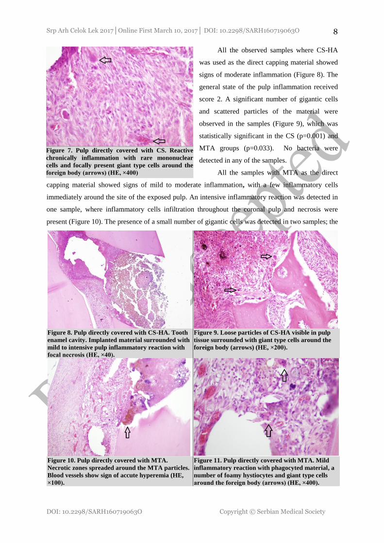
All the observed samples where CS-HA
was used as the direct capping material showed
signs of moderate inflammation (Figure 8). The
general state of the pulp inflammation received
score 2. A significant number of gigantic cells
and scattered particles of the material were
observed in the samples (Figure 9), which was
statistically significant in the CS (p=0.001) and
MTA groups (p=0.033). No bacteria were
detected in any of the samples.
All the samples with MTA as the direct
capping material showed signs of mild to moderate inflammation, with a few inflammatory cells
immediately around the site of the exposed pulp. An intensive inflammatory reaction was detected in
one sample, where inflammatory cells infiltration throughout the coronal pulp and necrosis were
present (Figure 10). The presence of a small number of gigantic cells was detected in two samples; the
Figure 8. Pulp directly covered with CS-HA. Tooth
enamel cavity. Implanted material surrounded with
mild to intensive pulp inflammatory reaction with
focal necrosis (HE, ×40).
Figure 9. Loose particles of CS-HA visible in pulp
tissue surrounded with giant type cells around the
foreign body (arrows) (HE, ×200).
Figure 10. Pulp directly covered with MTA.
Necrotic zones spreaded around the MTA particles.
Blood vessels show sign of accute hyperemia (HE,
×100).
Figure 11. Pulp directly covered with MTA. Mild
inflammatory reaction with phagocyted material, a
number of foamy hystiocytes and giant type cells
around the foreign body (arrows) (HE, ×400).
Figure 7. Pulp directly covered with CS. Reactive
chronically inflammation with rare mononuclear
cells and focally present giant type cells around the
foreign body (arrows) (HE, ×400)

Srp Arh Celok Lek 2017│Online First March 10, 2017│ DOI: 10.2298/SARH160719063O
DOI: 10.2298/SARH160719063O Copyright © Serbian Medical Society
9
particles of the phagocytized material were also observed (Figure 11). No bacteria were detected in
any of the samples.
DISCUSSION
At present an enormous progress has been achieved in synthesizing a number of new materials
used in the clinical practice.
The materials tested in this study were newly synthesized nanomaterials based on tricalcium
and dicalcium silicates and hydroxyapatites (CS and CS-HA). Their structure and biological
properties were compared with those of mineral trioxide aggregate (MTA), which is the golden
standard of tricalcium silicate cements.
Understanding the mechanism of interaction between biological fluids or cells and endodontic
materials is key to assessing new materials used in diagnostics and therapy, and to avoiding materials’
harmful reactions after their use [19]. Nanoparticles of different size and chemical structure typically
get deposited in mitochondria, causing considerable structural damage due to the reactive oxygen
species (ROS) synthesis that leads to oxidative stress. Mitochondria are the main locations of ROS
production in a cell, which can result in the generation of a hydroxyl radical (OH): one of the most
potent oxidising agents in nature. Oxidative stress is the most common cause of cell injury.
Nanostructured calcium silicates, CS and CS-HA (with particles from 117 to 477nm), in the
initial in vitro assays designed to assess genotoxicity and cytotoxicity showed the absence of harmful
cell effects within the parameters of the Comet and the MTT assays [20, 21].
In vitro studies are fundamentally different from in vivo ones where proteins, tissue fluids and
other factors can reduce the toxic effects of the material [22]. In vivo assays render more
comprehensive and clinically more relevant information about the tissue response over a protracted
time period. The soft tissues’ histological reaction to biomaterial has been a long established and
frequently used method of biocompatibility assessment [7, 9]. These assays are highly reliable in
evaluating the tissue irritation, and the interaction of tissue and biomaterial. Tissue’s reaction to an
implant is a cumulative pathophysiological consequence of a) the healing of an acute injury sustained
due to a surgical wound and the presence of implant, b) possible chronic inflammation, c) the
surrounding tissue repair while adapting to the implant.
This experiment monitored short-term, but also long-term, effects of the materials on the tissue,
since these data are relevant to the clinical use of the endodontic materials.
Monitoring of a specific response to the foreign body following the material implantation starts
with inflammation, continuing through the stages of wound healing with the involvement of various
cell types being specific indicators of the tissue repair stage. With the inflammatory process
attenuating, the total number of inflammatory cells decreases while the wound healing process moves
towards formation of granulation tissue and fibrous encapsulation of the implanted material.

Srp Arh Celok Lek 2017│Online First March 10, 2017│ DOI: 10.2298/SARH160719063O
DOI: 10.2298/SARH160719063O Copyright © Serbian Medical Society
10
The tissue surrounding the tested bioceramic materials (CS, CS-HA) showed the highest level
of inflammation in the first 15 days, with moderate disruption in the connective tissue structures. The
connective tissue around the MTA showed signs of the most severe inflammatory reaction with
diffuse and focal subcapsular and perivascular infiltrates, which was evaluated as moderate and severe
inflammation. Other researchers also reported such a powerful tissue response, observing even
coagulative necrosis and dystrophic calcifications [7]. A number of factors caused initially such a
severe inflammatory reaction to MTA. High pH values of the freshly mixed material, heat release
upon setting, and stimulation of inflammatory cytokines (interleukin 1 and interleukin 6) contribute to
such a powerful tissue response to MTA [18, 23, 24, 23].
Tissue reaction to empty tubes (negative control) in this experiment was similar to the findings
reported by other researchers [9, 22, 24]. It was most severe on the 7th day and the 15th day with
cellular infiltrate dominated by lymphocytes and plasmocytes, which is indicative of a chronic
inflammatory reaction. The gigantic cells detected in some sites suggest the tissue’s reaction to a
foreign body. This reaction could partly be a result of surgical trauma sustained upon implanting the
tubes in the tissue. At the end of weeks four and eight, the inflammatory infiltrate cells were
disappearing, and the tissue at the point of contact with the tube was encapsulated. This suggests the
body’s capacity to contain the inflammatory reaction thus preventing further tissue damage, as
confirmed in papers by Sumer [5] and Scarparo [6].
Further into the tissue repair process (after 30 and 60 days), significant decrease in the
inflammation intensity; disappearance of inflammatory infiltrate cells, but also tissue repair and
remodelling were observed in all tested materials. The inflammatory response after four and eight
weeks of the experiment can be explained by inducing the release of proinflammatory cytokines by
released particles of the hydroxyapatite layer formed on the surface of the bioceramic materials. This
also suggests good interaction between the material and the cells from the surrounding tissue, which is
a sign of good biocompatibility of the materials.
The pulp tissue response to the implanted material, usually, starts with an acute inflammation,
which need not be present in all cases [25]. The topography and chemistry of the surface of newly
synthetized materials plays an important role in odontoblast adhesion to biomaterial. It is a known fact
that micro- and nano-topography of the surface and adsorbed proteins have direct influence on the cell
behaviour and activity, primarily with respect to their adhesion and retention at the point of
application [26].
Nanostructured calcium silicate cements show increased osteoblast adhesion, proliferation and
differentiation, since the bone itself has nano-structure, and the crystal size and geometry can modify
the response of the surrounding tissue. The interaction between the direct capping material and the
injured pulp tissue, and the ways in which the healing and repair processes are initiated and developed
are still not fully clear. While many hypotheses exist, recent studies have accorded the main role to

Srp Arh Celok Lek 2017│Online First March 10, 2017│ DOI: 10.2298/SARH160719063O
DOI: 10.2298/SARH160719063O Copyright © Serbian Medical Society
11
growth factors in angiogenesis, mobilisation of progenitor cells, differentiation and, finally,
biomaterial-assisted mineralisation [27].
As a consequence of experimental perforation, but also of the initial effect of the tested
materials, a mild to moderate inflammatory reaction was detected in all observed teeth samples. The
inflammatory infiltrate was in close proximity of the implanted material and was not spreading further
into the coronal pulp. The weakest inflammatory reaction, sporadically with total absence of
inflammation, was observed in samples where calcium silicate cement (CS) was implanted as the
direct capping material, which agrees with the findings of other researchers who find up to 50%
samples with no signs of inflammation in the early stage [12, 28]. Moderate inflammation was
detected in the CS-HA and MTA samples, which is confirmed by similar experiments where
inflammation appeared in more than 62% of the MTA samples after two weeks, with the intensity
declining after eight weeks. This initially severe inflammatory response is a result of the pulp tissue
coagulative necrosis in contact with the MTA (pH is 9 - 10). This zone has a stimulating effect on the
surrounding vital pulp tissue, which initiates a string of healing processes. Due to bio-degradation of
the material in contact with the tissue fluids, Ca and P ions are released, creating alkaline
environment, which has a favourable effect on adhesion and proliferation of cells involved in the
healing processes [12, 29]. Asgary observes that new endodontic cements having similar structure as
the MTA, but improved physical and chemical properties – they include the nanomaterials tested –
show better pulp response (weaker inflammatory reaction) and thicker dentine bridge then the MTA at
the later stage [30].
Biomaterials are normally tested on animals, since they are a model of the environment one can
find in humans. Nevertheless, animas are characterised by a huge range of differences with respect to
anatomy, biochemistry, physiology, and other. In the absence of confirmation from human clinical
trials, it is often difficult to draw a conclusion solely on the basis of animal testing. Testing carried out
on live systems invariably leads to experimental variability. The more complex the system (human
cells versus microorganism cells) the higher statistical variability of testing results can be expected
[3].
CONCLUSION
The results shown in the present in vivo study on the animal model have proven that the
subcutaneous tissue of rats and the pulp tissue of rabbits have favourable biological response to newly
synthetized nanostructured biomaterials (CS and CS-HA). The inflammatory reaction in the
subcutaneous tissue was severe only in the initial days after the implantation and its intensity declined
as a function of time. In direct pulp capping there was a mild to moderate inflammatory reaction in the
close proximity of the implanted material. The tissues showed high tolerance to the implanted
materials, which confirms their biocompatibility, as in previous in vitro studies.

Srp Arh Celok Lek 2017│Online First March 10, 2017│ DOI: 10.2298/SARH160719063O
DOI: 10.2298/SARH160719063O Copyright © Serbian Medical Society
12
REFERENCES
1. Camargo SEA, Camargo CHR, Hiller K-A, Rode SM, Schweikl H, Schmalz G. Cytotoxicity and
genotoxicity of pulp capping materials in two cell lines. Int Endod J. 2009; 42: 227–37.
2. Nedel F, Soki FN, Conde MCM, Zeitlin BD, Tarquinio SBC, Nör JE, et al. Comparative analysis two
colorimetric assays in dental pulp cell density. Int Endod J. 2011; 44: 59–64.
3. Raković D, Uskoković D. Biomaterijali. Beograd: Institut tehničkih nauka SANU, Društvo za istraživanje
materijala Srbije; 2010. p. 221–40.
4. Modareszadeh MR, Chogle SA, Mickel AK, Jin G, Kowsar H, Salamat N. Shaikh S, Qutbudin S.
Cytotoxicity of set polymer nanocomposite resin root-end filling materials. Int Endod J. 2011; 44: 154–61.
5. Sumer M, Muglali M, Bodrumlu E, Guvenc T. Reactions of Connective Tissue to Amalgam, Intermediate
Restorative Material, Mineral Trioxide Aggregate, and Mineral Trioxide Aggregate Mixed With
Chlohexidine. J Endod. 2006; 32(11): 1094–96.
6. Scarparo RK, Haddad D, Acasigua GAX, Fossati ACM, Fachin EVF, Grecca FS. Mineral Trioxide
Aggregate-based Sealer: Analysis of Tissue Reactions to a New Endodontic Material. J Endod. 2010;
36(7): 1174–8.
7. Parirokh M, Mirsoltani B, Raoof M, Tabrizchi H, Haghdoost AA. Comparative study of subcutaneous
tissue responses to a novel root-end filling material and white and grey mineral trioxide aggregate. Int
Endod J. 2011; 44: 283–9.
8. Silva-Herzog D, Ramirez T, Mora J, Pozos AJ, Silva LAB, Silva RAB, et al. Preliminary study of the
inflammatory response to subcutaneous implantation of three root canal sealers. Int Endod J. 2011; 44:
440–6.
9. Gomes-Filho JE, Watanabe S, Lodi CS, Cintra LT, Nery MJ, Filho JA, et al. Rat tissue reaction to MTA
FILLAPEX. Dental Traumatology. 2012; 28: 452–56.
10. Zhang S, Yang X, Fan M. Bioaggregate, iRoot BP Plus optimize the proliferation and mineralization
ability of human dental pulp cells. Int Endod J. 2013; 46: 923–29.
11. Ando Y, Honda MJ, Ohshima H, Tonomura A, Ohara T, Itaya T, et al. The induction of dentin bridge-like
structures by constructs of subcultured dental pulp-derived cells and porous HA/TPC in porcine teeth. J
Med Sci. 2009; 71: 51–62.
12. Zarrabi MH, Javidi M, Jafarian AH, Joushan B. Histologic Assessment of Human Pulp Response to
Capping with Mineral Trioxide Aggregate and a Novel Endodontic Cement. J Endod. 2010; 36(11): 1778–
81.
13. Roberts HW, Toth JM, Berzins DW, Charlton DG. Mineral trioxide aggregate material use in endodontic
treatment: A review of the literature. Dental Materials. 2008; 24: 149–64.
14. Orhan EO, Maden M, Senguüven B. Odontoblast-like cell numbers and reparative dentine thickness after
direct pulp capping with platelet-rich plasma and enamel matrix derivative: a histomorphometric
evaluation. Int Endod J. 2012; 45: 317–25.
15. Güven EP, Taşli PN, Yalvac ME, Sofiev N, Kayahan MB, Sahin F. In vitro comparison of induction
capacity and biomineralization ability of mineral trioxide aggregate and a bioceramic root canal sealer. Int
Endod J. 2013; 46: 1173–82.
16. International Organization for Standardization. ISO 10993: 2009 ed.
17. International Standard Organisation. ISO 7405 Dentistry-Preclinical Evaluation of Biocompatibility of
Medical Device Used in Dentistry-Test method for Dental Material. Geneva, Switzerland; 1997
18. Lotfi M, Vosoughhosseini S, Saghiri MA, Mesgariabbasi M, Ranjkesh B. Effect of White Mineral
Trioxide Aggregate Mixed With Disodium Hydrogen Phosphate on Inflammatory Cells. J Endod. 2009;
35(5): 703–5.
19. Fenoglio I, Fubini B, Ghibaudi EM, Turci F. Multiple aspects of the interaction of biomacromolecules
with inorganic surface. Adv Drug Deliver Rev. 2011; 63: 1186–209.
20. Opačić-Galić V, Petrović V, Živković V, Jokanović V, Nikolić B, Knežević-Vukčević J, et al. New
nanostruktural biomaterials based on active silicate systems and hydroxyapatite: caracterization and
genotoxicity in human peripheral blood lymphocytes. International Endodontic Journal, 2013; 46: 506–16.
21. Petrović V, Opačić-Galić V, Živković S, Nikolić B, Danilović V, Miletić V, et al. Biocompatibility of new
nanostructural materials based on active silicate systems and hydroxyapatite: in vitro and in vivo study.
International Endodontic Journal 2015; 48: 966-75.
22. Yavari HR, Shahi S, Rahimi S, Shakouie S, Roshangar L, Abbasi MM, et al. Connective tissue reaction to
white and gray MTA mixed with distilled water or chlorhexidine in rats. Iranian Endod J. 2009; 4(1): 25–
30.
23. Camilleri J. A review of the methods used to study biocompatibility of Portland cement-derived materials
used in dentistry. Malta Medical Journal. 2006; 18(3): 9–14

Srp Arh Celok Lek 2017│Online First March 10, 2017│ DOI: 10.2298/SARH160719063O
DOI: 10.2298/SARH160719063O Copyright © Serbian Medical Society
13
24. Vosoughhosseini S, Lotfi M, Shabi S, Baloo H, Mesgariabbasi M, Saghiri MA, et al. Influence of White
versus Gray Mineral Trioxide Aggregate on Inflammatory Cells. J Endod. 2008; 34(6): 715–17.
25. Leprince JG, Zeitlin BD, Tolar M, Peters OA. Interaction between immune system and mesenchymal stem
cell in dental pulp and periapical tissues. Int Endod J. 2012; 45: 689–701.
26. Gandolfi MG, Ciapetti G, Perut F, Taddei P, Modena E, Rossi PL, et al. Biomimetic calcium-silicate
cement aged in simulated body solutions. Osteoblast response and analyses of apatite coating. Journal of
Applied Biomaterials & Biomechanics. 2009; 7(3): 160–70.
27. Laurent P, Camps J, About I. Biodentine induces TGF-β1 release from human pulp cells and early dental
pulp mineralization. Int endod J. 2012; 45: 439–48.
28. Da Silva GF, Guerreiro-Tanomaru JM, Sasso-Cerri E, Tanomaru-Filho M, Cerri PS. Histological and
histomorphometrical evaluation of furcation perforations filled with MTA, CPM and ZOE. Int Endod J.
2011; 44: 100–10.
29. Accorinte MLR, Holland R, Reis A, Bortoluzzi MC, Murata SS, Dezan E, et al. Evaluation of Mineral
Trioxide Aggregate and Calcium Hydroxide Cement as Pulp-Capping Agents in Human Teeth. J Endod.
2008; 34: 1–6.
30. Asgary S, Eghbal MJ, Parirokh M, Ghanavati F, Rahimi H. A comparative study of histologic response to
different pulp capping materials and a novel endodontic cement. Oral Surg Oral Med Oral Pathol Oral
Radiol Endod. 2008; 106: 609–14.