zastosowanie ultrasonografii w ocenie obrąbka stawu ramiennego
TRANSCRIPT

164
Zastosowanie ultrasonografii w ocenie obrąbka stawu ramiennego. Część I: Anatomia ultrasonograficzna i technika badania
The use of ultrasound in the assessment of the glenoid labrum of the glenohumeral joint. Part I: Ultrasound anatomy and examination technique
Wojciech Krzyżanowski
Specjalistyczna Praktyka Lekarska, Lublin, Polska Adres do korespondencji: Wojciech Krzyżanowski, ul. Głęboka 29, 20-612 Lublin, e-mail: [email protected]
StreszczenieStaw ramienny jest stawem kulistym o dużym zakresie ruchów w wielu płaszczyznach i zmniejszonej stabilności. Utrzymanie stabilności stawu jest uwarunkowane z jednej strony działaniem specyficznego układu mięśni okolicy barkowej, z drugiej – obecnością złożonego kompleksu więzadłowego wzmacniającego torebkę stawową, a także pogłę-bieniem panewki stawowej przez obrąbek. Uszkodzenia tych struktur wymagają dokład-nej diagnostyki przed podjęciem decyzji o leczeniu operacyjnym. Ultrasonografia należy do metod obrazowych szeroko stosowanych w ocenie różnych patologii okolicy barku. W opinii własnej autora pracy może mieć również zastosowanie w ocenie uszkodzeń obrąbka stawu ramiennego. Przyczepiając się na obwodzie panewki stawowej, obrąbek tworzy kołnierz pogłębiający panewkę i zwiększający jej pole kontaktu z głową kości ra-miennej. W celach topograficznych obrąbek stawowy dzieli się najczęściej na strefy. Więk-szość z nich jest widoczna w badaniu ultrasonograficznym. Badanie obrąbka w części tylnej przeprowadza się zwykle jednocześnie z oceną mięśnia podgrzebieniowego i obłego mniejszego. W części przedniej obrąbek wraz z kompleksem torebkowo-więzadłowym jest widoczny przy zarysie panewki stawowej pod mięśniem podłopatkowym. Badanie ultrasonograficzne obrąbka w części dolnej wykonuje się z dostępu pachowego. Obrąbek w części górnej jest tylko częściowo dostępny ocenie ultrasonograficznej. Kluczową część oceny ultrasonograficznej stanowi badanie dynamiczne. W pracy przedstawiono zarys prawidłowej anatomii ultrasonograficznej oraz techniki badania i oceny ultrasonogra-ficznej obrąbka stawu ramiennego.
Submitted: 02.02.2012Accepted: 14.02.2012
Słowa kluczoweobrąbek, staw
ramienny, anatomia ultrasonograficzna,
technika badania, diagnostyka
obrazowa stawu ramiennego
Praca poglàdowaReview
Journal of Ultrasonography 2012; 12: 164–177
165J Ultrason 2012; 12: 164–177
The use of ultrasound in the assessment of the glenoid labrum of the glenohumeral joint. Part I: Ultrasound anatomy and examination technique
AbstractThe glenohumeral joint is a spherical articulation with a remarkable range of motion in several planes and decreased stability. The maintenance of joint stability is influenced by the functioning of specific muscle groups in the shoulder region, a complex system of ligaments reinforcing the joint capsule, and the labrum which augments the glenoid fossa. Lesions of the aforementioned structures require accurate diagnosis prior to a decision for operative treatment. Ultrasound is one of the imaging methods that has been widely used in the assessment of various shoulder pathologies. In the author opinion, this imag-ing modality may also be applied for the evaluation of labral tears. Being attached along the glenoid rim, the labrum forms a collar deepening the glenoid fossa thus increasing area of its contact with the head of the humerus. To better describe the location of lesions, the glenoid labrum is usually divided into certain zones. Most of them may be visualized sonographically. The US examination of the posterior labrum can be performed during evaluation of the infraspinatus and teres minor muscles. The anterior labrum along with capsulolabral complex is seen at the glenoid edge under the subscapularis tendon. So-nographic examination of the inferior labrum is best performed using axillar approach. The superior labrum is only partially available for US examination. A crucial part of the sonographic assessment of the labrum is the dynamic examination during rotation of the upper extremity. The paper presents normal sonographic anatomy of the glenoid labrum and technique of the examination.
Key wordsglenoid labrum,
glenohumeral joint, ultrasound anatomy,
examination technique,
glenohumeral joint imaging
Wstęp
Staw łopatkowo-ramienny (w skrócie określany jako ramienny) z anatomicznego punktu widzenia jest stawem kulistym, utworzonym między głową kości ramiennej (główka stawowa) a wydrążeniem stawowym łopatki (panewka stawowa). Powierzch-nia stawowa głowy kości ramiennej jest mniej wię-cej czterokrotnie większa niż powierzchnia stawo-wa wydrążenia stawowego łopatki, dlatego często porównuje się ją do piłeczki golfowej (głowa) spo-czywającej na podstawce (panewka). Dzięki takiej budowie możliwy jest duży zakres ruchów w stawie ramiennym w różnych płaszczyznach, zwiększany dodatkowo przez ślizg łopatki względem ściany klat-ki piersiowej, a także ruch w stawie barkowo-oboj-czykowym i mostkowo-obojczykowym. Ta wyjątkowa ruchomość stawu ramiennego wiąże się jednak ze zmniejszoną stabilnością i większą podatnością na zwichnięcia i urazy od innych stawów. W warunkach prawidłowych podczas ruchu w stawie ramiennym istnieje pewna równowaga dynamiczna pomiędzy siłami dociskającymi głowę kości ramiennej do panewki a siłami odciągającymi. Stabilność stawu ramiennego jest uwarunkowana działaniem mecha-nizmów czynnych i biernych. Stabilizacja czynna wiąże się z funkcją mięśni, z wiodącą rolą mięśni rotatorów (obrotowych) oraz ścięgna głowy długiej mięśnia dwugłowego ramienia. Ścięgna mięśni ob-rotowych tworzą charakterystyczny „mankiet” osła-niający staw, połączony z torebką stawową („kabel rotatorów”). Stabilizacja bierna wynika z ukształ-towania, wielkości i ustawienia panewki stawowej, a także obecności złożonego układu torebkowo-wię-zadłowego wraz z obrąbkiem panewkowym. Obrą-
Introduction
The glenohumeral joint (in short referred to as the shoulder joint) is a ball-and-socket joint between the head of the humerus (the ball) and the glenoid fossa of the scapula (the socket). The joint surface area of the humeral head is approximately fourfold greater than that of the glenoid cavity; this is often compared to a golf ball resting upon the tee. Thanks to such morphology, a large range of motion of the shoul-der joint is possible in several planes, augmented by the sliding of the scapula with respect to the thoracic wall, as well as rotation within the acromioclavicular and sternoclavicular joints. This tremendous range of movement also makes the shoulder less stable and far more prone for dislocation and injury than other joints. However, during movements in the shoulder under normal circumstances, there exists a dynam-ic balance between the forces pushing the humeral head onto the glenoid fossa versus those forces pull-ing it away. The stability of the joint depends upon both active and passive mechanisms. Active stabiliza-tion is related to muscle function, most importantly the rotator muscles and the tendon of the long head of the biceps brachii muscle. The tendons of the rota-tor muscles form a characteristic “cuff” surround-ing the joint, along with the rotator cable of the joint capsule. Passive stabilization comes from the shape, size and positioning of the glenoid, as well as the presence of a capsular-ligamentous complex, and the glenoid labrum. The former plays a significant role. Being attached along the glenoid rim, the la-brum forms a collar deepening the glenoid fossa and increasing the area of its contact with the head of the humerus (fig. 1). This allows for the maintenance

166 J Ultrason 2012; 12: 164–177
Wojciech Krzy˝anowski
bek panewki pełni niebagatelną funkcję. Przycze-piając się na obwodzie panewki stawowej, tworzy pewnego rodzaju kołnierz pogłębiający panewkę i zwiększający jej pole kontaktu z głową kości ra-miennej (ryc. 1). Pozwala tym samym na utrzyma-nie ujemnego ciśnienia wewnątrz stawu, istotnego dla jego stabilizacji(1,2). Z histologicznego punktu widzenia obrąbek stawowy jest strukturą włóknisto-chrzęstną z różnym przebiegiem włókien kolageno-wych, głównie o układzie okrężnym, zasilaną w stre-fie zewnętrznej włóknami dochodzącymi od strony więzadeł obrąbkowo-ramiennych, torebki stawowej i otaczających ścięgien. Liczne włókna kolagenowe przebiegają w macierzy chrzęstnej/chrzęstno-włók-nistej, zawierającej drobne lakuny z chondrocytami. W strukturze obrąbka mogą występować także nie-liczne włókna elastyny. Unaczynienie obrąbka po-chodzi od rozgałęzień tętnicy nadłopatkowej, tętnicy okalającej łopatkę oraz tętnicy tylnej okalającej ra-mię, dochodzących do obrąbka od strony przyczepu torebkowego oraz od okostnej łopatki. Naczynia te odżywiają głównie obwodową strefę obrąbka, spora-dycznie dochodzą do części środkowej. Wraz z wie-kiem unaczynienie obrąbka (podobnie jak łąkotek) zmniejsza się(3,4). W dolnej połowie, zarówno w czę-ści przedniej, jak i tylnej, obrąbek stanowi przedłu-żenie chrząstki stawowej panewki z przejściową strefą chrzęstno-włóknistą. Z tego też względu wy-kazuje on wyraźnie ograniczoną ruchomość. Zarysy obrąbka w tej części są zwykle lekko zaokrąglone, choć istnieje duża zmienność anatomiczna w zakre-sie kształtu obrąbka(5). Dlatego nawet nieregularny zarys obrąbka od strony stawu nie powinien być traktowany bezwzględnie jako objaw patologiczny. W górnej połowie obrąbek ma nieco mniej zbitą strukturę włóknistą, luźniejszy przyczep panewko-wy i jest wobec tego bardziej mobilny. Na przekroju poprzecznym obrąbek ma kształt przypominający łąkotkę stawu kolanowego. Z obrąbkiem w części górnej łączy się ściśle ścięgno głowy długiej mięśnia dwugłowego ramienia (przyczepiające się, poza ob-rąbkiem, także do guzka nadpanewkowego łopatki), tworząc wspólny kompleks obrąbkowo-ścięgnisty, stanowiący przeplatające się włókna kolagenowe obu struktur(3). Na poziomie fuzji ze ścięgnem głowy długiej mięśnia dwugłowego ramienia między ob-rąbkiem a brzegiem panewki występuje niewielki za-chyłek maziowy (zachyłek podobrąbkowy)(1,5). Poni-żej – w strefie przednio-górnej – występuje niekiedy zupełny brak połączenia obrąbka z panewką (otwór podobrąbkowy), z obecną komunikacją stawu z za-chyłkiem podłopatkowym(1). Może występować także odcinkowy brak obrąbka w połączeniu z obecnością pogrubiałego więzadła obrąbkowo-ramiennego środ-kowego (kompleks Buforda)(6).
of negative intra-articular pressure, which is essen-tial for joint stability(1,2). Histologically, the glenoid labrum is composed of fibrocartilaginous tissue with a varied course of collagen fibers, mainly in a circu-lar arrangement, reinforced in the external zone by fibers from the glenohumeral ligaments, joint capsule and surrounding tendons. Numerous collagen fibers run through the fibrocartilaginous matrix, containing small lacunae with chondrocytes. There may also ap-pear a few elastin fibers within the glenoid labrum. The labrum is vascularized by branches of the supra-
Ryc. 1. Schemat kompleksu torebkowo-więzadłowo-obrąbkowego ze stożkiem rotatorów stawu ramiennego. Gl – panewka, L – obrąbek, LHB – ścięgno głowy długiej mięśnia dwu-głowego ramienia, więzadła obrąbkowo-ramienne (SGHL – górne, MGHL – środkowe, IGHL – dolne), CHL – więza-dło kruczo-ramienne, CAL – więzadło kruczo-barkowe, Sub – mięsień podłopatkowy, Sup – mięsień nadgrzebieniowy, Is – mięsień podgrzebieniowy, TM – mięsień obły mniejszy (dzięki uprzejmości: Michael Stadnick, Radsource)
Fig. 1. A scheme of the capsular-ligamentous-labral complex with the rotator cuff of the shoulder. Gl – the glenoid, L – the labrum, LHB – the tendon of the long head of the biceps brachii muscle; the superior – SGHL, middle – MGHL, and inferior – IGHL glenohumeral ligaments, CHL – the cora-cohumeral ligament, CAL – the coracoacromial ligament, Sub – the subscapularis muscle, Sup – the supraspinatus muscle, Is – the infraspinatus muscle, TM – the teres minor muscle (courtesy of: Michael Stadnick, Radsource)

167J Ultrason 2012; 12: 164–177
The use of ultrasound in the assessment of the glenoid labrum of the glenohumeral joint. Part I: Ultrasound anatomy and examination technique
Obwodowa część obrąbka łączy się z torebką stawo-wą i więzadłami obrąbkowo-ramiennymi, tworząc jednostkę anatomiczno-czynnościową, zwaną kom-pleksem torebkowo-więzadłowo-obrąbkowym(1). Przyczep torebki stawowej w części przedniej wyka-zuje pewną zmienność anatomiczną. Mniej więcej w połowie przypadków przednia torebka stawowa przyczepia się bezpośrednio do obrąbka stawowego i przyległej części panewki. W pozostałych przypad-kach przyczep torebki stawowej w części przedniej znajduje się za obrąbkiem na szyjce łopatki (naj-częściej w odległości do 1 cm od brzegu panewki, rzadko bardziej przyśrodkowo). Torebka w części tylnej jest zwykle zespolona z obrąbkiem(2,5,7,8). Z ob-rąbkiem stawowym łączą się także więzadła obrąb-kowo-ramienne. Więzadło obrąbkowo-ramienne górne (SGHL) przyczepia się zwykle do obrąbka górnego i częściowo do guzka nadpanewkowego łopatki. Więzadło obrąbkowo-ramienne środkowe (MGHL) przyczepia się na poziomie lub tuż poniżej przyczepu więzadła górnego w różny sposób: albo do panewki, albo do obrąbka, ewentualnie może posiadać wspólne odejście z więzadłem górnym. Niekiedy MGHL może być bardzo prominentną strukturą (jak w kompleksie Buforda), zdarzają się też przypadki, w których nie stwierdza się obecno-ści tego więzadła(5). Więzadło obrąbkowo-ramienne dolne (IGHL) jest utworzone przez dwa pasma – przednie i tylne – posiada wyraźny przyczep obrąb-kowy i panewkowy i stanowi istotne wzmocnienie torebki stawowej(1).
W celach topograficznych obrąbek stawowy dzie-li się na pewne strefy. Najczęściej stosuje się dwie alternatywne metody – jedną bazującą na tarczy zegarowej z podziałem na 12 stref obrąbkowych, drugą bardziej uproszczoną i znajdującą zasto-
scapular, the circum-scapular, and the posterior cir-cumflex arteries, which reach it from the capsule and the periosteum near the bony attachment. These ves-sels mainly supply the peripheral area of the glenoid labrum, while the vascularization of its middle part is rather poor. Similarly to menisci the vascularization of the glenoid labrum decreases with age(3,4). In its inferior half, both in the anterior and posterior parts, the labrum forms a continuation of the joint cartilage of the glenoid, with a transitional fibrocartilaginous zone. This explains the significantly limited mobility of this part of labrum. The outline of the labrum in this part is usually slightly rounded, although there exist variable anatomic variants in the shape of the labrum(5). Therefore, even irregular contours of the labrum should not be primarily considered patho-logical. On the other hand, the superior half of the glenoid labrum has a slightly less dense fibrous struc-ture, a looser bony attachment, and is thus more mobile. In the transverse section, the labrum has a shape resembling the meniscus of the knee joint. In its superior half, the glenoid labrum becomes firmly attached to the tendon of the long head of the biceps (which also attaches to the supraglenoid tubercle of the scapula), forming a common biceps-labral com-plex with the interweaving of collagen fibers of both structures(3). At the level of fusion of both structures, between the labrum and the glenoid rim, there exists a small synovial recess (sublabral recess)(1,5). In the anterosuperior area, there may be a complete dis-continuity between the labrum and the glenoid (sub-labral foramen), so that the joint communicates with the subscapularis recess(1). There may also be a lack of anterosuperior labrum combined with a cordlike thickening of the medial glenohumeral ligament (Bu-ford complex)(6).
The peripheral part of the glenoid labrum connects to the joint capsule and the glenohumeral ligaments, to form an anatomical-functional unit, called the labral capsular ligamentous complex(1). The glenoid attachment of the anterior joint capsule exhibits cer-tain anatomical variants. In more than 50 percent of cases, the anterior capsule attaches directly to the labrum and the adherent part of the glenoid. In the remaining cases, the insertion of the anterior cap-sule is located beyond the labrum, on the scapular neck (usually within 1 cm from the edge of the gle-noid, rarely more medially). The posterior capsule is usually attached directly to the glenoid labrum(2,5,7,8). The glenohumeral ligaments also join the glenoid la-brum. The superior glenohumeral ligament (SGHL) usually inserts to the superior part of the labrum and partially to the supraglenoid tubercle of the scapula. The middle glenohumeral ligament (MGHL) attaches
Ryc. 2. Topograficzny podział obrąbka panewkowego na 6 stref(8)
Fig. 2. The topographical division of the glenoid labrum into 6 areas(8)

168 J Ultrason 2012; 12: 164–177
Wojciech Krzy˝anowski
sowanie w codziennej praktyce – z podziałem na 6 stref(9) (ryc. 2). Obie skale są generalnie bardzo zbliżone i wyróżniają (idąc od góry do przodu i do dołu, zgodnie z ruchem wskazówek zegara dla stro-ny prawej i przeciwnie do ruchu wskazówek zegara dla strony lewej):
• strefę obrąbka górnego (między godziną 11. a 1.);• przednio-górnego (między godziną 1. a 3. dla stro-
ny prawej i 11. a 9. dla strony lewej);• przednio-dolnego (między godziną 3. a 5. dla stro-
ny prawej i 9. a 7. dla strony lewej);• dolnego (między godziną 5. a 7.);• tylno-dolnego (między godziną 7. a 9. dla strony
prawej i 5. a 3. dla strony lewej);• tylno-górnego (między godziną 9. a 11. dla strony
prawej i 3. a 1. dla strony lewej).
Czasami dzieli się dodatkowo obrąbek w górnej części na: górno-przedni oraz górno-tylny, a w dol-nej części – na dolno-przedni i dolno-tylny. Ma to większe znaczenie w przypadku obrąbka górnego ze względu na jego częstsze uszkodzenia (typu SLAP). Izolowane uszkodzenia obrąbka w części dolnej są niezwykle rzadkie. Można stosować także jeszcze bardziej uproszczony opis topograficzny obrąb-ka stawu ramiennego z podziałem na cztery strefy (kwadranty): przednio-górną, przednio-dolną, tylno-dolną i tylno-górną(10). Podział ten może być wyko-rzystywany w diagnostyce niestabilności przedniej i tylnej stawu ramiennego, natomiast nie jest satys-fakcjonujący w przypadku klasyfikacji uszkodzeń górnej części obrąbka.
Głównym badaniem obrazowym stosowanym do oceny obrąbka stawu ramiennego oraz więzadeł jest rezonans magnetyczny (RM), najlepiej z do-stawowym podaniem środka paramagnetycznego (artrografia RM)(2,5–7,11). Opcjonalnie wykorzystu-je się także artrografię tomografii komputerowej (artro-TK)(6). Dane z piśmiennictwa oraz kilkulet-nie osobiste doświadczenia autora wskazują, że także ultrasonografia (USG) może być przydatna w ocenie obrąbka stawowego(10,12–15). Ultrasonogra-fię powszechnie stosuje się w diagnostyce różnych patologii okolicy barku od wielu lat, a jej rola syste-matycznie wzrasta wraz z ciągłym postępem tech-nologicznym tej metody obrazowania(16–18). Badanie USG ma niekwestionowane znaczenie zwłaszcza w diagnostyce patologii mięśni stożka rotatorów. Włączenie do zakresu rutynowego badania USG barku oceny obrąbka stawu ramiennego uczyniło-by zatem całe badanie jeszcze bardziej komplek-sowym, ukierunkowującym dalsze postępowanie diagnostyczno-terapeutyczne.
at the level of the SGHL insertion or right below it, either to the glenoid, the labrum, or has a common origin with the SGHL. Sometimes the MGHL may be a very prominent structure (as in the Buford com-plex), but there are also cases in which this ligament is completely missing(5). The inferior glenohumeral ligament (IGHL) consists of two bands – the anterior and posterior, has distinct attachments to the labrum and the glenoid, and significantly reinforces the joint capsule(1).
To better describe the location of lesions, the glenoid labrum is usually being divided into certain zones. There are two main alternative mapping methods used for this purpose. One is based upon a clock face with 12 zones of the labrum, whereas the simpler and more practical second method, divides the labrum into 6 zones(9) (fig. 2). Both scales are quite similar to each other and both distinguish the following labral zones (going from superior to inferior anteriorly, clockwise for the right side, and counterclockwise for the left):
• the superior labrum (between 11 and 1 o’clock);• the anterosuperior labrum (between 1 and 3 o’clock
for the right shoulder, while between 11 and 9 o’clock for the left one);
• the anteroinferior labrum (between 3 and 5 o’clock for the right shoulder, while between 9 and 7 o’clock for the left one);
• the inferior labrum (between 5 and 7 o’clock);• the posteroinferior labrum (between 7 and
9 o’clock for the right shoulder, while between 5 and 3 o’clock for the left one);
• the posterosuperior labrum (between 9 and 11 o’clock for the right shoulder, while between 3 and 1 o’clock for the left one).
Sometimes the labrum’s superior half is further di-vided into superoanterior and superoposterior parts, while the inferior half into inferoanterior and infero-posterior parts. This subdivision has more relevance for the superior labrum which is more frequently in-jured (SLAP type injuries), whereas injuries to the inferior labrum alone are extremely rare. An even simpler topographical description divides the labrum into 4 zones (quadrants): the anterosuperior, antero-inferior, posteroinferior and posterosuperior ones(10). This classification may be used in diagnosing ante-rior and posterior instability of the shoulder, but is insufficient when describing injuries of the superior labrum.
The main imaging modality used to assess the glenoid labrum along with glenohumeral ligaments is mag-
169J Ultrason 2012; 12: 164–177
The use of ultrasound in the assessment of the glenoid labrum of the glenohumeral joint. Part I: Ultrasound anatomy and examination technique
Technika badania i anatomia USG obrąbka stawu ramiennego
Do badania USG obrąbka w stawie ramiennym sto-suje się głowicę liniową pracującą w zakresie często-tliwości 6–12 MHz (najlepiej szerokopasmową), za-pewniającą odpowiednią rozdzielczość i penetrację na głębokość kilku centymetrów. U chorych otyłych bądź z większą masą mięśniową można też zasto-sować głowicę o częstotliwości 5–6 MHz (np. typu convex). Niestety, nie zapewnia ona wystarczającej rozdzielczości do oceny niewielkich zmian pourazo-wych obrąbka. Obrąbek, będący strukturą włóknistą, podlega zjawisku anizotropii, podobnie jak torebka stawowa. Zarówno w części przedniej, jak i tylnej położony jest na głębokości zwykle nieprzekraczają-cej 4,0–5,0 cm, czasami nawet mniejszej (w prakty-ce autora 2,5–3,5 cm). Głębokość jest zmienna in-dywidualnie i zależy od grubości tkanki podskórnej i „mankietu” mięśniowego. Nie jest prawdą, że ob-rąbek w części przedniej jest znacznie głębiej poło-żony niż w części tylnej, co miałoby go czynić mniej dostępnym dla badania USG, jak sugerują niektórzy autorzy(18). Najczęściej różnica w głębokości położe-nia nie przekracza kilku milimetrów na niekorzyść obrąbka przedniego, co zdaniem autora nie ma istot-nego znaczenia.
Obrąbek w części tylnej
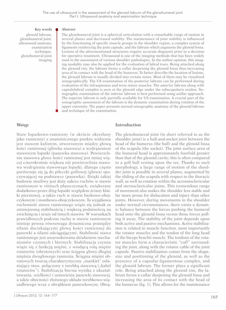
Badanie USG obrąbka w części tylnej przeprowadza się zwykle jednocześnie z oceną mięśnia podgrzebie-niowego i obłego mniejszego. Osoba badana siedzi wygodnie na stołku obrotowym, tyłem do osoby bada-jącej. Główną płaszczyzną obrazowania jest płaszczy-zna prostopadła do długiej osi ramienia (przekroje
netic resonance (MRI), especially with intra-articu-lar administration of paramagnetic contrast medium (MR arthrography)(2,5–7,11). Sometimes computed to-mography arthrography (CT arthrography) might be an option(6). Based upon published data, as well as the author’s personal experience of several years, ultra-sound (US) may also be valuable in the evaluation of the glenoid labrum(10,12–15). For many years, ultrasound has been widely used for diagnosing various pathol-ogies of the shoulder, and its role is still increasing with further technological progress of this imaging tool(16–18). The US study has an indisputable role, par-ticularly in diagnosing rotator cuff pathologies. The addition of an assessment of the glenoid labrum to the routine US imaging of the shoulder would make this examination even more complete, and influence further diagnostic-therapeutic proceedings.
Ultrasound anatomy and examination technique of the glenoid labrum
For the US examination of the glenoid labrum, a lin-ear probe with a frequency of 6–12 MHz (the broad-band probes are the best) is used, since it provides an appropriate resolution and penetration to the depth of a few centimeters. In obese patients or those with increased muscle mass, a probe of 5–6 MHz fre-quency (for example the convex type) may be used. Unfortunately, the latter does not provide enough resolution for the evaluation of small post-traumatic changes of the labrum. The glenoid labrum, being a fibrous structure, exhibits the anisotropy phenom-enon, as the joint capsule does. Both the anterior and posterior parts of the labrum are located at a depth usually not exceeding 4.0–5.0 cm, sometimes less (between 2.5 and 3.5 cm in the author’s experience). This depth varies, depending on the thickness of the subcutaneous fat tissue and the muscle “cuff”. Con-trary to the suggestions of some authors(18), it seems not to be true that the anterior part of the labrum is significantly more deeply located than its posterior part, which would make it less accessible for the US study. The difference in depth does not exceed a few millimeters, and has in author’s opinion little practi-cal significance.
The posterior part of the glenoid labrum
The US examination of the posterior part of the la-brum is usually carried out with the evaluation of the infraspinatus and teres minor muscles. The pa-tient sits comfortably on a rotating chair, with his/her back to the examiner. The main imaging plane is that
Ryc. 3. Ustawienie głowicy USG do badania obrąbka w części tylnejFig. 3. Positioning of the US probe for the evaluation of the poste-
rior part of the labrum

170 J Ultrason 2012; 12: 164–177
Wojciech Krzy˝anowski
osiowe) (ryc. 3). Uwidocznia się zarys tylnej części głowy kości ramiennej oraz panewki. Obrąbek sta-wowy jest widoczny najczęściej jako echogeniczna struktura trójkątnego kształtu, położona tuż przy brzegu panewki stawowej. Może być lekko zaokrąglo-ny od strony stawu, a nawet płaski, co jest związane ze zmiennością anatomiczną. Do obrąbka dochodzi i przyczepia się torebka stawowa, nad którą znajdują się mięśnie tylnej części stożka rotatorów (ryc. 4).
Obrąbek staramy się zobrazować możliwie w naj-większym zakresie, tj. od poziomu tuż poniżej grze-bienia łopatki aż do samego dołu panewki stawowej. Na poziomie grzebienia łopatki obrąbek nie jest do-stępny badaniu USG. Ważnym elementem badania jest ocena dynamiczna, polegająca na naprzemiennej rotacji wewnętrznej i zewnętrznej kończyny górnej w pozycji przywiedzenia. Przy rotacji wewnętrznej można zaobserwować napinanie się torebki stawowej z niewielkim pociąganiem obrąbka stawowego, nato-miast przy rotacji zewnętrznej torebka stawowa roz-luźnia się, zaś obrąbek nieco spłaszcza się i odchyla. W przypadku obecności wysięku w stawie ramien-nym podczas rotacji zewnętrznej uwydatnia się za-zwyczaj mniej lub bardziej wypełniony zachyłek tylny
perpendicular to the long axis of the forearm (axial cross-sections) (fig. 3). Both the posterior part of the humeral head and the glenoid rim should be first vi-sualized. The labrum is usually seen as a triangular echogenic structure, located right alongside the edge of the glenoid. It may be slightly rounded from the joint side, or may be flat, which is another anatomic variant. The joint capsule, which lies deep to the ten-dons of the posterior rotator cuff muscles, attaches to the labrum directly (fig. 4).
The labrum should be scanned as much as possible, from the level right below the scapular spine to the lower edge of the glenoid. The labrum is inaccessi-ble to US imaging at the level of the scapular spine. The dynamic assessment is an important aspect of the US examination, and consists of alternating in-ternal and external rotation of the upper extremity in the adduction position. In internal rotation, the tightening of the joint capsule may be observed, with slight pulling on the glenoid labrum. In external ro-tation, the joint capsule gets lax, while the labrum becomes slightly flattened and minimally bends in or out. If there is an effusion in the joint, a more or less filled posterior recess of the joint capsule is usually visible during external rotation. It extends medially from the glenoid rim and labrum along the scapular
Ryc. 4. Obrąbek w części tylnej stawu ramiennego (*), torebka sta-wowa (strzałki); H – głowa kości ramiennej, Gle – panewka, ISP – mięsień podgrzebieniowy
Fig. 4. The posterior labrum (*), the joint capsule (arrows); H – the head of the humerus, Gle – the glenoid, ISP – the infraspi-natus muscle
Ryc. 5. Uwypuklona torebka stawowa (strzałki) z bezechowym pły-nem wypełniającym tylny zachyłek stawu (+), nad zarysem zewnętrznym panewki (Gle) i obrąbkiem (*); H – głowa ko-ści ramiennej, ISP – mięsień podgrzebieniowy, D – mięsień naramienny
Fig. 5. The bulging joint capsule (arrows) with anechoic fluid fil-ling the posterior recess of the joint (+), above the external outline of the glenoid (Gle) and the labrum (*); H – the head of the humerus, ISP – the infraspinatus muscle, D – the deltoid muscle
171J Ultrason 2012; 12: 164–177
The use of ultrasound in the assessment of the glenoid labrum of the glenohumeral joint. Part I: Ultrasound anatomy and examination technique
torebki stawowej, sięgający przyśrodkowo od brzegu panewki i obrąbka stawowego (ryc. 5). Jest to miejsce pozwalające na wykrywanie nawet niewielkiej ilości płynu w stawie ramiennym, obecnego w przebiegu chorób tego stawu (drugim takim miejscem jest po-chewka ścięgna głowy długiej mięśnia dwugłowego ramienia).
Obrąbek w części przedniej
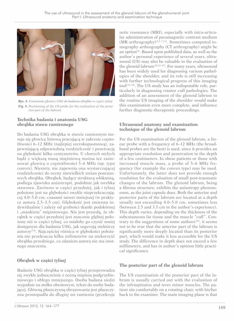
Badanie USG obrąbka w części przedniej jest nieco trudniejsze i wymaga pewnej wprawy. Badanie naj-lepiej wykonać u chorego leżącego na wznak, gdyż zapewnia to lepszą stabilizację sondy, szczególnie podczas oceny dynamicznej. Główną płaszczyzną obrazowania jest płaszczyzna prostopadła lub lek-ko skośna do długiej osi ramienia (przekroje osio-we) (ryc. 6). W badaniu należy uwidocznić przednią część głowy kości ramiennej oraz panewki stawowej. Budowa przedniego kompleksu torebkowo-obrąbko-wego jest bardziej złożona, dlatego obraz ultraso-nograficzny różni się nieco w porównaniu ze stroną tylną. Obrąbek stawowy jest widoczny przy brzegu panewki jako echogeniczna struktura razem z przy-ległą od strony przedniej torebką stawową, grubszą niż w części tylnej z powodu obecności przedniego pasma więzadła obrąbkowo-ramiennego dolnego (ryc. 7). Echogeniczność torebki jest zbliżona do echogeniczności obrąbka, stąd trudno oddzielić obie struktury, chociaż niekiedy daje się zauważyć w cza-sie badania dynamicznego niewielki ślizg torebki
neck (fig. 5). Incidentally, it is possible to visualize even small amounts of fluid in this location in the course of shoulder diseases (a second such place is the tendon sheath of the long head of the biceps).
The anterior part of the glenoid labrum
The US examination of the anterior part of the la-brum is somewhat more difficult and requires some practice. It is best to examine the patient in the su-pine position, as this ensures better stability of the probe, particularly during the dynamic assessment. The main imaging plane is that perpendicular or slightly oblique to the long axis of the arm (axial cross-sections) (fig. 6). Both the anterior part of the humeral head and the glenoid should be visualized. The structure of the anterior capsular-labral complex is more complicated than the posterior equivalent, thus their US image differs. The labrum is visible at the edge of the glenoid as an echogenic structure adjacent to the joint capsule, which is thicker than in the posterior part because of the presence of the anterior band of the IGHL (fig. 7). The echogenicity of the joint capsule is similar to that of the labrum, thus it is difficult to differentiate these structures, although sometimes during the dynamic examina-tion the joint capsule may slide against the labrum. Sometimes at the base of the labrum, there may be
Ryc. 6. Ustawienie głowicy USG do badania obrąbka w części przedniej
Fig. 6. Positioning of the US probe for the evaluation of the anterior labrum
Ryc. 7. Kompleks obrąbkowo-torebkowy w części przedniej stawu ramiennego (strzałki): obrąbek (*), przednie pasmo więza-dła obrąbkowo-ramiennego dolnego (+), H – głowa kości ramiennej, Gle – panewka, SSC – mięsień podłopatkowy
Fig. 7. The capsular-labral complex in the anterior part of the gle-nohumeral joint (arrows): the glenoid labrum (asterisk), the anterior band of the IGHL (+), H – the head of the humerus, Gle – the glenoid, SSC – the subscapularis muscle
172 J Ultrason 2012; 12: 164–177
Wojciech Krzy˝anowski
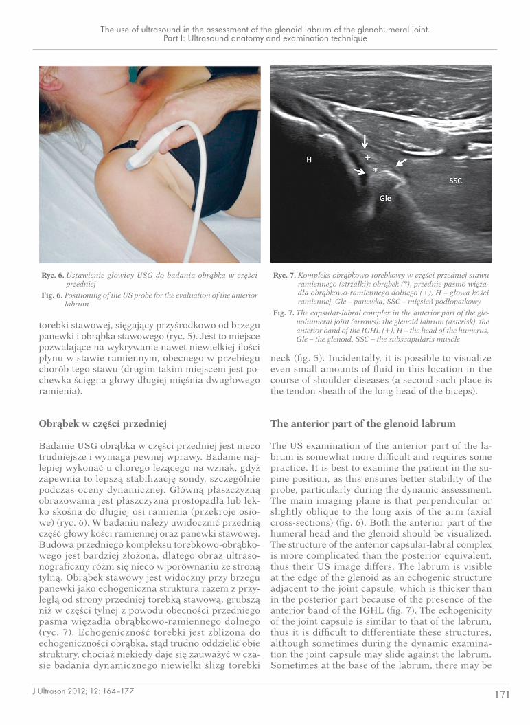
względem obrąbka. U podstawy obrąbka od strony panewki bywa czasami widoczna niewielka linijna strefa (szerokości <2 mm) o niższej echogeniczno-ści, odpowiadająca przejściowej warstwie chrząstki włóknistej między chrząstką stawową a obrąbkiem. Ocenę obrąbka przedniego powinno się zaczynać możliwie od najwyższej strefy dostępnej badaniu (poniżej wyrostka kruczego łopatki) i kontynuować do dolnej części panewki. W części przednio-górnej obrąbek kształtem i echogenicznością przypomina łąkotki kolana. W badaniu USG nie jest dostępna jego część przednio-górna na poziomie wyrostka kruczego łopatki, z kolei dolna część strefy obrąbka przednio-górnego – mniej więcej od godziny 2./10. – i cały obrąbek przednio-dolny są zwykle dobrze widoczne. Bardzo ważnym elementem badania USG jest ocena dynamiczna, przeprowadzana pod-
recognizable a thin linear area (width <2 mm) of lower echogenicity, which represents the transitional layer of fibrocartilage between the joint cartilage and the labrum. The assessment of the anterior labrum should be started from the highest part visible in the study (just below the level of the coracoid process of the scapula), and continued to the inferior edge of the glenoid. In the anterosuperior part, the labrum’s shape and echogenicity are quite similar to the me-nisci of the knee. The labrum is inaccessible to US imaging at the level of the coracoid process of the scapula, while the lower portion of the anterosupe-rior (approximately from 2/10 o’clock) and the entire anteroinferior parts of the labrum are usually well visible. As mentioned previously, the dynamic assess-ment is an important element of the US examination, and is performed during alternate external and inter-
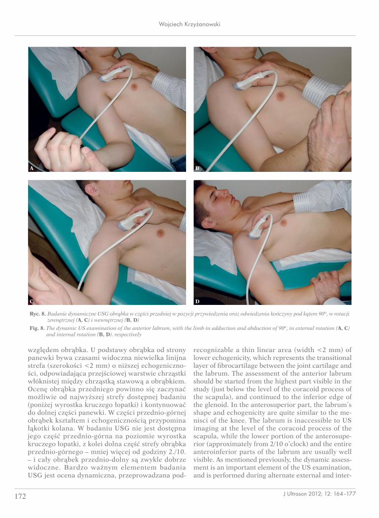
Ryc. 8. Badanie dynamiczne USG obrąbka w części przedniej w pozycji przywiedzenia oraz odwiedzenia kończyny pod kątem 90º, w rotacji zewnętrznej (A, C) i wewnętrznej (B, D)
Fig. 8. The dynamic US examination of the anterior labrum, with the limb in adduction and abduction of 90º, in external rotation (A, C) and internal rotation (B, D). respectively
A
D
B
C
173J Ultrason 2012; 12: 164–177
The use of ultrasound in the assessment of the glenoid labrum of the glenohumeral joint. Part I: Ultrasound anatomy and examination technique
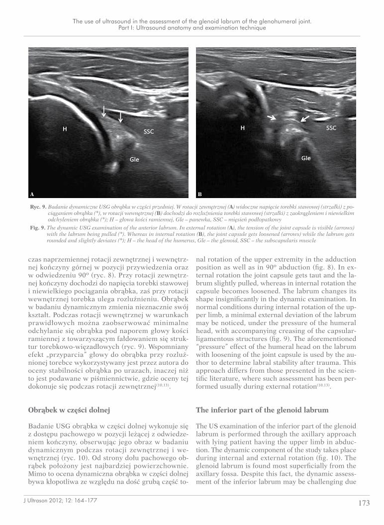
czas naprzemiennej rotacji zewnętrznej i wewnętrz-nej kończyny górnej w pozycji przywiedzenia oraz w odwiedzeniu 90º (ryc. 8). Przy rotacji zewnętrz-nej kończyny dochodzi do napięcia torebki stawowej i niewielkiego pociągania obrąbka, zaś przy rotacji wewnętrznej torebka ulega rozluźnieniu. Obrąbek w badaniu dynamicznym zmienia nieznacznie swój kształt. Podczas rotacji wewnętrznej w warunkach prawidłowych można zaobserwować minimalne odchylanie się obrąbka pod naporem głowy kości ramiennej z towarzyszącym fałdowaniem się struk-tur torebkowo-więzadłowych (ryc. 9). Wspomniany efekt „przyparcia” głowy do obrąbka przy rozluź-nionej torebce wykorzystywany jest przez autora do oceny stabilności obrąbka po urazach, inaczej niż to jest podawane w piśmiennictwie, gdzie oceny tej dokonuje się podczas rotacji zewnętrznej(10,13).
Obrąbek w części dolnej
Badanie USG obrąbka w części dolnej wykonuje się z dostępu pachowego w pozycji leżącej z odwiedze-niem kończyny, obserwując jego obraz w badaniu dynamicznym podczas rotacji zewnętrznej i we-wnętrznej (ryc. 10). Od strony dołu pachowego ob-rąbek położony jest najbardziej powierzchownie. Mimo to ocena dynamiczna obrąbka w części dolnej bywa kłopotliwa ze względu na dość grubą część to-
nal rotation of the upper extremity in the adduction position as well as in 90º abduction (fig. 8). In ex-ternal rotation the joint capsule gets taut and the la-brum slightly pulled, whereas in internal rotation the capsule becomes loosened. The labrum changes its shape insignificantly in the dynamic examination. In normal conditions during internal rotation of the up-per limb, a minimal external deviation of the labrum may be noticed, under the pressure of the humeral head, with accompanying creasing of the capsular-ligamentous structures (fig. 9). The aforementioned “pressure” effect of the humeral head on the labrum with loosening of the joint capsule is used by the au-thor to determine labral stability after trauma. This approach differs from those presented in the scien-tific literature, where such assessment has been per-formed usually during external rotation(10,13).
The inferior part of the glenoid labrum
The US examination of the inferior part of the glenoid labrum is performed through the axillary approach with lying patient having the upper limb in abduc-tion. The dynamic component of the study takes place during internal and external rotation (fig. 10). The glenoid labrum is found most superficially from the axillary fossa. Despite this fact, the dynamic assess-ment of the inferior labrum may be challenging due
Ryc. 9. Badanie dynamiczne USG obrąbka w części przedniej. W rotacji zewnętrznej (A) widoczne napięcie torebki stawowej (strzałki) z po-ciąganiem obrąbka (*), w rotacji wewnętrznej (B) dochodzi do rozluźnienia torebki stawowej (strzałki) z zaokrągleniem i niewielkim odchyleniem obrąbka (*); H – głowa kości ramiennej, Gle – panewka, SSC – mięsień podłopatkowy
Fig. 9. The dynamic USG examination of the anterior labrum. In external rotation (A), the tension of the joint capsule is visible (arrows) with the labrum being pulled (*). Whereas in internal rotation (B), the joint capsule gets loosened (arrows) while the labrum gets rounded and slightly deviates (*); H – the head of the humerus, Gle – the glenoid, SSC – the subscapularis muscle
A B

174 J Ultrason 2012; 12: 164–177
Wojciech Krzy˝anowski
rebkową całego kompleksu torebkowo-obrąbkowego oraz trudność w uzyskaniu optymalnego przekroju obrazowania (ryc. 11). Izolowane uszkodzenia ob-rąbka w części dolnej są jednak dużą rzadkością. Czasami od strony dołu pachowego udaje się uwi-docznić zmiany pourazowe dolnego więzadła obrąb-kowo-ramiennego (np. uszkodzenia typu HAGL) oraz obecność ciał wolnych w zachyłku pachowym stawu ramiennego.
to the rather thick capsular part of the capsular-labral complex, and difficulties in obtaining an optimal im-aging cross-section (fig. 11). However, isolated inju-ries to the inferior part of the labrum are very rare. Sometimes it is possible to visualize posttraumatic changes of the IGHL (ex. HAGL type injuries) using axillar approach, as well as the presence of loose bod-ies in the axillary recess of the glenohumeral joint.
The superior part of the glenoid labrum
The US study of the superior part of the labrum is somewhat problematic due to limited access to the parts of the labrum covered by bony structures (the acromion and the clavicle) as well as difficulties in good stabilizing the probe during the dynamic exami-nation. The examination is performed with the pa-tient sitting. The anterior part of the superior labrum (between 12 and 1 o’clock for the right shoulder, and 11 and 12 o’clock for the left one) is usually acces-sible for the assessment. The probe is applied where a minor depression of tissues is palpated, anterior to the distal part of the clavicle and medial to the anterior part of the acromion (fig. 12 A). This posi-tion of the probe should allow for visualization of the humeral head and the upper part of glenoid with adjacent triangular echogenic structure representing the labral-bicipital complex, consisting of the labrum on the bottom and the overlying tendon of the long head of the biceps. Above this complex the anterior part of the tendon/cord tendon-belly of the supraspi-natous muscle is present (fig. 13). In the author’s ex-perience, the labrum is best visualized with the upper limb internally rotated and adducted (ex. the Middle-
Ryc. 11. Obrąbek w części dolnej stawu ramiennego (*), kompleks torebkowo-więzadłowy (+++), H – głowa kości ramien-nej, Gle – panewka, SSC – mięsień podłopatkowy
Fig. 11. The inferior labrum (*), the capsular-ligamentous complex (+++), H – the head of the humerus, Gle – the glenoid, SSC – the subscapularis muscle
Ryc. 10. Badanie USG obrąbka z dostępu od strony dołu pachowego w pozycji odwiedzenia kończyny pod kątem 90º i rotacji zewnętrznej (A) oraz wewnętrznej (B)
Fig. 10. The US examination of the labrum from the axillary fossa approach with the limb in 90º abduction and being externally rotated (A) or internally rotated (B)
A B
175J Ultrason 2012; 12: 164–177
The use of ultrasound in the assessment of the glenoid labrum of the glenohumeral joint. Part I: Ultrasound anatomy and examination technique
Obrąbek w części górnej
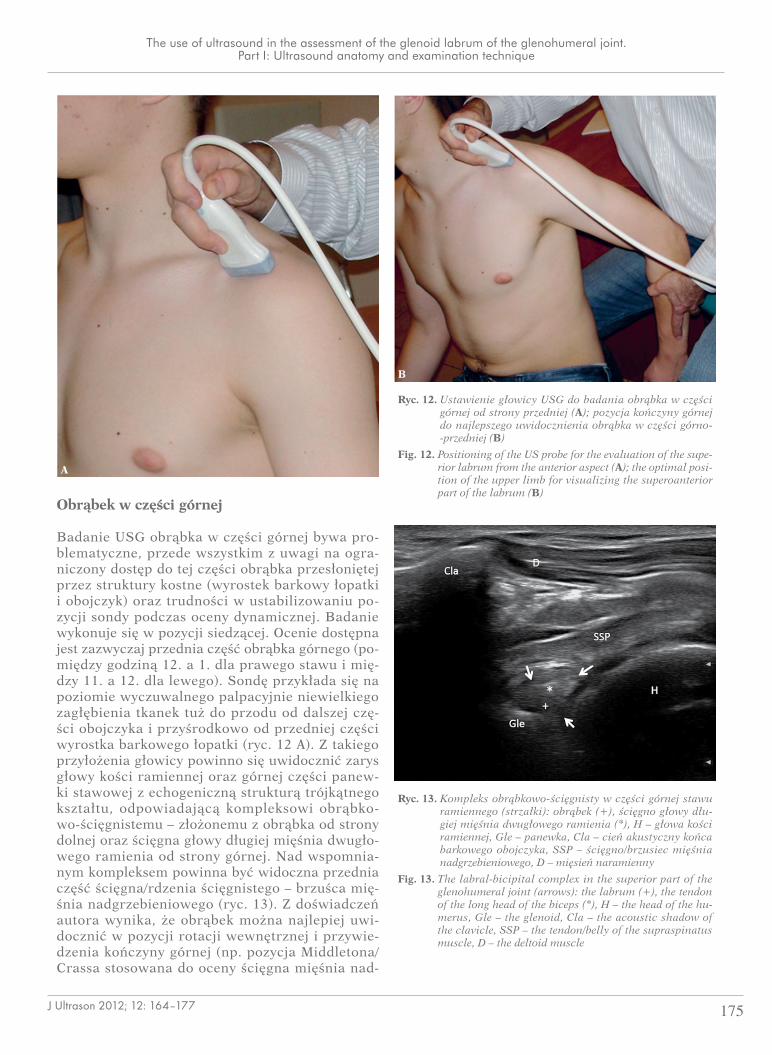
Badanie USG obrąbka w części górnej bywa pro-blematyczne, przede wszystkim z uwagi na ogra-niczony dostęp do tej części obrąbka przesłoniętej przez struktury kostne (wyrostek barkowy łopatki i obojczyk) oraz trudności w ustabilizowaniu po-zycji sondy podczas oceny dynamicznej. Badanie wykonuje się w pozycji siedzącej. Ocenie dostępna jest zazwyczaj przednia część obrąbka górnego (po-między godziną 12. a 1. dla prawego stawu i mię-dzy 11. a 12. dla lewego). Sondę przykłada się na poziomie wyczuwalnego palpacyjnie niewielkiego zagłębienia tkanek tuż do przodu od dalszej czę-ści obojczyka i przyśrodkowo od przedniej części wyrostka barkowego łopatki (ryc. 12 A). Z takiego przyłożenia głowicy powinno się uwidocznić zarys głowy kości ramiennej oraz górnej części panew-ki stawowej z echogeniczną strukturą trójkątnego kształtu, odpowiadającą kompleksowi obrąbko-wo-ścięgnistemu – złożonemu z obrąbka od strony dolnej oraz ścięgna głowy długiej mięśnia dwugło-wego ramienia od strony górnej. Nad wspomnia-nym kompleksem powinna być widoczna przednia część ścięgna/rdzenia ścięgnistego – brzuśca mię-śnia nadgrzebieniowego (ryc. 13). Z doświadczeń autora wynika, że obrąbek można najlepiej uwi-docznić w pozycji rotacji wewnętrznej i przywie-dzenia kończyny górnej (np. pozycja Middletona/Crassa stosowana do oceny ścięgna mięśnia nad-
Ryc. 12. Ustawienie głowicy USG do badania obrąbka w części górnej od strony przedniej (A); pozycja kończyny górnej do najlepszego uwidocznienia obrąbka w części górno--przedniej (B)
Fig. 12. Positioning of the US probe for the evaluation of the supe-rior labrum from the anterior aspect (A); the optimal posi-tion of the upper limb for visualizing the superoanterior part of the labrum (B)
Ryc. 13. Kompleks obrąbkowo-ścięgnisty w części górnej stawu ramiennego (strzałki): obrąbek (+), ścięgno głowy dłu-giej mięśnia dwugłowego ramienia (*), H – głowa kości ramiennej, Gle – panewka, Cla – cień akustyczny końca barkowego obojczyka, SSP – ścięgno/brzusiec mięśnia nadgrzebieniowego, D – mięsień naramienny
Fig. 13. The labral-bicipital complex in the superior part of the glenohumeral joint (arrows): the labrum (+), the tendon of the long head of the biceps (*), H – the head of the hu-merus, Gle – the glenoid, Cla – the acoustic shadow of the clavicle, SSP – the tendon/belly of the supraspinatus muscle, D – the deltoid muscle
A
B

176 J Ultrason 2012; 12: 164–177
Wojciech Krzy˝anowski
grzebieniowego) (ryc. 12 B). Po uwidocznieniu ob-rąbka wykonuje się następnie badanie dynamicz-ne, polegające na naprzemiennych ruchach rotacji kończyny w pozycji przywiedzenia i odwiedzenia,
ton/Crass position used to assess the tendon of the supraspinatus muscle) (fig. 12 B). After the labrum is identified, the dynamic examination is performed, relying on alternating rotations of the limb in adduc-tion and abduction, while trying to maintain the US probe stable. The structures are also evaluated during passive pulling of the upper limb downward.
In a favorable configuration of the acromion and in slender subjects, the posterior part of the superior la-brum may also be imaged at 11 o’clock (for the right shoulder) and 1 o’clock (for the left). The probe is positioned right behind the clavicle, medial to the ac-romion of the scapula (fig. 14 A). The outlines of the superior part of the glenoid and the labrum should be visualized, as well as a small part of the humeral head that is not covered by the acoustic shadow of the acromion. Above these described structures, the supraspinatus muscle is found (fig. 15). The dynamic examination relies on external and internal rotation of the limb in 90º abduction (fig. 14 B). The US ex-amination in this region is also used for diagnosing an internal posterosuperior glenoid impingement of the shoulder.
Summation
The diagnostics of injuries to the glenoid labrum and the capsular-ligamentous complex of the gleno-humeral joint, aside from the clinical assessment, is mainly based upon the MRI, especially after intra-articular contrast administration (MR arthrography).
Ryc. 14. Ustawienie głowicy USG do badania obrąbka w części górnej od strony tylnej (A); pozycja kończyny górnej do badania dynamicz-nego (B)
Fig. 14. Positioning of the US probe for the evaluation of the superior labrum from the posterior aspect (A); the positioning of the upper limb for the dynamic examination (B)
Ryc. 15. Obrąbek w górnej części stawu ramiennego od strony tyl-nej: obrąbek (*), Gle – panewka, H – głowa kości ramien-nej, Ac – cień akustyczny wyrostka barkowego łopatki, SSP – mięsień nadgrzebieniowy
Fig. 15. The superior labrum from the posterior aspect: the labrum (*), Gle – the glenoid, H – the head of the humerus, Ac – the acoustic shadow of the acromion, SSP – the supraspinatus muscle
A B

177J Ultrason 2012; 12: 164–177
The use of ultrasound in the assessment of the glenoid labrum of the glenohumeral joint. Part I: Ultrasound anatomy and examination technique
starając się cały czas utrzymywać stabilnie sondę USG. Przeprowadza się także ocenę podczas bier-nego pociągania kończyny ku dołowi.
Przy korzystnej konfiguracji wyrostka barkowego i u osób o smukłej budowie można zobrazować także tylną część obrąbka górnego na godzinie 11. (dla stro-ny prawej) i 1. (dla strony lewej). Sondę przykłada się tuż za obojczykiem przyśrodkowo od wyrostka barkowego łopatki (ryc. 14 A). Należy uwidocznić zarys górnej części panewki stawowej z obrąbkiem oraz niewielką część głowy kości ramiennej, nieprze-słoniętą przez cień akustyczny wyrostka barkowego łopatki. Nad opisywanymi strukturami znajduje się mięsień nadgrzebieniowy (ryc. 15). Ocena dynamicz-na polega na rotacji zewnętrznej i wewnętrznej koń-czyny górnej w pozycji odwiedzenia 90º (ryc. 14 B). Badanie w tej okolicy jest wykorzystywane także w diagnostyce ultrasonograficznej wewnętrznego konfliktu tylno-górnego w stawie ramiennym.
Podsumowanie
Diagnostyka uszkodzeń obrąbka oraz kompleksu to-rebkowo-więzadłowego stawu ramiennego opiera się w praktyce, poza oceną kliniczną, głównie na bada-niu RM, zwłaszcza po dostawowym podaniu środka kontrastowego (artrografia RM). Jak przedstawiono w niniejszej pracy, obrąbek jest w dużym zakresie do-stępny do oceny także w badaniu USG. Tym samym może to być wartościowa metoda w diagnostyce jego patologii, co zostanie przedstawione w II części pracy.
Piśmiennictwo/References
1. Di Giacomo G, Pouliart N, Constantini A, De Vita A: Atlas of Functional Shoulder Anatomy. Springer-Verlag, Rzym 2008: 110.
2. Van der Woude HJ, Vanhoenacker FM: MR arthrography in glenohu-meral instability. JBR-BTR 2007; 90: 377–383.
3. Cooper DE, Arnoczky SP, O’Brien SJ, Warren RF, DiCarlo E, Allen AA: Anatomy, histology, and vascularity of the glenoid labrum. An anatomi-cal study. J Bone Joint Surg Am 1992; 74: 46–52.
4. Prodromos CC, Ferry JA, Schiller AL, Zarins B: Histological studies of the glenoid labrum from fetal life to old age. J Bone Joint Surg, 1990; 72-A: 1344–1348.
5. Park YH, Lee JY, Moon SH, Mo JH, Yang BK, Hahn SH et al.: MR arthrography of the labral capsular ligamentous complex in the shoul-der: imaging variations and pitfalls. AJR Am J Roentgenol 2000; 175: 667–672.
6. Beltran J, Bencardino J, Mellado J, Rosenberg ZS, Irish RD: MR ar-thrography of the shoulder variants and pitfalls. Radiographics 1997; 17: 1403–1412.
7. Neumann CH, Petersen SA, Jahnke AH: MR imaging of the labral-capsular complex: normal variations. AJR Am J Roentgenol 1991; 157: 1015–1021.
8. Mohana-Borges AV, Chung CB, Resnick D: Superior labral anteropos-terior tear: classification and diagnosis on MRI and MR arthrography. AJR Am J Roentgenol 2003; 181: 1449–1462.
As this article has shown, the glenoid labrum is also visible in the US study. Therefore, this imaging tool may be valuable in the diagnostics of labral patholo-gies, which will be presented in part II of the article.
9. Taljanovic MS, Carlson KL, Kuhn JE, Jacobson JA, Delaney-Sathy LO, Adler RS: Sonography of the glenoid labrum: a cadaveric study with arthroscopic correlation. AJR Am J Roentgenol 2000; 174: 1717–1722.
10. Jee WH, McCauley TR, Katz LD, Matheny JM, Ruwe PA, Daigneault JP: Superior labral anterior posterior (SLAP) lesions of the glenoid labrum: reliability and accuracy of MR arthrography for diagnosis. Radiology 2001; 218: 127–132.
11. De Maeseneer M, Van Roy F, Lenchik L, Shahabpour M, Jacobson J, Ryu KN et al.: CT and MR arthrography of the normal and patho-logic anterosuperior labrum and labral-bicipital complex. Radiograph-ics 2000; 20: S67–S81.
12. Schydlowsky P, Strandberg C, Galatius S, Gam A: Ultrasonographic examination of the glenoid labrum of healthy volunteers. Eur J Ultra-sound 1998; 8: 85–89.
13. Hammar MV, Wintzell GB, Aström KG, Larsson S, Elvin A: Role of us in the preoperative evaluation of patients with anterior shoulder instabil-ity. Radiology 2001; 219: 29–34.
14. Sugimoto K: Ultrasonographic evaluation of the Bankart lesion. J Shoulder Elbow Surg 2004; 13: 286–290.
15. Schydlowsky P, Strandberg C, Galbo H, Krogsgaard M, Jørgensen U: The value of ultrasonography in the diagnosis of labral lesions in pa-tients with anterior shoulder dislocation. Eur J Ultrasound 1998; 8: 107–113.
16. Rutten MJ, Jager GJ, Kiemeney LA: Ultrasound detection of rotator cuff tears: observer agreement related to increasing experience. AJR Am J Roentgenol 2010; 195: W440–W446.
17. Martinoli C, Bianchi S, Prato N, Pugliese F, Zamorani MP, Valle M et al.: US of the shoulder: non-rotator cuff disorders. Radiographics 2003; 23: 381–401.
18. Bianchi S, Martinoli C: Ultrasound of the Musculoskeletal System. Springer Verlag, Berlin, Heidelberg 2007: 189.