الباطنة كلها بالتفصيل في 160 صفحة فقط لازم تحمل المذكرة...
DESCRIPTION
All medicine In 160 pages onlyTRANSCRIPT
INTERNAL MEDICINE LECTURES 2010 WITH MCQ QUESTIONS & ANSWERS NMT12
www.medadteam.org More Than You Dream Page 1
IInnddeexx
Topic Page
I. Cardiology:
1. Clinical Presentation of Cardiac Disease
2. Valvular Heart disease
3. Rheumatic Fever
4. Infective Endocarditis
5. Coronary (Ischemic) Heart Disease
6. Heart Failure
7. Hypertension
8. Hypotension
M.C.Q.
3
14
21
26
34
41
47
53
59
II. Chest:
1. Clinical presentations of chest diseases
2. COPD
3. Suppurative Lung Disease
4. Tuberculosis
5. Bronchial Asthma
6. Pulmonary Hypertension & Cor Pulmonale
M.C.Q
65
75
84
91
95
100
104
III. G.I.T.:
1. Clinical presentation of Abdominal diseases
M.C.Q.
107
113
IV. Hepatology:
1. Jaundice
2. Acute Viral Hepatitis
3. Liver Cirrhosis
4. Portal Hypertension
M.C.Q.
116
120
126
129
131

INTERNAL MEDICINE LECTURES 2010 WITH MCQ QUESTIONS & ANSWERS NMT12
www.medadteam.org More Than You Dream Page 2
V. Endocrinology:
1. Diabetes Mellitus
M.C.Q.
135
140
VI. Nephrology:
1. Acute Renal Failure
2. Chronic Renal Failure
M.C.Q.
142
147
150
VII. Hematology:
1. Anemia
M.C.Q.
153
155
VIII. Medical Ethics 157
NN..BB..:: TThhee ""TTrruuee"" aannsswweerrss iinn MM..CC..QQ..
qquueessttiioonnss aarree iinn tthhoossee iinn tthhee ""BBoolldd"" ssttyyllee..

INTERNAL MEDICINE LECTURES 2010 WITH MCQ QUESTIONS & ANSWERS NMT12
www.medadteam.org More Than You Dream Page 3
1. Clinical Presentation of Cardiac Disease
���� Structure & Function of the heart:
The heart is like any motor machine: a battery firing an engine to activate a pump.
- Pump (Cardiac muscles)
- Tubes (big vessels)
� On the right side:
1. Tubes to fill the Rt side (Sup. & inf. Vena cavae)
2. Tube to carry blood to lungs for oxygenation (Pulmonary artery)
� On the left side:
1. Tube to return oxygenated blood from the lungs to the left side. (Pulm. Vein)
2. Tube to distribute blood to all parts of the body. (aorta)
- Gates to control the passage of blood (valves)
- Battery to fire the engine (SAN)
- Cables and distributing station to distribute the electricity within the pump chambers.
(AVN and the Bundle of Hiss)
- Fuel to the pump.(coronary arteries)
- A rap to protect the heat,(pericardium)
- A communicating system with the brain (the neural supply of the heart)
���� Causes of heart diseases � Reduction in pump function.
� Obstruction to the inflow or the out flow tracts.
� Valvular heart diseases.
� Arrhythmias.
� Myocardial ischemia.
� Pericardial diseases.
� Dysregulation of the neural supply.
���� Clinical presentation of cardiac diseases � Chest pain. ���� Dyspnea.
Lectures objectives
1. List the common symptoms of cardiac diseases. 2. Define each symptom 3. List the commonest causes of each symptom 4. Explain the pathogenesis 5. Recognize the grades of dyspnea 6. Analyze each of the cardiac symptoms
INTERNAL MEDICINE LECTURES 2010 WITH MCQ QUESTIONS & ANSWERS NMT12
www.medadteam.org More Than You Dream Page 4
���� Any of these symptoms may also have extra-cardiac causes.
���� Clinical Presentations of Cardiac
����
� Palpitations. ���� Cough.
� Hemoptysis. ���� Syncope.
� Fatigue (effort intolerance). ���� Claudication.
� Edema of the lower limbs. ���� Dyspepsia and weight loss.
� Pain in the right hypochondrium. ���� Congested veins in the neck.
� Cyanosis or a bluish discoloration of the skin.
� Changes in the pulse volume, rate, rhythm or character.
NB:
Chest pain
���� Cardiac “major couses” Causes:
� Myocardial ischemia.
� Pericarditis.
� Pulmonary embolism.
� Aortic dissection.
� Noncardiac Causes:
� anxiety, cholecystitis, GERD, pleurisy, muscular pair, rib fracture or neuropathic pain.
���� In evaluating any pain or discomfort we need to know:
1. The anatomical site
2. The referral site, if any.
3. The character of pain
4. The severity of pain
5. What do precipitate or aggravates this pain?
6. What do relief this pain?
7. Accompanying symptoms or signs.
���� Definition of cardiac pain:
1. Precordial, usually sub-sternal or across the anterior chest.
2. Radiating to the left arm, back, neck, jaw, anterior chest or Rt arm.
3. Severe pain enforcing the patient to stop activity.
4. Squeezing, crushing. strangling, stabbing, compressing sensation, tightness, or other
discomfort rather than true sharp pain.
5. Precipitated by exertion, stress, heavy meals, going out in cold or sexual intercourse.
6. Relieved by rest, nitrates, beta blockers or calcium channel blockers.

INTERNAL MEDICINE LECTURES 2010 WITH MCQ QUESTIONS & ANSWERS NMT12
www.medadteam.org More Than You Dream Page 5
7. May be accompanied by dyspnea, palpitations, nausea, diaphoresis or abdominal
discomfort.
8. The frequency and duration of pain should be defined
���� Symptoms accompanying chest pain of cardiac origin:
1. Diaphoresis frequently occurs in acute myocardial infarction.
2. Palpitations, dyspnea and effort intolerance.
3. Cough, expectoration of frothy sputum.
4. May be nausea, and vomiting.
5.
Dyspnea
� The word dyspnea comes from the Greek :
"dys" = difficulty + "pnoia" = breathing
(dyspnea = difficult breathing )
(Difficult or labored breathing or shortness of breath.)
• Usually associated with disease of the heart or lungs but may be caused by general causes.
���� during intense physical exertion or at high normally It occurs
altitude.
���� finition of Dyspnea:
a subjective difficulty or distress in breathing concerning the rate or depth of respiration.
� At rest, an average 70 kg person breathes 12 to 15 times / minute with a tidal volume of
about 600 ml.
� A normal individual is not aware of his or her respiratory effort until ventilation is
doubled.
� Dyspnea is experienced only when the ventilation is tripled.
Dyspnea on exertion (exertional dyspnea) indicates dyspnea that occurs (or worsens) during
physical activity.
Dyspnea is a symptom caused by diseases of:
• The airway.

INTERNAL MEDICINE LECTURES 2010 WITH MCQ QUESTIONS & ANSWERS NMT12
www.medadteam.org More Than You Dream Page 6
• Lungs.
• Heart.
• General cause.
���� Grades:
� Grade I : Breathlessness during unaccustomed exercise
� Grade II : Breathlessness during accustomed exercise
� Grade III : Breathlessness during less than accustomed exercise
� Grade IV : Breathlessness at rest.
The type, the onset and the severity of dyspnea are important determining factors in the
evaluation of the patient.
���� Pathophysiology of dyspnea
- The control of the spontaneous initiation of breathing is in the medulla.
- This medullary center receives afferent neural input from receptors that monitor:
���� The rate and depth of breathing and
���� The levels of oxygen and carbon dioxide
���� The PH of the blood.
- These receptors are:
1. Mechanoreceptors in the muscles and tendons that participate in breathing.
2. Chemoreceptors in the carotid and aortic bodies (Hypoxia)
3. Airway and parenchymal receptors in the upper and lower airways and elsewhere
in the lungs themselves.
4. Receptors around the medullary center sensitive to changes in PCO2/pH.
���� Pathogenesis of cardiac dyspnea:
Increased afferent activity from any of these receptors,
will lead to change in the rate or depth of respiration .
1. muscle contraction (altered chest wall compliance)
2. airflow (increased resistance by spasm, edema, fibrosis, tumors or foreign body )
3. lung inflation or deflation (decreased lung compliance)
4. levels of PO2 and PCO2 in the blood
5. changes in PH
6. Medullary center (afferent input and efferent output)

INTERNAL MEDICINE LECTURES 2010 WITH MCQ QUESTIONS & ANSWERS NMT12
www.medadteam.org More Than You Dream Page 7
Pulmonary venous congestion increased pulmonary venous pressure transudation
of fluid into the interstitium transudation of fluid into the alveoli.
1. Congested lungs are more rigid needs more effort to expand. (decreased lung
compliance).
2. Congestion of the airways increase airway resistance.
3. Interstitial edema and alveolar transudation result in hypoxemia, and
ventilation/perfusion mismatch.
4. The juxtacapillary receptors (J-receptors), located in the alveolar interstitium are
stimulated by pulmonary congestion which activates the Hering–Breuer reflex.
5. This will lead to termination of the inspiratory effort before full inspiration is achieved,
resulting in rapid and shallow breathing.
���� Types of dyspnea and its major causes:
� Dyspnea of acute onset:
1. Bronchial asthma
2. Acute pulmonary edema
3. Massive pulmonary embolism
4. ARDS (Adult Respiratory Distress Syndrome or non cardiac pulmonary edema)
5. Foreign body inhalation.
6. Acute lung volume reduction ( Lobar pneonomia, Massive pnemothorax & Acute lung
collapse or Massive effusion or Hemothorax).
� Dyspnea of gradual onset:
Most of other conditions related to the heart or lungs first cause dyspnea with extreme
exertion and as the disease progresses, dyspnea appears with less exertion, and finally is
manifested at rest.
� Cardiac dyspnea:
1. It is characteristically related to effort.
2. In severe or advanced heart disease it may occur at rest.
3. It includs paroxysmal nocturnal dyspnea and orthopnea.
4. Rapid progression of an episode of respiratory distress may result in a very severe
form of dyspnea, acute pulmonary edema, i.e., "asthmatic" wheezes and a pink,
frothy sputum. (Cardiac asthma)
� Non cardiac, non pulmonary dyspnea can be either acute or chronic:
1. Anemia
1. Neuromuscular disease.

INTERNAL MEDICINE LECTURES 2010 WITH MCQ QUESTIONS & ANSWERS NMT12
www.medadteam.org More Than You Dream Page 8
2. Severe weight loss from malnutrition, malignancy or chronic disease may also cause
respiratory muscle weakness with associated dyspnea.
3. Renal disease leads to dyspnea from acidosis, anemia and volume overload.
4. Liver diseases and ascites.
5. Psychogenic dyspnea is usually a diagnosis of exclusion.
• Paroxysmal dyspnea:
• Bronchial asthma.
• Cardiac asthma (paroxysmal nocturnal dyspnea).
Positional dyspnea
Dyspnea on acquiring special positions.
• Orthopnea: Occurring within minutes or hours of becoming recumbent (Heart failure)
• Trepopnea : Present in the lateral decubitus position (Unilateral lung or pleural disease)
• Platypnea : Worse when assuming upright position. Platypnea in association with arterial
deoxygenation in the upright position as in several forms of cyanotic congenital heart
disease.
Orthopnea
���� Definition:
� Orthopnea is dyspnea on lying flat in bed.
� Orthopnea is caused by increased pulmonary congestion during recumbency.
���� Pathogenesis of orthopnea
- In the horizontal position there is redistribution of blood volume from the lower
extremities and splanchnic beds to the lungs.
- When the pulmonary circulation is already overloaded, this will make the situation worse.
- Reabsorption of edema fluid from previously dependent parts of the body.
- Elevation of the diaphragm by ascites and hepatomegaly decreases lung volumes.
- Less effective use of accessory muscles of respiration in the recumbent position.
- In normal individuals this has little effect, but in patients with decreased both vital capacity
and pulmonary compliance, from pulmonary congestion, this will result in shortness of breath.
- Pulmonary congestion decreases when the patient assumes a more erect position, and this
is accompanied by an improvement in symptoms.
- Initially, breathing at night is made easier by elevating the head on two or more pillows.

INTERNAL MEDICINE LECTURES 2010 WITH MCQ QUESTIONS & ANSWERS NMT12
www.medadteam.org More Than You Dream Page 9
Paroxysmal nocturnal dyspnea
- As heart failure progresses, the patient may have to sleep sitting up.
���� Definition:
- Paroxysmal nocturnal dyspnea is a cardinal feature of left sided hear failure.
- After sleep, the patient awakens suddenly with a feeling of severe anxiety and suffocation,
and has to sit upright for relief.
- Episodes are often accompanied by coughing and wheezing and may be extremely
frightening to the patient and family.
���� Pathogenesis of Paroxysmal nocturnal dyspnea:
- Paroxysmal nocturnal dyspnea is caused by mechanisms similar to those of orthopnea.
- During sleep additional mechanisms include:
1. Decreased responsiveness of the respiratory center in the brain.
2. Decreased adrenergic activity in the myocardium.
- When significant wheezing is associated with paroxysmal nocturnal dyspnea, it resembles an
acute asthmatic attack and may be referred to as cardiac asthma.
- Bronchospasm, which is caused by congestion of the bronchial mucosa and by interstitial
pulmonary edema compressing small airways, increases the work of breathing.
- It has to be differentiated from bronchial asthma.
Acute pulmonary edema
���� Definition:
Acute pulmonary edema occurs with marked elevation of the pulmonary capillary wedge
pressure leading to alveolar edema. The patient is extremely short of breath and coughs up
pink, frothy sputum. It needs urgent treatment as it can be fatal.
1. It can complicate paroxysmal nocturnal dyspnea.
2. Or it may occur as a primary manifestation of acute myocardial infarction, paroxysmal
or sudden onset rapid arrythmias or accelerated hypertension.
Palpitation
���� Definition:
Palpitations means an awareness of the heartbeat. Awareness occurs due to change in the
rate, rhythm or the force of contraction.
,So with palpitation, the pulse may be too slow, too fast, irregular, or at its normal frequency.

INTERNAL MEDICINE LECTURES 2010 WITH MCQ QUESTIONS & ANSWERS NMT12
www.medadteam.org More Than You Dream Page 10
���� Causes of palpitation:
1- Hyperdynamic circulation (Thyrotoxicosis Hypercapnia, Fever, Anemia, Overexertion , Pregnancy...)
2- Sympathetic overdrive (Panic disorders, Hypoglycemia, Hypoxia, Anemia, Heart Failure, Drugs as
adrenaline, caffeine, cocaine, amphetamines)
3- Arrhythmias.
���� Types of palpitation:
� Occasional or Persistent
� Regular or Irregular.
���� The patient feels as if:
1- The heart "stops"
o The feeling that the heart stops beating for a moment, and then starts again with a
"thump".
o This feeling is actually caused by a premature beat or extrasystole that happens earlier
than the next normal beat, and results in a compensatory pause.
o People are not usually aware of the early, extra beat, but may be aware of the pause,
which follows it (the heart seems to stop).
o The beat after the pause is more forceful than normal (due to filling with more blood than
usual during the compensatory pause), giving the "thumping" sensation.
2- The heart is “fluttering”.
o Any rapid tachycardia can give rise to this feeling ( rapid, regular, or rapid, irregular).
���� Palpitations can be associated with:
- tightness in the chest,
- shortness of breath,
- dizziness or light-headedness.
- actual blackouts.
According to the type and the etiology of the arrhythmia these
symptoms may be either a temporary event or a persistent complaint.
Actual blackouts or near blackouts, associated with palpitations, should be taken seriously
because they often indicate the presence of important underlying heart disease.
Cough
���� Definition:
A cough is a forceful release of air from the lungs that can be heard.
���� Coughing protects the respiratory system by clearing irritants and secretions.
���� It may be voluntarily.

INTERNAL MEDICINE LECTURES 2010 WITH MCQ QUESTIONS & ANSWERS NMT12
www.medadteam.org More Than You Dream Page 11
���� Pathophysiology of Cough:
1. Cough receptors are found at different parts of the respiratory system.
2. Cough is a reflex triggered when an irritant stimulates one or more of these receptors.
3. These receptors then send a message to the cough center in the brain, which in turn tells the
responsible muscles to do the job.
4. A cough begins with a deep breath in, followed by closure of the opening between the vocal cords
trapping the air in the lungs.
5. Then the glottis suddenly opens, producing an explosive outflow of air at high speeds caused by
diaphragmatic and inter-costal muscles contraction.
Sever bouts of cough may be accompanied by:
���� Dizziness, syncope, chest pain, or breathlessness.( prolonged expiratory effort decreased
cardiac filling)
���� Or it may interfere with sleep.
���� If a cough lasts more than three weeks it is considered a chronic cough.
����
���� Types of Cough
� A dry cough does not bring up sputum.
� Productive cough with sputum.
Productive cough
1. In the case of a bacterial infection, the sputum may be mucous, purulent greenish,
gray, or brown.
2. In the case of an allergy or viral infection it may be clear or white.
3. In the most serious conditions, the sputum may contain blood.
4. In lung congestion it might be pink and frothy.
Cough may be:
� Acute: usually begin suddenly. Typically, they do not last longer than two to three
weeks.
� Chronic: last longer than two to three weeks.
� Nocturnal cough: is related to lung congestion with orthopnea. It has the same
significance as orthopnea.
� Paroxysmal cough: with asthma and whooping cough.
� Postural cough: with suppurative lung diseases and cavitary lung diseases.
Hemoptysis
���� Definition:
Hemoptysis is the expectoration of blood or blood-tinged sputum from the lungs or
tracheobronchial tree.
INTERNAL MEDICINE LECTURES 2010 WITH MCQ QUESTIONS & ANSWERS NMT12
www.medadteam.org More Than You Dream Page 12
Coughing is important because nonpulmonary sources of bleeding are not usually associated
with hemoptysis.
Remember that the lung contains two separate vascular systems: the pulmonary and the
bronchial vessels. Either may be the source of hemoptysis.
���� D.D. :
� Spitting blood without coughing.
� Hematemesis.
���� Types of Hemoptysis:
1. Franck bright red blood or blood clots (as in adenoma and carcinoma of the lung, tuberculosis,
pulmonary embolism)
2. Blood-streaked, purulent sputum (as in bronchitis, bronchiectasis, or pneumonia)
3. Blood-tinged, white, frothy sputum or pink sputum (as in congestive heart failure).
4. Foul-smelling, bloody sputum (as in an anaerobic lung abscess)
���� Pathogenesis of hemoptysis:
• Hemoptysis may occur as a result of pulmonary parenchymal necrosis involving small blood
vessels due to severe infection or vasculitides.
• Vascular engorgement with erosion is the mechanism of hemoptysis as in lung congestion.
• Disruption of the pulmonary capillaries as a result of increased intravascular pressure (as in
mitral stenosis and left ventricular failure)
•
In all of these conditions the shearing force of coughing is the trigger for bleeding.
Edema
���� Definition:
Edema is a detectable excess of fluid in the interstitial spaces.
When due to cardiac disease, it is described as gravitational occurring mostly in dependant
parts according to patient’s position. (pedal, sacral or more on one side)
It is a sign of Rt. sided or congestive heart failure.
���� Pathogenesis of edema:
1. Increased hydrostatic pressure in the venous system as the Rt. side fails.
2. Accumulation of interstitial fluid is governed by Starling’s Equation: Hydrostatic gradient –
oncotic gradient

INTERNAL MEDICINE LECTURES 2010 WITH MCQ QUESTIONS & ANSWERS NMT12
www.medadteam.org More Than You Dream Page 13
• Edema of the gastrointestinal tract will result in anorexia, dyspepsia and early satiety
due to bowel congestion. These symptoms are nonspecific.
• Ascites results in an increase in abdominal girth.
• Unilateral or bilateral pleural effusions can aggravate dyspnea.
Syncope
���� Definition:
Syncope means transient loss of consciousness associated with loss of postural tone, due to
cardiovascular cause.
• "Blackout spells," "passing out," or "fainting" are terms occasionally used by patients.
These states should not be considered syncope unless the patient lost consciousness and
postural tone.
���� Pathogenesis of Syncope:
Syncope is caused by: a reduction in cerebral blood flow.
Common causes are:
• Inadequate cardiac output.
• Left ventricular outflow tract obstruction (e.g., aortic stenosis or hypertrophic
cardiomyopathy) commonly causes effort syncope.
• Arrhythmias that result in sudden decrease of cardiac output lead to syncopal episodes
either at rest or during activity.
Fatigue ���� Definition:
Fatigue is a feeling of tiredness, effort intolerance or lack of energy.
���� Pathogenesis: 1. The symptoms thought to be due to decreased cardiac output to exercising muscles.
2. Peripheral vasoconstriction, hypoxia, and altered skeletal muscle metabolism may also play
contributory roles.
3. Dilutional hyponatremia, volume depletion, and medications (e.g., β-blockers).
4. Insomnia due to orthopnea and paroxysmal nocturnal dyspnea and nocturia is a contributing
factor in day time fatigue.

INTERNAL MEDICINE LECTURES 2010 WITH MCQ QUESTIONS & ANSWERS NMT12
www.medadteam.org More Than You Dream Page 14
Cardiac Cachexia ���� Definition:
Chronic weight loss due to longstanding severe heart failure.
���� Pathogenesis:
Factors contributing to cardiac cachexia include:
���� Anorexia
���� Impaired absorption due to bowel wall edema.
���� Increased levels of circulating tumor necrosis factor-α.
Symptoms of Decreased Cardiac Output
These symptoms are non specific.
���� Symptoms related to decreased cardiac output can occur with right-sided or left-sided
heart failure but more commonly occur in patients with chronic biventricular failure.
���� Mental dullness and confusion, especially in older patients, may result from decreased
cerebral perfusion.
���� Oliguria due to water retention and decreased renal blood flow.
Manifestations of Systemic Venous Congestion
Right-sided heart failure or cardiac inflow obstruction will result in systemic venous
congestion.
The major manifestations are:
1. Edema of lower limbs in ambulant patients.
2. Pain in the right hypochondrium due to liver congestion and stretch of its capsule.
3. Dyspepsia & weight loss.
4. Peripheral cyanosis.
5. Congested neck veins
2. Valvular Heart disease
Lectures objectives
1) Define the aetiology and pathophysiology of different valve lesions
(mitral, aortic, pulmonary and tricuspid valve).
2) Explain the hemodynamic changes.
3) Describe the clinical manifestations specific to valve affection
4) List the complications.

INTERNAL MEDICINE LECTURES 2010 WITH MCQ QUESTIONS & ANSWERS NMT12
www.medadteam.org More Than You Dream Page 15
(A.) Mitral Stenosis
���� Etiology:
� Rheumatic heart disease (most of cases- 99%)
� Congenital
o 50% of sufferers have a history of rheumatic fever & chorea.
o The mitral valve is affected in 90% of those with rheumatic valvular heart disease
� Other causes: rare ( Infective endocarditis – SLE – Calcified mitral valve ring )
���� Pathophysiology/Hemodynamic changes:
� When the normal valve orifice area of 5cm² is reduced to 1 cm², severe mitral stenosis is
present.
� To maintain sufficient cardiac output, the left atrial pressure increases & L.A hypertrophy &
dilatation occurs. Atrial fibrillation may occur resulting in severe impairment of
hemodynamic functions
� Consequently, pulmonary venous, pulmonary arterial and right heart pressures also
increase.
� The increase in pulmonary capillary pressure is followed by the development of pulmonary
oedema, which is partially prevented by alveolar and capillary thickening and pulmonary
arterial vasoconstriction. (Pulm. HTN)
� Pulm. HTN leads to Right ventricular hypertrophy, dilatation and failure.
� R. V. dilatation results in Tricuspid regurge
� LV dysfunction occurs in 1/4 of patients with severe MS
� Low cardiac output in severe cases
���� Clinical Manifestations & complications:
� Symptoms:
- Stage 1: asymptomatic - Stage 2: Pulmonary congestion
- Stage 3: low cardiac output - Stage 4:systemic congestion
� No symptoms until valve orifice is moderately stenosed (area of 2 cm²).
� Because of pulm venous hypertension and recurrent bronchitis, progressively severe
dypsnea develops.
� A cough productive of blood tinged frothy sputum is quite common.
� Occasionally frank hemoptysis may occur.
� The development of Pulm. HTN eventually leads to R.V. failure with symptoms of
weakness, fatigue and abdominal and L.L swelling.
� The large left atrium favors atrial fibrillation; this may lead to symptoms as palpitation.
� A.F. may result in systemic embolization commonly to cerebral vessels leading to
neurological sequelae, renal, mesenteric & peripheral emboli are also seen.

INTERNAL MEDICINE LECTURES 2010 WITH MCQ QUESTIONS & ANSWERS NMT12
www.medadteam.org More Than You Dream Page 16
� Signs:
� General:
� Mitral facies (Malar flush) is present in cases of severe M.S with Pulm. HTN
� Pulse usually of small volume. Its regular early in the disease then it becomes
irregularly irregular when A.F. develops.
If R.V. failure develops there will be distended jugular veins.
� Inspection/Palpation:
� Apex beat is tapping in quality due to palpable 1st sound
� Parasternal sustained impulse due to R.V. enlargment may also be felt.
� Auscultation :
� Loud 1st sound if leaflets are pliable. This will not occur in calcific M.S.
� Opening snap occurs due to sudden opening of the valve under raised L.A.
pressure. This is followed by a low pitched ”rumbling mid-diastolic murmur
“best heard with the bell in the left lateral position.
� If the rhythm is sinus , the murmur becomes louder at the end of the diastole
due to atrial contraction (presystolic accentuation)
� N.B. How severe the M.S. is?
The severity of ms is judged clinically on basis of several criteria:
• The presence of Pulm. HTN, recognized by loud S2 over the pulmonary , signs of R.V
enlargement and failure. Pulm. HTN results in pulm. valve regurgitation which causes an
early diastolic murmur in the pulm area (graham steel murmur).
• The length of the mid diastolic murmur is proportional to the severity.
• The closeness of the opening snap to the 2nd sound is proportional to the severity.
• As the valve cusps become immobile, loud S1 softens and the opening snap disappears.
• Low cardiac output causes silent M.S.
���� Investigations:
� ECG
� Chest x-ray
� Echo:
- Thickened immobile cusps.
- Reduced valve area.
- Reduced rate of diastolic filling of lv.
� Doppler:
- Pressure gradient across the mitral valve.
- Pulmonary artery pressure.

INTERNAL MEDICINE LECTURES 2010 WITH MCQ QUESTIONS & ANSWERS NMT12
www.medadteam.org More Than You Dream Page 17
(B.) Mitral Regurgitation
���� Etiology:
• 50% of cases are due to rheumatic heart disease.
• The 2nd most common cause is prolapsing mitral valve( see later)
• Any disease that causes Lt Ventricular dilatation may cause mild M.R.:
o Aortic valve disease.
o Dilated cardiomypathy.
o IHD.
o Hypertensive HD
o Connective tissue disorders - SLE.
o Collagen abnormalities- Marfans syndrome.
���� Pathophysiology/ Hemodynamics • To and from movement across valve
o Regurgitant jet throughout systole
o Forward flow through aorta decreased
o Increased flow during diastole.
• In longstanding conditions regurgitation in the LT atrium produces LT atrial dilatation
but little increase in LT atrial pressure.
• Since a proportion of the stroke volume is regurgitated, the stroke volume increases to
increase the forward cardiac output and Lt V. enlarges.
• In acute MR the normal compliance of Lt A. doesn't allow much dilatation leading to
rise in Lt A. pressure and pulmonary oedema and shock.
���� Clinical Manifestations:
� Symptoms:
• Asymptomatic
• Palpitation due to increased stroke volume.
• Dyspnea & orthopnea may develop due to pulmonary venous hypertension as a dierct
result of MR (esp in acute cases ) & secondarily to LV failure.
• Fatigue & lethargy develop due to low CO.
• Symptoms of RV failure.
• Thromboembolic manifestations & infective endocarditis. (compare with Mitral
stenosis)
� Signs:
• Laterally displaced, hyperdynamic apex with systolic thrill.
• A soft 1st sound due to incomplete apposition of the valve cusps & their partial closure
when the ventricular systole begins.

INTERNAL MEDICINE LECTURES 2010 WITH MCQ QUESTIONS & ANSWERS NMT12
www.medadteam.org More Than You Dream Page 18
• Pansystolic murmer owing to the occurrence of regurgitation throughout the systole,
heard max at the apex & radiating to the axilla.
• Signs related to AF, PH, RT or LT ventricular HF may develop later in the disease.
���� Investigations
� Chest x-ray.
� ECG.
� Echo heart: dilated LT atrial and LT ventricle. There may be specific features of chordal
or papillary muscle rupture.
� Doppler detects and quantify regurgitation.
(C.) Prolapsing "Billowing" Mitral Valve
���� Also known as BARLOW'S syndrome or FLOPPY MR.
���� It’s due to excessively large mitral leaflets, an enlarged mitral annulus, abnormally long
chordae or disordered papillary muscle contraction.
���� Commonly seen in young women & has familial incidence.
���� It may be associated with marfan's syndrome , thyrotoxicosis or ischemic heart disease. It
also occurs as a part of hypertrophic cardiomypathy.
���� Pathophsiology:
� during Vent. systole, mitral valve leaflet prolapses into the Lt Atrium. This may result in
abnormal vent contraction & some mitral regurgitation.
���� Clinical Manifestations:
� Symptoms:
� atypical chest pain is the most common symptom& palpitation which may be due to
arrhythmia or abnormal vent contraction.
� Signs:
� mid systolic click due to sudden prolapse of the valve and the tensing of the chordae
tendinae that occurs during systole. This may be followed by a late systolic murmer
owing to some regurgitation.
���� Investigations:
� Echocardiography

INTERNAL MEDICINE LECTURES 2010 WITH MCQ QUESTIONS & ANSWERS NMT12
www.medadteam.org More Than You Dream Page 19
(D.) Aortic Stenosis
���� Etiology:
� Congenital
� Rhematic fever: results in progressive fusion, thickening & calcification of a previously
normal three-cusped valve.
� Arteriosclerotic: degeneration and calcification of the aortic valve results in leaflets
stiffness & reduced systolic opening.
���� Pathophysiology / Hemodynamics
� There is increased Lt Vent pressure leading to Lt Vent hypertrophy.
� In turn, this results in relative ischemia of Lt Ventricle myocardium, consequent angina,
arrhythmia and Lt Vent. failure.
� During exercise, cardiac output increases many folds. In severe aortic stenosis, the CO
can hardly increase leading to dropping of BP & worsening of ischemia.(syncope)
���� Clinical Manifestations:
� Symptoms :
� No symptoms occur until aortic orifice is 1/3 of its normal size.
� exertional Dyspnoea
� Angina
� Syncope: results from inability to raise cardiac output in the face of peripheral
vasodilatation or from arrhythmia
� Signs :
� Pulse: carotid pulse is of small volume & slowly rising.
� palpation:
The apex beat is not displaced. However, the pulsation is sustained & obvious.(heaving
apex)
� A systolic thrill may be felt in aortic area.
� Auscultation: ejection systolic murmer which is diamond shape (crescendo-
decrescendo)in aortic area radiating into the carotid arteries & precordium.The length
of the murmer- not the intensity-is proportionate to the severity.
� Decreased S2 over the aortic area.
���� Investigations
� Chest x-ray
� ECG ,Echocardiogram & Doppler:
o Thickened, calcified & immobile aortic valve cusps.
o Lt V. hypertrophy.
o The gradient across the valve can be estimated
� Cardiac catheterization:
o Is used to document the systolic gradient between Aorta & Lt V.
� Coronary angiography is important before recommending surgery.

INTERNAL MEDICINE LECTURES 2010 WITH MCQ QUESTIONS & ANSWERS NMT12
www.medadteam.org More Than You Dream Page 20
�
(E.) Aortic Regurgitation
���� Etiology:
� Rheumatic heart disease
� Infective endocarditis
� Trauma: surgical or blunt chest trauma
� Bicuspid aortic valve
� VSD
N.B.: Coexisting aortic stenosis essentially limits possible aetiologies to congenital and
rheumatic. Aortic valve disease without mitral valve disease less likely to be rheumatic
���� Pathophysiology / Hemodynamics
� There is reflux of blood through aortic valve into the Lt V. during diastole.
� This leads to increased volume of pumped blood through the aorta to maintain CO
leading to Lt V. enlargement.
� Because of the aortic run off during diastole, diastolic pressure falls & coronary
perfusion is decreased. Cardiac ischemia develops.
���� Clinical picture
� Symptoms:
� Significant symptoms occur late until Lt V. failure develops.
� Pounding of the heart due to increased lt V. Size.
� Angina is a frequent complaint.
� Varying degree of dyspnea depending on extent of Lt V. Dilatation&
dysfunction.(Orthopnoea, PND in LVF)
� Signs:
� They are numerous due to Lt Vent. Size & hyperdynamic circulation
� The apex is displaced laterally & downwards & hyperdynamic in quality.
� On auscultation: there is high- pitched early diastolic murmer heard on 3rd intercostal
space with the patient leaning forward & breath held in expiration.
� The pulse is bounding or collapsing.
� + Signs indicating a hyperdynamic circulation
���� Investigations
� ECG
� Chest x ray
� Echocardiogram & Doppler:
o Echo demonstrates vigorous cardiac contraction & dilated Lt ventricle.
o Doppler detects the regurgitant jet.
INTERNAL MEDICINE LECTURES 2010 WITH MCQ QUESTIONS & ANSWERS NMT12
www.medadteam.org More Than You Dream Page 21
�
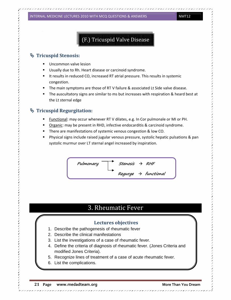
(F.) Tricuspid Valve Disease
���� Tricuspid Stenosis:
� Uncommon valve lesion
� Usually due to Rh. Heart disease or carcinoid syndrome.
� It results in reduced CO, increased RT atrial pressure. This results in systemic
congestion.
� The main symptoms are those of RT V failure & associated Lt Side valve disease.
� The auscultatory signs are similar to ms but increases with respiration & heard best at
the Lt sternal edge
���� Tricuspid Regurgitation:
� Functional: may occur whenever RT V dilates, e.g. In Cor pulmonale or MI or PH.
� Organic: may be present in RHD, infective endocarditis & carcinoid syndrome.
� There are manifestations of systemic venous congestion & low CO.
� Physical signs include raised jugular venous pressure, systolic hepatic pulsations & pan
systolic murmur over LT sternal angel increased by inspiration.
Pulmonary Stenosis � RHF
Regurge � functional
3. Rheumatic Fever
Lectures objectives
1. Describe the pathogenesis of rheumatic fever 2. Describe the clinical manifestations 3. List the investigations of a case of rheumatic fever. 4. Define the criteria of diagnosis of rheumatic fever. (Jones Criteria and
modified Jones Criteria). 5. Recognize lines of treatment of a case of acute rheumatic fever. 6. List the complications.

INTERNAL MEDICINE LECTURES 2010 WITH MCQ QUESTIONS & ANSWERS NMT12
www.medadteam.org More Than You Dream Page 22
���� Definition :
Non-suppurative inflammation disease occur as delayed sequel af group A β hemolytic
streptococcus pharyngitis (pathogenic serotypes 1,3,5,6,14,18,19,24,…….)
Two theories :
1. Toxic extracellular toxins of group A strept. on target organ
2. Abnormal immune response
���� Incidence:
� 5- 15 years old , rare < 4 years , uncommon >18 years
� Sex : no difference chorea more in females
� Familial susceptibility → significance geneXc / same housing
� Low socioeconomic status → overcrowdings & -- nutrition …recurrent chest infection
���� Pathology:
1. exudative lesions in serous membranes heal without residual effect
2. proliverative lesions in heart heal with fibrosis character of rheumatic affection Ashoff’s
nodules paravascules .central fibrinoid degeneration, ashoff cells, lymphocytes, fibroblasts
���� C/P:
���� Major criteria:
1. Arthritis : 75%
� Most common ( ++ in children )
� Large joints ( knee ,ankle ,wrist ,elbow) uncommon in small joints or central
"rheumatoid arthritis"
� D.D.:
++ Painful, tender, ± red, hot, swollen, effusions, limited mobility يتحرك عارف مش
Migratory to other big joint in upper/lower limbs "no sequale"
ماتمشى دبع تماما المفصل تسيب
Responds rapidly to salicylates (ARTHRITIS NOT ARTHLAGIA)
2. Carditis:
� +++ in young children & may be asymptomatic
� Long term disability &/or death
� Pancarditis:
• Endocarditis new murmur /change of present one
Altered similarity strept. Ag +human tissues
antigenic reaction
Ab against
Antigenic similarity Strept. antigen A b
Human tissue Ag
(immunological identical )

INTERNAL MEDICINE LECTURES 2010 WITH MCQ QUESTIONS & ANSWERS NMT12
www.medadteam.org More Than You Dream Page 23
o Mitral regurge : pansystolic murmur ,apical ,++common ,early in disease
o Mitral stenosis : mid- diastolic murmur ± presystolic (carey coomb’s
murmur ) disappear after acute stage at the apex .
o Aortic regurge → early diastolic murmur
• Myocarditis
o ++HR out of proportion of fever
o Cardiac enlargement
o CHF من البداية
o Arrhythmia & heart block
o Auscultation: weak heart sound , gallop ,functional MR,TR
o Tic-Tac rhythm S1=S2 due to loss of muscular component of S1
• percaditits :
o Pericardial friction rub or effusion → clinically ( confirmed by ECHO)
3. Chorea : 10- 15 %
� More in young females prepubertal in school age.
� Alone or with rheumatic manifestations may be 1st manifestation.
� Pt. present with difficult writing, speech ,impair by anxiety ,emotional instability
وتعيط تضحك شوية
� Purposeless rapid movements of arms, legs ↑↑ with anxiety بيراقبھا حد ان حست لو
& ↓↓ with sleep.
� Not responds to effort of closing of contraction
� Hypotonia & hyporeflexia are usually associated
� Quite room to ↓↓↓ anxiety
4. Subcutaneous nodules:
� firm ,painless ,not adherent to skin , at:
- extensor surface of large joints.
- bony prominences ( spine ,scapula, occiput).
- over tendons (Achilles) .
� for 1-2 ws , infrequent occurring & co-exist with severe carditis
5. Erythema marginatum:
� Transient ,serpiginous (↑↑ from periphery, ↓↓ from the centre ), flat ,non scarring
,non-purpuric , painless , erythm. rash on trunk
���� Others:
1-abdominal pain, 2- anorexia, 3- epistaxis, 4- fatigue, 5- fever >39oC characteristic pattern
N.B.: ARTHRALAGIA � pain in joint only, minor criteria, no inflammation.
���� Evidence of recent groupA strept. infection :
- recent scarlet fever.
- + ve culture of throat swab.
- ↑↑ ASO or other streprococcal Ab.
���� D.D.:
Other causes of :
� Fever SBE, any infection
� Acute arthritis

INTERNAL MEDICINE LECTURES 2010 WITH MCQ QUESTIONS & ANSWERS NMT12
www.medadteam.org More Than You Dream Page 24
� Heart failure
� Acute leukemia ,,H.S. purpura
���� Lab. Investigation:
� CBC : Anemia mild to moderate
- ↑↑ WBCs 12 – 20 ×10/ml
- ↑↑ plasma reactants
- ++ onset ,non specific
� For follow up with treatment : ESR >120 mm/h CRP ( non-specific )
� Recent strept. infection :
- Culture group A strept .
- ↑↑ ASO > 200 Todd’s U
- Others anti-DNAase, ASbase, anti-M protein Ab, anti hyalurodinase.
� Blood culture: rule & SBE ,bacteremia
� ECG: PR > 0,22 second ++ST Pericarditis , Chambers enlargement cardiac arrhythmia
� Chest X –ray :
Cardiomegaly detected clinically & confirmed by Echo & pulmonary congestion in Lt.
sided heart failure .
� Echo cardiography :
1- Valve lesion (MR, MS, AR) 2- pericardial effusion more sensitive
3- cardiac enlargement 4- confirm exclusion or diagnosis of vegetation of SBE
���� Diagnosis:
Modified John’s criteria
Major Minor
1. Polyarthritis Arthralgia
2. Carditis Fever
3. Chorea +ve acute phase reactants: ↑↑ESR & ↑↑CRP
4. S.C nodules Prior history of RF
5. Erythma marginatum Prolonged P-R in ECG
Two major or 1 major +2 minor indicate ++ probability or ARF provided by : evidence of recent
strept. infection (scarlet fever & culture & ++ASO).
���� Complication :
Immediate: - pancarditis → CHF, HB , pericardial effusion.
- chorea, debilitating
Long term:
� RHD * valvular disease – retraction → regurge – fibrosis → stenosis
* HF → ]t by surgery
� TED

INTERNAL MEDICINE LECTURES 2010 WITH MCQ QUESTIONS & ANSWERS NMT12
www.medadteam.org More Than You Dream Page 25
� Atrial arrhythmia( defect of contraction )
� Infective endocarditits
���� Treatment :
���� Prophylactic :
To Prevent Rheumatic Fever recurrence:
- long acting penicillin ( benzathine penicillin G 1.2 ×10 by deep IM into gluteal region
every 3 weeks شھر كل زمان كان
- erythromycin ( of allergy of penicillin) 250 mg/12h orally
- lifelong of carditits or minimum 10 years since last period .
���� Curative:
1. bed rest carditis ,chorea "quiet room" arthritis يتحرك عارف مش HF .
2. antibiotics for strept. phenoxy methyl penicillin 0.5 gm/day/10 days
Or: Benzathine single IM
Erythromycin if penicillin allergy.
3. Polyarthritis � salicylates 90 -120 mg /kg for 3 weeks , if satisfaction 60-70 mg /kg /day
to 6-9 days .
4. Carditis � salicylates.
if severe, HF � Steroids
* Prednisone ,upto 2 mg /kg /day (60mg /day ) in divided doses till:
1. ESR normal for 1 week.
2. CPR -ve.
* Gradual withdrawal 5mg /2days salicylates during withdrawal &after
disease for 3-6 weeks.
5. Chorea .
���� Quiet room
���� Diazepam
���� Halepritol
6. HF
���� Diuretics - O2 - bed rest -Na & fluid restriction
���� "Digoxin is contraindicated as it ++HB".
INTERNAL MEDICINE LECTURES 2010 WITH MCQ QUESTIONS & ANSWERS NMT12
www.medadteam.org More Than You Dream Page 26
4. Infective Endocarditis
���� Definition:
Infective endocarditis is defined as an infection, usually bacterial, of the endocardial surface of
the heart
���� Classified into four groups:
- Native Valve IE
- Prosthetic Valve IE
- Intravenous drug abuse (IVDA) IE
- Nosocomial IE
���� Further Classification:
���� Acute
• Affects normal heart valves
• Rapidly destructive ( very short course)
• Metastatic foci
• Very virulent( Commonly Staph.)
• If not treated, usually fatal within 6 weeks
• More in drug abusers (very virulent, Aggressive course, usually tricuspid (rt,
side) & unusual organsims
���� Subacute
• Often affects damaged heart valves
• Indolent nature
• If not treated, usually fatal by one year
• Usually mitral or aortic valves
���� Predisposing conditions:
- Mitral valve prolapse with murmur
- Rheumatic heart disease
Lectures objectives
1. Identify predisposing factors for infective endocarditis 2. Classify the causative organisms 3. Describe the clinical manifestations 4. List the complications 5. Construct a plan for management and treatment 6. Construct a plan for prophylaxis 7. Appreciate the importance of patient education.
INTERNAL MEDICINE LECTURES 2010 WITH MCQ QUESTIONS & ANSWERS NMT12
www.medadteam.org More Than You Dream Page 27
- Degenerative valvular disease
- Hypertrophic obstructive cardiomyopathy
- Intravenous drug use
- Prosthetic valve
- Pulmonary-systemic shunts
- Congenital abnormalities (valvular or septal defect)
- Coarctation of the aorta
- Previous endocarditis
- Complex cyanotic congenital heart disease
���� Microbiology
���� Native valve endocarditis
- Viridans streptococci - Enterococcus species
- Other streptococci - Staphylococcus aureus
- Coagulase negativeStaphylococcus - Gram negative bacilli
- Fungi
���� Intravenous Drug Abuse
- Risk is 2 – 5% per pt./year
- Underlying valve is normal in 75 – 93%
- Tendency to involve right-sided valves
- Distribution in clinical series:
� 46 – 78% tricuspid ▪ 24 – 32% mitral ▪ 8 – 19% aortic
- Staph. aureus predominant organism (>50%, 60-70% of tricuspid cases)
- Increased frequency of gram negative infection such as P. aeruginosa & fungal
infections
���� Pathophysiology
The disease follows a predictable sequence:
� Endocardial damage.
� Aggregation of platelets and fibrin to form a sterile vegetation.
� Transient bacteremia resulting in seeding of the vegetation.
� Microbial proliferation on and invasion of the endocardial surface.
� Metastatic infection to visceral organs and brain .
N.B.:
The surfaces of cardiac valves and vegetations are avascular, therefore healing is difficult.
���� Clinical Manifestations :
���� History:
The diagnosis requires a high index of suspicion because the initial presentation of the
disease varies enormously from patient to patient.

INTERNAL MEDICINE LECTURES 2010 WITH MCQ QUESTIONS & ANSWERS NMT12
www.medadteam.org More Than You Dream Page 28
Most patients complain of fever and nonspecific constitutional symptoms, such as
fatigue, malaise, and weight loss.
���� PHYSICAL EXAMINATION AND LABORATORY FINDINGS IN INFECTIVE ENDOCARDITIS:
Osler’s Nodes
• Painful and erythematous nodules
• Located on pulp of fingers and toes
• More common in subacute IE
Splinter Hemorrhages
• Nonspecific
• Non blanching
• Linear reddish-brown lesions found under the nail bed
• Usually do NOT extend the entire length of the nail
Janeway Lesions
• More specific
• Erythematous, blanching macules
• Nonpainful
• Located on palms and soles
���� Complications:
They are divided into four groups for ease of classification:
(1.) Direct valvular damage and consequences of local invasion,
(2.) Embolic complications,
(3.) Metastatic infections from bacteremia,
(4.) Immunologic phenomena
(1.) Direct valvular damage and consequences of local invasion
Finding % of Cases
Fever 80–95
Audible murmur 85
New or changed murmur 15–47
Neurologic abnormalities 20–40
Splenomegaly 0–60
Petechiae 20–40
Splinter hemorrhages 15
Osler's nodes 10–25
Janeway lesions <10
Roth's spots <5
Anemia of chronic disease 50–90
Leukocytosis 20–66
Elevated erythrocyte 90–100
Microscopic hematuria 50–70
Presence of rheumatoid factor 40–50
Abnormal chest x-ray (effusion, infiltrate, septic emboli) 67–85 (right-sided infective endocarditis)
INTERNAL MEDICINE LECTURES 2010 WITH MCQ QUESTIONS & ANSWERS NMT12
www.medadteam.org More Than You Dream Page 29
Local damage to the endocardium or myocardium is a dreaded complication that can be
difficult to diagnose and to treat.
- Infection may directly erode through the involved cardiac valve resulting in valvular
perforations or cardiac fistula
- Acute onset of heart failure
- Valve ring abscesses are more frequent in patients with prosthetic valves.
- A conduction defect on electrocardiography
- Frank myocardial abscess has been found in up to 20% of cases at autopsy
- Pericarditis is rare and is associated with myocardial abscess in most cases.
- Myocardial infarction, thought to be due to embolism of vegetative material in the
coronary arteries, has been found in 40 to 60% of cases at autopsy.
(2.) Embolic complications
Embolic events are less common now than in the preantibiotic era;
- The skin, the lungs (in right-sided endocarditis), kidneys, spleen, large blood vessels,
and central nervous system are common sites for embolisation
- Renal infarction is seen in more than 50% of cases at autopsy. Similarly, splenic
infarction occurs in up to 44% of autopsy
- Cerebrovascular accidents related to these emboli
(3.) Metastatic infections from bacteremia
- Osteomyelitis.
- Septic arthritis.
- Epidural abscess.
- Purulent meningitis.
- Intracranial abscesses.
(4.) Immunologic phenomena
- Hypocomplementemic glomerulonephritis.
- monorticular and oligoarticular arthritides.
���� Investigations:
���� Laboratory Findings:
- Complete blood count with differential
- Electrolyte determinations
- Measurement of renal function and urinalysis.
- Chest radiography.
- Electrocardiography.
- Echocardiography
- Erythrocyte sedimentation rate
- Rheumatoid factor

INTERNAL MEDICINE LECTURES 2010 WITH MCQ QUESTIONS & ANSWERS NMT12
www.medadteam.org More Than You Dream Page 30
(1.) Blood cultures:
- At least three sets of blood cultures should be obtained from 3 separate sites; each set
consists of one aerobic and one anaerobic bottle, with careful attention paid to aseptic
technique. Ideally, these sets are collected at least 1 hour apart to document
continuous bacteremia;
- In most cases of endocarditis, in the absence of prior antibiotic therapy, every blood
culture is positive because the bacteremia of endocarditis is continuous.
- Blood cultures are truly negative in less than 5% of cases of endocarditis
- The prior antibiotic administration results in most "culture-negative" conditions.
���� ORGANISMS CAUSING “CULTURE-NEGATIVE” ENDOCARDITIS:
• HACEK spp.( Haemophilus species, Actinobacillus actinomycetemcomitans,
Cardiobacterium hominis, Eikenella corrodens, and Kingella species)
• Nutritionally variant streptococci
• Coxiella burnetii (Q fever)
• Brucella spp.
• Bartonella spp.
• Chlamydia psittaci
• Legionella spp.
• Aspergillus and other noncandidal fungi
Non-bacterial thrombotic endocarditis:
Nonbacterial thrombotic endocarditis may occur spontaneously in patients with systemic illnesses (for instance, the marantic endocarditis of malignant disease or other wasting diseases and Libman-Sacks endocarditis in systemic lupus erythematosus). BUT
When transient bacteremia occurs, for example, as a result of distant infection or gingival disease, the previously sterile vegetation may be seeded
(2.) Imaging:
• Chest x-ray
Look for multiple focal infiltrates and calcification of heart valves
• ECG
Rarely diagnostic
Look for evidence of ischemia, conduction delay, and arrhythmias
• Echocardiography
• Transthoracic echocardiography (TTE)
o First line if suspected IE
o Native valves
• Transesophageal echocardiography (TEE)
INTERNAL MEDICINE LECTURES 2010 WITH MCQ QUESTIONS & ANSWERS NMT12
www.medadteam.org More Than You Dream Page 31
o Prosthetic valves
o Intracardiac complications
o Inadequate TTE
o Fungal or S. aureus or bacteremia
(3.) Diagnostic (Duke) Criteria
1. Definitive infective endocarditis
• Pathologic criteria microorganisms or pathologic lesions: demonstrated by culture or histology in a vegetation, or
in a vegetation that has embolized, or in an intracardiac abscess
• Clinical criteria
two major criteria, or one major and three minor criteria, or five minor criteria
2. Possible infective endocarditis
findings consistent of IE that fall short of “definite”, but not “rejected”
3. Rejected
-Alternate Ds
-resolution of manifestations of IE, with antibiotic therapy for ≤ 4 days
-no pathologic evidence of IE at surgery or autopsy, after antibiotic therapy for ≤ 4 days
Major criteria
• positive blood culture for IE
• evidence of endocardial involvement
Minor criteria
• predisposition (heart condition or IV drug use)
• fever
• vascular or immunologic phenomena
• microbiologic or echocardiographic evidence not meeting major criteria
���� Therapy:
(1.) Pre-antibiotic era : a death sentence is written
(2.) Antimicrobial therapy
Definitive antibiotic treatment of infective endocarditis is guided by antimicrobial
susceptibility testing of the responsible pathogen isolated from clinical culture
specimens. Frequently, however, it is advisable to begin empirical treatment before
definitive culture results are available
- Nafcillin-penicillin-gentamicin is suitable in most cases of suspected native valve
endocarditis, providing optimal coverage for streptococci, staphylococci,
enterococci, and HACEK organisms.
- or vancomycin-gentamicin
- If S. aureus is an important consideration, as in drug users, empirical therapy
should be with vancomycin and gentamicin
INTERNAL MEDICINE LECTURES 2010 WITH MCQ QUESTIONS & ANSWERS NMT12
www.medadteam.org More Than You Dream Page 32
(3.) Surgery: For intracardiac complications.
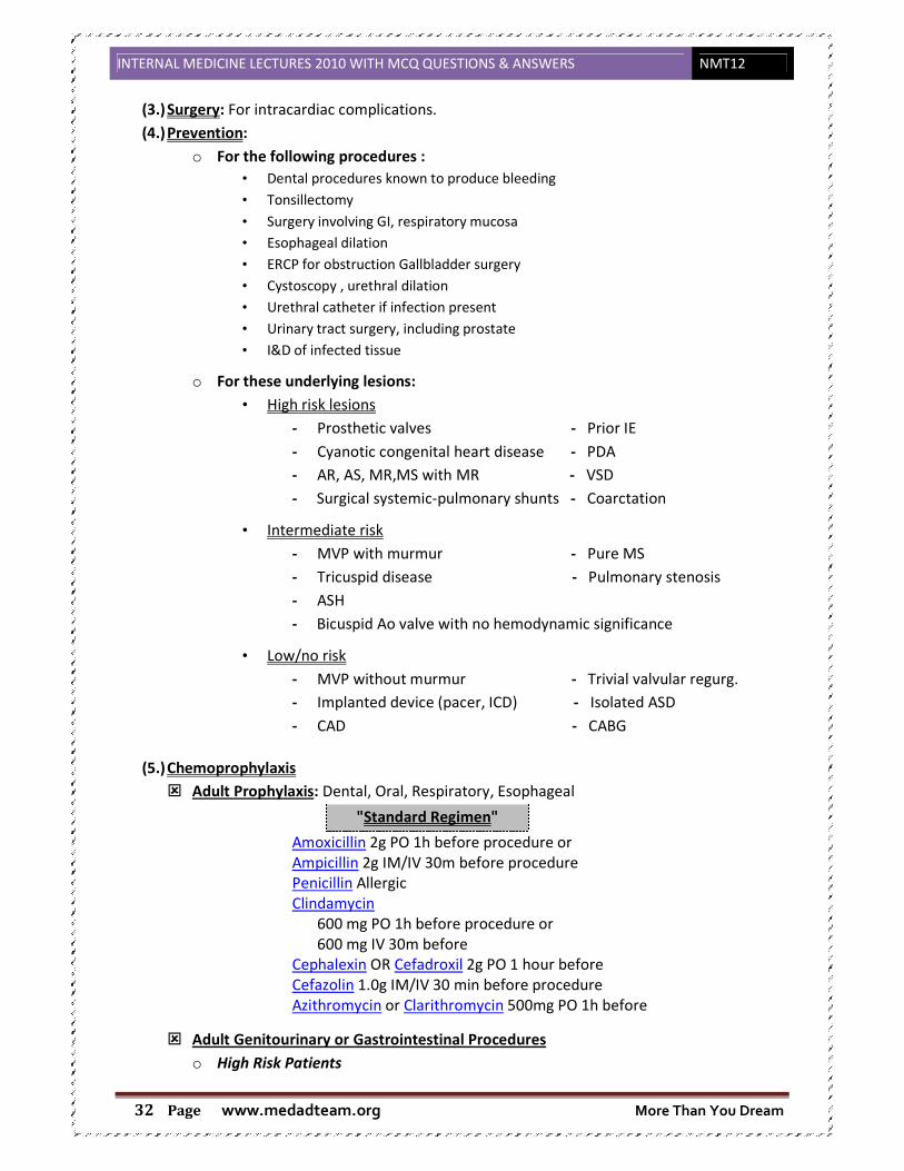
(4.) Prevention:
o For the following procedures :
• Dental procedures known to produce bleeding
• Tonsillectomy
• Surgery involving GI, respiratory mucosa
• Esophageal dilation
• ERCP for obstruction Gallbladder surgery
• Cystoscopy , urethral dilation
• Urethral catheter if infection present
• Urinary tract surgery, including prostate
• I&D of infected tissue
o For these underlying lesions:
• High risk lesions
- Prosthetic valves - Prior IE
- Cyanotic congenital heart disease - PDA
- AR, AS, MR,MS with MR - VSD
- Surgical systemic-pulmonary shunts - Coarctation
• Intermediate risk
- MVP with murmur - Pure MS
- Tricuspid disease - Pulmonary stenosis
- ASH
- Bicuspid Ao valve with no hemodynamic significance
• Low/no risk
- MVP without murmur - Trivial valvular regurg.
- Implanted device (pacer, ICD) - Isolated ASD
- CAD - CABG
(5.) Chemoprophylaxis
� Adult Prophylaxis: Dental, Oral, Respiratory, Esophageal
"Standard Regimen"
Amoxicillin 2g PO 1h before procedure or
Ampicillin 2g IM/IV 30m before procedure
Penicillin Allergic
Clindamycin
600 mg PO 1h before procedure or
600 mg IV 30m before
Cephalexin OR Cefadroxil 2g PO 1 hour before
Cefazolin 1.0g IM/IV 30 min before procedure
Azithromycin or Clarithromycin 500mg PO 1h before
� Adult Genitourinary or Gastrointestinal Procedures
o High Risk Patients
INTERNAL MEDICINE LECTURES 2010 WITH MCQ QUESTIONS & ANSWERS NMT12
www.medadteam.org More Than You Dream Page 33
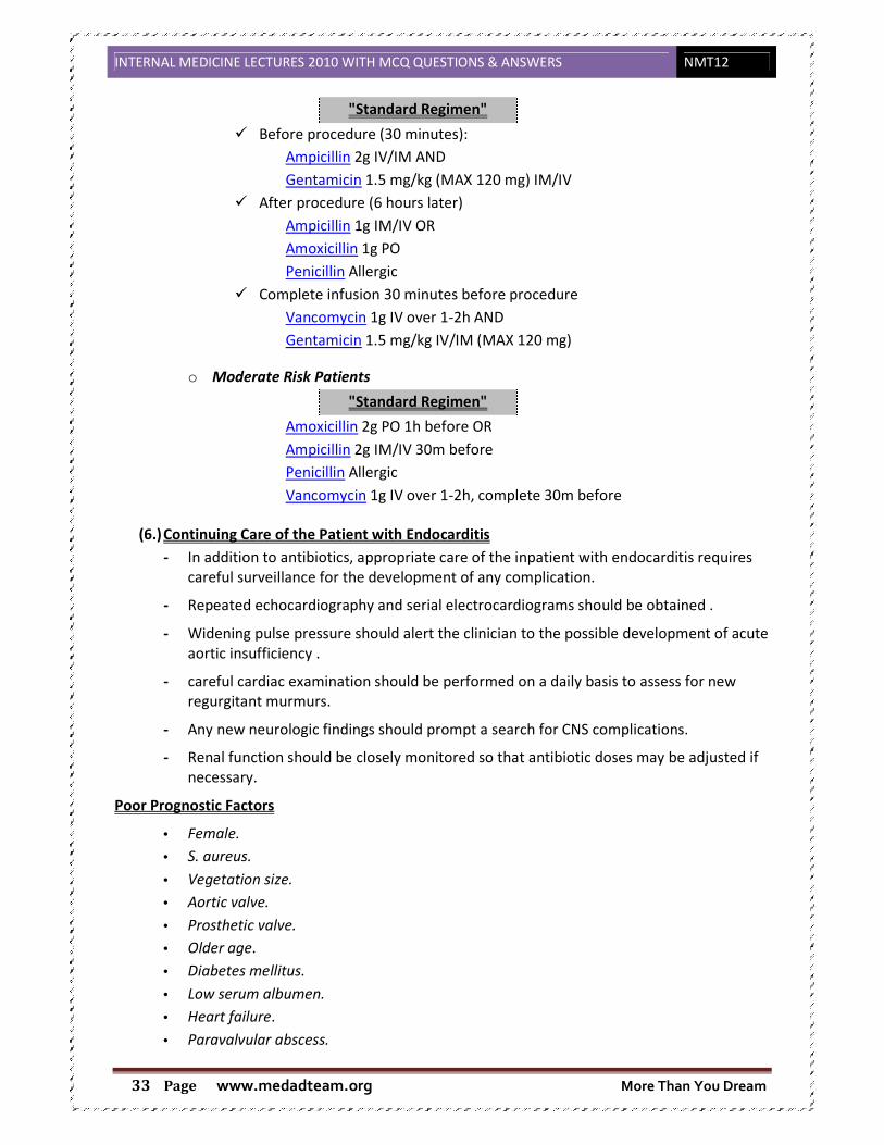
"Standard Regimen"
� Before procedure (30 minutes):
Ampicillin 2g IV/IM AND
Gentamicin 1.5 mg/kg (MAX 120 mg) IM/IV
� After procedure (6 hours later)
Ampicillin 1g IM/IV OR
Amoxicillin 1g PO
Penicillin Allergic
� Complete infusion 30 minutes before procedure
Vancomycin 1g IV over 1-2h AND
Gentamicin 1.5 mg/kg IV/IM (MAX 120 mg)
o Moderate Risk Patients
"Standard Regimen"
Amoxicillin 2g PO 1h before OR
Ampicillin 2g IM/IV 30m before
Penicillin Allergic
Vancomycin 1g IV over 1-2h, complete 30m before
(6.) Continuing Care of the Patient with Endocarditis
- In addition to antibiotics, appropriate care of the inpatient with endocarditis requires
careful surveillance for the development of any complication.
- Repeated echocardiography and serial electrocardiograms should be obtained .
- Widening pulse pressure should alert the clinician to the possible development of acute
aortic insufficiency .
- careful cardiac examination should be performed on a daily basis to assess for new
regurgitant murmurs.
- Any new neurologic findings should prompt a search for CNS complications.
- Renal function should be closely monitored so that antibiotic doses may be adjusted if
necessary.
Poor Prognostic Factors
• Female.
• S. aureus.
• Vegetation size.
• Aortic valve.
• Prosthetic valve.
• Older age.
• Diabetes mellitus.
• Low serum albumen.
• Heart failure.
• Paravalvular abscess.

INTERNAL MEDICINE LECTURES 2010 WITH MCQ QUESTIONS & ANSWERS NMT12
www.medadteam.org More Than You Dream Page 34
• Embolic events.
5. Coronary (Ischemic) Heart Disease
���� Definition:
� Coronary artery disease (CAD) (or atherosclerotic heart disease) is the end result of the
accumulation of atheromatous plaques within the walls of the coronary arteries that
supply the myocardium (the muscle of the heart) with oxygen and nutrients.
� Ischaemic or ischemic heart disease (IHD), or myocardial ischaemia, is a disease
characterized by reduced blood supply to the heart muscle, usually due to coronary artery
disease (atherosclerosis of the coronary arteries).
� Myocardial ischemia is a condition in which oxygen deprivation to the heart muscle is
accompanied by inadequate removal of metabolites because of reduced blood flow or
perfusion.
� Myocardial ischaemia occurs when there is an imbalance between the supply of oxygen
(and other essential myocardial nutrients) and the myocardial demand for these
substances.
���� Causes:
1. Coronary blood flow to a region of the myocardium may be reduced by a mechanical
obstruction that is due to:
• Atheroma
• Thrombosis
• Spasm
• Embolus
• Coronary arteritis (e.g. In SLE).
2. There can be a decrease in the flow of oxygenated blood to the myocardium that is due to:
• Anaemia
• CO poisoning
Lectures objectives
1. Define coronary heart disease 2. Explain the aetiology and pathogenesis of different types of ischemic
heart disease 3. Enumerate risk factors of coronary atherosclerosis 4. Describe the C/P of stable angina and the acute coronary syndromes
and interpret the DD 5. List the different investigations 6. Explain the management of different clinical types with special
reference to the emergency situation. 7. Outline appropriate management plan

INTERNAL MEDICINE LECTURES 2010 WITH MCQ QUESTIONS & ANSWERS NMT12
www.medadteam.org More Than You Dream Page 35
• Hypotension causing decreased coronary perfusion pressure.
3. An increased demand for oxygen may occur owing to an increase in cardiac output (e.g.
thyrotoxicosis) or myocardial hypertrophy (e. g. from aortic stenosis or hypertension).
���� Risk factors for coronary atherosclerosis:
Risk factors
Modifiable Non-modifiable
���� Fixed (Non-modifiable) risk factors:
1. Age: CAD rates increase with age. Atherosclerosis is rare in childhood
2. Male sex: Men have a higher incidence of coronary artery disease than premenopausal
women. However, after the menopause the incidence of atheroma in women
approaches that in men.
3. Race: Significant racial variations exist in the incidence, prevalence, presentations, and
response to therapy for CAD.
4. Positive family history
5. Genetic: A number of genetic factors have been linked with coronary artery disease.
���� Potentially changeable with treatment:
1- Hyperlipidaemia: High serum cholesterol, a low value of high-density lipoproteins
(HDL), high serum triglyceride (TG) are strongly associated with coronary
atheroma...Familial hypercholesterolaemia is also a risk factor
2- Cigarette smoking: In men, the risk of developing CAD is directly related to the number
of cigarettes smoked. Evidence suggests that each person stopping smoking will reduce
his/her own risk by 25%. The risk from smoking declines to almost normal after 10
years of abstention.
3- Hypertension: Both systolic and diastolic hypertension are associated with an increased
risk of CAD.
4- Diabetes mellitus: Diabetes, an abnormal glucose tolerance or raised fasting glucose is
strongly associated with vascular disease. Diabetes not only increases the risk of CAD
but also magnifies the effect of other risk factors for CAD such as raised cholesterol
levels, raised blood pressure, smoking and obesity.
5- Lack of exercise: Lack of exercise is an independent risk factor for CAD equal to
hypertension, hyperlipidaemia and smoking.

INTERNAL MEDICINE LECTURES 2010 WITH MCQ QUESTIONS & ANSWERS NMT12
www.medadteam.org More Than You Dream Page 36
6- Blood coagulation factors - high fibrinogen, factor VII: Serum fibrinogen is strongly,
consistently, and independently related to CAD risk. The pathophysiological mechanism
is related to its effect on the coagulation cascade, platelet aggregation, endothelial
function and smooth muscle cell proliferation and migration. High levels of coagulation
factor VII are also a risk factor.
7- C-reactive protein: CRP is linked with future risk of coronary events
8- Homocysteinaemia: Homocysteinaemia is a major risk factor in the pathogenesis of
CAD and a strong predictor of mortality in this group.
9- Personality: Type A personality has been found to be most consistently associated with
an increased risk of CAD
10- Diet and obesity: Diets high in fats are associated with ischemic heart disease, as are
those with low intakes of antioxidants (i.e. fruit and vegetables). Patients who are
overweight and those who are obese have an increased risk of CAD. The adverse effect
of excess weight is more pronounced when the fat is concentrated mainly in the
abdomen.( trunkal obesity )
11- Contraceptive pills
12- Heavy alcohol consumption: Moderate alcohol consumption (one or two drinks per
day) is associated with a reduced risk of CAD. At high levels of intake the risk of CAD is
increased risk'.
13- Infections implicated in development of atherosclerosis:
• Chlamydia pneumoniae
• Helicobacter pylori
• Herpes simplex virus
���� Pathogenesis of coronary atherosclerosis:
Coronary atherosclerosis is a complex inflammatory process characterized by the accumulation
of lipid, macrophages and smooth muscle cells in intimal plaques in the large and medium-
sized coronary arteries.
1. Endothelial dysfunction/injury: Mechanical shear stresses (e.g. from morbid
hypertension), biochemical abnormalities (e.g. elevated and modified LDL, diabetes
mellitus, elevated plasma homocysteine), immunological factors (e.g. free radicals from
smoking), inflammation (e.g. infection such as Chlamydia pneumonia and Helicobactor
pylori) and genetic alteration may contribute to the initial endothelial 'injury' or
dysfunction, which is believed to trigger atherogenesis.
2. Increased permeability: Endothelial dysfunction, with increased permeability to and
accumulation of oxidized lipoproteins, which are taken up by macrophages at focal sites
within the endothelium to produce lipid-laden foam cells. Macroscopically, these lesions
are seen as flat yellow dots or lines on the endothelium of the artery and are known as
'fatty streaks'

INTERNAL MEDICINE LECTURES 2010 WITH MCQ QUESTIONS & ANSWERS NMT12
www.medadteam.org More Than You Dream Page 37
3. Mediators: Release of cytokines such as platelet-derived growth factor and transforming
growth factor-(5 (TGF-P) by monocytes, macrophages or the damaged endothelium
promotes further accumulation of macrophages as well as smooth muscle cell migration
and proliferation. Collagen is produced in larger and larger quantities by the smooth
muscle and the whole sequence of events cumulates as an 'advanced plaque'.
4. Thrombosis: The 'advanced plaque' may grow slowly and encroach on the lumen or
become unstable, undergo thrombosis and produce an obstruction (complicated plaque).
N.B. Mechanism for thrombosis on plaques:
Superficial endothelial injury, subendocardial connective tissue matrix is then exposed and platelet
adhesion occurs because of reaction with collagen. The thrombus is adherent to the surface of the plaque.
Angina & Acute Coronary Syndromes
���� Etiology and Pathogenesis:
� Angina: Classical or exertional (stable) angina pectoris, decubitus angina , variant
(prinzmetal' s) angina , nocturnal angina
� Acute Coronary Syndromes: include ST-elevation myocardial infarction (STEMI), non-ST-
elevation myocardial infarction (NSTEMI), and unstable angina Angina (chest pain)
• The diagnosis of angina is largely based on the clinical history.
• The chest pain is generally described as 'heavy', 'tight' or 'gripping'.
• Typically, the pain is central/ retrosternal and may radiate to the jaw and/or arms,
also to the back or epigastric region. Angina can range from a mild ache to a most severe pain
that provokes sweating and fear. There may be associated breathlessness.
(A.) Angina
���� Types:
1. Classical or exertional (Stable) angina pectoris: is provoked by physical exertion, especially
after meals and in cold, windy weather, and is commonly aggravated by anger or
excitement. The pain fades quickly (usually within minutes) with rest.
2. Decubitus angina is that occurring on lying down. It usually occurs in association with
impaired left ventricular function, as a result of severe coronary artery disease.
3. Nocturnal angina occurs at night and may wake the patient from sleep. it tends to occur in
patients with critical coronary artery disease and may be the result of vasospasm.
4. Variant (Prinzmetal) angina refers to an angina that occurs without provocation, usually at
rest, as a result of coronary artery spasm. It occurs more frequently in women.
5. Unstable angina: refers to angina of recent onset (less than 1 month), worsening angina or
angina at rest (see Acute coronary syndrome)

INTERNAL MEDICINE LECTURES 2010 WITH MCQ QUESTIONS & ANSWERS NMT12
www.medadteam.org More Than You Dream Page 38
���� Examination and diagnosis:
� Usually from history and symptoms ( about nature of attacks and presence of risk factors )
see before
� There are usually no abnormal findings in angina signs to suggest anaemia, thyrotoxicosis
or hyperlipidaemia (e.g. lipid arcus, xanthelasma, tendon xanthoma) should be sought.
� It is essential to exclude aortic stenosis (i.e. slow-rising carotid impulse and ejection systolic
murmur radiating to the neck) as a possible cause for the angina.
� The blood pressure should be taken to identify coexistent hypertension
���� D.D:
Other causes of chest pain (see chest and cardiac symptomatology)
���� Investigations for angina:
1. Resting ECG: This is usually normal between attacks. Evidence of old myocardial infarction
(e.g. pathological Q waves), left ventricular hypertrophy or left bundle branch block may be
present.
2. Exercise ECG: Exercise testing can be very useful both in confirming the diagnosis of angina
and in giving some indication as to the severity of the CAD.
3. Echocardiography: This can be used to assess ventricular wall involvement and ventricular
function.
4. Coronary angiography: Not used routienly. More often, the test is performed in patients
being considered for revascularization (i.e. coronary artery bypass grafting or coronary
angioplasty).
5. Lab: Blood picture, Lipid profile, Thyroid function, Diabetes mellitus, Homocystein level, C
reactive protein, Cardiac enzymes
6. Radiology: to exclude other causes of chest pain.
���� Treatment of angina:
1. General management
� Patients should be informed as to the nature of their condition and reassured that
the prognosis is good (annual mortality less than 2%).
� Underlying problems, such as anaemia or hyperthyroidism, should be treated.
� Management of coexistent conditions, such as diabetes and hypertension, should
be optimized.
� Risk factors should be evaluated and steps made to correct: smoking must be
stopped, hypercholesterolaemia should be treated, weight loss, and regular
exercise should b encouraged.
� Choosing between medical therapy and revascularization (coronary artery bypass
grafting and angioplasty)
2. Medical treatment

INTERNAL MEDICINE LECTURES 2010 WITH MCQ QUESTIONS & ANSWERS NMT12
www.medadteam.org More Than You Dream Page 39
a. Prognostic therapies:
� Aspirin reduces the risk of coronary events in patients with coronary artery
disease. All patients with angina, therefore, should take aspirin (75 mg daily is
probably adequate) unless contraindicated.
� Lipid-lowering therapy statins ,fibrates
b. Symptomatic treatment:
� Glyceryl tri-nitrate (GTN) used sublingually, either as a tablet or as a spray, gives
prompt relief (peak action 4.8 minutes and lasts 20.30 minutes). It can be used
prior to performing activities that the patient knows will provoke angina.
Transdermal GTN preparations last up to 24 hours.
� Beta-blockers reduce the heart rate (-ve chronotropic effect) and the force of
ventricular contraction (-ve inotropic effect), both of which reduce myocardial
oxygen demand, especially on exertion. Atenolol, 50-100 mg daily, is the most
commonly prescribed. Beta-blockers may aggravate coronary artery spasm.
� Long-acting nitrates (e.g. isosorbide mononitrate) Sildenafil (or other PDE5
inhibitors) should not be given to patients taking nitrates.
� Calcium-channel blockers, they relax coronary arteries, cause peripheral
vasodilatation and reduce the force of left ventricular contraction, thereby
reducing the oxygen demand of the myocardium.
� Nicorandil is a potassium-channel activator with a nitrate component; it has both
arterial and venous vasodilating properties. Not used as a first-line drug
3. Coronary angioplasty:
� Percutaneous transluminal coronary angioplasty (PTCA) refers to the technique of
dilating coronary atheromatous obstructions by inflating a balloon within the
obstruction
� Intra-coronary stents
4. Surgical management: Coronary Artery Bypass Grafting (CABG):
Indications:
� Symptom control in patients who remain symptomatic despite optimal medical
therapy and whose disease is not suitable for PTCA.
� Improved survival in patients with severe three-vessel CAD.
(B.) Acute Coronary Syndromes
� Acute coronary syndromes (ACS) include ST-elevation myocardial infarction (STEMI),non-
ST-elevation myocardial infarction (NSTEMI), and unstable angina.
� Myocardial infarction occurs when cardiac myocytes die due to myocardial ischaemia,
INTERNAL MEDICINE LECTURES 2010 WITH MCQ QUESTIONS & ANSWERS NMT12
www.medadteam.org More Than You Dream Page 40
���� Pathophysiology:
The common mechanism to all ACS is erosion of the fibrous cap of a plaque. This leads to
platelet aggregation and adhesion, localized thrombosis, vasoconstriction, and distal
thrombus embolization. This results in myocardial ischaemia due to reduction of coronary
blood flow.
���� Diagnosis:
� Clinical presentation ( unstable angina / infarction )
� Patients with an ACS may complain of a new onset of chest pain, chest pain at rest, or
deterioration of preexisting angina. However, some patients present with atypical features
including indigestion, pleuritic chest pain or dyspnoea.
� Physical examination to exclude other diagnoses of chest pain such as aortic dissection,
pulmonary embolism or peptic ulceration
���� Investigations:
1. ECG:
� May be normal in patients with an ACS, ST depression and T-wave inversion are highly
suggestive of an ACS, particularly if associated with anginal chest pain
� N.B.: transient ST elevation is seen with coronary vasospasm (Prinzmetal's angina).
2. Biochemical markers:
� Creatinine-kinase -MB level was until recently the standard marker for myocyte death
used in ACS. However, the presence of CKMB in the serum of normal individuals and in
patients with significant skeletal muscle damage, has limited its accuracy.
� The cardiac troponin complex The cardiac troponins are not detectable in normal
people and so monoclonal antibody tests for cardiac specific troponin I and cardiac-
specific troponin T are highly sensitive markers of myocyte necrosis.
� Myoglobin may be useful for a rapid diagnosis of ACS, as the levels become elevated
very early in the time course of an MI, but because of the presence of myoglobin in
skeletal muscle the test has poor specificity for ACS.
� New markers are becoming available, e.g.: myeloperoxidase or glutathione peroxidase 1
���� Therapy in Acute Coronary Syndromes:
1. Oxygen: 35-50%
2. Antiplatelet: Aspirin , Clopidogrel
3. Analgesia: Diamorphine
4. Myocardial energy consumption: B-blockers
5. Coronary vasodilatation Glyceryl tri-nitrate 2-10 mg/h I.V./buccal/or sublingual

INTERNAL MEDICINE LECTURES 2010 WITH MCQ QUESTIONS & ANSWERS NMT12
www.medadteam.org More Than You Dream Page 41
6. Plaque stabilization/ventricular remodeling HMG-CoA reductase inhibitors (statins) ,
ACEinhibitors e.g. Ramipril.
For myocardial infarction (NSTEMI): Add
7. Antithrombin: Low-molecular-weight heparins
8. Glycoprotein IIB/IIIA inhibitors: Abciximab Eptifibatide Tirofiban
& "5" is used If coronary intervention likely within 24 h
& "6" is indicated in high-risk patients managed without coronary intervention
Plus for ST-elevation myocardial infarction (STEMI): Add
9. Thrombolysis: Streptokinase , Alteplase (rt-PA) Tenecteplase (TNKase) , Reteplase.
+ Coronary interventions:
Coronary revascularization is recommended in high-risk patients with ACS.
Post-ACS:
---- Risk factor modification is necessary to reduce future cardiovascular events.
���� Patients should be encouraged to stop cigarette smoking and should be referred to a
smoking cessation clinic.
���� The patient should maintain optimal weight, exercise daily for, and have a healthy diet.
���� Hypertension should be treated
���� In patients with diabetes, glycaemic control should be very tight
���� Low-fat diets should be combined with HMG-CoA reductase inhibitors to reduce LDL
cholesterol.
---- Follow up medications: should include aspirin (clopidogrel), statin, beta-blocker, and an
ACE inhibitor, with a nitroglycerine spray for symptomatic relief of angina.
6. Heart Failure
Lectures objectives
1. Define heart failure. 2. Identify the aetiology of heart failure (acute and chronic). 3. Recognize the precipitating causes of heart failure 4. Describe the Pathophysiology 5. Describe clinical syndromes of heart failure and their causes. 6. Outline the appropriate management plan for a case of heart failure.
INTERNAL MEDICINE LECTURES 2010 WITH MCQ QUESTIONS & ANSWERS NMT12
www.medadteam.org More Than You Dream Page 42
���� Definition:
Reduction of cardiac output (COP) below normal in spite of normal venous return (as low VR
may cause collapse and HF but heart is normal)
���� Etiology of heart failure (Acute and Chronic):
� Acute heart failure
� The term "acute heart failure" is often used exclusively to mean acute (cardiogenic)
dyspnoea characterized by signs of pulmonary congestion.
� It is preferable to use the term 'acute pulmonary oedema' or where applicable
'cardiogenic shock'.
� Acute failure of the heart most commonly occurs in the setting of acute myocardial
infarction when there is extensive loss of ventricular muscle.
� The condition may also occur with rupture of the interventricular septum producing a
ventricular septal defect, or be due to acute valvular regurgitation.
� Common examples of valvular regurgitation are papillary or chordal rupture producing
mitral regurgitation, or sudden aortic valve regurgitation in infective endocarditis.
� Other causes of acute heart failure include obstruction of the circulation by acute
pulmonary embolus and cardiac tamponade. In each case severe cardiac failure can
occur with a relatively normal heart size.
� Chronic heart failure
� Chronic heart failure is often referred for an abnormality of cardiac function is
responsible for the failure of the heart to maintain a physiological circulation
� At present, no simple objective definition of heart failure is available, since there is no
cut-off value for cardiac or ventricular dysfunction or change in flow, pressure,
dimension or volume that can be used reliably to identify patients with heart failure.
When there is gradual impairment of cardiac function, a variety of compensatory
changes may take place.
���� Causes of heart failure
� Main causes
� Ischaemic heart disease (35-40%)
� Cardiomyopathy (dilated) (30-34%)
� Hypertension (15-20%)
� Other causes
� Cardiomyopathy (undilated): hypertrophic/obstructive, restrictive (amyloidosis, sarcoidosis)
� Valvular heart disease (mitral, aortic, tricuspid)
� Congenital heart disease (ASD, VSD)
� Alcohol and drugs (chemotherapy)
� Hyperdynamic circulation (anaemia, thyrotoxicosis, haemochromatosis, Paget's disease)
� Pulmonary causes ( RVF) (pulmonary hypertension, pulmonary embolism, cor pulmonale
[COPD])
� Arrhythmias (atrial fibrillation, bradycardia [complete heart block, the sick sinus syndrome])
� Pericardial disease (constrictive pericarditis, pericardial effusion)
INTERNAL MEDICINE LECTURES 2010 WITH MCQ QUESTIONS & ANSWERS NMT12
www.medadteam.org More Than You Dream Page 43
� Factors that may precipitate or aggravate "Heart failure" in patients with pre-existing heart
disease:
1. Myocardial ischemia or infarction
2. Inter current illness, e.g. infection
3. Arrythmia, e.g. atrial fibrillation
4. Inappropriate reduction of therapy
5. Administration of a drug with negative inotropic properties (e.g. β-blocker) or fluid-
retaining properties (e.g. non-steroidal anti-inflammatory drugs, corticosteroids)
6. coronary embolism
7. conditions associated with increased metabolic demand, e.g. pregnancy, thyrotoxicosis,
anemia
8. Intravenous fluid overload, e.g. post-operative I.V. infusion
���� Pathophysiology:
� When the heart fails, considerable changes occur to the heart and peripheral vascular
system in response to the haemodynamic changes associated with heart failure
� These physiological changes are compensatory and maintain cardiac output and
peripheral perfusion. However, as heart failure progresses, these mechanisms are
overwhelmed and become pathophysiological.
1. Venous return (preload)
� Myocardial failure leads to a reduction decrease cardiac output and thus increase in
the volume of blood remaining after systole.
� This increased diastolic volume stretches the myocardial fibers and, as Starling's law of
the heart would suggest, myocardial contraction is restored.
� Sinus tachycardia also ensures that any reduction of stroke volume is compensated for
by the increase in heart rate; cardiac output (stroke volume x heart rate) is therefore
maintained.
� The increased venous pressure contributes to the development of dyspnoea, owing to
the accumulation of interstitial and alveolar fluid, and to the occurrence of hepatic
enlargement, ascites and dependent oedema, due to increased systemic venous
pressure.
N.B.: The cardiac output at rest may not be much depressed, but myocardial and
haemodynamic reserve is so compromised that a normal increase in cardiac output cannot be
produced by exercise. In very severe heart failure the cardiac output at rest is depressed,
despite high venous pressures.
� The inadequate cardiac output is redistributed to maintain perfusion of vital organs,
such as the heart, brain and kidneys, at the expense of the skin and muscle.
2. The sympathetic nervous system
� is activated in heart failure via baroreceptors as an early compensatory mechanism,
which provides inotropic support and maintains cardiac output.

INTERNAL MEDICINE LECTURES 2010 WITH MCQ QUESTIONS & ANSWERS NMT12
www.medadteam.org More Than You Dream Page 44
3. Neurohormonal and sympathetic system activation:
� salt and water retention : the increase in venous pressure that occurs when the
ventricles fail leads to retention of salt and water and their accumulation in the
interstitium, producing many of the physical signs of heart failure.
� Reduced cardiac output also leads to diminished renal perfusion, activating the renin-
angiotensin system and enhancing salt and water retention , which further increases
venous pressure .
� The retention of sodium is in part compensated by the action of circulating atrial
natriuretic peptides and antidiuretic hormone.
4. Long term changes:
� Increased ventricular wall stress promotes ventricular dilatation and further worsens
contractile efficiency. In addition, prolonged activation of the sympathetic nervous and
renin-angiotensin-aldosterone systems exerts direct toxic effects on myocardial cells.
� Myocardial remodelling : is a process of progressive alteration of ventricular size,
shape, and function owing to the influence of mechanical, neurohormonal, and possibly
genetic factors
� Summary
� Ventricular dilatation
� Cardiac Myocytes changes(remodeling) ( Hypertrophy – Sarcoplasmic changes –
collagen synthesis – Myosin gene expression )
� Increased Atrial naturetic peptide secretion
� Salt and water retention
� Sympathetic stimulation
� Peripheral vasoconstriction ( redistribution of cardiac output)
���� Clinical manifestations:
According to classification of heart failure:
� Left sided heart failure:
Characterized by:
� Low C.O. � Pulmonary congestion. � Cardiac signs.
1. Low CO manifestations:
� Fatigue
� Claudications
� Dizziness, blurring of vision
� In severe cases: syncope, oliguria, angina
� Pallor, coldness, peripheral cyanosis
N.B: Earliest symptom of low CO is fatigue
2. Pulmonary congestion manifestations:
� Dyspnea
� Cough
� Haemoptysis
� Chest infections(due to decreased blood flow)
� Bilateral basal crepitations (basal due to gravity and becomes generalized in late stages)
� Acute pulmonary edema(patient is drowning in his own secretions)
Left sided
Right sided
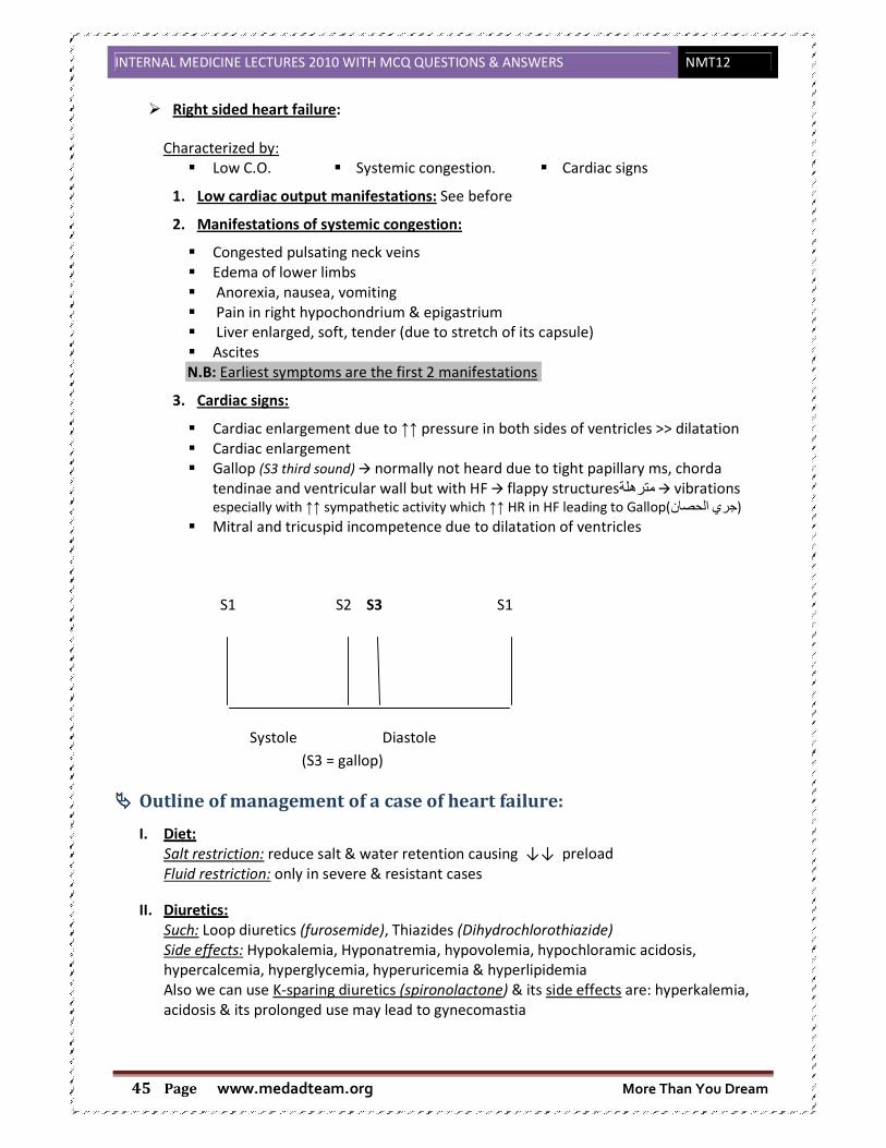
INTERNAL MEDICINE LECTURES 2010 WITH MCQ QUESTIONS & ANSWERS NMT12
www.medadteam.org More Than You Dream Page 45
� Right sided heart failure:
Characterized by:
� Low C.O. � Systemic congestion. � Cardiac signs
1. Low cardiac output manifestations: See before
2. Manifestations of systemic congestion:
� Congested pulsating neck veins
� Edema of lower limbs
� Anorexia, nausea, vomiting
� Pain in right hypochondrium & epigastrium
� Liver enlarged, soft, tender (due to stretch of its capsule)
� Ascites
N.B: Earliest symptoms are the first 2 manifestations
3. Cardiac signs:
� Cardiac enlargement due to ↑↑ pressure in both sides of ventricles >> dilatation
� Cardiac enlargement
� Gallop (S3 third sound) � normally not heard due to tight papillary ms, chorda
tendinae and ventricular wall but with HF � flappy structuresمترھلة � vibrations especially with ↑↑ sympathetic activity which ↑↑ HR in HF leading to Gallop( الحصان جري )
� Mitral and tricuspid incompetence due to dilatation of ventricles
S1 S2 S3 S1
Systole Diastole
(S3 = gallop)
���� Outline of management of a case of heart failure:
I. Diet:
Salt restriction: reduce salt & water retention causing ↓↓ preload
Fluid restriction: only in severe & resistant cases
II. Diuretics:
Such: Loop diuretics (furosemide), Thiazides (Dihydrochlorothiazide)
Side effects: Hypokalemia, Hyponatremia, hypovolemia, hypochloramic acidosis,
hypercalcemia, hyperglycemia, hyperuricemia & hyperlipidemia
Also we can use K-sparing diuretics (spironolactone) & its side effects are: hyperkalemia,
acidosis & its prolonged use may lead to gynecomastia

INTERNAL MEDICINE LECTURES 2010 WITH MCQ
www.medadteam.org Page 46
III. Vasodilators: e.g: ACE inhibitor
(Nitroglycerine & Na nitroprusside)
IV. Inotropic agents: e.g: Digitalis
decrease cardiac automaticity & conductivity so it is contraindicated in heart block
Aminophylline: Bronchodilator, vasodilator,
its side effects are irritability, insomnia, hypotension & arrhythmia
V. Treatment of acute pulmonary edema: Hospitalization - Morphine Vasodilators –
� Important Definition: Diastolic heart failure
It is a syndrome consisting of symptoms and signs of heart failure with preserved left
ventricular ejection fraction above 45
assessed by echocardiography.
There is increased stiffness in the ventricular wall and
compliance, leading to impairment of diastolic ventricular filling and hence decreased
cardiac output. Diastolic heart failure is more common in elderly hypertensive patients
but may occur with primary cardiomyopathies
infiltrative).
CTURES 2010 WITH MCQ QUESTIONS & ANSWERS
www.medadteam.org
e.g: ACE inhibitor (Captopril) , Alpha blockers (Prazocin) & Direct vasodilators
(Nitroglycerine & Na nitroprusside)
Digitalis which increase cardiac contractility & excitability and
decrease cardiac automaticity & conductivity so it is contraindicated in heart block
Bronchodilator, vasodilator, positive inotropic & has mild diuretic effect &
its side effects are irritability, insomnia, hypotension & arrhythmia
Treatment of acute pulmonary edema: Hospitalization - O2 inhalation - Frusemide Amiophylline - Positive inotropics
Diastolic heart failure
is a syndrome consisting of symptoms and signs of heart failure with preserved left
ventricular ejection fraction above 45–50% and abnormal left ventricular relaxation
assessed by echocardiography.
There is increased stiffness in the ventricular wall and decreased left ventricular
compliance, leading to impairment of diastolic ventricular filling and hence decreased
cardiac output. Diastolic heart failure is more common in elderly hypertensive patients
r with primary cardiomyopathies (hypertrophic, restrictive, and
NMT12
& Direct vasodilators
which increase cardiac contractility & excitability and
decrease cardiac automaticity & conductivity so it is contraindicated in heart block
positive inotropic & has mild diuretic effect &
Frusemide
is a syndrome consisting of symptoms and signs of heart failure with preserved left
50% and abnormal left ventricular relaxation
decreased left ventricular
compliance, leading to impairment of diastolic ventricular filling and hence decreased
cardiac output. Diastolic heart failure is more common in elderly hypertensive patients
restrictive, and

INTERNAL MEDICINE LECTURES 2010 WITH MCQ
www.medadteam.org Page 47
���� Definition:
Persistent elevation of blood pressure above normal v
diagnosed as hypertension
N.B.: for diagnosis reading must be repeated 2
The patient should be informed that a single elevated reading does not constitute a diagnosis
of HTN but is a sign for further observation
���� Classification
���� Etiology :
1. Essential: > 95 % No definite cause
2. Secondary: < 5 % Due to other causes
3. White Coat hypertension or office hypertension: HTN upon visiting the doctor or
hospital admission
A.) Essential hypertension: 95%
1. Genetic factors: Blood pressure tends to run in families.
2. Environmental factors:
i. Obesity: Fat people have higher blood pressures than thin people.
1. Define normal BP and hypertension.2. Describe the possible mechanisms causing primary (essential) hypertension. 3. List the causes of secondary hypertension. 4. Assess patients with hypertension for end organ damage5. Recognize the updated Guidelines for the treatment of hypertension6. Explain management of patients with hypertensive emergencies
CTURES 2010 WITH MCQ QUESTIONS & ANSWERS
www.medadteam.org
7. Hypertension
Persistent elevation of blood pressure above normal value bl.pressure more than 140/90
for diagnosis reading must be repeated 2-3 times at subsequent visits.
The patient should be informed that a single elevated reading does not constitute a diagnosis
further observation
No definite cause
Due to other causes
White Coat hypertension or office hypertension: HTN upon visiting the doctor or
95% no definite cause but it has a multifactorial aetiology
Blood pressure tends to run in families.
Fat people have higher blood pressures than thin people.
Lectures objectives Define normal BP and hypertension. Describe the possible mechanisms causing primary (essential)
List the causes of secondary hypertension. patients with hypertension for end organ damage
Recognize the updated Guidelines for the treatment of hypertensionExplain management of patients with hypertensive emergencies
NMT12
alue bl.pressure more than 140/90
The patient should be informed that a single elevated reading does not constitute a diagnosis
White Coat hypertension or office hypertension: HTN upon visiting the doctor or
aetiology
Describe the possible mechanisms causing primary (essential)
Recognize the updated Guidelines for the treatment of hypertension Explain management of patients with hypertensive emergencies

INTERNAL MEDICINE LECTURES 2010 WITH MCQ QUESTIONS & ANSWERS NMT12
www.medadteam.org More Than You Dream Page 48
There is a risk, however, of overestimation if the blood pressure is measured with
a small cuff. Sleep disordered breathing often seen with obesity may be an
additional risk factor.
ii. Alcohol intake: Most studies have shown a close relationship between the
consumption of alcohol and blood pressure level.
iii. Stress: uncertain.
3. Humoral mechanisms:
The autonomic nervous system, as well as the renin - angiotensin, natriuretic peptide
and kallikrein - kinin system, plays a role in the physiological regulation of short-term
changes in blood pressure and have been implicated in the pathogenesis of essential
hypertension.
• A low renin, salt –sensitive individuals, essential hypertension in which patients have
renal sodium and water retention has been described.
• Sympathetic over activity.
4. Increased sodium intake: Populations with higher sodium intake have higher average
blood pressures than those with lower intake
5. Decreased potassium &calcium intake: There is some evidence that a high - potassium
diet can protect against the effects of a high sodium intake.
6. Insulin resistance: An association between diabetes and hypertension has long been
recognized and a syndrome has been described of hyperinsulinaemia, glucose
intolerance, reduced levels of HDL cholesterol, hypertriglyceridaemia and central
obesity (all of which are related to insulin resistance) in association with hypertension.
This association (also called the 'metabolic syndrome') is a major risk factor for
cardiovascular disease
7. Imbalance: between endothelial V.D and V.C agents
B.) Secondary hypertension: less than 5% (Curable causes)
1. Renal :
i. Parenchymal disease: polycystic kidney ,chronic nephritis glomerulonephritis
ii. Renovascular disease: Renal artery stenosis , collagen vascular disease
2. Endocrinal causes:
"Hyperfunction of all endocrine glands"
a. 1ry aldosteronism
b. Cushing syndrome
c. Pheochromocytoma
d. Congenital adrenal hyperplasia
e. Acromealy
f. Thyrotoxicosis
3. Pregnancy Induced Hypertension .
4. Coarctation of aorta.
5. Increased intracranial tension.
6. Drugs : "oral contraceptive pills, NSAIDs, corticosteroids, sympathommimetics"
N.B.:
INTERNAL MEDICINE LECTURES 2010 WITH MCQ QUESTIONS & ANSWERS NMT12
www.medadteam.org More Than You Dream Page 49
a. Licorice سوس العرق also increases blood pressure
b. Arteriovenous shunt : Increase systolic pressure
c. Paget’s disease "hyperkinetic circulation"
d. Beri Beri
���� Diagnosis :
A. Baseline arterial blood pressure measurement
B. Screen 2ry HTN
C. Identify other risk factor
D. Degree of end organ damage
� History:
1. Duration of HTN
2. Indication of 2ry
HTN:
- Patient younger than 30 years old
- Older patient develop new onset Hypertension
- Inadequate drug therapy
3. Risk factors:
- Family history of cardiovascular diseases, DM, Dyslipidemia
- Dietary habits
- Snoring and Obstructive sleep apnea
- Smoking
- D.M.
4. Organ damage (complications of hypertension)
- Brain : Headache , vertigo, impaired vision , transient ischemic attack (TIA), Sensory
& motor affection
- Heart : Palpitation ,chest pain ,shortness of breath ,edema at ankle
- Kidnery: Polyuria
- Peripheral arteries : Cold extremities
� Examination :
1. Blood pressure measurement
- Standard BP measurement: under precautions ( standing and lying down)
- Ambulatory BP measurement :
Indications:
o Paroxysmal pheochromocytoma
o White coat HTN
o To assess the effect of drugs
- Home BP:
To assess the effect of drug all through 24 hours and for adjustment of the dose
2. Features of Cushing syndrome
3. Neurofibromatosis (café au lait skin patches)
4. Palpation of enlarged kidney
5. Auscultation of abdominal murmur ( renal artery stenosis)
6. Auscultation of heart murmur
7. Delayed or reduced femoral pulse (coarctation of aorta)

INTERNAL MEDICINE LECTURES 2010 WITH MCQ QUESTIONS & ANSWERS NMT12
www.medadteam.org More Than You Dream Page 50
���� Investigations:
• Fasting blood glucose
• Lipid profile: Serum cholesterol ,LDL,HDL,fastig serum triglycerides
• Serum creatinine and creatinine clearance
• Serum uric acid
• Serum K+
• Urine analysis
• Hemoglobin ,hematocrite value
• Other Recommended invsetigtions:
o Echo TIA
o Carotid ultrasound
o ECG
o Glucose tolerance test FBG >100
o Search for causes of 2ry HTN when suspected
o 24h ambulatory BP monitoring
• Searching for organ damage:
o Heart ECG, Echo
o Bl. Vessels Doppler Ultrasound
o Kidney micro albuminuria (urine test)
o Fundus examination Hge. ,papilledama ( retinopathy)
o Brain MRI &CT (silent brain infarction)
���� Treatment:
1. Life style change (applied to all patients): Smoking cessation, weight reduction, decrease salt intake & decrease saturated fat & total fat
intake, increase fruits & vegetables intake (rich in potassium), ++physical activity, -- alcohol intake .
2. Drugs:
a. Recommendations:
- Previous favorable or unfavorable experience of the patient for certain
treatment or drug
- Effect of drug in CVS
- Subclinical organ damage
- Cost of drug
- Presence of disorder or disease ( B- blockers are Contra indicated in BA )
- Drug interactions
- Side effect of the drug
- Lowering effect should last 24 hours( lifelong therapy – for better compliance)
عشان جرعات من بدل واحد قرص ياخد العيان كتيرة
b. Drug groups :
- Thiazide diuretics.
- Angiotensin 2 receptor blockers (ARBS)
- B-Blockers (BB)
- Direct vasodilators
- Calcium channel blocker

INTERNAL MEDICINE LECTURES 2010 WITH MCQ QUESTIONS & ANSWERS NMT12
www.medadteam.org More Than You Dream Page 51
- α blockers
- Angiotensin converting enzyme inhibitors (ACEI)
- Centrally acting drugs(Reserpine)
c. Preferred drugs:
- In HTN associated with Heart failure >> give Diuretics & BB, antialdosterone
(spironolcatone).
- In HTN associated with Left ventricular hypertrophy: ACEI, ARBs, CCB (calcium
channel blockers)
- In HTN associated with Myocardial infarction >> give BB, ACEI, ARBS
- In HTN associated with Angina pectoris: BB, CCB
- In HTN associated with peripheral artery disease: CCB ( BB are CI)
- In HTN associated with Diabetes >> give ARBS, ACEI
- In HTN associated with Chronic kidney disease >> give BB & ARBS
- In HTN associated with Asthma >> give diuretics ( BB are CI )
- In HTN associated with Atrial fibrillation >> give BB
- Previous stroke: Any drug lowering BP
- Atherosclerosis CCB , ACEI
- Angina BB,CCB
- Isolated systole HTN Diuretics, CCB
- Pregnancy : alpha methyl dopa
- Blacks: Diuretics
d. Side effects:
- Thiazide Diuretics :
o Skin rash, muscle cramps
o Ototoxicity
o Gynecomastia
o Metabolic disorders: hyponatremia, hypokalemia, hyperglycemia &
hypercholesterolemia, hypomagnisemia
- Beta Blockers:
o Fatigue & impotence
o Insomnia & depression
o Dyslipidemia
o Bronchospasm & cold extremities
o Heart block
o Mask symptoms of hypoglycemia( in diabetics)
- Angiotensin 2 receptor blockers (ARBs) :
o Hyperkalemia
o Skin rash
o Leucopenia
o Deterioration of kidney function if bilateral renal artery stenosis is present
o Cough

INTERNAL MEDICINE LECTURES 2010 WITH MCQ QUESTIONS & ANSWERS NMT12
www.medadteam.org Page 52
o Altered taste
- ACEI: as ARBS
- Calcium channel blockers
o Flushing
o Headache
o Palpitations - reflecting the reflex tachycardia in response to vasodilatation.
o ankle oedema
o dilatation of the precapillary "sphincters" NOT generally a reflection of
worsening heart failure!
o May worsen heart failure due to their negative inotropic effect
�
�
Hypertensive Crisis
���� Diastolic B.P. > 120 - 130 mmHg.
���� With or without target organ damage
���� Approach to patient:
� History taking: Stress on symptoms of organ damage brain ,heart ,kidney
� Examination: Mental status ,Fundal examination ,Evaluation of Lt. ventricle size, peripheral
vessels
� Investigations: Chest X-ray ,Urine analysis ,Kidney function test
� TTT :
1. Clonidine (oral ) Urgency
2. Sodium nitroprusside, nitroglycerin ,esmolol ,hydralazine ,labetalol (I.V. infusion)
Emergency
N.B.:
- Don’t use diuretics unless there is volume overload
H.F. يف لو "إ Diuretics ـال عن نبعد يستحسن
- NEVER sublingual Nifidipine (severe hypotension).

INTERNAL MEDICINE LECTURES 2010 WITH MCQ QUESTIONS & ANSWERS NMT12
www.medadteam.org More Than You Dream Page 53
Aim of TTT :
- Is to reduce of BP to 150-160/100-110 mmHg
it is unwise to reduce the blood pressure too rapidly since this may lead to:
stasis, thrombosis cerebral, renal, retinal or myocardial infarction
8. Hypotension
� Blood pressure is maintained at levels sufficient to permit adequate perfusion of the
capillary networks in the systemic vascular bed
���� Blood pressure control count:
� Rapid reduction of blood pressure diminishes the stimulation of pressor receptor
� When blood pressure falls → ↑↑ adrenal medullary, ADH. ACTH, rennin and aldosterone
secretion in order to control the decrease in Bl.pressure
Chronic hypotension
� Blood pressure below the normal range
� Systolic blood pressure = 90-100 is a form of hypotension
� Patients are normal, have no symptoms, may have longer life expectancy
� True chronic hypotension: Patients complain from fatigability, weakness, dizziness,
faintness & lack of concentration
���� Causes of chronic hypotension:
1. ↓↓ Cardiac output
2. ↓↓ glucocorticoids, mineralocorticoids lead to ↓↓ of intravascular & interstitial fluid
3. Malnutrition
4. Cachexia
5. Chronic bed rest
6. Neurological disturbance
Lectures objectives 1. Define Hypotension 2. List its causes 3. Define shock (circulatory failure) 4. List the main causes of shock 5. Describe pathogenesis and clinical features of each type 6. Describe the management of a case presenting with circulatory failure

INTERNAL MEDICINE LECTURES 2010 WITH MCQ QUESTIONS & ANSWERS NMT12
www.medadteam.org More Than You Dream Page 54
Neurological disorders
(A.)Idiopathic orthostatic hypotension
� Occurs in old age males
� Degeneration of central and/or peripheral autonomic nervous system structures
� Syncope or seizures may occur on standing, also there may be anhydrosis, loss of hair,
signs like mxyedema ( ↓↓ BMR "basal metabolic rate") & ↓↓ salivary secretions
(B.) Shy-Dragger syndrome
� Form of orthostatic hypotension
� Degeneration of extra-pyramidal tract & basal ganglion
���� Treatment of orthostatic hypotension:
1. Increase salt intake
2. Sympathomimetic amines
3. Full length elastic stockings طويل شراب يلبس
4.
5. No treatment for orthostatic hypotension due to neurological disorders
Circulatory Failure (Shock)
���� Definition:
Profound & wide spread reduction of the effective tissue perfusion leading to tissue hypoxia &
dying
���� Causes:
A. Hypovolemic causes
B. Cardiogenic
C. Extra-cardiac obstruction
D. Distributive
(A.) Hypovolemia
1- Hemorrhage: Trauma, GIT, retroperitoneal causes
2- Fluid depletion:
� External: dehydration or vomiting & diarrhea
� Interstitial: fluid redistribution
(B.) Cardiac
1- Myopathic: myocardial infarction, myocardial contusion & septic myocarditis
2- Mechanical: valvular (stenosis,regurge), hypertrophy & cardiomyopathy
3- Arrhythmia

INTERNAL MEDICINE LECTURES 2010 WITH MCQ QUESTIONS & ANSWERS NMT12
www.medadteam.org More Than You Dream Page 55
(C.) Extra-cardiac obstruction
1- Impaired diastolic filling:
� Direct venous obstruction: intrathoracic tumour
� Intrathoracic pressure
� Mechanical ventilation
� Asthma
� Cardiac compliance
� Impaired systolic contraction of Rt. ventricle
2- Lt. ventricle: Aortic dissection
(D.) Distributive
1- Septic.
2- Toxic shock.
3- Anaphylactic.
���� Pathophysiology:
1) Sympatho-adrenal response
2) Neuro-endocrine response
3) Compensatory mechanism
↓↓ Bl. Pressure → ↓↓ pulse → ↓↓ Bl.flow & Bl.volume → vasoreceptors &
chemoreceptors on vessels will be stimulated → sXmulate VMC "Vasomotor center" in
brain stimulation of sympathetic nervous activity → Adrenaline & Nor-adrenaline → ↑↑
heart rate & myocardial contractility → ↑↑ stroke volume → venocomplexion →
contraction of arterioles → maintain COP, maintain venous return & maintain pressure
� Mediators:
� Vaso-active materials.
� Adhesion molecules from micro-organisms & their toxic products cause vasodilatation.
� Activation of complement cascade cytokines.
� Compensatory mechanism: "Sensing mechanisms"
1) Pressure receptor: found in atrium, aortic arch, pulmonary artery & carotid.
2) Chemoreceptor: sensitive to concentration of CO2, O2.
� Cellular dysfunction:
Shock produce cellular dysfunction through:
� Cellular ischemia.
� Inflammatory mediators.
� Free radial injury.
(1) Cellular ischemia
� Anaerobic glycolysis give 2 ATP instead of 36 ATP in aerobic glycolysis for every
glucose molecule.

INTERNAL MEDICINE LECTURES 2010 WITH MCQ QUESTIONS & ANSWERS NMT12
www.medadteam.org More Than You Dream Page 56
� Intracellular acidosis due to accumulation of lactic acid.
(2) Inflammatory mediators
� Cytokines (TNF, interleukin I) produce dysfunction of transmembrane.
(3) Free radical injury
� Inactivate protein.
� Damage DNA.
���� Clinical picture:
���� Symptoms & signs:
� Dizziness, anxiety & restlessness,
� Hypothermia: due to decreased perfusion and evaporation of sweat,
� Rapid and shallow respirations: due to sympathetic nervous system stimulation and
acidosis
� Cool, clammy skin: due to vasoconstriction and stimulation of vasoconstriction
� A rapid, weak, thready pulse: due to decreased blood flow combined with tachycardia
���� Management: Immediately:
� Raise the patient legs up على العيان رجلين إرفع�شوية
� Restore delivery of O2
� Intubation and mechanical ventilation may be necessary in some cases with massive
secretions
���� Control:
� Control bleeding disorders and other causes of hypovolemic shock
� Continuous measuring Bl.pressure by using Swan Gans Catheter to estimate pulmonary
artery pressure
Myocardial Ischaemia
� Site
Jaw, retrosternal, left submammary
� Radiation
Left chest, left arm, jaw….mandible, teeth, palate
� Quality/severity
tightness, heaviness, compression…clenched fists
� Precipitating factors
physical exertion, cold windy weather, emotion
� Relieving factors
rest, sublingual nitrates

INTERNAL MEDICINE LECTURES 2010 WITH MCQ QUESTIONS & ANSWERS NMT12
www.medadteam.org More Than You Dream Page 57
� Autonomic symptoms
sweating, pallor, peripheral vasoconstriction, nausea and vomiting
���� Differential diagnosis � Cardiac pathology
Pericarditis, aortic dissection
� Cardiac pathology
Pericarditis, aortic dissection
� Pulmonary pathology
Pulmonary embolus, pneumothorax, pneumonia
� Gastrointestinal pathology
Peptic ulcer disease, reflux, pancreatitis.
� Musculoskeletal pathology
Trauma.
���� Acute Myocardial Infarction
� 250,000 deaths per year.
� 150,000 presentations to hospital.
� 30% of deaths occur in the first 2 hours.
� (Cardiac muscle death occurs after 45 mins of ischaemia)
���� Assessment
� Symptoms and signs of myocardial ischaemia
� Sudden death
� Myocardial infarction
� Stable angina pectoris
� Heart failure
� Arrhythmia
� Asymptomatic
� Acute coronary syndrome
� Also : HR , Rhythm & BP
���� Confirming the diagnosis
� Typical chest pain
� Electrocardiographic changes
� ST elevation
� new LBBB
� Myocardial enzyme elevation
� Creatine kinase (CK-MB)
� Troponin
���� Treatment
� Stop dental treatment
� Call for help
� Rest, sit up and reassure patient
� Oxygen
� Analgesia (opiate, sublingual nitrate)
� Aspirin
� Prepare for basic life support

INTERNAL MEDICINE LECTURES 2010 WITH MCQ QUESTIONS & ANSWERS NMT12
www.medadteam.org More Than You Dream Page 58
���� Medical treatment
� Rest, oxygen, analgesia, aspirin
� Thrombolysis
� Beta-Blockers
� ACE inhibitors
���� Complications
� Death (18% within 1 hour, 36% within 24 hours)
� Non-fatal arrhythmia
� Acute left ventricular failure
� Cardiogenic shock
� Papillary muscle rupture and mitral regurgitation
� Myocardial rupture and tamponade
� Ventricular aneurysm and thrombus
Cardiopulmonary resuscitation
Adult basic life support
� Check responsiveness > Open airway > Check breathing > Breathe Assess (10 sec only)
� Circulation: if No circulation > Compress chest 100 pm, 15:2
� Continue rescue breathing Check
circulation every minute
Importance :
� Ensure safety of rescuer and victim
� Check responsiveness
� Shout for help if not responsive
� Open airway
� head tilt
� chin lift
� Check breathing
� look for chest movement
� listen over mouth
� feel air on cheek
� Breathe
� pinch nose closed and open mouth using chin lift
� Assess circulation
� carotid pulse only if confident…don’t waste time
� Circulation present
� continue breathing, check each minute
�
� No circulation
� start chest compressions @ 15:2
� heal of hand over sternum, straight arms
� depress 4-5cm, 100 bpm
� continue until responsive/help/exhausted

INTERNAL MEDICINE LECTURES 2010 WITH MCQ QUESTIONS & ANSWERS NMT12
www.medadteam.org More Than You Dream Page 59
M.C.Q.
Section A: Read each question carefully and record the answer "TRUE" or "FALSE":
1. The pain of myocardial ischemia: aa)) IIss ttyyppiiccaallllyy iinndduucceedd bbyy eexxeerrcciissee aanndd rreelliieevveedd bbyy rreesstt..
b) Radiates to the neck and jaw but not teeth.
c) Rarely lasts longer than 10 seconds after resting.
d) Is easily distinguished from esophageal pain.
ee)) IInnvvaarriiaabbllyy wwoorrsseennss aass eexxeerrcciissee ccoonnttiinnuueess..
2. The pulse characteristic listed below are typical features of the following disorders: a) Pulsus bisferiens-combinfed, mitral stenosis and regurgitation.
b) Pulsus paradoxus- aortic regurgitation.
cc)) CCoollllaappssiinngg ppuullssee--sseevveerree aanneemmiiaa..
d) Pulsus altemans-extrasystoles every alternate beat.
e) Slow rising pulse-mitral stenosis.
3. The following statements about the jugular venous pressure (JVP) are true: a) The external Jugular vein is a reliable guide to right atrial pressure.
b) The JVP Is conventionally measured from the suprasternal notch.
c) The normal JVP, unlike the blood pressure, does not rise with anxiety.
d) The normal JVP does not rise on abdominal compression.
ee)) TThhee nnoorrmmaall JJVVPP ffaallllss dduurriinngg iinnssppiirraattiioonn..
4. The auscultatory findings listed below are associated with the following
phenomena: a) Third heart sound-opening of mitral valve.
bb)) VVaarryyiinngg iinntteennssiittyy ooff ffiirrsstt hheeaarrtt ssoouunndd--aattrriioovveennttrriiccuuiiaarr ddiissssoocciiaattiioonn..
c) Soft first heart sound-mitral stenosis.
dd)) RReevveerrsseedd sspplliittttiinngg ooff sseeccoonndd hheeaarrtt ssoouunndd--lleefftt bbuunnddllee bbrraanncchh lloocckk..
e) Fourth heart sound-atrial fibrillation.
5. The cardiac drugs listed below are associated with the following adverse effects:- aa)) DDiiggooxxiinn--aaccuuttee ccoonnffuussiioonnaall ssttaattee..
bb)) VVeerraappaammiill--ccoonnssttiippaattiioonn..
cc)) AAmmiiooddaarroonnee--pphhoottoosseennssiittiivviittyy..
d) Propafenone-comeal microdeposits.
ee)) LLiiggnnooccaaiinnee--ccoonnvvuullssiioonnss..
6. The pulse characteristics listed below are typical features of the following disordes:- a) pulsus bisfertens � combined mitral stenosis and regurgitation
b) pulsus paradoxus � aortic regurgitation
cc)) ccoollllaappssiinngg ppuullssee � sseevveerree aannaaeemmiiaa
d) pulsus altemans � exlrasystoles every alternate beat
e) slow rising pulse � mitral stenosis
7. The abnormalities of the Jugular venous pulse listed below are associated
with the disorders:- a) cannon waves � pulmonary hypertension.
bb)) ggiiaanntt aa wwaavveess � ttrriiccuussppiidd sstteennoossiiss..
cc)) vv--wwaavveess � ttrriiccuussppiidd rreegguurrggiittaattiioonn..
dd)) iinnssppiirraattoorryy rriissee iinn jjuugguullaarr vveennoouuss pprreessssuurree � ppeerriiccaarrddiiaall TTaammppoonnaaddee..
e) absent a waves � atrioventricular dissociation.

INTERNAL MEDICINE LECTURES 2010 WITH MCQ QUESTIONS & ANSWERS NMT12
www.medadteam.org More Than You Dream Page 60
8. The auscultatory findings listed below are associated with the following phenomena a) third heart sound � opening of mitral valve
b) varying intensity of first heart sound � atrioventricular dissociation
c) soft first heart sound � mitral stenosis
dd)) rreevveerrsseedd sspplliittttiinngg ooff sseeccoonndd hheeaarrtt ssoouunndd � lleefftt bbuunnddllee bbrraanncchh bblloocckk
e) fourth heart sound � atrial fibrillation
9. In patients with atrial fibrillation (AF) a) aspirin therapy alone does not reduce the risk of stroke.
bb)) tthhee rraaddiiaall ppuullssee iiss ttyyppiiccaallllyy iirrrreegguullaarrllyy iirrrreegguullaarr..
cc)) tthhee rreessppoonnssee iinn ccaarrddiiaacc oouuttppuutt ttoo eexxeerrcciissee iiss rreedduucceedd dduuee ttoo tthhee aabbsseennccee ooff aattrriiaall ssyyssttoollee..
d) elective direct current (DC) cardioversion is contraindicated during anticoagulant therapy.
ee)) aallccoohhooll aabbuussee sshhoouulldd bbee ccoonnssiiddeerreedd aass aa lliikkeellyy ccaauussee..
10. Digoxin: a) Shortens the refractory period of conducting tissue.
b) Usually converts atrial flutter to sinus rhythm.
cc)) aaccttss pprriimmaarriillyy oonn cceellll mmeemmbbrraannee iioonniicc ppuummppss..
d) Effects are potentiated by hyperkalemia.
ee)) IIss aa rreeccooggnniizzeedd ccaauussee ooff vveennttrriiccuullaarr aarrrrhhyytthhmmiiaass..
11. In a patient with central chest pain at rest: aa)) iinnttrraassccaappuullaarr rraaddiiaattiioonn ssuuggggeessttss tthhee ppoossssiibbiilliittyy ooff aaoorrttiicc ddiisssseeccttiioonn..
bb)) ppoossttuurraall vvaarriiaattiioonn iinn ppaaiinn ssuuggggeessttss tthhee ppoossssiibbiilliittyy ooff ppeerriiccaarrddiittiiss..
cc)) cchheesstt wwaallll tteennddeerrnneessss iiss aa ttyyppiiccaall ffeeaattuurree ooff TTiieettzzee''ss ssyynnddrroommee..
d) relief of pain by nitrates excludes an oesophagealcause.
e) features of autonomic disturbance are specific to cardiac pain.
12. In a patient with cardiogenic shock due to acute myocardial infarction: aa)) tthhee aabbsseennccee ooff ppuullmmoonnaarryy ooeeddeemmaa ssuuggggeessttss rriigghhtt vveennttrriiccuullaarr iinnffaarrccttiioonn
b) the central venous pressure is the best index of left ventricular filling pressure
cc)) ddooppaammiinnee iinn llooww ddoossee iinnccrreeaasseess rreennaall bblloooodd ffllooww
dd)) hhiigghh ffllooww,, hhiigghh ccoonncceennttrraattiioonn ooxxyyggeenn iiss iinnddiiccaatteedd
e) colloid Infusion is indicated if oliguria and pulmonary oedema develop
13. In the treatment of cardiac failure associated with acute pulmonary oedema: a) Controlled oxygen therapy should be restricted to 28% oxygen in patents who smoke.
bb)) MMoorrpphhiinnee rreedduucceess aannggoorr aanniimmii aanndd ddyyssppnnooeeaa..
cc)) FFrruusseemmiiddee tthheerraappyy ggiivveenn iinnttrraavveennoouussllyy rreedduucceess pprreellooaadd aanndd aafftteerrllooaadd..
d) Nitrates should be avoided if the systolic blood pressure <140 mmHg.
e) ACE inhibitors decrease the afterload but increase the preload.
14. In patients with significant mitral stenosis: aa)) TThhee mmiittrraall vvaallvvee oorriiffiiccee iiss rreedduucceedd ffrroomm 55 ccmm22 ttoo aabboouutt 11 ccmm
22..
b) a history of rheumatic fever or chorea is elicited in over 90% of patients.
c) left atrial enlargement cannot be detected on the chest X-ray.
d) the risk of systemic emboli is trivial In sinus rhythm.
ee)) mmiittrraall bbaalllloooonn vvaallvvuullooppllaassttyy iiss nnoott aaddvviissaabbllee iiff tthheerree iiss aallssoo ssiiggnniiffiiccaanntt mmiittrraall rreegguurrggiittaattiioonn..
15. Disorders typically producing the sudden onset of symptomatic mitral
regurgitation Include: a) Marian's syndrome
bb)) aaccuuttee mmyyooccaarrddiiaall iinnffaarrccttiioonn HHbbWW
cc)) aaccuuttee rrhheeuummaattiicc ffeevveerr
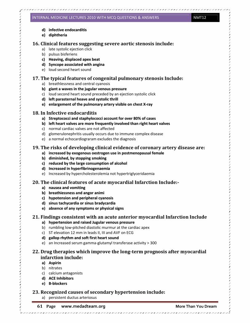
INTERNAL MEDICINE LECTURES 2010 WITH MCQ QUESTIONS & ANSWERS NMT12
www.medadteam.org More Than You Dream Page 61
dd)) iinnffeeccttiivvee eennddooccaarrddiittiiss
ee)) ddiipphhtthheerriiaa
16. Clinical features suggesting severe aortic stenosis include: a) late systolic ejection click
b) pulsus bisferiens
cc)) HHeeaavviinngg,, ddiissppllaacceedd aappeexx bbeeaatt
dd)) SSyynnccooppee aassssoocciiaatteedd wwiitthh aannggiinnaa
e) loud second heart sound
17. The typical features of congenital pulmonary stenosis Include: a) breathlessness and central cyanosis
bb)) ggiiaanntt aa wwaavveess iinn tthhee jjuugguullaarr vveennoouuss pprreessssuurree
c) loud second heart sound preceded by an ejection systolic click
dd)) lleefftt ppaarraasstteerrnnaall hheeaavvee aanndd ssyyssttoolliicc tthhrriillll
ee)) eennllaarrggeemmeenntt ooff tthhee ppuullmmoonnaarryy aarrtteerryy vviissiibbllee oonn cchheesstt XX--rraayy
18. In Infective endocarditis aa)) SSttrreeppttooccooccccii aanndd ssttaapphhyyllooccooccccii aaccccoouunntt ffoorr oovveerr 8800%% ooff ccaasseess
bb)) lleefftt hheeaarrtt vvaallvveess aarree mmoorree ffrreeqquueennttllyy iinnvvoollvveedd tthhaann rriigghhtt hheeaarrtt vvaallvveess
c) normal cardiac valves are not affected
d) glomerulonephritis usually occurs due to immune complex disease
e) a normal echocardiograram excludes the diagnosis
19. The risks of developing clinical evidence of coronary artery disease are: aa)) iinnccrreeaasseedd bbyy eexxooggeennoouuss ooeessttrrooggeenn uussee iinn ppoossttmmeennooppaauussaall ffeemmaallee
bb)) ddiimmiinniisshheedd,, bbyy ssttooppppiinngg ssmmookkiinngg
cc)) rreedduucceedd bbyy tthhee llaarrggee ccoonnssuummppttiioonn ooff aallccoohhooll
dd)) IInnccrreeaasseedd iinn hhyyppeerrffiibbrriinnooggeennaaeemmiiaa
e) Increased by hypercholesterolemia not hypertriglyceridaemia
20. The clinical features of acute myocardial Infarction Include:- aa)) nnaauusseeaa aanndd vvoommiittiinngg
bb)) bbrreeaatthhiieessssnneessss aanndd aannggoorr aanniimmii
cc)) hhyyppootteennssiioonn aanndd ppeerriipphheerraall ccyyaannoossiiss
dd)) ssiinnuuss ttaacchhyyccaarrddiiaa oorr ssiinnuuss bbrraaddyyccaarrddiiaa
ee)) aabbsseennccee ooff aannyy ssyymmppttoommss oorr pphhyyssiiccaall ssiiggnnss
21. Findings consistent with an acute anterior myocardial Infarction Include aa)) hhyyppeerrtteennssiioonn aanndd rraaiisseedd JJuugguullaarr vveennoouuss pprreessssuurree
b) rumbling low-pitched diastolic murmur at the cardiac apex
c) ST elevation 12 mm in leads II, III and AVF on ECG
dd)) ggaalllloopp rrhhyytthhmm aanndd ssoofftt ffiirrsstt hheeaarrtt ssoouunndd
e) an Increased serum gamma-glutamyl transferase activity > 300
22. Drug therapies which improve the long-term prognosis after myocardial
infarction include: aa)) AAssppiirriinn
b) nitrates
c) calcium antagonists
dd)) AACCEE IInnhhiibbiittoorrss
e) BB--bblloocckkeerrss
23. Recognized causes of secondary hypertension include: a) persistent ductus arteriosus

INTERNAL MEDICINE LECTURES 2010 WITH MCQ QUESTIONS & ANSWERS NMT12
www.medadteam.org More Than You Dream Page 62
bb)) pprriimmaarryy hhyyppeerraallddoosstteerroonniissmm
cc)) aaccrroommeeggaallyy
dd)) ooeessttrrooggeenn--ccoonnttaaiinniinngg oorraall ccoonnttrraacceeppttiivveess
ee)) tthhyyrroottooxxiiccoossiiss
24. Complications of systemic hypertension include:- a) Retinal microaneurysms
bb)) DDiisssseeccttiinngg aanneeuurryyssmm ooff tthhee aasscceennddiinngg aaoorrttaa
cc)) RReennaall aarrtteerryy sstteennoossiiss
dd)) llaaccuunnaarr ssttrrookkeess ooff tthhee iinntteerrnnaall ccaappssuullee
e) subdural haemorrhage
25. Accelerated phase or malignant hypertension Is suggested by hypertension and a) a loud second heart sound
b) a heaving apex beat
cc)) HHeeaaddaacchhee
dd)) rreettiinnaall ssoofftt eexxuuddaatteess oorr hhaaeemmoorrrrhhaaggeess
ee)) rreennaall oorr ccaarrddiiaacc ffaaiilluurree
26. Important explanations for hypertension refractory to medical therapy Include:- aa)) ppoooorr ccoommpplliiaannccee wwiitthh ddrruugg tthheerraappyy..
bb)) iinnaaddeeqquuaattee ddrruugg tthheerraappyy..
cc)) PPhhaaeeoocchhrroommooccyyttoommaa ..
dd)) pprriimmaarryy hhyyppeerraallddoosstteerroonniissmm..
ee)) rreennaall aarrtteerryy sstteennoossiiss..
27. The following statements about shock syndromes are correct a) In severe hypovolamia, a source of fluid loss is invariably apparent clinically
b) In cardiogenic shock, the peripheries are characteristically warm
c) AA mmaassssiivvee ppuullmmoonnaarryy eemmbboolliissmm ttyyppiiccaallllyy pprreesseennttss wwiitthh sshhoocckk
d) Anaphylactic shock is associated with profound allergen-induced systemic vasoconstriction
ee)) AArrtteerriioovveennoouuss sshhuunnttiinngg iiss aa ssiiggnniiffiiccaanntt ccoonnttrriibbuuttoorryy ffaaccttoorr iinn sseeppttiicc sshhoocckk
28. Typically clinical features of acute circulatory failure of anaphylactic shock include a) Elevated jugular venous pressure
b) Warm dry skin
cc)) SSttrriiddoorr
dd)) CCoonnffuussiioonn
e) Polyuria
29. Acute circulatory failure with an elevated central venous pressure are typical
finding in: a) Acute pancreatitis
bb)) MMaassssiivvee ppuullmmoonnaarryy eemmbboolliissmm
c) Rupture ectopic pregnancy
dd)) AAccuuttee rriigghhtt vveennttrriiccuullaarr iinnffaarrccttiioonn
ee)) PPeerriiccaarrddiiaall ttaammppoonnaaddee
30. In a patient suspected with septic shock: a) The lower urinary tract is the commonest cause of infection
b) A normal transthoracic electrocardiogram exclude endocarditis
c) Intravenous access sites need only be changed if cutaneous evidence of infarction is visible
dd)) PPrriioorr ttrreeaattmmeenntt wwiitthh hhiissttaammiinnee rreecceeppttoorr aannttaaggoonniisstt mmaakkeess ppnneeuummoonniiaa aa mmoorree lliikkeellyy ccaauussee
e) CCoorrttiiccoosstteerrooiidd tthheerraappyy iiss ooff nnoo pprroovveenn bbeenneeffiitt
INTERNAL MEDICINE LECTURES 2010 WITH MCQ QUESTIONS & ANSWERS NMT12
www.medadteam.org More Than You Dream Page 63
31. The expected effect of the following vasoactive drugs include aa)) SSooddiiuumm nniittrroopprruussssiiddee--rreedduuccttiioonn iinn ssyysstteemmiicc vvaassccuullaarr rreessiisstteennccee
b) Prostacyclin- increase pulmonary vascular resistence
cc)) IIssoopprreennaalliinnee-- ssiinnuuss ttaacchhyyccaarrddiiaa
d) Dopamine- sinus bradycardia
e) Adrenaline- increase splanchnic blood
32. Echocardiography is the most sensitive method of diagnosing aa)) PPrreesseennccee aanndd ddeeggrreeee ooff mmiittrraall sstteennoossiiss
b) Evaluation of left ventricular function
cc)) DDeetteeccttiioonn ooff vvaallvvuullaarr vveeggeettaattiioonnss
dd)) DDeetteeccttiioonn ooff ppeerriiccaarrddiiaall eeffffuussiioonn
e) Assessing the degree of coronary stenosis
SECTION B: only one item is appropriately applies to the statement.
33. All the following signs BUT one are suggestive of left ventricular hypertrophy aa)) SSllaappppiinngg aappeexx
b) Apical displacement downward and outward
c) Hyperkinetic apex
d) Heaving apex
e) Localized apex
34. Percussion of the heart may be useful in the diagnosis of the following
condition, except: a) Pulmonary hypertension
bb)) AAnnggiinnaa ppeeccttoorriiss
c) Pericardial effusion
d) Right atrial enlargement
e) Aortic aneurysm
35. Splitting of the second heart sound occur in a) Mitral incompetence
b) Left bundle branch block
c) Sever aortic stenosis
d) Atrial septal defect
ee)) AAllll ooff tthhee aabboovvee
36. An abnormal early diastolic sound heard at the apex and lower sternal
border can be: a) Loud P2
b) S3 gallop
c) Opening snap
d) All of the above
ee)) NNoonn ooff tthhee aabboovvee
37. Graham Steell murmur is: aa)) AAnn eeaarrllyy ddiiaassttoolliicc mmuurrmmuurr
b) A pansystolic murmur
c) An Austin Flint murmur
d) A subvulvular murmur
e) A pericardial murmur

INTERNAL MEDICINE LECTURES 2010 WITH MCQ QUESTIONS & ANSWERS NMT12
www.medadteam.org More Than You Dream Page 64
38. In sever mitral stenosis, the following occur except a) Pulmonary hypertension
bb)) WWiiddeerr AA22-- OOSS ttiimmee iinntteerrvvaall
c) Long mid diastolic murmur
d) Low cardiac output
e) Valve area less than approximately 1.0 cm2/m2 body surface area
39. In aortic regurgitation, the following occur except a) Quinck's sign
b) Duroziez sign
cc)) TTrraauubbee''ss aarreeaa
d) Corrigan's sign
e) Austin Flint murmur
40. Diastolic heart failure is characterized by all the following, except: aa)) PPoooorr vveennttrriiccuullaarr ccoonnttrraaccttiilliittyy
b) Impaired compllance of the ventricle
c) High pulmonary venous pressure
d) High EDP
41. ACEI IS regarded the first line of treatment in heart failure because it: a) Decreases preload
bb)) DDeeccrreeaasseedd bbootthh pprree aanndd aafftteerr llooaadd
c) Increased contractility
d) Potent loop diuretic
42. ACEI are contraindicated in: aa)) BBiillaatteerraall rreennaall aarrtteerryy sstteennoossiiss
b) Hypokalemia
c) Ejection fraction <40%
d) Diastolic heart failure
43. Beta blockers in heart failure a) Absolutely contraindicated in CHF
b) Prescribed only in patient with IV heart failure
cc)) CCaann bbee pprreessccrriibbeedd wwiitthh AACCIISS iinn ccllaassss IIII aanndd IIIIII hheeaarrtt ffaaiilluurree
d) Safe and beneficial in corpulmonal with respiratory failure

INTERNAL MEDICINE LECTURES 2010 WITH MCQ QUESTIONS & ANSWERS NMT12
www.medadteam.org More Than You Dream Page 65
1. Clinical presentations of chest diseases
���� Chest diseases: diseases that affect respiratory system (below the larynx)
Airway parenchyma of the lung covers of the lung (pleura)
���� Airway diseases: from trachea downward to terminal bronchioles
• Trauma (physical, chemical)
• Irritation
• Allergy (bronchial asthma)
• Infection
• neoplasm
���� Parenchymal diseases:
• Trauma (stab)
• Inflammation Infectious: bacterial, viral, fungal
Non infectious: Immune (e.g. systemic lupus)
• Neoplasm(alveolar carcinoma)
���� Pleura diseases:
• Trauma (blunt→spasm-respiratory distress,
stab→ Pnuemothorax)
• Inflammation (Infection: Familial mediterain fever→ Acute Inflammation of the
serous membranes
(not infection :immune reaction)
Lectures objectives
1. Describe the underlying pathogenic mechanisms of main presentations of chest diseases.
2. List the causes of dyspnea (acute and chronic). 3. Describe different grades of dyspnea. 4. Explain the mechanism of chest pain. 5. List the different causes of chest pain. 6. List the causes of cough 7. List the causes of haemoptysis

INTERNAL MEDICINE LECTURES 2010 WITH MCQ
www.medadteam.org Page 66
• Hydrothorax
N.B.: Starling's law:
Generalized ansarca (Hydrothorax, Ascites, Edema L.L.)
• Pnuemothorax
• Hydropneumothorax
• Neoplasm(mesothelioma
���� Cardinal chest symptoms:
• Cough.
• Expectoration.
• Hemoptysis.
• Dyspnea.
• Asthmatic attacks & wheezing of chest
• Chest pain.
���� Minor chest symptoms:
• Toxemia (e.g.TB.)
• Mediastinal compres
• Respirtory failure (Hypoxia, Hypercapnia)
� Hypoxia: Irritable, Cyanosed, Dyspn
� Hypercapnia: very drowsy, headache, flapping
• Corpulmonale (2ry to pulmonary hypertension )
• Jaundice
� Hepatic congestion
� Lobar pneumonia
• Cyanosis
Reflex
CTURES 2010 WITH MCQ QUESTIONS & ANSWERS
www.medadteam.org
exudates
transudes liver cirrhosis, nephritic syndrome
s law: ↓↓ osmotic pressure � ↑↑ permeability from capillary
Generalized ansarca (Hydrothorax, Ascites, Edema L.L.)
Pnuemothorax stab (Open pnuemothorax)
Rupture of emphysematous bulla (emphysema, COPD)
(Tension pneumothorax)
Hydropneumothorax Suction of pleural fluid by wrong technique
Gas forming organisms
mesothelioma)
Cardinal chest symptoms:
Asthmatic attacks & wheezing of chest.
Mediastinal compression (↑↑ intrathoracic pressure)
(Hypoxia, Hypercapnia)
: Irritable, Cyanosed, Dyspnea
: very drowsy, headache, flapping
(2ry to pulmonary hypertension )
Hepatic congestion Cholestatic jaundice
Lobar pneumonia (heamolysis) Haemolytic jaundice
1. Cough
Psychogenic
(Hysterical)
NMT12
nephritic syndrome
permeability from capillary �
Rupture of emphysematous bulla (emphysema, COPD)
Suction of pleural fluid by wrong technique
Haemolytic jaundice
Central

INTERNAL MEDICINE LECTURES 2010 WITH MCQ QUESTIONS & ANSWERS NMT12
www.medadteam.org More Than You Dream Page 67
Etiology:
1. Respiratory
• Airway dis. : e.g. Bronchitis
• Parenchymatous lung dis.: e.g. Lung abscess
• Pleural dis.: e.g. Pleurisy
2. Extra-respiratory
• Cardiovascular dis. : e.g. Pulm congestion (LSHF)
• Other causes: e.g. ACE – I
APPROACH TO THE PATIENT
1. Is the cough ACUTE or CHRONIC ??
ACUTE (< 3 weeks): e.g. Pneumonia, Pulmonary embolism.
CHRONIC (> 3 weeks): e.g. COPD, GORD.
2. Is the cough DRY or PRODUCTIVE ??
DRY: e.g. Pleurisy,Psychogenic, DRUG (ACE – I).
PRODUCTIVE: e.g. Chronic bronchitis, Bronchiectasis, Lung abscess.
3. Is there PERIODICITY ?? (DIURNAL or SEASONAL)
DIURNAL
(Early morning): e.g. Bronchitis, Br. asthma, Bronchiectasis.
(Nocturnal): e.g. Cardiac asthma.
SEASONAL (winter): e.g. Bronchitis, Br. asthma, Bronchiectasis.
4. Is there associated WHEEZING ??
GENERALISED: e.g. Bronchitis, Br. asthma, Bronchiectasis, COPD.
LOCALISED: e.g. Br. carcinoma.
5. Is the patient taking an ACE – I ??
Types of Cough:
• Pharyngeal Pharyngitis
• Laryngeal Laryngitis
• Tracheal Tracheaitis
• CVS
• Encephalitis
• Brain tumours
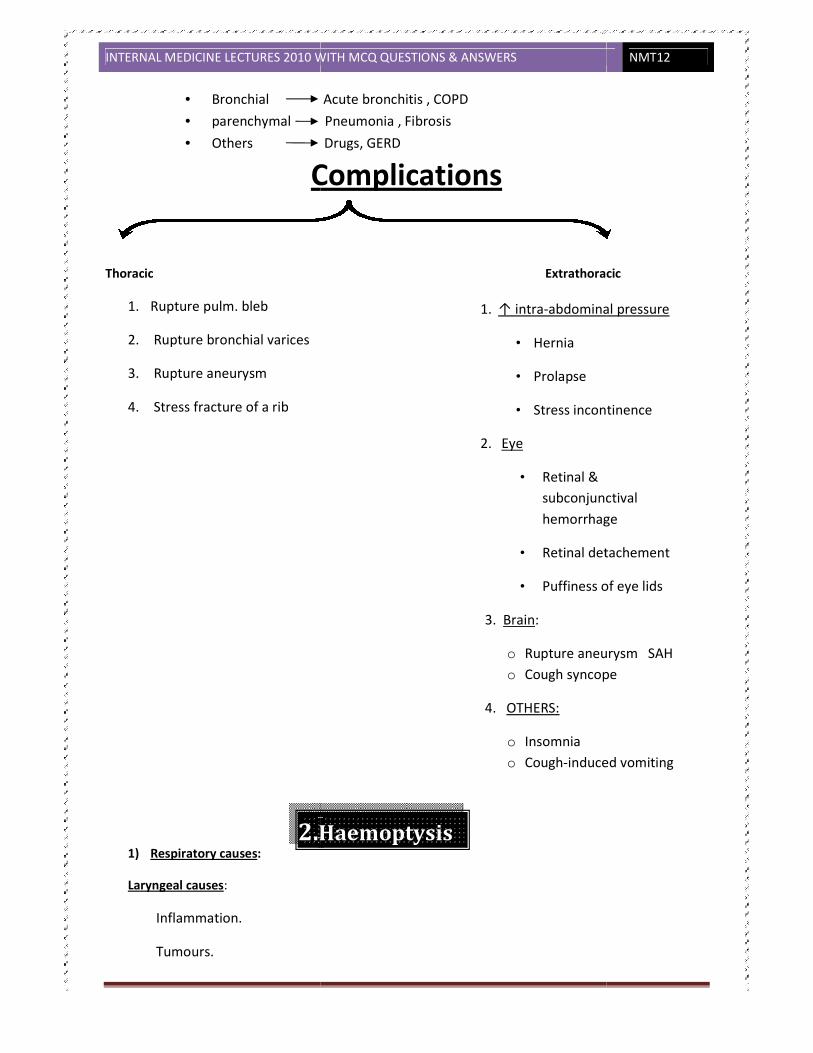
INTERNAL MEDICINE LECTURES 2010 WITH MCQ
www.medadteam.org Page 68
• Bronchial Acute bronchitis , COPD
• parenchymal Pneumonia , Fibrosis
• Others Drugs, GERD
Complications
Thoracic
1. Rupture pulm. bleb
2. Rupture bronchial varices
3. Rupture aneurysm
4. Stress fracture of a rib
1) Respiratory causes:
Laryngeal causes:
Inflammation.
Tumours.
2.Haemoptysis
CTURES 2010 WITH MCQ QUESTIONS & ANSWERS
www.medadteam.org
Bronchial Acute bronchitis , COPD
parenchymal Pneumonia , Fibrosis
Others Drugs, GERD
Complications
Thoracic Extrathoracic
1. ↑ intra-abdominal pressure
• Hernia
• Prolapse
• Stress incontinence
2. Eye
• Retinal &
subconjunctival
hemorrhage
• Retinal detachement
• Puffiness of eye lids
3. Brain:
o Rupture aneurysm SAH
o Cough syncope
4. OTHERS:
o Insomnia o Cough-induced vomiting
Haemoptysis
NMT12
Extrathoracic
abdominal pressure
Stress incontinence
subconjunctival
hemorrhage
Retinal detachement
Puffiness of eye lids
Rupture aneurysm SAH Cough syncope
induced vomiting

INTERNAL MEDICINE LECTURES 2010 WITH MCQ QUESTIONS & ANSWERS NMT12
www.medadteam.org More Than You Dream Page 69
Tracheo-bronchial causes:
Acute Bronchitis. (the most common cause)
Bronchiectasis.
Bronchial carcinoma.
Bronchial adenoma.
Pulmonary causes:
Pulmonary TB.
Pulmonary embolism.
Pneumonia.
Lung abscess.
Goodpasture syndrome.
2) Cardiovascular causes:
Pulmonary congestion, e.g. MS & acute pulm oedema
Pulmonary infection.
Pulmonary infarction.
Rupture of bronchial varices.
Rupture of aortic aneurysm.
3) Systemic diseases:
Hemorrhagic blood diseases, e.g.
Hemophilia,Purpura.

INTERNAL MEDICINE LECTURES 2010 WITH MCQ QUESTIONS & ANSWERS NMT12
www.medadteam.org More Than You Dream Page 70
MANAGEMENT OF HEMOPTYSIS
I. Diagnosis:
1. Differentiation between hemoptysis & hematemesis:
Hemoptysis Hematemesis
Past history
Chest disease GIT disease
The attack Cough Vomiting
Blood Bright red, frothy Dark red, food particles
After the attack Streaked sputum Melena
Examination
Chest signs Abdominal signs
Investigations Chest or heart disease GIT disease
2 Exclusion of false hemoptysis:
Examination of the upper resoiratory tract usually reveals the cause.
3 Detection of the cause of hemoptysis:
Clinical picture Investigations
I I.Treatment:
1. Treatment of the cause.
2. Cases of massive hemoptysis require:
Hospitalization.
Sedation.
Blood transfusion & anti-shock measures

INTERNAL MEDICINE LECTURES 2010 WITH MCQ QUESTIONS & ANSWERS NMT12
www.medadteam.org More Than You Dream Page 71
Etiology:
Cardiovascular causes:
o Myocardium: CAD (angina or infarction). o Pericardium: Pericarditis or pericardial effusion. o Endocardium: Mitral valve prolapse. o Aorta Aortic aneurysm & Aortic dissection. o Pulmonary: Pulmonary embolism & Pulmonary infarction. o Pain of cardiac neurosis. o Huge cardiomegaly.
Respiratory causes:
o Pleural disease: pleurisy, pleural effusion, pneumothorax, hydropneumothorax. o Pulmonary disease extending to the pleura , e.g. pneumonia & lung abscess. o Acute massive lung collapse.
Chest wall causes:
o Skin: wounds. o Breast: mastitis, tumour. o Ribs:osteomyelitis,tumour,fracture,Tietze syndrome. o Intercostal muscles: myositis, muscle strain (from severe cough). o Intercostal nerves: nerve root pain (herpes zoster).
Mediastinal causes:
Tumours, LN ,Mediastinitis.
GIT:
GORD, peptic ulcer ,cholecystitis.
CAUSES OF ACUTE CHEST PAIN:
o MYOCARDIUM: angina pectoris & acute myocardial infarction. o PERICARDIUM: acute pericarditis. o ENDOCARDIUM: mitral valve prolapse. o AORTA: dissecting aneurysm of the aorta. o PULMONARY: massive pulmonary embolism & pulmonary infarction. o CARDIAC NEUROSIS. o Pleural disease: acute pleurisy & pneumothorax. o Lung disease: acute massive lung collapse. o Musculo-skeletal disorders: Tietze syndrome.
3.Chest Pain

INTERNAL MEDICINE LECTURES 2010 WITH MCQ QUESTIONS & ANSWERS NMT12
www.medadteam.org More Than You Dream Page 72
o Nerve root pain: Herpetic neuralgia. o GIT: GORD, peptic ulcer, cholecystitis.
Etiology:
o Cardiovascular causes:
o Pulmonary congestion: Left-sided heart failure.
o Pulmonary embolism.
o Pericardial effusion.
Pathogenesis of cardiac dyspnea
1) Pulm congestion → ↓ alveolar compliance due to intersXXal oedema.
2) Low CO → faXgue & weakness of the respiratory muscles. 3) Pleural effusion → mechanical compression of the lungs. 4) Churchill Cope reflex. 5) Hypoxia → stimulates the respiratory centre.
Types of Cardiac Dyspnea:
o Exertional
o Dyspnea at rest o Orthopnea o PND
Chest causes:
Laryngeal: - Foreign body, Tumours. Tracheo-bronchial: - Bronchitis, Br. asthma, Bronchiectasis, COPD.
Lung: - Consolidation, Collapse. - Fibrosis (pulmonary, ILD). Pleural: - Pleurisy, pleural effusion, pneumothorax, hydropneumothorax. Chest wall: - Chest deformities, scleroderma, marked obesity (Pick syndrome).
Abdominal causes:
- Abdominal distension, e.g. marked ascites.
4.Dyspnea

INTERNAL MEDICINE LECTURES 2010 WITH MCQ QUESTIONS & ANSWERS NMT12
www.medadteam.org More Than You Dream Page 73
General causes:
- Anemia.
- Hemorrhage & shock.
- Acidosis.
Psychogenic (Hysterical)
Neurological causes:
1- Diaphragmatic paralysis.
2- Disorders of Neuro-muscular apparatus:
CNS: Head injury, CVS, Drugs (opiates & barbiturates).
AHCs: MND, Poliomyelitis.
Peripheral nerve: Polyneuropathy, e.g.GBS.
NMJ: Myasthenia gravis.
Muscle: Myopathies.
���� Slow onset
Wheezes No wheezes
Atopic sputum+ smoking sputum
Asthma COPD bronchoactasis
Occupational history cruckles pleurisy+heamoptysis
Pneumoconiosis interstitial lung pulmonary embolism
diseases
INTERNAL MEDICINE LECTURES 2010 WITH MCQ QUESTIONS & ANSWERS NMT12
www.medadteam.org More Than You Dream Page 74
CAUSES OF ACUTE DYSPNEA
Acute massive myocardial infarction, (Acute LVF).
Acute pulmonary embolism.
Acute massive lung collapse.
Pneumothorax (Tension).
Pneumonia.
Pleurisy.
Bronchus: Bronchial asthma.
Larynx: Inhaled FB, laryngeal spasm, laryngeal oedema.
Hemorrhage, shock, acidosis.
Hysterical.
���� Amount (large : suppartive lung disease)
���� Color Whitish Bronchitis ,APO
Yellowish LRTI, suppartive lung disease
Greenish Retained pus, peudomonus
Rusty pnumonia
Chocolate Amoebic abscess
Red currant jelly Freidlander pneumonia, BC
���� Odor
���� Relation to posture
� Related: localized bronchial disease
� Not related: Generalized bronchial disease
���� Aspect:
� Serous
� Visid Mucoid (whitish), Mucopurulent (whitish yellow),
Purulent (yellow or green)
5. Expectoration

INTERNAL MEDICINE LECTURES 2010 WITH MCQ QUESTIONS & ANSWERS NMT12
www.medadteam.org More Than You Dream Page 75
2.COPD (Chronic Obstructive Pulmonary Disease)
Normal ventilatory function
Diaphragm contracts and descends, rib cage moves upwards and outward.
Pressure in the thorax is less than in the mouth so air flow into the lungs occurs.
In expiration diaphragm relaxes and moves upwards, the rib cage moves inward.
Expiration is passive so no muscular contraction is needed.
Lung tissue is intrinsically elastic and has a natural ability to recoil.
During exercise expiration is aided by the contraction of abdominal and thoracic expiratory
muscles.
Contractions generate positive pressure in the thorax pushing air out.
Definition:
• Progressive, non-reversible, obstructive airway disease leading to damaged alveolar
walls and inflammation of the conducting airways
• Some part of the airway becomes obstructed or no longer functions efficiently
• Preventable and treatable disease characterized by air flow limitation, not fully
reversible, usually progressive.
• The term COPD bring together a variety of clinical syndromes :
� Chronic bronchitis
� Emphysema
� Obstructive bronchiolitis
Etiolgy and pathogenesis:
Cigarette smoking by far the most common cause of COPD accounting for >90% of cases in
developed countries.
Infections:
� The role of infections in development of the disease is not clear, however it causes acute
exacerbations and prognosive decline in Lung Function.
� Prompt use of antibiotics and influenza vaccine are appropriate.

INTERNAL MEDICINE LECTURES 2010 WITH MCQ
www.medadteam.org Page 76
Pathology
Chronic bronchitis
Hypertrophy and increase
number of mucus secreting
goblet cells.
Infiltration of bronchi and
bronchioles with acute and
chronic inflammatory cells (neutrophilis
Ulceration of epithelial layer.
Inflammation followed by scarring and remodeling process that thicknes the walls and lead
to wide spread airway narrowing.
Obstructive bronchiolitis:
Early stages inflammation is reversible this accounts for improvement in airway function with early
smoking cessation.
Late Stages :
Fibrosis – squamous metaplasia and progressive airway limitation
Noxious particles
Lung inflammation
COPD pathology
Oxidative stress
Anti-oxidants
CTURES 2010 WITH MCQ QUESTIONS & ANSWERS
www.medadteam.org
chronic inflammatory cells (neutrophilis – CD8 – T-Lymphocytes).
Ulceration of epithelial layer.
Inflammation followed by scarring and remodeling process that thicknes the walls and lead
to wide spread airway narrowing.
Early stages inflammation is reversible this accounts for improvement in airway function with early
squamous metaplasia and progressive airway limitation
Noxious particles
and gases
Lung inflammation
Host factors
COPD pathology
Proteinases
Anti-proteinases
Repair mechanisms
NMT12
Inflammation followed by scarring and remodeling process that thicknes the walls and lead
Early stages inflammation is reversible this accounts for improvement in airway function with early
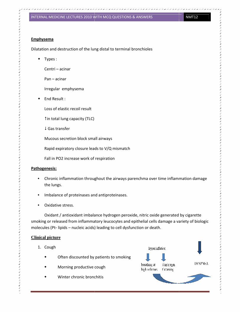
INTERNAL MEDICINE LECTURES 2010 WITH MCQ
www.medadteam.org Page 77
Emphysema
Dilatation and destruction of the lung dis
Types :
Centri – acinar
Pan – acinar
Irregular emphysema
End Result :
Loss of elastic recoil result
�in total lung capacity (TLC)
� Gas transfer
Mucous secretion block small airways
Rapid expiratory closure leads to
Fall in PO2 increase work of respiration
Pathogenesis:
• Chronic inflammation throughout the airways parenchma over time inflammation damage
the lungs.
• Imbalance of proteinases and antiproteinases.
• Oxidative stress.
Oxidant / antioxidant imbalance hydrogen peroxide, nitric oxide generated by cigarette
smoking or released from inflammatory leucocytes and epithelial cells damage a variety of biologic
molecules (Pt- lipids – nucleic acids) leading to cell dysfunction or death.
Clinical picture
1. Cough
Often discounted by patients to smoking
Morning productive cough
Winter chronic bronchitis
CTURES 2010 WITH MCQ QUESTIONS & ANSWERS
www.medadteam.org
Dilatation and destruction of the lung distal to terminal bronchioles
Mucous secretion block small airways
Rapid expiratory closure leads to V/Q mismatch
Fall in PO2 increase work of respiration
Chronic inflammation throughout the airways parenchma over time inflammation damage
Imbalance of proteinases and antiproteinases.
lance hydrogen peroxide, nitric oxide generated by cigarette
smoking or released from inflammatory leucocytes and epithelial cells damage a variety of biologic
nucleic acids) leading to cell dysfunction or death.
Often discounted by patients to smoking
Morning productive cough
Winter chronic bronchitis
NMT12
Chronic inflammation throughout the airways parenchma over time inflammation damage
lance hydrogen peroxide, nitric oxide generated by cigarette
smoking or released from inflammatory leucocytes and epithelial cells damage a variety of biologic

INTERNAL MEDICINE LECTURES 2010 WITH MCQ QUESTIONS & ANSWERS NMT12
www.medadteam.org More Than You Dream Page 78
2. Dyspnea
Insidious or on exertion
Major source of disability
3. Chest pain (strain of intercoastal muscles)
4. Lower limb edema (cor pulmonale)
Causes of dyspnea in COPD:
narrowed airways (bronchospasm, increased compliance, airway secretions, airway thickening,
increased cholinergic tone).
WHO classification of COPD:
Stage o:At risk ,chronic cough
Stage 1:mild COPD ,cough, change in pulmonary function
Stage2:shortness of breath , airflow limitation
Stage 3:Severe COPD ,impaired quality of life
Signs:
Mild disease: No signs
Severe disease:
Lip pursing
Supra sternal & intercostal retraction
Cyanosis & flapping tremors
Tachypnea
- Respiratory failure
Barell shaped chest with enchroachment on hepatic and cardiac dullness
Ronchi throughout the chest
Maybe basal crepitations
Physical signs:
Large barrel shaped chest (hyperinflation)
Prominent accessory respiratory muscles in neck and use of accessory muscle in respiration
INTERNAL MEDICINE LECTURES 2010 WITH MCQ QUESTIONS & ANSWERS NMT12
www.medadteam.org More Than You Dream Page 79
Low, flat diaphragm
Diminished breath sound
Complications:
Repeated chest infection
Respiratory failure PO2 < 60 mm Hg
PCO2 < 33 mm Hg
Corpulmonal, pulmonary hypertrension
Rt ventricular failure
Pneumothrax
The complications that can be associated with COPD, which may include:
• Heart failure
• Nutritional deficiencies
• COPD exacerbations - a sudden increase or intensification of COPD symptoms which
often lead to hospitalization
• Skeletal muscle wasting
• Worsening of pre-existing conditions such as cardiovascular disease, diabetes,
osteoporosis, and mood disorders
• Increased susceptibility to lung diseases such as pneumonia, respiratory failure, and
end-stage lung disease.
Chest X-ray.
Lung function test.
Spirometry reduced FEV1
FEV1 / FVC < 70%
FV1 improve by bronchodilator > 15%

INTERNAL MEDICINE LECTURES 2010 WITH MCQ QUESTIONS & ANSWERS NMT12
www.medadteam.org More Than You Dream Page 80
Stage of COPD:
Stage FV1 FV1/FVC
0 At risk >80 Normal
1 Mild >80 < 70%
2 Moderate 50-80 < 70%
3 Severe 30-50 <70%
4 Very severe
< 30 < 70%
High resolution C.T
E.C.G (P-pulmonale – right ventricular enlatgement - RBBB).
Treatment
Objectives:
Prevent disease progression
Relieve symptoms
Improve exercise tolerance
Improve health status
Prevent and treat exacerbations
Prevent and treat complications
Reduce mortality
Minimize side effect from medication
Current therapeutic Current therapeutic Current therapeutic Current therapeutic management of COPDmanagement of COPDmanagement of COPDmanagement of COPD
• COPD remains under diagnosed and under treated
• Bronchodilators are the only approved treatments
• Additional treatment options are needed
Efficacy measures used to assess treatment response in COPDEfficacy measures used to assess treatment response in COPDEfficacy measures used to assess treatment response in COPDEfficacy measures used to assess treatment response in COPD
Spirometry (FEV1)

INTERNAL MEDICINE LECTURES 2010 WITH MCQ QUESTIONS & ANSWERS NMT12
www.medadteam.org More Than You Dream Page 81
• Objective, reproducible
• Diagnostic and prognostic
Other measures
• Health status (QOL)
• Symptoms
• Exacerbations
Smoke cessation:
Vital even at a late stage of the disease this may slow down the rate of deterioration and
death occur.
Drug therapy:
� Bronchodilators
� Corticosteroids � Antibiotics � Vaccines � Alpha-1 antitrypsin replacement.
Bronchodilators:
� Adrenergic agents :
� Beta-agonists bind to B2 receptors on airway and result in smooth muscle relaxation and
bronchodilation
� Inhaled route is preferred
� Acute relief of symptoms
� Anti-cholinergic agents
• Bind to acetylcholine receptors and result in bronchodilation (of mostly larger airways)
• Reduces sputum production
• Inhaled route is preferred
� Methylxanthines (i.e. theophylline)
• Weak bronchodilator
• Delays respiratory muscle fatigue
• Reduces trapped lung gas
• Improves respiratory muscle mechanics Corticosteroids
• Reduce airway inflammation

INTERNAL MEDICINE LECTURES 2010 WITH MCQ QUESTIONS & ANSWERS NMT12
www.medadteam.org More Than You Dream Page 82
Mucolytics • Alter viscosity of sputum
• May reduce symptoms in some patients
• Must be used carefully (i.e. avoiding hypotension)
Antibiotics
Shortens exacerabtions should always be given in acute exacerbation,
preventing further lung damage.
Vaccines
Yearly influenza vaccine, pneumoccocal vaccine
Stem cell transplation
Oxygen Therapy
• Rehabilitation
• Reduce symptoms – improve quality of life increase physical and emotional participation
• Oxygen Therapy:
� Increase survival
� Improve exercise capacity � Improve mental status � Decrease pulmonary arterial pressure
� Can be administrated by:
i. Long term continuous
ii. During exercise
iii. relieve acute dyspnea
Indications for o2 therapy
PaO2 55 mmHg or less
PaO2 56 – 59 mmHg with complication, such as
erythrocytosis or cor pulmonale
SaO2 88% or less
EXERCISE
Increase exercise tolerance
Increase quality of life
Improve co-ordination and efficiency of movement
Improve strength particularly respiratory muscles
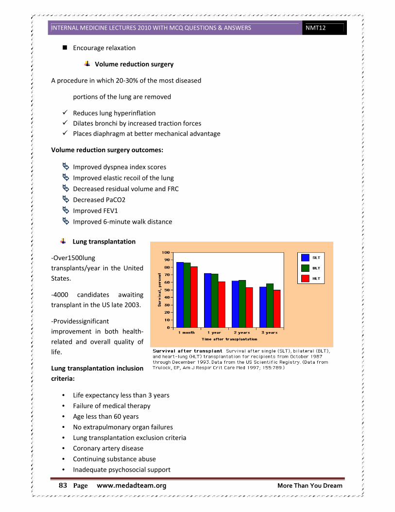
INTERNAL MEDICINE LECTURES 2010 WITH MCQ QUESTIONS & ANSWERS NMT12
www.medadteam.org More Than You Dream Page 83
Encourage relaxation
Volume reduction surgery
A procedure in which 20-30% of the most diseased
portions of the lung are removed
� Reduces lung hyperinflation � Dilates bronchi by increased traction forces � Places diaphragm at better mechanical advantage
Volume reduction surgery outcomes:
���� Improved dyspnea index scores
���� Improved elastic recoil of the lung
���� Decreased residual volume and FRC
���� Decreased PaCO2
���� Improved FEV1
���� Improved 6-minute walk distance
Lung transplantation
-Over1500lung
transplants/year in the United
States.
-4000 candidates awaiting
transplant in the US late 2003.
-Providessignificant
improvement in both health-
related and overall quality of
life.
Lung transplantation inclusion
criteria:
• Life expectancy less than 3 years
• Failure of medical therapy
• Age less than 60 years
• No extrapulmonary organ failures
• Lung transplantation exclusion criteria
• Coronary artery disease
• Continuing substance abuse
• Inadequate psychosocial support

INTERNAL MEDICINE LECTURES 2010 WITH MCQ QUESTIONS & ANSWERS NMT12
www.medadteam.org More Than You Dream Page 84
• Extreme cachexia or obesity
• Recent malignancy (<3 years)
• Long term, high dose corticosteroid use
3. Suppurative Lung Disease
���� Definition:
It is the expectoration of excessive amount of purulent sputum, usually foetid and related to
posture.
It occurs in the following conditions:
- Bronchiectasis.
- Cystic lung (infected).
- Lung abscess.
- Empyema with bronchopleural fistula.
( a.) Bronchiectasis
���� DEFINITION:
Permanent abnormal dilatation of the bronchi due to destruction of the elastic and muscular
components of the bronchial wall.
���� ETIOLOGY:
Two factors are required
- Impairment of drainage
- Infection
1. Impairment of drainage:
Leads to retention of secretion which enhances bacterial infection
- Air way obstruction:
� Tuberculus enlargement of hilar lymph nodes (particularly around the origin of the
middle lobe bronchus)
� Inhaled F.B (in children)
� Bronchial carcinoma
� Chronic obstruction by viscid mucus in cystic fibrosis
- Defect in host defense:
Lectures objectives
1. Define Suppurative lung DS 2. List the causes and explain the aetiology of the different types of the
disease. 3. Describe the C/P of the various types of the disease. 4. List the appropriate investigations. 5. List the complications.

INTERNAL MEDICINE LECTURES 2010 WITH MCQ QUESTIONS & ANSWERS NMT12
www.medadteam.org More Than You Dream Page 85
Congenital dysfunction of the cilia e.g. Kartagner's syndrome: sinusitis, transposition of the
viscera, bronchiectasis, and immotile cilia.
N.B.: Post obstructive bronchiectasis is often localized. Diffuse bronchiectasis is associated with
diffuse obstruction of airways in patients with chronic bronchitis, atopic asthma and cystic
fibrosis.
2. Infection:
Leads to destruction of the bronchial wall and supporting tissue (leading to abnormal dilatation).
- Secondary to severe bacterial infection in childhood often as a complication of whooping
cough or measles (causing pneumonia).
- Hypogammaglobulinaemia causing repeated infection.
���� CLINICAL FEATURES:-
Bronchiectasis may involve any part of the lung but the more efficient drainage by gravity of the upper
lobes usually produces less serious symptoms and complications than when bronchiectasis involves the
lower lobes.
���� SYMPTOMS:-
- Due to accumulation of pus in dilated bronchi:
� chronic productive cough, worse in the morning brought on by changes of posture
� sputum often copious and purulent, green or yellow
- Due to inflammatory changes in lung &pleura surrounding the dilated bronchi:
� fever and malaise
� increase cough & sputum volume when spread of infection cause pneumonia which is
frequently associated with pleurisy
� recurrent pleurisy in the same side
- Hemoptysis: (if necrosis of the mucosa is severe)
� slight or massive often recurrent
� usually associated with purulent sputum
� can be the only symptom in the so called dry bronchiectasis
- General health:
� Weight loss, anorexia, night sweat and halitosis.
���� PHYSICAL SIGNS:-
- In the chest:
� May be unilateral or bilateral, usually basal.
� In the presence of large amount of secretion, coarse crepitation will be heard over the
affected area.
� There may be signs of collapse.
� If no secretion, no collapse, there will be no abnormal physical signs.
- General ill health and Finger clubbing
���� INVESTIGATIONS:-
� Chest X-Ray:
� may be normal
� in advanced disease the cystic bronchiectatic spaces may be visible associated pulmonary
infection and or collapse are evident
� CT scanning: Bronchial wall thickening
� Bronchogrames: very rarely done
� Bronchoscopy: in localized bronchiectasis for diagnosis of obstruction by foreign body, lymph
nodes or tumor
INTERNAL MEDICINE LECTURES 2010 WITH MCQ QUESTIONS & ANSWERS NMT12
www.medadteam.org More Than You Dream Page 86
� Sputum examination: culture & sensitivity
� Major pathogens are: staph. aureus, pseudomonas aeruginosa, hemophilus influenza,
anaerobes
� Others: streptococcus pneumonia, klebsiella pneumonia, aspergillus fumigatus
� Serum immunoglobulins : for Hypogammaglobulinaemia
� Sweat test: The sweat test measures the concentration of chloride and sodium that is excreted
in sweat. It is used to diagnose or exclude cystic fibrosis (CF).
���� TREATEMENT:-
� Postural drainage
The aim is to keep the dilated bronchus emptied of secretions
It is of value in reducing the amount of cough & sputum and in preventing recurrent episodes
of bronchopulmonary infection
� Antibiotics: To eradicate infections
� Bronchodilator
� Surgery: For localized cases
� TTT For hemoptysis: Bed rest - Antibiotics
���� COMPLICATIONS:-
� Pneumonia- pneumothorax- empyema
� Metastatic cerebral abscess
� Severe life threatening hemoptysis
� Respiratory failure and corpulmonale
� Amyloidosis
( b.) Cystic Fibrosis
���� Definition
Dysfunction of all exocrine glands resulting in abnormal mucus production
The classical form includes:
- Bronchopulmonary infection
- Pancreatic insufficiency
- High sweat sodium & chloride concentration
���� ETIOLOGY & PATHOGENESIS:-
OBSTRUCTION � INFECTION � CYSTIC DILATATION OF DUCTS
- Autosomal recessive genetic disease caused by mutation in the gene encoding the cystic
fibrosis transmembrane conductance regulator (CFTR)
- This protein defect produces an abnormality in the regulation of chloride channel in the cell
membrane
- This basic defect in all exocrine glands produces thick viscid secretions causing obstructions of
ducts or passages in the respiratory, salivary glands, digestive & biliary tracts, pancreas &
genitourinary tract

INTERNAL MEDICINE LECTURES 2010 WITH MCQ QUESTIONS & ANSWERS NMT12
www.medadteam.org More Than You Dream Page 87
���� CLINICAL FEATURES:-
C.F. is the commonest cause of recurrent bronchopulmonary infection in childhood & early adult
life
���� Symptoms:
- Cough, productive cough with thick purulent sputum
- Finger clubbing
- Hemoptysis
- Breathlessness
���� Signs:
- Chest examination � Rales & Rhonchi, usually more on one side
Older children may develop nasal polyps
- Delayed puberty& skeletal maturity
- Male infertility due to failure of development of the vas deference & epididymis
Female are able to conceive, often develop secondary amenorrhea as the disease progress
- Pancreatic exocrine insufficiency:
Steatorrhea - Peptic ulcer - Diabetes mellitus
- Meconium ileus in the newborn (blockage of the lumen of the intestine due to the viscoid
consistency of the meconium)
- Cholesterol gall stones
- Biliary cirrhosis develop in 5% of older patients due to blockage of biliary ducts
���� INVESTIGATIONS:-
- Sputum culture & sensitivity
- X-Ray � soap bubble appearance
- CT chest � early changes of bronchiectasis
- Genetic test � analysis for CFTR gene defect
- Sweat test: A high sweat sodium concentration over 60 mmol/L
���� TREATMENT:-
- Respiratory disease: as for bronchiectasis (antibiotics, bronchodilators, physiotherapy)
+ Human deoxyribonuclease: degrade DNA helping cough clearance of sputum resection only in
localized cases.
- TTT Pancreatic insufficiency.
- Heart-lung transplantation.
( c.) Lung Abscess
���� Definition:
Severe localized suppuration in the lung, associated with cavity formation, often with the presence of
fluid level (on chest X-Ray), that is not due to tuberculosis
���� CAUSES:
� The commonest is aspiration
- Alcoholics following aspiration pneumonia
- In altered consciousness (coma & anesthesia)
- Oropharyngeal & and esophageal dysfunction

INTERNAL MEDICINE LECTURES 2010 WITH MCQ QUESTIONS & ANSWERS NMT12
www.medadteam.org More Than You Dream Page 88
- Following inhalation of foreign body into a bronchus
- Bronchial obstruction (e.g. bronchial carcinoma or lymph node)
� During the course of specific pneumonia (pneumonia caused by staphylo coccus pyogenes or
klebsiella pneumoniae)
� Septic emboli, usually staphylococci, resulting in multiple lung abscesses
� Pulmonary infarction may cavitate and rarely becomes infected
� Amebic abscesses may develop in the right lower lobe following transdiphragmatic spread from an
amebic liver abscess
N.B.: CAUSATIVE ORGANISMS:
- Aerobic bacteria: streptococcus pneumonia, staph aureus, H-influenza
- Anaerobic bacteria
���� CLINICAL FEATURES:
- Are those of persisting and worsening pneumonia with production of large quantities of
sputum, which is often foul smelling. Hemoptysis may occur.
- Pleuretic chest pain.
- Fever, chills, night sweat, anorexia.
Chest signs may be few (deep central abscess)
Superficial abscess usually reveals signs of consolidation and signs of cavitation may be found, pleural
rub is common
Clubbing usually develops quickly (10- 14 days)
���� Investigations
- Chest X- Ray: Cavitary lesion with an air fluid level with or without surrounding infiltrate
- Chest CT
- Sputum culture & sensitivity
���� TREATMENT:-
- Antimicrobial therapy
- Postural drainage is of great value
- Surgical resection of necrotic lung may be needed
���� COMPLICATIONS:-
- Empyema formation resulting from a bronchopleural fistula
- Massive hemoptysis
- Spontaneous rupture into uninvolved lung segments
- Non-resolution of abscess cavity, with residual fibrosis and bronchiectasis
( d.) Empyema with bronchopleural fistula
Empyema
���� Definition
Empyema means the presence of pus within the pleural cavity.

INTERNAL MEDICINE LECTURES 2010 WITH MCQ QUESTIONS & ANSWERS NMT12
www.medadteam.org More Than You Dream Page 89
���� Aetiology:
This usually arises from bacterial spread from a severe pneumonia or after the rupture of a lung
abscess into the pleural space
���� Clinical presentation
���� Symptoms
It depends on the underlying cause of the empyema.
- Most patients report dyspnea with little exertion
- low-grade fever early in the course
- Pleuretic chest pain and a feeling of heaviness on the affected side of the chest.
- Purulent sputum.
���� Signs
- Breath sounds are decreased on the involved side of the chest.
- The affected side may be less mobile than the other
���� Investigations
- Chest radiographs are the appropriate first study and usually show opacifications and may show
air-fluid levels.
- CT scans valuable to detect loculation and to direct appropriate drainage of the area.
- Bacteriological investigation of lung abscess and empyema is best conducted on specimens
obtained by transtracheal aspiration, bronchoscopy or percutaneous transthoracic aspiration with
ultrasound or CT guidance.
- Bronchoscopy is helpful to exclude carcinomas and foreign bodies.
���� Treatment
- Tube drainage or by rib resection and drainage of the empyema cavity under ultrasound control.
- Appropriate antibiotic treatment is given for up to 6 weeks.
Bronchopleural fistula
���� Definition
A bronchopleural fistula (BPF) is a fistula between the pleural space and the bronchial tree
���� Aetiology
Bronchopleural fistula, although reduced in incidence in recent years, remains a grave complication of
pulmonary disease and of pulmonary resection
Most common causes are:
1. pulmonary infection
2. previous surgical resection of lung
- These represent 70-80% of empyemas
- Those patients with empyema treated by plastic surgeons commonly have undergone
previous surgical resection and have developed a bronchopleural fistula.
Bronchopleural fistulas occur following pulmonary resections because of failure of the bronchial stump
to heal and may lead to empyema when not quickly recognized and treated.
This failure to heal may be from improper initial closure, inadequate blood supply, infection at the
bronchial stump, or residual malignant tumor at the bronchial stump.

INTERNAL MEDICINE LECTURES 2010 WITH MCQ QUESTIONS & ANSWERS NMT12
www.medadteam.org More Than You Dream Page 90
���� Clinical picture:
Presence of fistula:
The purulent expectoration is having the same character as the pleural fluid
Expectoration is related to posture, being increased when the affected side is upper most
Manifestations of pyopneumothorax in the affected side (evidence of fluid & air in the pleura): shifting
dullness, sucussion splash & transverse fluid level
Amphoric breathing due to the presence of bronchopleural fistula
N.B.:
ssuuccccuussssiioonn ssppllaasshh:: A splashing sound produced by the presence of air and liquid in the chest. It may
be elicited by gently shaking the patient during auscultation. This sound nearly always indicates either a
hydropneumothorax or a pyopneumothorax, although it has also been detected over very large
cavities. The presence of air and liquid in the stomach produces similar sounds, large hiatal hernia,
intestinal or pyloric obstruction.
AAmmpphhoorriicc bbrreeaatthhiinngg:: Produced by, or indicating, a cavity in the lungs, not filled, and giving a sound
like that produced by blowing into an empty jar or a coke can. It is loud, with a prolonged, hollow
expiration and metallic quality. It is heard in bronchiectatic cavities or pneumothorax when the opening
to the lung is patulous; in the consolidation area near a large bronchus; and sometimes over a lung
compressed by a moderate effusion.
Supplementary Information
Patient education:
- Early recognition of exacerbation
- Postural drainage & physical therapy
- Vaccination against measles &whooping cough
Primary care doctor:
- Antibiotics should be started early in the course of
exacerbation
- Prompt diagnosis & treatment of obstruction
Screening:
- Genetic counseling to minimize the incidence of cystic
fibrosis
INTERNAL MEDICINE LECTURES 2010 WITH MCQ QUESTIONS & ANSWERS NMT12
www.medadteam.org More Than You Dream Page 91
- HbA1C should be followed routinely in patients with cystic
fibrosis since DM occur in nearly 10% of patients.
4. Tuberculosis
� It is a Granulomatous response with intense tissue inflammation & damage leading to
prominent pulmonary diseases ,other organs can be involved
� Increased due to : AIDS, use of immuno-suppressive drugs, decreased socio-economic
conditions, as well as increased immigration of persons from areas of high endemicity.
� TB is the most common disease which leads to death from infection.1.7 million deaths
(without HIV) in 2004 (why??) Because
1. Inadequate programs for disease control with poorly supervised ttt.
2. misuse of drugs:
- multidrug resistant T.B.(MDRTB):resistant for rifampcin ,isoniazed
- Extensively drug resistant T.B.(XDRTB):resistant also for quinolones & injectable
second line agents
3. Co-infection with HIV
4. rapid rise in world’s population of young adults (highest mortality from T.B.)
5. overcrowding
6. poor nutrition
���� Etiology:
Caused by mycobacterium tuberculosis (slowly growing Bactria, facultative intercellular organisms) .
���� Mode of infection:
- Exogenous: . Inhalation (droplet)
. Ingestion (contaminated milk)
. cutaneous (very rare)
. congenital (very rare)
Lectures objectives
1. Identify the causative organism and mode of transmission of TB. 2. Recognize the natural history of development of the disease. 3. Describe the underlying pathogenic mechanisms and pathologic
characteristics of Tuberculosis. 4. Describe the clinical picture of different presentations of
Tuberculosis.(initial primary and post-primary TB infection) 5. Construct a management plan both diagnostic (laboratory and imaging)
and therapeutic :( prophylactic and curative).
6. Recognize the importance of prophylactic measures of T B .

INTERNAL MEDICINE LECTURES 2010 WITH MCQ QUESTIONS & ANSWERS NMT12
www.medadteam.org More Than You Dream Page 92
- Endogenous: . Activation (relapse)
. Dissemination (haematogenic or Bronchogenic)
���� Predisposing factors:
1. Race: more common in Negros
2. Age:
- Below 5 yrs � high susceptibility to infection
- 5 - 15 yrs � relatively resistance
- Above 15 yrs � high susceptibility to progressive pulmonary disease
3. Sex: equal below 40 & more in males after that age
4. Environment: Bad housing & poor nutrition are risk factor.
���� Risk group: Children, Elderly, organ transplantation, Immunocomprised patient, AIDS, malnutraion ,
chronic illness, chemotherapy
���� Pathophysiology: � Transmitted by air borne nuclei expelled from an active infected host
� development of an infection depends on prolonged exposure (on order of weeks)
� Fates: - Killed by immune system � multiply( 1ry T.B.)
� dormant (within macrophages)
� proliferate after a latency period (reactivation )
� The response of the body to the initial infection is called "THE PRIMARY COMPLEX" which
is formed of: � Regional lymphadenitis
� Lymphangitis in the intervening lymph vessels
� Gohn’s focus: It is an aggregation of tubercles � Tubercle is a granuloma usually sub-
pleural in the mid to upper zones of lung & formed of central caseation surrounded
by epithelioid cells & Langhan’s giant cells with multiple nuclei, both cells are derived
from macrophages, Lymphocytes are present & varying degree of fibrosis then
calcification occurs.
� Fate of the primary complex: A. Healing
B. Progression
���� Clinical picture of TB:
Primary TB:
� In many cases primary T.B is asymptomatic
� But it may be symptomatic with malaise, cough, wheeze, night fevers, loss of weight,
erythema nodula (nodosum) which appears in the chin of tibia due to allergic reaction
� Pleural effusion: it may occur if immunity is low � effusion is characterized by being
massive, hemorrhagic & rapidly accumulated
� 2ry infection: Collapse or bronchiectasis often in the middle lobe � because of hilar
lymph nodes enlargement due to infection � will compress the bronchus (Brock’s
syndrome )

INTERNAL MEDICINE LECTURES 2010 WITH MCQ QUESTIONS & ANSWERS NMT12
www.medadteam.org More Than You Dream Page 93
� Tuberculous pneumonia: mainly in children if the T.B spread in lung
� Miliary T.B: - acute diffuse dissemination of tubercle bacilli via blood stream
- difficult to make diagnosis especially in older people (partially covert)
- lead to 1ry infection or reactivation of a latent focus as in
immunosuppressed patients.
- Fatal with ttt
Post primary TB:
� When reactivation occurs � Cough, expectoration, malaise, night fever, cachexia &
consolidation may occur
� Tuberculin Toxemia: a condition at which patient is suffering from cough, night fever,
expectoration & loss of weight
Extra-pulmonary TB:
� Intestine: chronic diarrhea, iliocecal fibrosis & obstruction
� Peritoneum: Tuberculous ascites Hemorrhage
Recollected
Massive
� CNS: meningitis
� Joints: Tuberculous arthritis
� Female genital system: Salpingitis & salpingoophritis � Fibrosis � sterility.
� Bones: Pott's disease of thoracic spines � deformity � compress on nerves � paraplegia
& also T.B can cause osteomyelitis
� Eye: phlycten, iritis � so fundus examination for military TB should be done
� Pericardium: effusion � fibrosis
� Suprarenal: Addison’s disease
���� Investigations:
1. X-ray:
Lesion appears in clusters even if no symptoms appears, But x-ray can’t differentiate
between past & active infection
2. Sputum & sedimentation rate � to detect the active cases
3. Culture � is the gold standard diagnosis
4. Tuberculin test: (mantoux test)
� Reaction more than 10 cm in diameter is positive
� Negative test � means no TB (- ve in 30-50 % of people with severe disease)
5. CT scanning :lung paranchymal abnormalities
6. Trans-bronchial biopsies
Vaccinated
Dormant Past Infection
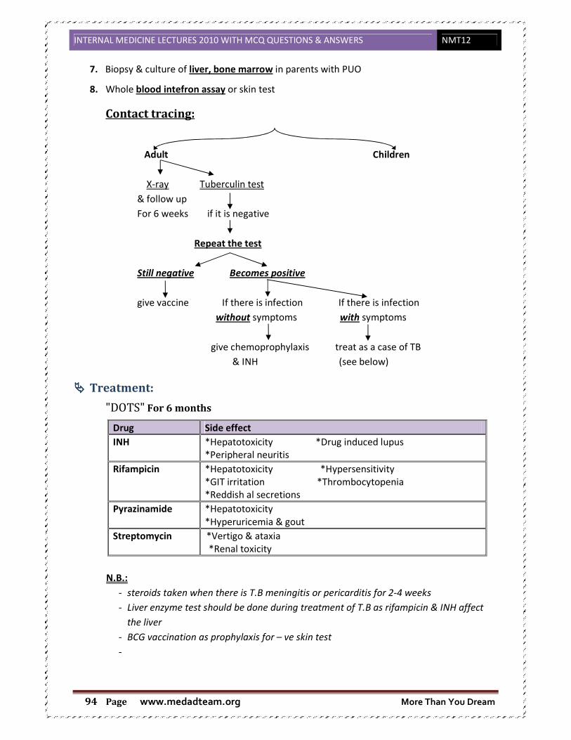
INTERNAL MEDICINE LECTURES 2010 WITH MCQ QUESTIONS & ANSWERS NMT12
www.medadteam.org More Than You Dream Page 94
7. Biopsy & culture of liver, bone marrow in parents with PUO
8. Whole blood intefron assay or skin test
Contact tracing:
Adult Children
X-ray Tuberculin test
& follow up
For 6 weeks if it is negative
Repeat the test
Still negative Becomes positive
give vaccine If there is infection If there is infection
without symptoms with symptoms
give chemoprophylaxis treat as a case of TB
& INH (see below)
���� Treatment:
"DOTS" For 6 months
Drug Side effect
INH
*Hepatotoxicity *Drug induced lupus
*Peripheral neuritis
Rifampicin *Hepatotoxicity *Hypersensitivity
*GIT irritation *Thrombocytopenia
*Reddish al secretions
Pyrazinamide *Hepatotoxicity
*Hyperuricemia & gout
Streptomycin *Vertigo & ataxia
*Renal toxicity
N.B.:
- steroids taken when there is T.B meningitis or pericarditis for 2-4 weeks
- Liver enzyme test should be done during treatment of T.B as rifampicin & INH affect
the liver
- BCG vaccination as prophylaxis for – ve skin test
-

INTERNAL MEDICINE LECTURES 2010 WITH MCQ QUESTIONS & ANSWERS NMT12
www.medadteam.org More Than You Dream Page 95
5. Bronchial Asthma
���� Pathophysiology:
� Activated eosinophils have IgE receptors � release leukotrines which sustain inflammatory
reaction and that lead to bronchial asthma
� The activated eosinophils are not like the normal ones as their life span is > 14 day
� Role of eosinophils in asthma is the same like the role of mast cells
� Activation of eosinophils in allergy occurs through interleukins which are secreted from
Th2
���� , So Definition of asthma is:
- Asthma is a common chronic inflammatory condition of the lung airways in which many cell types
are involved and whose cause is incompletely understood. ( In 1990s )
- Symptoms are cough, wheeze, chest tightness and shortness of breath, often worse at night.
It has three characteristics:
* Airflow limitation which is usually reversible spontaneously or with treatment
* Airway hyper responsiveness to a wide range of stimuli
* Inflammation of the bronchi with eosinophils, T-lymphocytes and mast cells with associated
plasma exudation, oedema, marked smooth muscle hyper trophy, mucus plugging and
epithelial damage.
- In chronic asthma, inflammation may be accompanied by irreversible airflow limitation.
N.B.: Evalution of our understanding of asthma pathophysiology :
In 1960s: Bronchial hyper responsiveness + reversible airway obstruction
Autonomic imbalance
In late 60s: Discovery of the IgE/ Mast cells disease
In 1970s: Mast cell diseases/ Mediators
Characterization of Leukotrienes (LTB4, LTC4, LTD4, LTE4)
In 1980s: Eosinophils discovered as central cells in pathogenesis
Normal Eosinophils Activated Eosinophils
Little Leukotrienes formation High Leukotrienes formation
Low cytotoxicity High cytotoxicity
No IgE receptor expression High IgE receptor expression
Life span = 48 hrs Life span more than 14 days
Lectures objectives
1. Define bronchial asthma 2. Classify bronchial asthma 3. Identify the common allergens and triggering factors 4. Construct a plan for management of asthma according to severity 5. Discuss the pharmacological therapy of asthma including management of
acute attacks. 6. Appreciate the role of primary physician in patients and family education

INTERNAL MEDICINE LECTURES 2010 WITH MCQ QUESTIONS & ANSWERS NMT12
www.medadteam.org More Than You Dream Page 96
Normal Eosinophils Activated Eosinophils
T- cells are principle immunoregulators
So, Since there are multiple cells involved with many mediators ……. Treatment with mast cells stailizres
or anti leukotrienes or anti- histaminics alone are non- effective or of little value if used alone.
���� Classification:
Asthma can be divided into:
- Extrinsic: implying a definite external cause
- Intrinsic or cryptogenic: when no causative agent can be identified.
Extrinsic asthma occurs most frequently in atopic individuals who show positive skin-prick reactions to
common inhalant allergens. Positive skin-prick tests to inhalant allergens are shown in 90% of children
and 50% of adults with persistent asthma. Childhood asthma is often accompanied by eczema. An
overlooked cause of late-onset asthma in adults is sensitization to chemicals or biological products in
the workplace.
Intrinsic asthma often starts in middle age ('late onset'). Nevertheless, many patients with adult-onset
asthma show positive skin tests and on close questioning give a history of respiratory symptoms
compatible with childhood asthma. Non- atopic individuals may develop asthma in middle age from
extrinsic causes such as sensitization to occupational agents or aspirin intolerance. Extrinsic causes must
be considered in all cases of asthma and, where possible, avoided.
���� Aetiology and pathogenesis:
There are two major factors involved in the development of asthma and many other stimuli that can
precipitate attacks
Atopy and allergy
- to run in families
- to have characteristic wealing skin reactions to com mon allergens in the environment
- to have circulating antibody in their serum that could be transferred to the skin of non-
sensitized individuals
Increased responsiveness of the airways of the lung (airway hyper responsiveness)
- Attacks of asthma only on extreme exertion
- Wheezing or prolonged periods of coughing following a viral infection
- Cough variant asthma
- Seasonal wheeze in pollen season
- Allergic rhinitis, but not complaining of any lower respiratory symptoms until specifically
questioned
- Some subjects with no respiratory symptoms.
IL-5

INTERNAL MEDICINE LECTURES 2010 WITH MCQ QUESTIONS & ANSWERS NMT12
www.medadteam.org More Than You Dream Page 97
Mechanism:
This lead to change in the treatment modality of asthma as Inhaled steroids are used for long term to
control the chronic inflammation
Risk group:
1. Genetics
2. Allergy � inhaled allergen (pollen or dust)
3. Viral disease
4. Exercise
5. Emotional asthma
Common allergens that exacerbates asthma
1- Allergens: Dust mites, moulds , Pollen, cats , cockroaches
2- Air pollutants ( Dust, cigarette smoke , car fumes , NO2, SO2,
3- Respiratory infections
4- Exercise , Hyperventilation
5- Weather changes ( Cold)
6- Drugs e.g B- Blockers are absolutely contra indicated
NSAIDs (Asprin) being inhibiting the cyclooxygenase pathway exacerbates asthma
7- Food, additives (least important)
Histologic changes:
The epithelium: the epithelium of the conducting airways is stressed and damaged with loss of ciliated
columnar cells onto the lumen. Metaplasia occurs with a resultant increase in the number and activity
of mucus-secreting goblet cells. The epithelium is a major source of mediators, cytokines and growth
factors that serve to enhance inflammation and promote tissue remodelling. Damage and activation of
the epithelium make it more vulnerable to infection by common respiratory viruses, e.g. rhinovirus,
coronavirus, and to the effects of air pollutants.
Epithelial basement membrane: A pathognomonic feature of asthma is the deposition of repair
collagens beneath the basement membrane. This causes the appearance of a thickened basement
membrane observed by light microscopy in asthma. This collagen deposition reflects activation of an
underlying sheath of fibroblasts
Smooth muscle: A prominent feature of asthma is hyperplasia of the helical bands of airways smooth
muscle.
N.B.: In Late stages of Asthma, airway narrowing is irreversible, so early treatment is very important in
asthma.
Risk factors for
asthma development
Inflammation
Air way
Hyper-response
Airflow
obstruction
Risk factors for
exacerbation Symptoms
INTERNAL MEDICINE LECTURES 2010 WITH MCQ QUESTIONS & ANSWERS NMT12
www.medadteam.org More Than You Dream Page 98
���� Clinical picture:
� Symptoms:
� During attack of asthma � wheezy chest, marked dyspnea & cough with
expectoration of small amount of viscid sputum
� Signs:
� During attack:
• Chest becomes emphysematous
• Vesicular breathing with prolonged expiration
• Rhonchi are found all over the chest
• Tachycardia & working accessory respiratory muscles
� In between attack: chest is completely free unless bronchial asthma is accompanied
with bronchitis
���� Investigations:
� Respiratory Function tests ( FEV1/FVC) = normally more than 70 ,it will be reduced in
asthma
• Measurements of peak expiratory flow (PEF) on waking, prior to taking a bronchodilator
and before bed after a bronchodilator, are particularly useful in demonstrating the
variable airflow limitation that characterizes the disease.
• Diffusion:
� CO Transfer factor � Normal in asthma
• Arterial Blood Gases:
� ↓↓ PO2
� ↑↑ PCO2
� Exercise test
� Histamine or methacholine bronchial provocation
� Trial of corticosteroids
� Blood and sputum tests: Increased Eosinophils
� Chest X-ray: Nothing characteristic – exclude other diseases and complications
� Skin tests: Allergy is specific – exposure dependent
� Serum IgE: ↑↑ in most cases of extrinsic asthma
���� Treatment:
� Aim of treatment � Control disease
1. No or minimal ( less than twice a week ) daytime symptoms
2. No nocturnal symptoms ( less than twice a month )
3. No Limitation of activity
4. No or minimal( less than twice a week ) need for rescue medications
5. Normal lung functions
6. No Exacerbations

INTERNAL MEDICINE LECTURES 2010 WITH MCQ QUESTIONS & ANSWERS NMT12
www.medadteam.org More Than You Dream Page 99
� Asthma grades:
Comparison Controlled Partially Uncontrolled
Day symptoms None Less than 2 / week 3 or more
features of
partially
controlled
asthma in
any week
Nocturnal symptoms None Any symptoms / any week
Activities Any symptoms / any week
Need for rescue Meds None (less than 2/week) More than 2 / week
Lung functions Normal (more than 70–80 %) Less than 70 %
Exacerbations None One or more / year
N.B.: Old method of treatment was using bronchodilators alone for long period � lead to
irreversible obstruction
� Drugs that is contraindicated in asthma:
� NSAID (Non-steroidal anti-inflammatory drugs)
� B.B. (Beta blockers)
� Drug families used in treatment of asthma:
Short -acting bronchodilator treatment taken at any step on as-required basis
� Management of acute attacks: (Severe)
� At home
1. The patient is assessed. Tachycardia, a high respiratory rate and inability to speak in
sentences indicate a severe attack.
Relievers Controllers
• Short acting B- agonists
e.g. salbutamol ,
Terbutaline
• Anti- cholinergic e.g
Ipratropium , oxytropium
Theophylline preparations
• Systemic steroids ( oral)
• Inhaled Corticosteroids e.g. Fluticasone
• Leukotrienes Modifiers e.g Zafirlucast , Montlukast ,
• Long acting inhaled B2- agonists e.g Salmetrol , Formetrol
• Theophylline
• Mast cell stabilizers ( cromones )
• Anti- IgE ( omalizumab ) – only in very severe cases
• N.B. All drugs are used in addition to the Inhaled steroids.
Step Treatment
1 Occasional symptoms As-required bronchodilators
If used more than once daily, move to step 2
2 Daily symptoms Low dose ICS
Leukotrienes modifiers
Sodium cromoglycate
If not controlled, move to step 3
3 Severe symptoms Low dose ICS
Long acting B2 agonists
4 Severe symptoms uncontrolled with
high-dose
Medium or high dose ICS
Long acting B2 agonists
5 Severe symptoms deteriorating Oral glucocorticoids e.g. prednisolone
Lowest dose
6 Severe symptoms deteriorating in spite
of prednisolone
Prednisolone increased dose
Anti- IgE

INTERNAL MEDICINE LECTURES 2010 WITH MCQ QUESTIONS & ANSWERS NMT12
www.medadteam.org More Than You Dream Page 100
2. Nebulized salbutamol 5 mg or terbutaline 10 mg is administered.
3. Hydrocortisone sodium succinate 200 mg i.v. is given. If severe and no
improvement
4. Oxygen 40-60% is given if available.
5. Prednisolone 60 mg is given orally.
� At hospital
1. The patient is reassessed.
2. Oxygen 40-60% is given.
3. The PEFR is measured , Measure O2 saturation with a pulse oximeter.
4. Nebulized salbutamol 5 mg or terbutaline 10 mg is repeated and administered 4-
hourly.
5. Add nebulized ipratropium bromide 0.5 mg to nebulized salbutamol/terbutaline.
6. Hydrocortisone 200 mg i.v. is given 4-hourly for 24 hours.
7. Prednisolone is continued at 60 mg orally daily for 2 weeks.
8. Arterial blood gases are measured
9. A chest X-ray is performed to exclude pneumothorax.
10. One of the following intravenous infusions is given if no improvement is seen:
salbutamol or terbutaline or magnesium sulphate.
N.B.: When can we say that the patient is in control state?
I. ↓↓ Day time symptoms < 2 times/week
II. ↓↓ Need of Ventolin (short acting B-agonist) < 2 times/week
III. Patient can do exercise
6. Pulmonary Hypertension & Cor Pulmonale IV.
V.
VI.
VII. VIII.
IX.
X.
XI.
XII. XIII.
Definition:
Elevation of pulmonary arterial pressure above the upper limit of normal, which is estimated 25
mmHg.It may be the result of left heart failure, pulmonary parenchymal or vascular disease,
thrombo-embolism or a combination of these factors.
Lectures objectives
1. Define pulmonary hypertension 2. Explain the aetiology and mechanism of pulmonary hypertension (primary
and secondary) 3. List the causes of secondary pulmonary hypertension 4. Describe the clinical manifestations of pulmonary hypertension 5. Define Cor pulmonale 6. List the etiology 7. Describe the clinical picture
(A.) Pulmonary Hypertension
INTERNAL MEDICINE LECTURES 2010 WITH MCQ QUESTIONS & ANSWERS NMT12
www.medadteam.org More Than You Dream Page 101
Primary Pulmonary Hypertension (PPH):
The precise mechanism is unknown, Presumed mechanisms include the following:
• Endothelial dysfunction resulting in pulmonary vasoconstriction.
• Voltage gated K channel: A defect in this ion channel changes leading to pulmonary
vasoconstriction.
• Thrombosis insitu.
The etiologies of PPH include the following:
� Use of appetite suppressants such as fenfluramine and dexfenfluramine.
� PPH may be also inherited as autosomal dominant trait.
Secondary Pulmonary Hypertension:
Mechanisms of Secondary Pulmonary Hypertension are often multifactorial,
depending on the underlying etiology:
� Hypoxic vasoconstriction:e.g. Chronic obstructive pulmonary disease, restrictive lung
disease, obesity and sleep apnea.
� Decreased area of the pulmonary vascular bed: chronic thrombotic and/or embolic
disease.Pulmonary embolism is the most common etiology and characterized by occlusion
of the pulmonary arterial system.
� Volume/pressure overload may also be a factor.
� Pulmonary venous hypertension: the most common is due to left sided heart failure
secondary to coronary artery disease, HT & valvular diseases. Less commonly atrial and
ventricular septal defects are common.
Clinical picture:
The interval between the onset of symptoms and diagnosis is about 2 years (i.e may be initially
asymptomatic ).
The most common presenting symptoms are the following:
Patients with idiopathic PPH are usually young females;
� Exertional dyspnea.
� Fatigue & lethargy.
� Angina.
� Syncope.
� Raynaud phenomenon.
� Edema.
� Less common symptoms include: cough, hemoptysis & hoarseness.
Signs:
- Each underlying or associated condition affect the clinical findings e.g.COPD.
- Signs of right ventricular hypertrophy and right ventricular failure secondary to pulmonary
hypertension. On examination of the jugular venous pressure in the neck, the following
may be observed:
� Prominent A wave with right ventricular hypertrophy.
� Prominent V wave in acute right ventricular failure, leading to tricuspid
regurgitation.
- Low-volume carotid arterial pulse with a normal upstroke.

INTERNAL MEDICINE LECTURES 2010 WITH MCQ QUESTIONS & ANSWERS NMT12
www.medadteam.org More Than You Dream Page 102
Precordial examination:
• Palpable S2 from the increased intensity of the pulmonic component.
• Left parasternal heaving from right ventricular hypertrophy.
• Increased P2, narrowly split
• Right ventricular third heart sound.
• Right ventricular fourth heart sound.
• Systolic ejection murmur ( a high pitched tricuspid regurgitation murmur).
• a high pitched early diastolic murmur of pulmonic regurgitation.
Extracardiac physical examination, One may observe the following:
• Hepatosplenomegaly, pulsatile liver, ascites , peripheral edema.
Investigations:
The electrocardiogram is useful for demonstrating signs of right ventricular hypertrophy& strain.
The signs include the following:
Right-axis deviation.
R/S ratio greater than one in lead V l.
Right atrial enlargement as indicated by an increased P-wave amplitude in lead l l.
Right bundle branch block.
Chest x-ray: shows enlarged central pulmonary arteries. The lung fields may or may not reveal
other pathology.
High resolution chest CT: is abnormal restrictive lung disease.
Pulmonary function tests: are helpful in documenting underlying obstructive airways diseases.
Echocardiography: demonstrates right ventricular and right atrial enlargement, a reduction in left
ventricular (LV) cavity size, and a tricuspid regurgitant jet that can be used to estimate right
ventricular systolic pressure.
Ventilation perfusion scanning: is abnormal in with thrombo-embolic hypertension.
Computed topographic pulmonary angiography (CTPA).
Cardiac catheterization : for accurate measurement of pulmonary artery pressure, cardiac output
& LV filling pressure as well as for exclusion of an underlying cardiac shunt.
Treatment of primary pulmonary hypertension:
Reducing pulmonary artery pressure by vasodilating drugs:
Intravenous drugs ( Prostaglandins), Prostacyclin or Prostacyclin analogue.
Oral drugs:
o Bosentan ( Endothelin antagonist) orally.
o Sildenafil ( nitric oxide synthase inducer).
o Calcium channel antagonists , α blockers.
Inhaled drugs (ileoprost): inhaled prostaglandins.
Heart lung transplantation in specialized centers for patients with idiopathic
Treatment of secondary pulmonary Hypertension:
Treatment of the underlying etiology.

INTERNAL MEDICINE LECTURES 2010 WITH MCQ QUESTIONS & ANSWERS NMT12
www.medadteam.org More Than You Dream Page 103
Right ventricular enlargement with or without failure secondary to pulmonary vascular or
parenchymatous disease after exclusion of left sided heart disease and/or congenital heart
disease.
Etiolgy:
A. Acute corpulmonale : acute pulmonary embolism.
B. Chronic corpulmonale:
• Recurrent pulmonary emboli , pulmonary vasculitis.
• Chronic parenchymatous disease, chronic bronchitis and emphysema.
• Diffuse interstitial disease i.e idiopathic pulmonary fibrosis , pulmonary tuberculosis and
pneumoconiosis.
• Muscloskeletal disorder like kyphoscoliosis.
• Neuromuscular disorder like poliomyelitis & multiple sclerosis.
Pathophysiology:
Pulmonary hypertension develops as a result of:
���� Alveolar hypoxia leads to increase of pulm. Vascular resistance, secondary to pulm.
Vasoconstriction.
���� Increase of blood viscosity secondary to polycythemia.
���� Peripheral vasodilatation and increase in the venous return, with the resultant increase in
pulm. Vascular flow.
���� Anatomic reduction in the pulmonary vascular bed either by the expanding alveoli as in
emphysema or replacement by fibrosis in interstitial pulm. Fibrosis or obstruction of the
pulm. artery in pulm. vascular disease.
���� Pulm. hypertention in the acute stage results in right ventricular dilatation and in the
chronic stage results in right ventricular hypertrophy which eventually leads to right
ventricular failure.
Clinical picture:
Symptoms of corpulmonale generally are non specific.
� Fatigue, tachpnea, exertional dyspnea and cough.
� Anginal chest pain may be due to right ventricular ischemia or pulm. artery
stretching.
� Hemoptysis may occur because of rupture of a dilated or atherosclerotic pulm.
artery.
� A variety of neurologic symptoms due to decreased cardiac output and hypoxia.
(B)Corpulmonale

INTERNAL MEDICINE LECTURES 2010 WITH MCQ QUESTIONS & ANSWERS NMT12
www.medadteam.org More Than You Dream Page 104
� In advanced stages, passive hepatic congestion secondary to severe right
ventricular failure, may lead to anorexia, right upper quadrant abdominal
discomfort and jaundice.
Signs :may reflect the underlying lung disease or pulmonary hypertension, right ventricular
enlargement and right ventricular failure.
Treatment: medical treatment for chronic cor pulmonale is generally focused on:
• Treatment of the underlying pulm. disease e.g. pulm. embolism , COPD.
• Improving oxygenation.
• Improving right ventricular function by increasing right ventricular contractility and
decreasing pulm. vasoconstriction.
Antifailure measures: Diuretics for right ventricular failure.Digoxin is effective only if patients have
concomitant left ventricular dysfunction ; caution is required because patients with COPD are
sensitive to Digoxin's effects.
ـــــــــــــــــــــــــــــــــــــــــــــــــــــــــــــــــــــــــــــــــــــــــــــــــــــــــــــــــــــــــــــــــــــــــــــــــــــــــــــــــــ
M.C.Q.
Section A: Read each question carefully and record the answer "TRUE" or "FALSE":
1. Finger clubbing is atypical finding in:
a) Chronic bronchitis
bb)) BBrroonncchhiieeccttaassiiss
cc)) PPrriimmaarryy bbiilliiaarryy cciirrrrhhoossiiss
dd)) CCrryyppttoo ggeenniiee ffiibbrroossiinngg aallvveeoolliittiiss
e) Ventricular septal defect
2. The suppurative syndrome is characterized by
aa)) EExxcceessssiivvee eexxppeeccttoorraattiioonn
bb)) FFooeettiidd ssppuuttuumm
cc)) FFiinnggeerr cclluubbbbiinngg
d) Multiple peripheral abscesses
e) Bronchial hyper- reactivity
3. An increase in ventilatory rate is associated with
aa)) LLaaccttiicc aacciiddoossiiss
bb)) RReessppiirraattoorryy aallkkaalloossiiss
cc)) EExxeerrcciissee
d) Fever
e) Decrease in arterial Paco2
4. The following statement about pulmonary function tests are true
a) Over 80% of vital capacity can normally be expelled in 1 second
b) The transfer factor is measured using inspired oxygen
cc)) RReessiidduuaall vvoolluummee iiss iinnccrreeaasseedd iinn cchhrroonniicc bbrroonncchhiittiiss aanndd eemmpphhyysseemmaa

INTERNAL MEDICINE LECTURES 2010 WITH MCQ QUESTIONS & ANSWERS NMT12
www.medadteam.org More Than You Dream Page 105
dd)) TThhee ffoorrcceedd eexxppiirraattoorryy vvoolluummee ((FFEEVV))// ffoorrcceedd vviittaall ccaappaacciittyy ((FFVVCC)) rraattiioo iiss uussuuaallllyy nnoorrmmaall iinn
aannkkyylloossiinngg
e) Peak expiratory flow rates accurately reflect the severity restrictive lung disorders
5. Recognized features of milltary tuberculosis include
aa)) SSeevveerr ssyysstteemmiicc uuppsseett wwiitthh ffeevveerr iinn cchhiillddhhoooodd
bb)) BBlloooodd ddyyssccrraassiiaass aanndd hheeppaattoosspplleennoommeeggaallyy
cc)) NNeeggaattiivvee ttuubbeerrccuulliinn tteesstt
dd)) IInnccooiinnssppiiccuuooss pphhyyssiiccaall ssiiggnnss iinn tthhee cchheesstt
ee)) CChhaarraacctteerriissttiicc ggrraannuulloommaattaa oonn lliivveerr aanndd bboonnee bbiiooppssyy
6. Recognized complication of post- primary tuberculosis include
aa)) AAssppeerrggiilllloommaa
bb)) AAmmyylloollddoossiiss
cc)) MMiilliiaarryy ttuubbeerrccuulloossiiss
dd)) BBrroonncchheeccttaassiiss
ee)) PPaarraapplleeggiiaa
7. Prophylactic antituberculosis drug therapy is indicated in the following
tuberculin positive subjects:
a) Insulin dependent diabetics
bb)) PPaattiieennttss rreecceeiivviinngg lloonngg tteerrmm iimmmmuunnoossuupppprreessssaanntt ddrruugg
cc)) HHIIVV aannttiibbooddyy ppoossiittiivvee ssuubbjjeeccttss
dd)) CChhiillddrreenn aaggee << 33 yyeeaarrss wwhhoo hhaavvee nnoott hhaadd BBCCGG iimmmmuunniizzaattiioonn
ee)) AAdduulltt wwhhoo hhaavvee rreecceennttllyy bbeeccoommee ttuubbeerrccuulliinn ppoossiittiivvee
8. Typical features of late onset bronchial asthma include:
a) Invariable history of cigarette smoking
b) Multiple allergen are often identifiable
cc)) EExxppoossuurree ttoo aassppiirriinn aanndd cceerrttaaiinn cchheemmiiccaallss iinncclluuddee aattttaacckkss
dd)) AAsstthhmmaa iiss mmoorree oofftteenn cchhrroonniicc tthhaann eeppiissooddiicc
ee)) SSeerruumm iiggEE ccoonncceennttrraattiioonn iiss oofftteenn nnoorrmmaall
9. Typical features of asthma include:
aa)) EEoossiinnoopphhiilliicc bbrroonncchhiiaall iinnffiillttrraattee
bb)) IInnccrreeaassee aaiirrwwaayy mmaacchhrroopphhaaggee
cc)) GGoobblleett cceellll hhyyppeerrppllaassiiaa
dd)) EEppiitthheelliiaall sshheeddddiinngg
ee)) SSuubbeennddootthheelliiaall ffiibbrroossiiss
10. In the management of chnmic persistent asthma
aa)) IInnhhaalleedd BB22-- aaggoonniissttuussee mmoorree tthhaann oonnccee ppeerr ddaayy iiss aann iinnddiiccaattiioonn ffoorr iinnhhaalleedd sstteerrooiidd tthheerraappyy
b) Cromoglycate therapy is often useful as an alternative to inhaled steroids in adults
cc)) PPaattiieennttss ttaakkiinngg hhiigghh ddoosseess ooff iinnhhaalleedd sstteerrooiiddss sshhoouulldd uussee aa ssppaacceerr ddeevviiccee
d) Lucotriene antagonists are valuable substitutes for inhaled steroids
e) Anticholinergic agents should be avoided
11. Mediastinal opacification on the chest X-ray is atypical features of
aa)) TThhyymmoommaa
bb)) RReettrroosstteerrnnaall ggooiitteerr
c) Pancoast tumour

INTERNAL MEDICINE LECTURES 2010 WITH MCQ QUESTIONS & ANSWERS NMT12
www.medadteam.org More Than You Dream Page 106
dd)) HHiiaattuuss hheerrnniiaa
ee)) NNeeuurrooffiibbrroommaa
12. Recognized causes of bronchiectasis include:
aa)) PPrriimmaarryy hhyyppooggaammmmaagglloobbiinneemmiiaa
bb)) AAnn iinnhhaalleedd ffoorreeiiggnn bbooddyy
cc)) CCyyssttiicc ffiibbrroossiiss
dd)) AAsstthhmmaattiicc ppuullmmoonnaarryy eessiinnoopphhiilliiaa
e) Sarcoidosis
Section B: Only one item appropriately applies to the statement:
13. Cavernous breathing:
a) Is a form of bronchial breathing
b) Is usually associated with whispering pectoriloque
cc)) IIss iinnvvaarriiaabbllyy aassssoocciiaatteedd wwiitthh iinnccrreeaasseedd ttaaccttiillee vvooccaall ffrriimmiittuuss
d) Is indicative of underlying pulmonary cavitations
e) All of the above
14. Dullness in traube's area may occur in the following conditions, except:
aa)) BBaassaall lleefftt ppnneeuummoonniiaa
b) Left pleural effusion
c) Large pericardial effusion
d) Huge splenomegaly
e) Left lobe hematoma
15. Asthma may be a manifestation of:
a) Atopy
b) Mitral stenosis
c) Chronic bronchitis
dd)) IIsscchheemmiicc hheeaarrtt ddiisseeaassee
e) All of the above
16. Bronchogenic carcinoma may present with:
a) Hypercalcemia
b) Esinophilia
c) Hemoptysis
dd)) AAllll ooff tthhee aabboovvee

INTERNAL MEDICINE LECTURES 2010 WITH MCQ QUESTIONS & ANSWERS NMT12
www.medadteam.org More Than You Dream Page 107
1. Clinical presentation of Abdominal diseases
���� Common Symptoms of GIT Diseases:
• Oral
• Oesophageal
• Gastric
• Intestinal
• Colonic
• Liver
• Gall Bladder
���� Symptoms of upper gastrointestinal diseases:
1) Painful mouth
2) Bad breath (halitosis)
3) Altered taste
4) Heart burn
5) Dysphagia
6) Odynophagia
7) Retrosternal chest pain
8) Dyspepsia
9) Vomiting of fresh blood (hematemesis)
10) Vomiting of altered blood (melenemesis)
11) Nausea & vomiting
Lectures objectives
1. List the common symptoms of GIT diseases.
2. Define each symptom
3. List the commonest causes of each symptom
4. Analyze each of the GIT symptoms
5. Define haematemesis, hematochezia and melena
6. List the commonest causes of upper and lower GIT bleeding.
7. Define acute abdomen and list its common causes
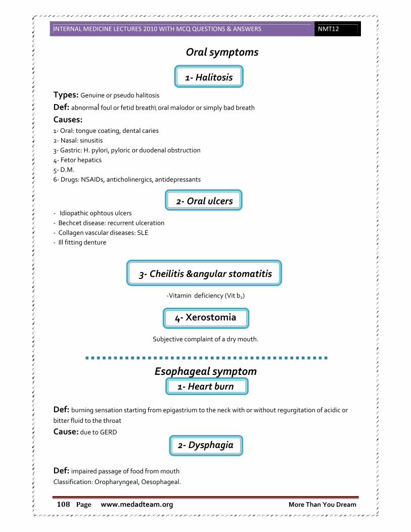
INTERNAL MEDICINE LECTURES 2010 WITH MCQ QUESTIONS & ANSWERS NMT12
www.medadteam.org More Than You Dream Page 108
1- Halitosis
Oral symptoms
Types: Genuine or pseudo halitosis
Def: abnormal foul or fetid breath\ oral malodor or simply bad breath
Causes: 1- Oral: tongue coating, dental caries
2- Nasal: sinusitis
3- Gastric: H. pylori, pyloric or duodenal obstruction
4- Fetor hepatics
5- D.M.
6- Drugs: NSAIDs, anticholinergics, antidepressants
2- Oral ulcers - Idiopathic ophtous ulcers
- Bechcet disease: recurrent ulceration
- Collagen vascular diseases: SLE
- Ill fitting denture
3- Cheilitis &angular stomatitis
-Vitamin deficiency (Vit b2)
4- Xerostomia
Subjective complaint of a dry mouth.
Esophageal symptom
1- Heart burn
Def: burning sensation starting from epigastrium to the neck with or without regurgitation of acidic or
bitter fluid to the throat
Cause: due to GERD
2- Dysphagia
Def: impaired passage of food from mouth
Classification: Oropharyngeal, Oesophageal.

INTERNAL MEDICINE LECTURES 2010 WITH MCQ QUESTIONS & ANSWERS NMT12
www.medadteam.org More Than You Dream Page 109
Causes: a- Upper oropharyngeal
• Neurological: bulbar muscles affection in strock usually bilateral, multiple sclerosis, myasthenia
gravis.
• structural: neoplasm , zenker diverticulum
b- Lower esophageal
1- Functional
- achalasia : no relaxation of esophageal sphincter
- diffuse esophageal spasm
- sclerodema: collagen disease affect esoph. Muscles
2- Organic
- Intrinsic esophageal disorders:
1- Long standing GERD lead to ulceration then fibrosis
2- Benign: post corrosive, post sclerotherapy.
3- Malignancies e.g. oesophageal adenocarcinoma.
- Extrinsic esophageal compression: Mediastinal tumors.
Evaluation of dysphagia
-Level: upper or lower
-Fluid or solid
-Age: young: corrosive - old: cancer
-Duration:
-Long: non malignant
-Course:
-Progressive: post corrosive, cancer
-Regressive: inflammatory
-Weight loss:
-Functional causes: not associated with weight loss
-Malignant: associated with weight loss
-Associated anemia or GIT bleeding:
GERD and malignancy are associated with bleeding
Esophageal spasm not associated with bleeding
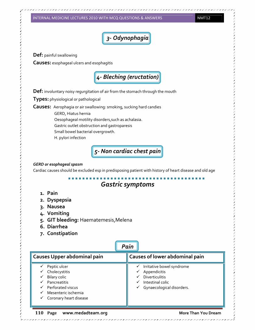
INTERNAL MEDICINE LECTURES 2010 WITH MCQ QUESTIONS & ANSWERS NMT12
www.medadteam.org More Than You Dream Page 110
3- Odynophagia
Def: painful swallowing
Causes: esophageal ulcers and esophagitis
4- Bleching (eructation)
Def: involuntary noisy regurgitation of air from the stomach through the mouth
Types: physiological or pathological
Causes: Aerophagia or air swallowing: smoking, sucking hard candies
GERD, Hiatus hernia
Oesophageal motility disorders,such as achalasia.
Gastric outlet obstruction and gastroparesis
Small bowel bacterial overgrowth.
H. pylori infection
5- Non cardiac chest pain
GERD or esophageal spasm
Cardiac causes should be excluded esp in predisposing patient with history of heart disease and old age
Gastric symptoms 1. Pain
2. Dyspepsia
3. Nausea
4. Vomiting
5. GIT bleeding: Haematemesis,Melena 6. Diarrhea
7. Constipation
Pain
Causes of lower abdominal pain Causes Upper abdominal pain
� Irritative bowel syndrome
� Appendicitis
� Diverticulitis
� Intestinal colic
� Gynaecological disorders.
� Peptic ulcer
� Cholecystitis
� Bilary colic
� Pancreatitis
� Perforated viscus
� Mesenteric ischemia
� Coronary heart disease

INTERNAL MEDICINE LECTURES 2010 WITH MCQ QUESTIONS & ANSWERS NMT12
www.medadteam.org More Than You Dream Page 111
Cholecystitis: pain radiating to the shoulder or the back after fatty meals
Biliary colic: pain in the RT hypocondrium or epigatrium
May be associated with vomiting , jaundice or change in color of urine
Pancreatitis: sever epigastric pain radiating to the back relieved or decreased by leaning forward
Perforated viscus: sever abdominal pain may be associated with peritonitis
Mesenteric ischemia: Manifestation appear after meal (digestion need more oxygen) Sometimes called abdominal angina
Sometimes associated with GIT bleeding, weight loss (the patient refuse to
eat to avoid the pain)
Type of patient: old age, diabetic and hypertensive
May be mistaken with malignancy
Appendicitis: acute pain at the RT quadrant associated with guarding, rigidity and fever
More common in old age
Diverticulitis: mostly in old age
Long standing constipation lead to increase in colonic pressure lead to pouch formation
Inflammation of the pouch leads to diverticulitis
More common in the left side
Diarrhea
Def: stool weight > 25 g/24 hours
Patient description Increase frequency of bowel movement
Increase stool liquidity
Sense of fecal urgency
Incontinence
Types
Acute: inflammatory (Viral,bacterial and protozoal ) and non inflammatory
Chronic due to
Osmotic, motility, inflammatory bowel, malabsorbtion and secretory
Causes of acute abdomen
Appendicitis
Perforated viscus
Mesenteric vascular occlusion
Peritonitis bacterial or familial Mediterranean fever (recurrent with family history)
Systemic diseases: Diabetes mellitus,Porphyria.

INTERNAL MEDICINE LECTURES 2010 WITH MCQ QUESTIONS & ANSWERS NMT12
www.medadteam.org More Than You Dream Page 112
Constipation
Def: <3 bowel motion /week
Causes:
Habitual
Poor dietary habits (low dietary fibers)
Obstruction (cancer colon)
Antispasmodic medication
GIT Bleeding
Hematemsis
Def: Vomiting of blood
Causes Esophageal varices e.g. hepatitis
Peptic ulcer
Neoplastic: cancer esophagus , cancer stomach
Gastritis
Esophagitis
Mallory- Weisslears syndrome: frequent severe vomiting and straining lead to injury to
esophageal mucosa lead to bleeding
Angiodysplasia: rupture of dilated capillaries in the stomach of duodenum
Hematochazia
Def: fresh blood per rectum which is bright red
Causes: Hemorrhoids
Cancer colon
Colonic polyps.
Inflammatory bowel syndrome
Diverticulosis
Angiodysplasia in the colon
Severe massive GIT bleeding.
Melena: Passage of black tarry stools.

INTERNAL MEDICINE LECTURES 2010 WITH MCQ QUESTIONS & ANSWERS NMT12
www.medadteam.org More Than You Dream Page 113
M.C.Q.
Section A: Read each question carefully and record the answer "TRUE" or "FALSE":
1. Ultrasonography is the diagnostic method of choice in: aa)) DDeetteeccttiinngg ggaallllbbllaaddddeerr ssttoonneess
b) Searching for a mass in the lower part of common bile duct
cc)) DDiiaaggnnoossiiss ooff vveeggeettaattiioonnss ooff ssuubbaaccuuttee bbaacctteerriiaall eennddooccaarrddiittiiss
dd)) DDeetteeccttiinngg aasscciitteess
e) Diagnosis of broncogenic carcinoma
Section B: Only one item appropriately applies to the statement:
2. Dyspepsia includes all of the following, except: a) Upper abdominal discomfort
b) Upper abdominal pain
c) Change of bowel habits
dd)) NNaauusseeaa && vvoommiittiinngg
e) Eructation
3. In functional dyspepsia, we should: a) Exclude gastric & duodenal pathology
b) Exclude gall bladder disease
c) Exclude uremia
d) Exclude diabetes
ee)) AAllll ooff tthhee aabboovvee
4. The following is implicated in the pathogenesis of functional dyspepsia: a) Delayed gastric motility
b) Helicobacter pylori infection
c) Erosive gastritis
dd)) PPssyycchhoollooggiiccaall ssttrreessss
e) Antroduodenal reflux
5. Symptoms of ulcer-like dyspepsia include the following, except: a) Pain in the epigastrium
b) Pain on fasting or after meals
cc)) PPaaiinn rreelliieevveedd bbyy aannttaacciidd oorr pprroottoonn ppuummpp iinnhhiibbiittoorr ((PPPPII))
d) Pain associated with hematemesis
e) Pain associated with heart burn
6. Dysmotility-like dyspepsia includes the following: a) Nausea & vomiting
b) Eructations
c) Upper abdominal discomfort
d) Bloating
ee)) AAllll ooff tthhee aabboovvee
7. Warning sign of functional dyspepsia include the following, except: a) Hematemesis
b) Age above 50 years
c) Loss of weight
dd)) FFeemmaallee
e) Elevated ESR

INTERNAL MEDICINE LECTURES 2010 WITH MCQ QUESTIONS & ANSWERS NMT12
www.medadteam.org More Than You Dream Page 114
8. Investigations of functional dyspepsia should be done in the following causes: a) Melena
b) Cachexia
c) Recent onset
dd)) AAllll ooff tthhee aabboovvee
e) None of the above
9. Goals of treatment of functional dyspepsia include the following, except: a) Reassurance
b) Understanding the patient concerns
c) Relieving the symptoms
dd)) EErraaddiiccaattiioonn ooff tthhee ssyymmppttoommss
e) All of the above
10. Investigations for dyspepsia include: a) Upper endoscopy
b) Abdominal ultrasound
c) Blood urea
d) Fasting & 2hrs postprandial blood sugar
ee)) AAllll ooff tthhee aabboovvee
11. In cholestatic liver disease, indications of liver transplantation are all of the
following, except: a) Hyperbilirubinemia
bb)) HHyyppeerrlliippiiddeemmiiaa
c) Bony diseases
d) Growth retardation
e) Intactable pruritis
12. Common causes of dysphagia include all, except: a) Cancer
bb)) CCeerrvviiccaall ssppoonnddyylloossiiss
c) Achalasia
d) Post-corrosive
13. GERD may present by: a) Heart burn
b) Chest pain
c) Epigastric pain
d) Bronchial asthma
ee)) AAllll ooff tthhee aabboovvee
14. Mechanisms of diarrhea include the following, except: a) malabsorption
bb)) IInnccrreeaasseedd fflluuiidd iinnttaakkee
c) Mucosal injury
d) Motility disorders
15. Osmotic Gap increase in: a) Malabsorption diarrhea
bb)) MMoottiilliittyy ddiissoorrddeerr
c) Secretory diarrhea
d) Osmotic diarrhea

INTERNAL MEDICINE LECTURES 2010 WITH MCQ QUESTIONS & ANSWERS NMT12
www.medadteam.org More Than You Dream Page 115
16. First battery of investigations in chronic diarrhea include all, except:
aa)) ooccccuulltt bblloooodd
b) barium follow through
c) Sigmoidoscopy
d) Stool electrolytes
17. The following are symptoms of constipation except a) Hard stools > ¼ times of defecation.
b) Incomplete evacuation > ¼ times of defecation.
cc)) SSooiilliinngg ooff ccllootthhss..
d) Straining in > ¼ times of defecation hard stools > ¼ times of defecation.
18. The following may be a cause of constipation except a) Ignoring urge
b) Antispasmodics
c) Irritable bowel syndrome
dd)) TThhyyrroottooxxiiccoossiiss

INTERNAL MEDICINE LECTURES 2010 WITH MCQ
www.medadteam.org Page 116
���� Definition:
Yellowish discoloration of skin, mucous membranes & the eyes (more sen
Due to accumulation of bilirubin > 3mg/dl
The yellow pigment is from bilirubin, a byproduct of old red blood cells.
1. Define jaundice.
2. Explain bilirubin metabolism
3. Classify jaundice and explain the pathogenesis of each type and list the
commonest causes of each type.
4. Describe the clinical and laboratory features of each type of jaundice
5. Differentiate between extra hepatic and intrahepatic cholestasis and list the
commonest causes of each
CTURES 2010 WITH MCQ QUESTIONS & ANSWERS
www.medadteam.org
1. Jaundice
Yellowish discoloration of skin, mucous membranes & the eyes (more sensitive than others),
Due to accumulation of bilirubin > 3mg/dl
The yellow pigment is from bilirubin, a byproduct of old red blood cells.
Lectures objectives
Explain bilirubin metabolism
Classify jaundice and explain the pathogenesis of each type and list the
commonest causes of each type.
the clinical and laboratory features of each type of jaundice
Differentiate between extra hepatic and intrahepatic cholestasis and list the
commonest causes of each
NMT12
sitive than others),
……Normally it
is <1mg/dl.
Classify jaundice and explain the pathogenesis of each type and list the
the clinical and laboratory features of each type of jaundice
Differentiate between extra hepatic and intrahepatic cholestasis and list the
INTERNAL MEDICINE LECTURES 2010 WITH MCQ QUESTIONS & ANSWERS NMT12
www.medadteam.org More Than You Dream Page 117
���� Sources of UCB (Un-conjugated Bilirubin):
� Hb breakdown(normal life span of RBCS = 120 days)
� Catabolism of other heme containing protein(e.g.myoglobin& cytochrome containing
enzymes)
� Infective erythropoiesis.
���� Rate of production:
� Bilirubin in Blood is normally almost all unconjugated
� Rate of production of UCB 250-350 mg/day (important) from catabolism of heam after
removal of its iron component.
� UCB:
- toxic
- Not water soluble
- Bound to albumin
- Can’t pass to urine
� While conjugate(mono- & di-) are:
- non toxic
- water soluble
- not bound to albumin
- pass to urine
• N.B: In the endoplasmic reticulum :UCB by glucoronyl transferase become bilirubin mono
and di glucoronide.
• In the colon conjugated bilirubin is metabolized by colonic bacteria forming stercobilinogen
which is further oxidized into stercobilin.
• Both stercobilinpgen and stercobilin are excreted in the stools.
Small amount of stercobilinogen (4 mg./d) is absorbed from the bowel , pass through the
liver and excreted in urine (urobilinogen ) which is further oxidized into urobilin.
���� Classification of Jaundice:
1. Pre-hapatic (haemolytic)
2. Hepatic
3. Post-hapatic (obstructive)
���� Mechanism of jaundice:
� +++Production of bilirubin à haemolysis
� Impaired excretion:
I. Congenital non hemolytic hyperbilirubinemia:
1. Gilbert syndrome.
2. Crigler-Najar.
3. Dubin Johnson syndrome.
4. Rotor syndrome.
II. Hepatocellular jaundice:
1. Acute paranchymal liver disease.
2. Chronic paranchymal liver disease.

INTERNAL MEDICINE LECTURES 2010 WITH MCQ QUESTIONS & ANSWERS NMT12
www.medadteam.org More Than You Dream Page 118
III. Cholestasis:
1. Extra-hepatic cholestasis (surgical).
2. Intra-hepatic cholestasis (medical).
(A.) Hemolytic Jaundice
���� Etiology:
� Due to increase destruction of RBCs leading to bilirubin production.
- Usually mild
- Healthy liver can excrete a billirubin load up to 6 times (important) greater than normal
before UCB accumulates in the plasma.
� In haemolytic jaundice +++ UCB, Uroblinogen, Stercoblinogen
���� Clinical features:
� No stigmata of chronic liver disease,only features as ablood disease
� Pallor due to anemia
� ↑↑ excretion of bilirubin � ↑↑ stercobilinogen � Normal color stool or dark stool
� ↑↑ Urobilinogen excretion � Urine darkens on standing.
� Splenomegaly due to excess RE activity
� Pigment stones
� Leg ulcers
���� Investigations:
� Biochemical tests:
- Pl. billirubin < 6 % (indirect)
- Liver function tests à Normal
- Urine : no bilirubinuria ( unconjugated bilirubin is water insoluble).
Excess urobilinogen
� Blood count: anemia
� Blood film: haemolysis
(B.) Hepatocellular jaundice
���� Etiology:
� Inability of liver to transport bilirubin into bile as a result of parenchymal liver diesase
� Billirubin transport across the hepatocytes may be impaired at any point between uptakes of unconjugated bilirubin into the cells and transport of conjugated bilirubin into canaliculi.
� In addition: swelling of cells & edema.
� Increase:unconjugated bilirubin,conjugated bilirubin,stercobilinogen and urobilinogen.
���� Clinical Manifestations:
� Lemon yellow jaundice.
� Dark urine.

INTERNAL MEDICINE LECTURES 2010 WITH MCQ QUESTIONS & ANSWERS NMT12
www.medadteam.org More Than You Dream Page 119
� +/- Manifestations of chronic liver diseases:
- Ascites
- Circulatory changes: palmar eryth.,spider naev
- Endocrin.: Loss of lipido,hair loss,gynecomast.
- Haemorrahgic tendency
- Manifestations of hepatic encephalopathy.
- Manifestations of portal hypertension.
- Others: pigmentations,clubbing,low grade fever.
���� Investigations:
� Biochemical tests:
- Pl. billirubin: biphasic increase both direct & indirect
- Increase enzymes
- Evidence of chronic liver diseases:
o Decrease palsma proteins (More sensitive)
o Increase prothrombin time
o Decrease concentration
(C.) Cholestatic (obstructive) jaundice
���� Etiology:
� Conjugated bilirubin is unable to enter bile canaliculi(due to inflamed hepatocytes
compressing them) & pass back to blood
� Failure of clearance of unconjugated bilirubin arriving at the liver cells.
� Features of elevation of UCB due to tumor obstruction of canlicular system
Causes: failure of hepatocytes to generate bile flow.
Obstruction to the bile flow in bile ducts in the portal tracts.
Obstruction to bile flow in the extrahepatic bile ducts between porta hepatica and
papillae of vater.
���� Causes of intra-hepatic obstruction:
� 1ry biliary cirrhosis.
� Auto-immune hepatitis
� 1ry sclerosing cholangitis:
- Alcohol
- Drug
- Preganancy(important)
- Viral hepatitis
- Severe bacterial infection
���� Causes of extra hepatic obstruction:
- Cholethiasis
- Carcinoma
- Parasitic infection
- Traumatic: biliary stricture

INTERNAL MEDICINE LECTURES 2010 WITH MCQ QUESTIONS & ANSWERS NMT12
www.medadteam.org More Than You Dream Page 120
� In cholestatic jaundice:
- ↑↑ S.Bilirubin mainly direct
- ↑↑ Direct bilirubin in urine
- ↓↓ Stercoblinogen
- ↓↓ Stercoblin
���� Clinical features:
� Cholestasis
� Early:
- Jaundice, dark urine, pale stool(clay coloured), purities.
� Late:
- Xanthelasma & Xanthomata(due to +++ cholesterol)
- Mal-absorption
- Weight loss
- Steatorrhea(due to malabsorbtion of fat)
- Osteomalacia
- Bleeding tendancy
� + Clinical picture of cholangitis
� + Underlying chronic Liver disease
���� Investigations:
� Biochemical:
- Increase direct bilirubin
- Greater increase gamma GT& Alkaline phosphatase compared toaminotransferase.
� Ultrasound: to differentiate intra from exta hepatic biliary obstruction
- Intrahepatic ---- no dilated bile canaliculi
- Extrahepatic ---- dilatation of all bile canaliculi
[also can be detected by (ERCP)endoscopic retrogradecholaggio- pancreatography]
� CT & MRI: may be helpful.
2. Acute Viral Hepatitis
���� What is hepatitis?
• Hepatitis is the Latin word for liver inflammation.
• It is characterized by the destruction of a number of liver cells and the presence of
inflammatory cells in the liver tissue.
���� Viral hepatitis:
� Hepatitis A through E (more than 95% of viral cause).
� Herpes simplex, Cytomegalo-virus, Epstein-Barr, yellow fever virus, adenoviruses.
• Non viral infection: toxoplasma, Leptospira, Q fever, rocky mountain spotted fever
• Alcohol
• Toxins: Amanita toxin in mushrooms, carbon tetrachloride.
• Drugs: Paracetamol, antituberculosis medicines, minocycline.

INTERNAL MEDICINE LECTURES 2010 WITH MCQ QUESTIONS & ANSWERS NMT12
www.medadteam.org More Than You Dream Page 121
• Ischemic hepatitis (circulatory insufficiency)
• Pregnancy
• Auto immune conditions, e.g., Systemic Lupus Erythematosus (SLE)
• Metabolic diseases, e.g., Wilson's disease, Hemocromatosis
���� Etiology of acute viral Hepatitis
• HAV / HBV / HCV / HDV / HEV / HGV / TTV
• Acute viral hepatitis is a systemic infection affecting the liver predominantly.
Hepatitis A & E • Incubation period: 2 – 4 weeks
• Transmission: Feco-oral
• Prevention: Vaccine against A
• Chronicity: no
• Treatment: non specific
���� HAV Transmission
• Primarily transmitted via fecal-oral route
• Highly infectious and stable in environment for months
• Most common transmission through close personal contact with an infected person
• Vaccine is available to prevent infection
� Hepatitis B, C & D
• Incubation period: 4 – 24 weeks.
• Transmission: Blood & body secretion.
• Prevention: Vaccine against B.
• Chronicity: all can leads to chronic liver.
• Treatment: non specific.
HBV • Transmitted by blood, semen, vaginal fluids
• Highly infectious, stable in environment for at least 7 days
• Most common transmission through:
⇒ perinatal (mom to baby)
⇒ unprotected sex
⇒ percutaneous (through opening in skin)
• Vaccine is available to prevent infection
���� Hepatitis B: Virion Structure
1- HBsAg, envelope glycoprotein
2- HBeAg (Core + PreCore)
3- HBcAg, nucleocapsid
4- DNA polymerase

INTERNAL MEDICINE LECTURES 2010 WITH MCQ QUESTIONS & ANSWERS NMT12
www.medadteam.org More Than You Dream Page 122
5- dsDNA
HCV • HCV is an enveloped positve RNA virus
• Six genotypes
• Genotype 1 predominant in Europe and North America
• Genotype 4 in Egypt
• Genotype 1 and 4 most difficult to treat
���� HCV research
• Unknowns
• No cell culture system
• No small animal model
���� HCV Transmission
• Transmitted by direct blood-to-blood contact
• Highly infectious, stable in environment for at least 16 hours but not longer than 4
days
• Most common transmission through sharing of injection drug.
• Also blood transfusions & products before 1992
– Perinatal transmission (4% chance)
– Needle stick/health care exposure (1.8%)
– Sexual transmission
– Other blood risks low/unknown risk: tattooing/piercing intranasal cocaine use,
shared personal items
���� Pathology of Acute Hepatitis
• Diffuse inflammation lead to hepatic cell necrosis associated with inflammatory cell
infiltration, Kuppfer cell hyperplasia.
• In severe cases with massive necrosis, there is marked reduction in liver size and
development of fibrous septa (post – necrosis scarring).
���� Clinical picture of acute hepatitis
1. Asymptomatic
2. Non Icteric: flu – like symptoms, GI symptoms.
3. Typical icteric attak.
� Typical icteric attack:
1. Prodromal phase
2. Icteric phase
3. Recovary phase
� Complications:
1. Prolonged Cholestasis
2. Acute Liver Failure
� Prodromal phase (3 – 7 d)
• Malaise – ANV – fever – Headache.
INTERNAL MEDICINE LECTURES 2010 WITH MCQ QUESTIONS & ANSWERS NMT12
www.medadteam.org More Than You Dream Page 123
� Icteric phase:
• Jaundice
• Dark urine
• Pale stools
• Fever decrease
• Tender mildly enlarged liver
• Enlarged spleen (25 %)
� Recovary phase
• Jaundice disappears .
• Improve general condition.
• Lassitude persist.
���� Prolonged cholestasis
• Jaundice deepen
• Itching
• Last up to 6 months
• Complete recovery
• More with HAV
���� Acute liver failure
• Rare serious complication.
• Failure of the liver functions.
• Bleeding – Confusion – Coma - Death.
���� Investigations for acute hepatitis
• Urine
++ bilirubin
• Bilirubin level
++ both direct and indirect bilirubin (hepatocellular)
• Liver Enzymes
++ AST, ALT. up to 100 folds – particularly in HAV.
• Serological Diagnosis:
Serological Diagnosis
• HAV
• Virus can be detected PCR in stool (not used)
• HAV Ab: IgM – IgG
• HBV
• HBsAg: appears 3 weeks, disappears months, if persists it implies a carrier state.
• HBsAb: appears 3 m, persists for several years and implies immunity.
• HBeAg: indicate viral replication and infectivity, persistence indicate chronicity.
• HBeAb: evidence that the patient recovery.
• Cont. HBV
• HBcAg: can not be detected in blood (Biobsy)
• HBcAb: acute hepatitis persistence indicate chronicity

INTERNAL MEDICINE LECTURES 2010 WITH MCQ QUESTIONS & ANSWERS NMT12
www.medadteam.org More Than You Dream Page 124
• HBV DNA by PCR: indicate active viral infection, most reliable test.
• HCV
o HCV Ab:
o Detected by ELISA after 10 weeks.
o HCV RNA by PCR:
o To confirm the diagnosis (qualitative) and to assess viral load (quantitative).
HDV
• The delta agent is a defective virus which shows similarities with the viroids in
plants.
• The agent consists of a particle 35 nm in diameter consisting of the delta antigen
surrounded by an outer coat of HBsAg.
• The genome of the virus is very small and consists of a single-stranded RNA
���� Hepatitis D
Clinical Features
� Coinfection
– Severe acute disease.
– Low risk of chronic infection.
� Superinfection
– Usually develop chronic HDV infection.
– High risk of severe chronic liver disease.
– May present as an acute hepatitis.
� Modes of Transmission
– Percutanous exposures
– injecting drug use
– Permucosal exposures
– sex contact
� Prevention
HBV-HDV Coinfection
Pre or postexposure prophylaxis to prevent HBV infection.
HBV-HDV Superinfection
Education to reduce risk behaviors among persons with chronic HBV infection.
HEV
• Calicivirus-like viruses
• unenveloped RNA virus, 32-34nm in diameter
• +ve stranded RNA genome, 7.6 kb in size.
• very labile and sensitive
• Can only be cultured recently
INTERNAL MEDICINE LECTURES 2010 WITH MCQ QUESTIONS & ANSWERS NMT12
www.medadteam.org More Than You Dream Page 125
� Clinical Features
Incubation period: Average 40 days
Case-fatality rate: Overall, 1%-3%,Pregnant women, 15%-25%
Illness severity: Increased with age
Chronic sequelae: None identified
� Epidemiologic Features
Most outbreaks associated with faecally contaminated drinking water.
Several other large epidemics have occurred since in the Indian subcontinent and
the USSR, China, Africa and Mexico.
In the United States and other nonendemic areas, where outbreaks of hepatitis E
have not been documented to occur, a low prevalence of anti-HEV (<2%) has been
found in healthy populations. The source of infection for these persons is unknown.
� Prevention and Control Measures for Travelers to HEV-Endemic Regions
Avoid drinking water of unknown purity, uncooked shellfish, and uncooked
fruit/vegetables not peeled or prepared by traveler.
� Treatment of acute viral hepatitis
• Prophylaxis
• Bed Rest
• Convalesence
• Diet
• Drugs
� Prophylaxis
• Good hygienic measures.
• Isolation
• Immunisation
Passive: Anti-HAV Ig, Anti-HBV Ig.
Active: HAV, HBV.
Bed Rest:
Until the patient feels well
Convalesence:
Twice the period of bed rest, till bilirubin less than 1.5 mg/dl
Diet
• No specific diet
• Low fat (nausiating)
Drugs
• Avoid hepatotoxic drugs
• Symptomatic treatmen
INTERNAL MEDICINE LECTURES 2010 WITH MCQ QUESTIONS & ANSWERS NMT12
www.medadteam.org More Than You Dream Page 126
3. Liver Cirrhosis & Portal Hypertension
���� Definition:
-Diffuse hepatic disease characterized by (Triad)
Fibrosis(like wound healing)
distortion of hepatic architecture
formation of regenerative nodules (micro,macro or mixed)
-Cirrhosis is usually the end stage of any Chronic injury, chronic injury to the liver results in
inflammation, necrosis and eventually fibrosis.
-Role of stellate cells
���� Etiology:
1. Chronic viral hepatitis: is the most important cause of liver cirrhosis. o HCV (the commonest form of hepatic viral infections in Egypt)
o HBV (less common than HCV)
o HDV (on top of HBV)
o CMV and EBV
2. Fatty liver: NASH (Non Alcoholic SteatoHepatitis)
o it is one of a group of diseases named Non alcoholic fatty liver diseases
(NAFLD) and is related to metabolic syndrome and insulin resistance.
3. Metabolic:
o Wilson's disease: an inherited disorder of copper homeostasis leading to
copper deposition into tissues usually presented in young ages and
adolescence. � TTT: is chelating of copper by D-penicillamine and trientine.
o Hereditary hemochromatosis (HH): a common inherited disorder of iron
metabolism leading to iron deposition in tissues (ex: liver leading to liver
cirrhosis), this disease is usually asymptomatic and is presented clinically in the
3rd or 4th decade of life
o α1 antitrypsin deficiency.
o NASH
N.B.: in Metabolic disorders early detection and control is the major line of prevention
of liver cirrhosis in such patients.
4. Alcoholic liver disease: Not common due to cultural variations.
Lectures objectives
1. Identify the classification of portal hypertension 2. Describe the underlying pathogenic mechanisms of portal hypertension. 3. List the causes of portal hypertension. 4. Describe different presentations of portal hypertension (varices,
splenomegaly, encephalopathy, and ascites). 5. Enumerate the different investigations of a case of portal hypertension 6. Outline the management protocol of variceal haemorrhage

INTERNAL MEDICINE LECTURES 2010 WITH MCQ QUESTIONS & ANSWERS NMT12
www.medadteam.org More Than You Dream Page 127
5. Biliary:
o Primary and secondary
6. Autoimmune hepatitis.
7. Drug or toxin induced liver injury, Ex: prolonged use of Methotrexate (MTX) which is a folic
acid antagonist used for ttt of malignancies.
8. Vascular:
o Chronic right side heart failure
o Budd-Chiari syndrome (symptomatic obstruction or occlusion of hepatic vein)
���� Clinical picture:(It is a very imp. Oral Q. in clinical exam)
� Symptoms of liver cirrhosis:
1-Asymptomatic (Except after occurrence of liver cell failure)
2-General
o Weakness, Malaise, Anorexia, Fatigue, Weight loss.
3-Liver cell failure:
o Hypoalbuminemia:
� Albumin is responsible for oncotic pressure of blood; decrease of serum
albumin will lead to decrease of oncotic pressure, hence exudation of the
intravascular fluid into interstitial fluid leading to:oedema and ascites.
o Coagulopathy:
� Bleeding tendency: is due to deficient coagulation factors
� Bleeding gums (most common early presentation of the disease),
Epistaxis, Menorrhagia, Ecchymosis
� Also bleeding tendency occurs due to hypersplenism which is a condition
characterized by exaggeration of the suggested inhibitory or destructive
functions of the spleen, resulting in a deficiency of the peripheral blood
elements, singly or in combination, hypercellularity of the bone marrow, and
usually, but not always, splenomegaly.
� Over destruction of RBCs by spleen will cause anemia while over destruction
of platelets will lead to coagulopathy (thrombocytopenia).
o Endocrine changes:
Loss of libido,hair loss
Men:Gynecomastia,testicular atrophy,impotence
Women:Breast atrophy,irregular menses,amenorrhoea
Hepatic encephalopathy:

INTERNAL MEDICINE LECTURES 2010 WITH MCQ QUESTIONS & ANSWERS NMT12
www.medadteam.org More Than You Dream Page 128
i. Subclinical: sometimes patient are alert and conscious, however,
clinically these condition may be elicited by certain clinical test (You can
ask your patient to connect points drawn on a paper).
ii. Flapping tremors: also known as liver flap (i.e., a sudden forward
movement of the wrist) the patient fails to keep his extended arms and
wrist infront of him in the same position. It is a sign of end-organ failure
most commonly liver cell failure.
iii. Lethargy: feeling of tiredness and laziness.
iv. Disturbed sleep rhythm and conscious level.
v. Personality changes.
4-Portal Hypertension
� Signs of liver cirrhosis:
1. Protein-calorie malnutrition
2. Skin manifestations:
3. Spider navei
4. Palmer & plantar erythema
5. Clubbing of fingers
6. Manifestations of hyper-dynamic circulation
7. Abdominal examination:
a- Ascites
b- Umbilical & abdominal wall hernia
c- Palpable, firm in consistency, tender & enlarged liver
� Complications of liver cirrhosis
A. Hepatocellular carcinoma
Patient is present by general manifestations of malignancy.
B. Spontaneous Bacterial peritonitis (SBP)
SBP is a common and severe complication of ascites characterized by spontaneous
infection of the ascitic fluid without an intraabdominal source, patient is presented by
fever and other manifestations of peritonitis.
C. Hepatorenal syndrome(HRS)
D. Others:Portopulmonary hypertention(PPH)
:Hepatopulmonary syndrome(HPS)
� Clinical Examination
A. Jaundice: increased serum bilirubin due to inability of the liver to excrete bilirubin
(Normal bilirubin level is not more than 1mg/dl)
B. Palmar erythema: persistant redness of the palms, it is not a pathognomonic sign for
liver cirrhosis as it occurs in different diseases and may occur normally during
pregnancy.
C. Spider Naevi: (also known as spider angiomata and also present normally during
pregnancy, not a pathognomonic sign)
Feminization

INTERNAL MEDICINE LECTURES 2010 WITH MCQ QUESTIONS & ANSWERS NMT12
www.medadteam.org More Than You Dream Page 129
Portal hypertention
Definition:Portal hypertension is defined as the elevation of the hepatic venous pressure
gradient (HVPG) to >5 mmHg.
Normal portal pressure:2-5mmHg
Clinical features if > 12 mmHg
Etiology and pathogenesis:-
1-portal blood flow versus portal vascular resistance
2-Increased vascular resistance (the main cause)
3-Cirrhosis(sinusoidal) >90% of causes
Increased intrahepatic vascular resistance
A-Mechanical>>> intrahepatic fibrosis
-Framwork distortion
-Regenerative nodules
-Collagen deposition in the space of disse
B-Dynamic>>>Vasoconstriction in portal venules
-Imbalance between vasodilators (e.g NO) and vasoconstrictors (e.g NA)
N.B; PORTAL HYPERTENTION
A-Pre-sinusoidal:
1-Intra hepatic: Portal vein thrombosis or compression
2-Extra hepatic:bilharziasis(priportal fibrosis)
B-Sinusoidal:
1-Liver cirrhosis
C-Post-sinusoidal:
1-Intra-hepatic:Veno-occlusive disease
2-Extra-hepatic:Budd-chiari syndrome
Clinical presentation of PH:- (Important oral Q. for clinical exam)
I. Asymptomatic (mainly)
II. Splenomegaly:
o the most common symptom is pain in the left hypochondrial pain
o Hypersplenism.
III. Ascites
o an increase in intrahepatic resistance, causing increased portal pressure

INTERNAL MEDICINE LECTURES 2010 WITH MCQ QUESTIONS & ANSWERS NMT12
www.medadteam.org More Than You Dream Page 130
o there is also vasodilatation of the splanchnic arterial system, which in turn
results in an increase in portal venous inflow
o Both of these abnormalities result in increased production of splanchnic
lymph.
o V.D. factors such as nitric oxide.
o These hemodynamic changes result in sodium retention by causing
activation of the renin-angiotensin-aldosterone system with the
development of hyperaldosteronism
o The renal effects of increased aldosterone leading to Na retention
o Na retention causes fluid accumulation and expansion of the extracellular
fluid volume.
o Hypoalbuminemia and reduced plasma oncotic pressure also contribute to
the loss of fluid from the vascular compartment into the peritoneal cavity.
o Hypoalbuminemia is due to decreased synthetic function in a cirrhotic liver.
Development of ascites in liver cirrhosis
Cirrhosis
Portal hypertension
Splanchnic vasodilatation
Increase splanchnic pressure Arterial underfilling
Lymph formation activation of vasoconstrictors
Formation of ascites & antinatriuretic factors
Plasma volume expansion sodium retention
IV. Porto-systemic shunt:
o Encephalopathy
o Normally, liver receives blood from gut via the portal vein, Liver
detoxifies blood from its toxins (ex: ammonia), when pressure inside the
portal vein increases, blood shifts into other anastomosis and collaterals
bypassing the liver, hence, blood remains full of toxins, when these toxins
reaches cerebral circulation they cause symptoms of hepatic
encephalopathy.
o Collaterals: Varices, Caput medusae.
V. GIT bleeding:
o Oesophagial variceal bleeding (the commonest GIT bleeding, presented
clinically as hematemesis and/or melena)
o Hemorrhoids

INTERNAL MEDICINE LECTURES 2010 WITH MCQ QUESTIONS & ANSWERS NMT12
www.medadteam.org More Than You Dream Page 131
� Management of bleeding…… (Acute Variceal Bleeding)
A. Patient stabilization:In ICU>>>> IV fluids. Packed RBCs.
B. Patient assessment and insure open airway,avoid aspiration.IV lines
C. Aim:
1-correction of hypovolemic shock
2-prevent complications associated with GIT bleeding
3-stop bleeding by hemostasis at the bleeding sites
Specific treatment:-
A-Endoscopic management:
o Band ligation: treatment of varices by binding them at the base with rubber bands
so that the distal portion sloughs away within several days.
o Sclerotherapy: injection of a chemical irritant into veins to produce inflammation
and eventual fibrosis and obliteration of the lumen.
o Variceal obstruction by glue
B.Pharmacologic: Vasopressin, Somatostatin analogues.
C.Variceal tamponade: Sengstaken-Blakemore tube (rarely used)
D. Porto-systemic shunt: very rarely used
o Sugical
o TIPSS (Transjugular Intrahepatic PortoSystemic Shunt): it is a very invasive surgical
procedure and is considered the last option of treatment after failure of any other
line of management.
M.C.Q.
Section A: Read each question carefully and record the answer "TRUE" or "FALSE":
1. In the normal liver: aa)) TThhee ssppaaccee ooff DDiissssee sseeppaarraatteess tthhee hheeppaattooccyytteess ffrroomm ssiinnuussooiiddaall eennddootthheelliiuumm..
bb)) TThhee hheeppaattiicc aarrtteerryy ssuupppplliieess 5500%% ooff tthhee ttoottaall hheeppaattiicc ooxxyyggeenn ssuuppppllyy..
cc)) KKuuppffffeerr cceellllss aarree ddeerriivveedd ffrroomm bblloooodd mmoonnooccyytteess..
dd)) IIttoo cceellllss aarree rreessppoonnssiibbllee ffoorr tthhee uuppttaakkee aanndd ssttoorraaggee ooff vviittaammiinn DD..
e) The right and left hemilivers are divided into 10 segments.
2. Bilirubin is: a) Derived exclusively from the breakdown of hemoglobin.
b) Bound in the unconjugated form to plasma B-globin.
cc)) CCoonnjjuuggaatteedd iinn tthhee mmiiccrroossoommeess ooff tthhee hheeppaattooccyytteess..
d) Reabsorbed in the small bowel as bilirubin diglucronide.
e) Normally excreted as stercobilinogen in the feces and as urobilinogen in the urine.
3. Hepatic encephalopathy in cirrhosis is typically precipitated by: aa)) IInnffeeccttiioonn..
bb)) HHyyppookkaalleemmiiaa..
cc)) AAbbddoommiinnaall ssuurrggeerryy..
dd)) GGaassttrrooiinntteessttiinnaall bblleeeeddiinngg..
e) Lactulose therapy.
INTERNAL MEDICINE LECTURES 2010 WITH MCQ QUESTIONS & ANSWERS NMT12
www.medadteam.org More Than You Dream Page 132
4. Characteristic features of Glibert's syndrome include: a) An autosomal recessive mode of inheritance.
bb)) DDeeccrreeaasseedd hheeppaattiicc ggllooccuurroonnyyll ttrraannssffeerraassee aaccttiivviittyy..
cc)) UUnnccoonnjjuuggaatteedd hhyyppeerrbbiilliirruubbiinneemmiiaa << 110000 ppmmooll//LL..
dd)) SSeerruumm bbiilliirruubbiinn ccoonncceennttrraattiioonn iinnccrreeaassee bbyy ffaassttiinngg..
e) increased serum bile acide concentrations.
5. The following features suggest extrahepatic cholestasis rather than viral hepatitis: aa)) aa ppaallppaabbllee ggaallll bbllaaddddeerr..
b) right hypochondrial tenderness.
cc)) sseerruumm aallkkaalliinnee pphhoosspphhaattaassee ccoonncceennttrraattiioonn >> 22..55 ttiimmeess nnoorrmmaall..
dd)) pprruurriittuuss aanndd rriiggoorrss..
ee)) ppeerriipphheerraall bblloooodd ppoollyymmoorrpphh lleeuuccooccyyttoossiiss..
6. The histopathological characteristics of acute hepatitis include: a) Polymorph leucocyte infiltration of the lobules.
b) Sparing of the centrilobular areas.
cc)) EEnnllaarrggeemmeenntt ooff tthhee ppoorrttaall ttrraaccttss..
dd)) HHeeppaattooccyyttee nneeccrroossiiss wwiitthh ddeeeeppllyy--ssttaaiinneedd aacciiddoopphhiilliicc bbooddiieess..
e) Fatty infiltration.
7. The typical features of type A viral hepatitis (HAV) include: a) Picornavirus infection spread by the feco-oral route.
b) An incubation period of 3 months.
c) A greater risk of acute liver failure in the young than in the old.
dd)) RRiigghhtt hhyyppoocchhoonnddrriiaall ppaaiinn aanndd tteennddeerrnneessss..
e) Progression to cirrhosis if cholestasis is prolonged.
8. Circulating hepatitis B surface antigen (HBsAg) is: aa)) DDeetteeccttaabbllee dduurriinngg tthhee pprrooddrroommee ooff aaccuuttee ttyyppee BB hheeppaattiittiiss..
bb)) aa DDNNAA vviirraall ppaarrttiiccllee ttrraannssmmiissssiibbllee iinn aallll bbooddyy fflluuiiddss..
c) Likely to persist in about 50% of adults following acute type B hepatitis.
d) Invariably present in a patient with jaundice attributable to type B hepatitis infection.
e) Commoner in asymptomatic subjects in the Western rather than the Eastern hemisphere.
9. The typical features of type B viral hepatitis (HBV) include: a) An incubation period of 1 month.
bb)) HHiissttoorryy ooff eexxppoossuurree ttoo uunnssaaffee sseexx oorr ddrruugg aabbuussee..
cc)) PPrrooddrroommaall iillllnneessss wwiitthh ppoollyyaarrtthhrraallggiiaa..
dd)) HHeeppaattiittiiss iillllnneessss mmoorree sseevveerree tthhaann wwiitthh ttyyppee AA vviirruuss..
e) Absence of progression to chronic hepatitis.
10. The typical features of acute hepatic failure include: aa)) OOnnsseett wwiitthhiinn 88 wweeeekkss ooff tthhee iinniittiiaall iillllnneessss..
b) Hepatosplenomegaly and ascites.
cc)) EEnncceepphhaallooppaatthhyy aanndd ffeettoorr hheeppaattiiccuuss..
dd)) NNaauusseeaa,, vvoommiittiinngg aanndd rreennaall ffaaiilluurree..
ee)) CCeerreebbrraall ooeeddeemmaa wwiitthhoouutt ppaappiillllooeeddeemmaa..
11. Typical liver function values in acute hepatic failure include: a) Hypoalbuminemia.
bb)) HHyyppooggllyycceemmiiaa..
cc)) PPrroolloonnggeedd pprrootthhrroommbbiinn ttiimmee..
d) Serum alkaline phosphatase > three times normal.
INTERNAL MEDICINE LECTURES 2010 WITH MCQ QUESTIONS & ANSWERS NMT12
www.medadteam.org More Than You Dream Page 133
e) Peripheral blood lymphocytosis.
12. The clinical features of autoimmune hepatitis include: aa)) PPrreeddoommiinnaannccee ooff ffeemmaalleess aaggeedd 2200--4400 yyeeaarrss..
bb)) AAccuuttee oonnsseett ssiimmuullaattiinngg vviirraall hheeppaattiittiiss iinn 2255%% ooff ppaattiieennttss..
cc)) AArrtthhrraallggiiaa,, ffeevveerr aanndd aammeennoorrrrhheeaa..
dd)) SSppiiddeerr tteelleennggiieeccttaassiiaa aanndd hheeppaattoosspplleennoommeeggaallyy..
ee)) CCuusshhiinnooiidd ffaacciieess,, hhiirrssuuttiissmm aanndd aaccnnee..
13. In patients with hepatic cirrhosis: a) Central cyanosis responds well to oxygen therapy.
bb)) IInnccrreeaassiinngg jjaauunnddiiccee ssuuggggeessttss pprrooggrreessssiivvee lliivveerr ffaaiilluurree..
c) The peripheral blood flow is typically reduced.
dd)) TThhee gglloommeerruullaarr ffiillttrraattiioonn rraattee iiss ddeeccrreeaasseedd..
ee)) OOeessoopphhaaggeeaall vvaarriicceess iinnddiiccaattee ppoorrttaall hhyyppeerrtteennssiioonn..
14. Hepatic encephalopathy due to progressive liver failure is suggested by: aa)) DDyyssaarrtthhrriiaa aanndd cchhoorreeaa..
b) Focal neurological signs.
cc)) YYaawwnniinngg aanndd hhiiccccoouugghhiinngg..
d) Serum aminotransferase activity > 10 times normal.
e) Epilepsy and disorientation .
15. Hepatic encephalopathy in cirrhosis is typically precipitated by: aa)) IInnffeeccttiioonn..
bb)) HHyyppookkaalleemmiiaa..
cc)) AAbbddoommiinnaall ssuurrggeerryy..
dd)) GGaassttrrooiinntteessttiinnaall bblleeeeddiinngg
e) Lactulose therapy.
16. The management of severe hepatic encephalopathy should include: aa)) WWiitthhddrraawwaall ooff ddiieettaarryy pprrootteeiinn iinnttaakkee..
b) Sedatives to minimize neuropsychiatric symptoms.
cc)) nneeoommyycciinn ttoo rreedduuccee ccoolloonniicc bbaacctteerriiaall fflloorraa..
d) diuretic therapy with potassium supplementation.
ee)) eenneetteerraall oorr ppaarreenntteerraall gglluuccoossee 330000 gg//ddaayy..
17. In the management of acute bleeding from oesophageal varices due to
hepatic cirrhosis: aa)) tthhee mmoorrttaalliittyy rraattee ooff tthhee ffiirrsstt bblleeeedd iiss aabboouutt 4400%%..
b) variceal banding or sclerotherapy are contraindicated.
cc)) ssoommaattoossttaattiinn aanndd vvaassoopprreessssiinn bbootthh rreedduuccee ppoorrttaall vveennoouuss pprreessssuurree..
dd)) bbaalllloooonn ttaammppoonnaaddee iiss bbeetttteerr ddeeffeerrrreedd uunnttiill eennddoossccooppiicc ccoonnffiirrmmaattiioonn ooff bblleeeeddiinngg vvaarriicceess..
e) transjugular intrahepatic portosystemic stent shunting (TIPSS) is contraindicated in hepatic failure.
18. Prevention of recurrent variceal bleeding is achievable using: a) somatostatin (octreotide) therapy.
bb)) ttrraannssjjuugguullaarr iinnttrraahheeppaattiicc ppoorrttoossyysstteemmiicc sstteenntt sshhuunnttiinngg ((TTIIPPSSSS))..
cc)) BB--aaddrreennoorreecceeppttoorr aannttaaggoonniisstt ttrreeaattmmeenntt..
dd)) vvaarriicceeaall bbaannddiinngg..
ee)) SScclleerrootthheerraappyy..
19. Causes of ascites in the absence of intrahepatic liver disease include: aa)) CCoonnggeessttiivvee ccaarrddiiaacc ffaaiilluurree..
bb)) NNeepphhrriittiicc ssyynnddrroommee..
INTERNAL MEDICINE LECTURES 2010 WITH MCQ QUESTIONS & ANSWERS NMT12
www.medadteam.org More Than You Dream Page 134
cc)) PPeerriittoonneeaall ttuubbeerrccuulloossiiss..
dd)) LLyymmpphhaattiicc oobbssttrruuccttiioonn..
ee)) BBuudddd--cchhiiaarrii ssyynnddrroommee..
20. The typical features of hepatocellular carcinoma include: aa)) FFeevveerr,, wweeiigghhtt lloossss && aabbddoommiinnaall ppaaiinn..
bb)) AAsscciitteess && iinnttrraa--aabbddoommiinnaall bblleeeeddiinngg..
c) Venous hum over the liver.
dd)) SSeerruumm aallpphhaa--ffeettoopprrootteeiinn iinn hhiigghh lliittrree ..
e) Surgically resectable disease in 50 % of patients.
21. Section B: Only one item appropriately applies to the statement:
22. Chronic hepatitis can result from all the following, except: a) Alpha methyl dopa
b) Methotrexate
c) Hepatitis D virus
dd)) HHeeppaattiittiiss EE vviirruuss..
e) INH
23. Buff chiari syndrome presents with the all of the following, except: a) Jaundice
b) Ascites
c) Enlarged tender liver
dd)) PPoossiittiivvee hheeppaattoojjuugguullaarr rreefflluuxx..
e) Liver failure
INTERNAL MEDICINE LECTURES 2010 WITH MCQ QUESTIONS & ANSWERS NMT12
www.medadteam.org More Than You Dream Page 135
1. Diabetes Mellitus
���� Some points in the start:
� There are disorders of metabolism (carbohydrates., protein & fat)
� Due to absolute/ relative decrease in insulin.
� Characterized by hyperglycemia.
� Clinically (3Ps): Polyuria, Polydepsia, Polyphagia.
� Incidence is increasing alarmingly.
� Most common non-communicable disease.
� High morbidity & mortality.
� DM shortens life span by 15 years.
� Leading cause of blindness & kidney disease.
� Insulin - Anabolic steroid:
� Transmembrane transport of glucose
� Liver, muscles & fat � ↓↓ Bl. Glucose
� Liver, skeletal muscles � ↑↑ Glycogen
� Converts glucose to triglycerides
� Nucleic acid & protein synthesis
� Diabetes � ↑↑ catabolism
� Hyperglycemia, ↓↓ protein synthesis, lipolysis, wasting & weight loss.
Lectures objectives
1. Define DM
2. Define Pre-diabetic state (IGT, IFG)
3. List the types of DM
4. List causes of secondary DM.
5. Explain pathophysiology of DM
6. Enumerate acute and chronic complications of DM
7. Explain the clinical and laboratory features of DM.
8. Outline a general plan for treatment of diabetes.
9. Diagnose acute complications

INTERNAL MEDICINE LECTURES 2010 WITH MCQ QUESTIONS & ANSWERS NMT12
www.medadteam.org More Than You Dream Page 136
� Blood Glucose & Hormones:
Hormones Action
Insulin ↓↓ Glucose level
Glucocorticoids ↑↑ Glucose level
Glycogen ↑↑ Glucose level
Growth hormone ↑↑ Glucose level
Epinephrine ↑↑ Glucose level
� Cellular Glucose Uptake:
Insulin requiring Non-insulin requiring
Striated muscles Bl. vessels
Cardiac muscles Nerves
Fibroblasts Kidneys
Fat Eye lens
����
���� Classification:
� Primary DM (no other disease)
Type I Type I-IDDM/ juvenile onset 10%
Type II Type II-NIDDM/ adult onset 80%
MODY 5% maturity onset-Genetic
Gestational diabetes ويخفي الحمل مع يظھر
الو)دة بعد
� Secondary DM “secondary to other disease”
� Pancreatitis/tumors/hemochromatosis
� Infectious � congenital Rubella, CMV
� Endocrinopathy, downs
� Drugs � corticosteroids
���� Pathogenesis:
� Type I
• Genetic "HLA-DR314"
• Environmental "Viral infection"
Type I/IDDM insulin
B cell destruction
Autoimmune insulitis
Ab to B cells/ insulin

INTERNAL MEDICINE LECTURES 2010 WITH MCQ QUESTIONS & ANSWERS NMT12
www.medadteam.org Page 137
� Type II
Genes Insulin resistance Life style & diet
Normal B-cells Abnormal B-cell
Function function
Compensatory relative ↓↓ insulin
Hyperinsulinemia accelerated atherogenesis hyperglycemia
Normoglycemia Type II
� Insulin action defects:
1. Hepatocyte in liver
2. Myocyte in Skeletal muscles
3. Adipocyte in fat
� Defects in insulin secretion:
� Loss of 1st
phase insulin secretion
No Diabetes Type II
IV
glucose stimulus IV glucose stimulus
Type I Type II
• Less common
• Children <25years
• Insulin dependant
• Onset: acute, days
• Autoantibodies: √
• Family history: X
• Insulitis: √
• Level of insulin : very low
• 50% in twins
• Acute complication: DKA
"Diabetic Keto-acidosis"
• More common
• Adults>25
• Could require insulin
• Months or years
• X
• √
• X
• Normal or low
• 60-80% in twins
• HNS

INTERNAL MEDICINE LECTURES 2010 WITH MCQ QUESTIONS & ANSWERS NMT12
www.medadteam.org More Than You Dream Page 138
� The ADA recommendations for diagnosis of DM:
� Fasting Bl. glucose is equal or > 126 mg/dl
� DM symptoms exist & casual plasma glucose is equal or > 200 mg/dl
� Plasma glucose is = or > 200 mg/dl during an oral glucose tolerance test. If any of these test results occur, testing should be repeated on different day to confirm diagnosis.
If causal plasma glucose = 200mg/dl or above, the repeated test should be done by estimating
fasting Bl. Glucose level
� Who should be tested?
ADA recommends that everyone aged 45yr & over should be tested & if results are normal, re-
tested every 3 years
Exceptions: � You have apparent with DM:
• overweight
• Gestational DM or baby above 9 pounds
• HDL cholesterol Levels are 35mg/dl
• TG3. 250
• High Bl. Pressure
���� Complications:
� Acute (metabolic):
1. Hypoglycemia
2. Diabetic Keto-Acidosis (DKA).
3. Hyperglycemic Hyperosmolar Non-Ketotic Syndrome (HHNS).
4. Lactic acidosis
� Chronic (Angiopathy): وعية الدموية�مراض اأ
1. Micropathology � Retinopathy, nephropathy, Neuropathy.
2. Macroangiography � Atherosclerosis.
Hypoglycemia
Syndrome characterized by: Triad
1. Low Bl. Sugar
2. Symptoms pressure
3. Reversal of symptoms when sugar is restores to normal
� Symptoms due to sympatho-adrenal activation:
• Sweating, shakiness, tachycardia, anxiety & sensation of hunger.
• Neuroglycopenic symptoms: weakness, tiredness or dizziness, inappropriate behavior
• Convulsions, coma & death.
INTERNAL MEDICINE LECTURES 2010 WITH MCQ QUESTIONS & ANSWERS NMT12
www.medadteam.org More Than You Dream Page 139
DKA
� Acute, major, life threatening complication.
� DKA in patients with Type I & may be in some Pts. Of Type II
� Clinically: acute state of severe uncontrolled DM that requires insulin & IVF.
� Biochemically:
- ↑↑ serum conc. Of ketons > 5mEq/L
- ↑↑ Bl.G. >250mg
- ↓↓ PH : 7.2
- Biocarbonates: 18 mEq/L or less
� Signs of dehydration: weak & rapid pulse, dry tongue & skin, hypotension & increase
capillary refill time.
� Pt. odor: characteristic acetone odor.
� Signs of acidosis.
� Signs of intercurrent illness.
�
HHNS
� Acute complication
� Impaired mental status & increase plasma osmolarity in pt. with hyperglycemia.
� More in type II
� Criteria:
1. Serum osmolarity of 320 mOsm/kg
2. BL. Gl.: >600 mg/dl
3. Profound dehydration
4. No ketocidosis
5. PH: of 7.3
6. HCO3-: greater than 15 mEq/L
7. Absence of severe ketosis
� Microangiopathy pathogenesis:
� Chronic hyperglycemia
� Glycosylation of BM "Basement membrane"
Protein � leaky Bl.Vessels
� Excess deposition of protein � Glycosylation cycle � Thick & leaky Bl. Vessels
- Neuropathology:
o Sensory � motor (myelin)
o Peripheral � Bilateral – symmetric
- Neuropathic ulcer:
o Painless surrounded by callus
o Pulsation محسوسة
o May not be associated with gangrene
� All Patients of DM should:

INTERNAL MEDICINE LECTURES 2010 WITH MCQ QUESTIONS & ANSWERS NMT12
www.medadteam.org More Than You Dream Page 140
� DPN at time of diagnosis of type II then every year.
� Hallmark of diabetic nephropathy proteinuria
� Retinopathy (proliferative – Non-proliferative)
� Macroangiogiopathy atherosclerosis:
� Dyslipedemia
� Decrease HDL
� Non-enzymatic glycosylation
� Increase platelets adhesion
� Increase thromboxane A2
� Decrease prostacyclin
� Endothelial damage � atherosclerosis
���� Glycemic goals of therapy:
To reach blood glucose levels of about [Fasting � 110 mg/dl] & [2hrs postprandial � 140 mg/dl]
� TTT of type I & Gestational:� only insulin
� TTT of type II:
The primary treatment for type 2 diabetes is exercise and diet.
The long-term goals of treatment are to prevent diabetes-related complications.
M.C.Q.
Section A: Read each question carefully and record the answer "TRUE" or "FALSE":
1. Secondary DM could caused by: aa)) TThhiiaazziiddee ddiiuurreettiicc tthheerraappyy..
bb)) HHaaeemmoocchhrroommaattoossiiss..
cc)) PPrriimmaarryy hhyyppeerraallddoosstteerroonniissmm..
dd)) PPaannccrreeaattiicc ccaarrcciinnoommaa..
ee)) TThhyyrroottooxxiiccoossiiss..
2. In the diagnosis of DM: a) Glycated haemoglobin(Hb AIC) is a sensitive screening test
b) Absence of glycosuria excludes diabetes
c) Glycosuria is usually due to reduced renal threshold in young patients
d) 2 % of patients have significant diabetic complications at presentation
ee)) PPllaassmmaa gglluuccoossee ccoonncceennttrraattiioonn aarree 1155 %% hhiigghheerr tthhaann wwhhoollee bblloooodd lleevveellss
3. Typical presentations of DM include aa)) WWeeiigghhtt lloossss aanndd nnooccttuurriiaa..
bb)) BBaallaanniittiiss oorr pprruurriittiiss vvuullvvaaee..
cc)) EEppiiggaassttrriicc ppaaiinn && vvoommiittiinngg..
dd)) LLiimmbb ppaaiinnss wwiitthh aabbsseenntt aannkkllee rreefflleexxeess..
ee)) AAssyymmppttoommaattiicc ggllyyccoossuurriiaa iinn tthhee eellddeerrllyy..
4. Adverse effects of oral corticosteroid therapy include: a) Peptic ulceration

INTERNAL MEDICINE LECTURES 2010 WITH MCQ QUESTIONS & ANSWERS NMT12
www.medadteam.org More Than You Dream Page 141
bb)) HHyyppeerrtteennssiioonn..
cc)) AAvvaassccuullaarr bboonnee nneeccrroossiiss..
d) Pseudo-gout
ee)) IInnssoommnniiaa..
5. The insulin-induced hypoglycemia stimulation test is a) Mandatory in the confirmation of secondary hypoadrenalism
bb)) BBeesstt tteerrmmiinnaatteedd wwhheenn tthhee ppllaassmmaa gglluuccoossee ffaallllss bbeellooww 22..22 mmmmooll//LL..
cc)) CCoonnttrraaiinnddiiccaatteedd iinn iisscchheemmiicc hheeaarrtt ddiisseeaassee && eeppiilleeppssyy..
dd)) CCoonnttrraaiinnddiiccaatteedd iinn aaddvvaanncceedd hhyyppooppiittuuiittaarriissmm..
e) An unreliable test of hypothalamic function
6. The following statements about diabetes mellitus are true: aa)) TThhee UUKK pprreevvaalleennccee iiss aapppprrooxxiimmaatteellyy 11 %% ..
b) The disorder is more common in nulliparous than multiparous women f4
c) Type IJODMJs typically inherited as an autosomal dominant trait f 2
dd)) TTyyppee IIJJJJNNIIDDDDMM iinnccrreeaasseess iinn pprreevvaalleennccee wwiitthh aaddvvaanncciinngg aaggee..
e) Hyperglycaemia occurs only after 50 % reduction in islet cell mass
7. Secondary diabetes is associated with: aa)) TThhiiaazziiddee ddiiuurreettiicc tthheerraappyy
bb)) HHaaeemmoocchhrroommaattoossiiss
cc)) PPrriimmaarryy hhyyppeerraallddoosstteerroonniissmm
dd)) PPaannccrreeaattiicc ccaarrcciimmoonnaa
ee)) TThhyyrroottooxxiiccoossiiss,, pphheeoocchhrroommooccyyttoommaa && aaccrroommeeggaallyy
8. In the dietry management of DM: a) 75% of patients also require hypoglycemic drug therapy
bb)) CCaarrbboohhyyddrraattee iinnttaakkeess sshhoouulldd bbee 5500 %% ooff ttoottaall ccaalloorriiee iinnttaakkee..
c) Ice cream & chocolates should never be consumed
dd)) FFaatt iinnttaakkeess sshhoouulldd nnoott eexxcceeeedd 3355%% ooff ttoottaall ccaalloorriiee iinnttaakkee..
e) In obese patients, calorie intake should not exceed 600 Kcal/day
9. Typical symptoms of hypoglycemia in diabetic patients include aa)) FFeeeelliinnggss ooff ffaaiinnttnneessss && hhuunnggeerr..
bb)) TTrreemmoorr ,,ppaallppiittaattiioonn && ddiizzzziinneessss..
cc)) HHeeaaddaacchhee,, ddiippllooppiiaa && ccoonnffuussiioonn..
d) Abnormal behavior despite plasma-glucose consistently >5 mmol/L
ee)) NNooccttuurrnnaall sswweeaattiinngg ,,nniigghhttmmaarreess && ccoonnvvuullssiioonnss..
10. In the management of diabetes mellitus. During pregnancy: aa)) TThheerree iiss aann iinnccrreeaasseedd ppeerriinnaattaall mmoorrttaalliittyy rraattee..
b) The baby is usually smaller than expected from gestational age
c) Delivery should be undertaken by caesarian section at week 36
d) Mild diabetes responds well to sulphonylurea & diet therapy
e) Insulin requirements usuall decrease throughout pregnancy
Section B: Only one item appropriately applies to the statement:
11. Hypocalcemia can cause all the following, except: a) Carpopedal spasm
b) Convulsions
c) Calcification of the basal ganglia.
dd)) NNeepphhrrooccaallcciinnoossiiss && rreennaall ssttoonneess..

INTERNAL MEDICINE LECTURES 2010 WITH MCQ QUESTIONS & ANSWERS NMT12
www.medadteam.org More Than You Dream Page 142
Acute kidney injury
Acute kidney injury (AKI)
Previously referred to as acute renal failure, is a disease carrying up to 50% chance of mortality.
Despite significant improvements in therapeutics, the mortality and morbidity associated with AKI
remain high.
Impact of acute renal failure:
• Increased mortality
50% of mortality rate seems to have remained unchanged, despite technical progress in
management over the last 50 years.
Higher if dialysis is required male, black, older, oliguric, CKD, cardiac/lung diseases.
• Causes of death?
CVD, infection, underlying illness, MSOF.
• Longer and more costly hospitalization:
Also more short-/ long-term care needs after discharge.
Definition:
• Renal failure over the course of hours to days.
• Hard to define: in 26 studies, no two used the same definition!!!
– Oliguria = U.O.≤ 400 ml/day
– Anuria = U.O. ≤ 100 ml/day
• The result will be failure to excrete nitrogenous waste and electrolyte imbalance.
Classic laboratory definition
• Cr increase of 0.5 mg / dl.
• Increase in more than 50% over baseline Cr.
• Decreased in calculated Cr Clearance by more than 50%.
• Any decrease in renal function that requires dialysis.
Basic Differential Diagnosis
• Pre-Renal:
• Decreased renal perfusion without cellular injury.
– 70% of community acquired cases.
– 40% of hospital acquired cases.
– Can cause Intra-Renal failure.
• Intra-Renal:
– ATN: Ischemic, toxic insult to the renal tubule. Tubular
– AIN: Inflammation and edema.
– GN: Injury to the filtering mechanism.
• Post-Renal: obstruction the urinary outflow tract.
INTERNAL MEDICINE LECTURES 2010 WITH MCQ QUESTIONS & ANSWERS NMT12
www.medadteam.org More Than You Dream Page 143
Prerenal Failure
• Often rapidly reversible if we can identify this early.
• The elderly at high risk because of their predisposition to hypovolemia and renal
atherosclerotic disease.
• This is by definition rapidly reversible upon the restoration of renal blood flow and
glomerular perfusion pressure.
• THE KIDNEYS ARE NORMAL.
• This will accompany any disease that involves hypovolemia, low cardiac output, systemic
dilation, or selective intrarenal vasoconstriction.
Prerenal Failure
• Hypovolemia
– GI loss: vomiting, diarrhea
– Renal loss: diuresis, hypo adrenalism, osmotic diuresis (DM)
– Sequestration: pancreatitis, peritonitis, trauma, low albumin.
– Hemorrhage, burns, dehydration.
• Low COP
– Myocardial diseases
– Valvular heart disease
– Pericardial disease
– Tamponande
– Pulmonary HTN
– +ve pressure mechanical ventilation
• Renal vasoconstriction: hyper Ca, norepi, epi, cyclosporine,
• tacrolimus, ampho B.
• Systemic vasodilation: sepsis, medications, anesthesia, anaphylaxis.
• Cirrhosis with ascites
• Cardiorenal syndrome
• Impairment of autoregulation: NSAIDs, ACE, ARBs.
• Hyperviscosity syndromes: MM, WM, PCV
Fractional excretion of Na
• In the case of prerenal disease Na is actively reabsorbed to restore intravascular volume.
• This is not the case in renal injury (absorptive mechanisms are broken). In either case Cr is
NOT reabsorbed. So we have the makings of a comparative ratio. The cut off is 1%.
• U Na X P Cr
• P Na X U Cr
• Clearance of Na+ = Urine [Na+] x Urine Volume
• Serum [Na+]
• Clearance of Creatinine = Urine Creatinine x Urine Volume
• Serum Creatinine

INTERNAL MEDICINE LECTURES 2010 WITH MCQ QUESTIONS & ANSWERS NMT12
www.medadteam.org More Than You Dream Page 144
Intrinsic Renal Disease
• ARF DOES NOT EQUAL ATN.
• Causes.
– Glomerulus : oliguria, smoky urine,
proteinuria>0.5gms/day,hypertension,dysmorphic RBCs, RBC casts
– Vessels
– Interstitum: white blood cell cast, eosinophiluria
– Tubules: epithelial casts.
GN
• Red blood cell casts are the classicfinding.
• Dysmorphic RBCs.
• These indicate glomerular injury.
• These are rarely seen in acute ATN.
• May also see proteinuria: > 0.5 g /day.
AIN
• White cell / granular casts.
• Eosinophiluria (> 5%) is a classic finding (Wright’s Stain) – especially in acute allergic
interstitial nephritis AIN.
ATN
• Ischemic Injuries to the renal tubule:
– Takes 1-2 weeks to recover after perfusion has been normalized.
– In the extreme form this can lead to bilateral renal cortical necrosis
• Three phases:
– Oliguric phase
– Diuretic phase. tubular epithelial cell repair and regeneration.
– Recovery phase
• ATN: Ischemic
• Hypovolemia: loss of fluids, plasma or blood
• Low cardiac output
• Renal vasoconstriction
• Systemic dilation
ATN: Toxic
Exogenous Endogenous
– Radiocontrast – Myoglobin
– CSP – Hemoglobin
– TAC – Uric acid
– Amino glycosides – Oxalate

INTERNAL MEDICINE LECTURES 2010 WITH MCQ QUESTIONS & ANSWERS NMT12
www.medadteam.org More Than You Dream Page 145
– Chemotherapy – Light chains
– Ethylene glycol
– Tylenol
ATN : Toxic facts
• Contrast – toxicity is worst in patients with CRI, DM, MM, CHF, hypovolemia. This is dose
related.
• Cisplatin (mitochondrial injury).
• Myoglobin and hemoglobin will both increase epithelial cell oxidative stress. They also
inhibit NO _vasoconstriciton.
• Light chains: can form intratubular casts and are directly toxic. UA crystal deposition.
Post Renal Causes
• If we can identify this early, this can be readily reversible. This accounts for fewer
• than 5% of cases of
• Introduction
• ADQI, 2002
• R Risk (increase of s.cr. 50%, decrease in GFR of 25% or U.O. of 0.5 ml/kg per h
for 6 h).
• I Injury (doubling of s.cr. Or U.O. <0.5 ml/kg per h for12 h.)
• F Failure (s.cr. X3 Or ¯in GFR of 75% or U.O. < 0.3 ml/ kg perh for 24 h or anuria
for12 h.)
• L Loss (longstanding need for RRT (4 wk)
• E End-stage renal disease
Pitfalls
• Rhabdo: dip is pos for heme, neg for RBCs
• MM: dip is neg for protein, + for light chains on UPEP
• TLS: uric acid crystals (can also be a normal variant of concentrated urine)
Management of Acute Kidney Injury
• Early diagnosis is of utmost importance to improve outcome, this might be done by looking
at biomarkers in urine, regular monitoring of urine output and GFR in high risk patients.
• Exclude retention of urine ( percuss the urinary bladder and perform ultrasonograhy).
• Try to reach a cause from clinical circumstances and sonograhy.
• Differentiate pre-renal causes from ATN
• Estimation of Fractional Excretion of sodium (FENa), if less than 1% it means pre-renal
cause.
• Estimation of Fractional Excretion of urea (FEurea), if more than 40% it means intrinsic
renal disease.

INTERNAL MEDICINE LECTURES 2010 WITH MCQ QUESTIONS & ANSWERS NMT12
www.medadteam.org More Than You Dream Page 146
• Weight, BP, creatinine, potassium, HCO3, fluid balance should be recorded daily.
• Diet: proteins are restricted 0.8 gm/ Kgm/day, Potassium and phosphorous are restricted.
Calcium is allowed liberally. The amount of fluids given is guided by the amount of urine
plus 500ml. If temperature is high compensate for it.
• Dietary regulations are of less importance if the patient is regularly dialyzed.
• Drugs: the doses of all drugs administered to the patient should be adjusted to the level of
kidney functions. Hyperkalemia is treated by potassium restriction and the administration
of cation exchange resins. Hyperphosphatemia by protein restriction and calcium
carbonate which acts as phosphate binder and supplies calcium. Acidosis is combated by
Na bicarbonate. Anemia if severe is treated by I.V. iron and recombinant Erythropoietin.
• Symptomatic treatment for CNS and CVS manifestations is important.
• Treatment of the original cause of renal failure should be prompt.
• Dialysis
Treatment
• Prevention is the key.
– Appropriate volume resuscitation.
– Renal dosing of potentially toxic meds
– To estimate GFR : MDRD formula
– When appropriate follow serum drug levels for dosage adjustment.
– Use of NSAIDS, ACIs, ARBs, diuretics should be used sparingly in patients who are
• hypovolemic or have renovascular disease.
– Allopurinol / IVFs use in patients high risk for TLS.
– Ethanol for EG toxicity / NAC for tylenol toxicity.
– Alkalinization of urine : to prevent MTX toxicity.
Prerenal disease
• IVFs: keep in mind where the loss is coming from and administer fluids accordingly.
• Inotropes, preload / after-load reduction, antiarrythmics, mechanical aids in CHF.
• Large volume paracentesis: to decrease intraabdominal pressure and increase venous
return from the kidneys.
Post Renal Treatment
• Foley catheter
• Nephrostomy tube
• Stenting
• 5% will develop a salt wasting diuresis.
Intrinsic Renal Disease
• Intrinsic renal disease: NO SPECFIC REVERSING THERAPIES FOR
• ISCHEMIC AND NEPHROTOXIC DISEASE. SUPPORTIVE CARE.
• Follow electrolytes. Avoid further insult.
• GN: may respond to steroids, alkylating agents, plasmapheresis.
• AIN: glucocorticoidsmay be of use.
• Malignant HTN: control of blood pressure.
• Scleroderma: HTN and ARF may be responsive to ACE.
INTERNAL MEDICINE LECTURES 2010 WITH MCQ QUESTIONS & ANSWERS NMT12
www.medadteam.org Page 147
ATN-Prevention
• Contrast induced nephropathy (CIN) – increase of serum creatinine> 44.2µ mol/L or 25%
from baseline.
• High risk patients (DM, CKD, CCF, RAS, Elderly)
• Non-ionic iso-osmolal contrast.
• N-Acetylcysteine and saline. Meta analysis of 30 trials suggests benefit over saline alone.
However surrogate endpoint CIN.
• Stop NSAIDS/ Diuretics/ Metformin/ ACEI/ ARB temporarily if no contraindication.
• Saline / Isotonic sodium bicarbonate hydration (1 trial)
• Tumour lysis syndrome (TLS)
• Precipitation of urate crystals in tubules with 2⁰ ATN.
• Allopurinol (inhibiting the enzyme xanthine oxidase) prevents formation of uric acid but
does not degrade it.
• Rasburicase = recombinant urate oxidase converts urate to allantoin (5X more soluble)
• Forced alkaline diuresis.
Chronic Kidney Injury (CKI)
Definition
• The National kidney foundation (NKF) Kidney Disease Outcome Quality Initiatives (KDOQI)
has replaced the term of chronic renal failure by the new name (Chronic Kidney Disease)
which was staged to 5 stages according to the estimated GFR (eGFR).
• In order for any patient to be stratified in any of these stages his eGFR should be within this
stage for 3 consequetive monthes.
• In addition to eGFR, patients in stage 1 and 2 must have other functional (e.g.
microalbuminuria or overt proteinuria) and / or structural renal abnormalities to be
categorized in these stages.
Stages of CKD
Common Causes of CKD
• Diabetes: most common cause ESRD .
• HTN

INTERNAL MEDICINE LECTURES 2010 WITH MCQ QUESTIONS & ANSWERS NMT12
www.medadteam.org More Than You Dream Page 148
• Glomerulonephritis accounts for ~10% cases
• Polycystic Kidney Disease - about 5% of cases
• Renal deposition diseases
• Renal Vascular Disease - renal artery stenosis
• Analgesic Nephropathy
Pathophysiology
• The single nephron filtration rate (SNGFR) is increased in the remaining glomeruli which
overwhelm the tubular capacity to concentrate or dilute urine hence there is polyuria with
a fixed specific gravity at 1010.
• As a result of diminished number of nephrons the global GFR is reduced with subsequent
retention of uremic toxins.
• Uremic toxins include urea, creatinine, guanidines, amines and phenolic acids
• Tubular dysfunction leads to acidosis.
Renal Osteodystrophy : Uremic osteodystrophy results from:
• secondary hyperparathyroidism and hypocalcaemia
• aluminum and amyloid bone deposition in dialyzed patients
Anemia is mainly due to ineffective erythropoietin production by the interstitium.
• Other causes include bone marrow suppression from retained uremic toxins
• Hemolytic element
• Chronic blood loss from the GIT , bleeding tendency or during dialysis Iron
deficiency
• Decreased protein intake
CVD
• Hypertension
• Proteinuria
• Left ventricular hypertrophy
• Anemia
• Calcification
• dyslipidemia
Clinical Picture:
• CNS: headache, insomnia, reversed sleep rhythm, tremors, myoclonic jerks, convulsions
and coma. Sensory-motor PN may be present.
• CVS: hypertension, cardiomegaly, cardiomyopathy, heart failure, accelerated
atherosclerosis, coronary heart disease, pericarditis, heart block in hyperkalemia.
• Chest: pleurisy, effusion, congestion, pulmonary oedema and pneumonia.
• GIT: anorexia, nausea, vomiting, dyspepsia, constipation, hematemesis, melena,
hematochezia.
• Skin: pallor, earthy look, dryness, itching.
• Blood: anemia, thrombocytopenia, leucocytosis and purpura.
• Skeletal system: aches, easy fractures with delayed union, gouty arthropathy.
INTERNAL MEDICINE LECTURES 2010 WITH MCQ QUESTIONS & ANSWERS NMT12
www.medadteam.org More Than You Dream Page 149
Evaluation
1. Search for underlying reversible causes:
– Abd.U.S.
– Doppler of renal Aa
2. Laboratory
– S.Creat , Urea, eGFR, Na, K, ABG
– Calcium, phosphate, uric acid, magnesium and albumin
– Urinalysis, microscopic exam, quantitation of protein in urine (protein:creatinine
ratio)
– Complete blood count
– Consider complement levels, protein electrophoresis, antinuclear antibodies, ANCA
3. Renal biopsy - particularly in mixed or idiopathic disease.
GENERAL MANAGEMENT OF CHRONIC KIDNEY DISEASE
1-Treatment of reversible causes of renal dysfunction:
Decreased renal perfusion
– Hypovolemia (vomiting, diarrhea, diuretic use, bleeding)
– hypotension (due to myocardial dysfunction or pericardial disease)
– infection (such as sepsis)
– administration of drugs which lower the GFR (NSAIDs and ACE inhibitors)
– Administration of nephrotoxic drugs
– aminoglycoside antibiotics (particularly with unadjusted doses)
– nonsteroidal antiinflammatory drugs
– radiographic contrast material, particularly in diabetics.
Urinary tract obstruction
– Ultrasonography is often performed to exclude urinary tract obstruction in patients
with an unexplained elevation in the serum creatinine.
2-Preventing or slowing the progression of renal disease
• Use of RAS blockers in proteinuric patients.
• A reduction in protein excretion to less than 500 mg/day; or at least 60 percent of
baseline values.
• A reduction in blood pressure to less than 130/80 mmHg.
• Smoking cessation should be encouraged, with smoking stoppage being associated
with a reduced rate of progression of renal failure
• Sodium Bicarbonate?
3-Treatment of the complications of renal dysfunction
• Renal osteodystrophy
• Diet
• Phosphate binders
• Vit D
• Anemia
• IV iron
• EPO

INTERNAL MEDICINE LECTURES 2010 WITH MCQ QUESTIONS & ANSWERS NMT12
www.medadteam.org More Than You Dream Page 150
4-Optimizing the Outcomes of ESRD
• Renal replacement therapy
Hemodialysis : Indications
a. Uremia with symptoms and/or signs
b. GFR ± 5 ml/minute
c. Severe Hyperkalemia (K 6 ≥ m.mol/L)
d. Volume Overload - usually with congestive heart failure (pulmonary edema)
e. Severe metabolic acidosis
f. Toxin Removal theophylline overdose, etc.
An arterio-venous fistula in the arm is created surgically
Renal transplantation: can be done pre-emptive (before starting hemodialysis) or
after starting regular dialysis. Pre-emptive transplant carries better patient and
graft survival. Donor either living (related or unrelated) or cadaveric.
• Nutrition
• Management of renal anemia
• Management of renal osteodystrophy
• Optimizing vascular access
• Infection control
• Prophylaxis and treatment of coronary heart disease in patients with ESRD
M.C.Q.
Section A: Read each question carefully and record the answer "TRUE" or "FALSE":
1. In a normal 65 kg man ,the following statements are true: aa)) TToottaall bbooddyy wwaatteerr iiss aapppprrooxxiimmaatteellyy 4400 lliitteerrss..
bb)) 7700 %% ooff tthhee ttoottaall bbooddyy wwaatteerr iiss iinntteerrcceelllluullaarr..
c) 75 % of extracellular water is intracellular.
d) Sodium ,bicarbonate & chloride ions are mainly intercellular.
e) Potassium ,magnesium ,phosphate & sulphate ions are mainly extracellular.
2. Within the normal kidney aa)) 3333 %% ooff tthhee ffiilltteerreedd wwaatteerr iiss rreeaabbssoorrbbeedd iinn tthhee pprrooxxiimmaall ttuubbuulleess..
bb)) AAnnttiiddiiuurreettiicc hhoorrmmoonnee ((AADDHH)) iinnccrreeaasseess tthhee wwaatteerr ppeerrmmeeaabbiilliittyy ooff tthhee ddiissttaall ttuubbuulleess..
c) The glomerular filtrate contains about 200 mg protein per litre.
d) 33 % of the filtered sodium is reabsorbed in the proximal tubules.
e) The juxtaglomerular apparatus comprises specialized cells of the lateral arterioles.
3. The kidney produces the following substances: aa)) EErryytthhrrooppooiieettiinn..
b) 25-hydroxycholecalciferol.
cc)) PPrroossttaaggllaannddiinnss PPGGEE,, && PPGGII..
dd)) AAnnggiiootteennssiinn--ccoonnvveerrttiinngg eennzzyymmee..
e) Aldosterone.
INTERNAL MEDICINE LECTURES 2010 WITH MCQ QUESTIONS & ANSWERS NMT12
www.medadteam.org More Than You Dream Page 151
4. Proteinuria in excess of 3.5 g per day is a typical feature of: a) Cardiac failure.
b) Polycystic renal disease.
cc)) RReennaall aammyyllooiiddoossiiss..
dd)) MMiinniimmaall cchhaannggee nneepphhrrooppaatthhyy..
e) Chronic pyelonephritis.
5. Microscopic heamaturia would be an expected finding in: aa)) UUrriinnaarryy ttrraacctt iinnffeeccttiioonn..
bb)) RReennaall ppaappiillllaarryy nneeccrroossiiss..
c) Membranous glomerulonephritis.
dd)) IInnffeeccttiivvee eennddooccaarrddiittiiss..
ee)) RReeaannll iinnffeeccttiioonn..
6. Typical features of the acute glomerulonephritis syndrome include: a) Bilateral renal angle pain & tenderness.
bb)) HHyyppeerrtteennssiioonn && ppeerriioorrbbiittaall ffaacciiaall ooddeemmaa..
cc)) OOlliigguurriiaa << 880000 mmll && hhaaeemmaattuurriiaa..
d) Highly selective proteinuria.
e) History of allergy with edema of the lips.
7. Typical features of the nephritic syndrome include: a) Bilateral renal angle pain.
bb)) GGeenneerraalliizzeedd ooeeddeemmaa && pplleeuurraall eeffffuussiioonnss..
cc)) HHyyppooaallbbuummiinnaaeemmiiaa && pprrootteeiinnuurriiaa >> 33..55 gg//ddaayy..
d) Hypertension & polyuria.
e) Urinary sodium concentration >20 mmol/L.
8. Characteristic features of minimal change nephropathy are: a) Occurrence in adults usually follows an acute infection.
b) Marked mesangial cell proliferation on renal biopsy.
c) Nephritic syndrome with unselective proteinuria.
d) Hypertension & microscopic haematuria.
e) Progression to chronic failure in patients not responding to corticosteroid therapy.
9. The typical features of lower urinary tract infections include: a) Rigors ,renal pain & renal impairment .
bb)) SSuupprraappuubbiicc ppaaiinn ,,ddyyssuurriiaa && hhaaeemmaattuurriiaa..
c) Progression to acute pyelonephritis if untreated.
dd)) MMiiddssttrreeaamm uurriinnee ccuullttuurree pprroodduucciinngg EEsscchheerriicchhiiaa ccoollii >> 110000000000//mmll..
e) The drug of choice for the majority is ciprofloxacin.
10. Complications of chronic renal failure include: a) Macrocytic anaemia.
bb)) PPeerriipphheerraall nneeuurrooppaatthhyy ..
cc)) BBoonnee ppaaiinn..
dd)) PPeerriiccaarrddiittiiss..
e) Metabolic alkalosis.
11. Typical biochemical features of chronic renal failure include: aa)) IImmppaaiirreedd uurriinnaarryy ccoonncceennttrraattiinngg aabbiilliittyy..
b) Hypophosphatemia.
c) Hypercalcemia.
dd)) MMeettaabboolliicc aacciiddoossiiss..

INTERNAL MEDICINE LECTURES 2010 WITH MCQ QUESTIONS & ANSWERS NMT12
www.medadteam.org More Than You Dream Page 152
e) Protenuria > 3.5 g/day.
12. The typical features of acute pyelonephritis in adults include: aa)) NNoorrmmaall aannaattoommyy ooff tthhee uurriinnaarryy ttrraacctt..
bb)) VVoommiittiinngg ,,rriiggoorrss && rreennaall aannggllee tteennddeerrnneessss..
c) Renal angle pain is usuall bilateral.
d) Evidence of efflux on isotope renography.
e) Loin pain in the flank.
13. During the diuretic phase of acute renal failure: a) The blood urea concentration decreases.
b) Increase in the dietary protein intake should be avoided.
cc)) SSooddiiuumm && bbiiccaarrbboonnaattee ssuupppplleemmeennttaattiioonn iiss rreeqquuiirreedd..
d) Fluid restriction should br maintained.
e) Renal medullary dysfunction typically persists for 2-3 months.
14. The typical features of established acute renal failure include: a) Oliguria <800 ml per day indicates irreversibility.
b) Systemic hypertension with significant renal ischemia.
c) Urinary osmolality >600 mosm/kg indicates acute tubular necrosis.
d) Urinary sodium concentration <20 mmol/L indicates irreversibility.
e) Anaemia with a haemoglobin concentration< 80 g/L.
15. Treatment of the oliguric phase of acute renal failure includes: aa)) RReessttrriiccttiioonn ooff ddiieettrryy pprrootteeiinn ttoo 4400 gg ppeerr ddaayy..
bb)) CCaallcciiuumm ooffrreessoonniiuumm oorraallllyy &&//oorr rreeccttaallllyy ttoo rreedduucceerr hhyyppeerrkkaallaaeemmiiaa..
cc)) RReessttrriiccaattiioonn ooff fflluuiidd iinnttaakkee ttoo tthhee ttoottaall vvoolluummee ooff ddiiaallyy lloosssseess..
d) Tetracycline therapy if enterocolitis supervenes.
e) Avoidance of dialysis if pulmonary oedema supervenes.
16. The following drugs should be avoided in moderate or severe renal failure: aa)) GGeennttaammyycciinn..
bb)) OOxxyytteettrraaccyycclliinnee..
c) Morphine.
dd)) MMeessaallaazziinnee..
e) MMeettffoorrmmiinn.

INTERNAL MEDICINE LECTURES 2010 WITH MCQ QUESTIONS & ANSWERS NMT12
www.medadteam.org More Than You Dream Page 153
1. Anemia
Introduction:
Red cells production then after 120 days destruction.
In bone marrow cells: production and proliferation.
Reduction of hemoglobin Definition of Anemia:
- < 13 gm/dl in males
- < 12 gm/dl in females
Etiology:
1. Decreased red cell production
2. Increased red cell destruction: Hemolytic anemia
3. Increased red cell loss: Hemorrhagic anemia
Decreased red cells production
1- Decreased proliferation:
• BM aplasia e.g. drugs, toxins, viruses.
• BM infiltration anemia.
• Organ failure e.g. renal failure.
2- Decreased maturation:
• Hypochromic anemia e.g. Fe deficiency
• Megaloblastic anemia e.g.B12 deficiency
• Myelodysplastic syndromes (MDS)
Causes of hemolysis
• Corpuscular causes:
Membrane defects: hereditary spherocytosis, PNH.
Hemoglobin defects: Thalassemia and sickle cell anemia.
Enzyme defects: G6PD deficiency
• Extra corpuscular causes:
Immune disorders: Alloimmune hemolytic anemia (incompatible transfusion, hemolytic
disease of the newborn) and autoimmune hemolytic anemia.
Infections: Malaria
Hypersplenism
Red cell trauma: Microangiopathic hemolytic anemia (DIC, TTP), Mechanical cardiac valve
and March hemoglobin.
Toxins: Amphoterecin B, Snake venom.
Classification of anemia: Etiological (as before) and morphological
Morphological classification:
• Microcytic: Iron deficiency anemia, thalassemia and anemia of chronic disease.
• Normocytic: Hemolytic anemia, hemorrhagic anemia, aplastic anemia and renal failure.

INTERNAL MEDICINE LECTURES 2010 WITH MCQ QUESTIONS & ANSWERS NMT12
www.medadteam.org More Than You Dream Page 154
• Macrocytic anemia: Megaloblastic anemia, reticulocytosis e.g. (hemolysis,hemorrhage),
MDS and liver failure.
Clinical picture:
1- Manifestation of anemia:
• Symptoms: General (Fatigue, weakness)
• Cardiovascular: (Dyspnea, palpitation, heart failure)
• Neurological; (Headache, dizziness, blurring of vision, lack of concentration and syncope)
• Genital: (Menstrual irregularities, impotence)
• Signs: Pallor , hyperdynamic circulation.
2- Manifestation of the cause: e.g
• In Iron deficiency anemia: Pica, koilonychias, history of blood loss
• In aplastic anemia: Bleeding tendency, fever and recurrent infections.
• In leukemia: Bleeding tendency, fever, recurrent infections,
lymphadenopathy&hepatosplenomegaly, bone pains&tenderness, organ infiltration.
• In hemolytic anemia: Jaundice, hepatosplenomegaly, gall stones and hemolytic anemia.
Initial investigations:
1- CBC:
• HB, Hematocrit, RC count
• MCV, MCH & MCHC
• TLC & differential count
• Platelets
• Blood cells morphology
2- Reticulocytic count:
• Reticulocytosis: hemolytic anemia, hemorrhagic anemia.
• Reticulocytopenia: Decreased red cell production e.g. aplastic anemia.
Additional investigations
1- Iron studies: serum iron, ferritin & TIBC.
2- Test for hemolysis:
• LDH, indirect bilirubin, reticulocytes,
• hemoglobinemia, hemoglobinuria & haptoglobon.
• Red cell morphology
• Incubated osmotic fragility test
• Hemoglobin electrophoresis
• G6PD deficiency
• Coomb’s test
• Abdominal imaging& tests for hypersplenism.
• Ham test
• Test for thrombotic microangiopathy
3- Serum B12& folate
4- Bone marrow examination
(Indications)
• Bi- or pancytopenia
• Blasts
INTERNAL MEDICINE LECTURES 2010 WITH MCQ QUESTIONS & ANSWERS NMT12
www.medadteam.org More Than You Dream Page 155
• Anemia of unknown cause
• Anemia not responding to adequate therapy.
(Diagnosis)
• BM aplasia and infiltration.
• MDS, Megaloblastic anemia.
Treatment:
1- Correction of the cause
2- Transfusion therapy
Packed red cells:
• Hb < 7-8 gm/dl
• Anemic heart failure
• Marked symptoms
M.C.Q.
Section A: Read each question carefully and record the answer "TRUE" or "FALSE":
1. Some features of the beta-thalassemia include: a) Macrocytic anemia
bb)) HHeeppaattoosspplleennoommeeggaallyy
cc)) PPiiggmmeenntt ggaallll ssttoonneess
d) Neonatal hemolytic anemia
ee)) CChhrroonniicc lleegg uullcceerraattiioonn
2. Pernicious anemia is characterized by: aa)) MMaaccrrooccyyttiicc aanneemmiiaa
bb)) PPyyrraammiiddaall lleessiioonnss
c) Splenomegaly
dd)) DDeeeepp sseennssoorryy lloossss
ee)) CCeerreebbeellllaarr aattaaxxiiaa
3. Hypochromic microcytic anemia is a recognized finding in: a) Hemolytic anemia
bb)) PPrriimmaarryy ssiiddrrooppllaassttiicc aanneemmiiaa
c) Hypothyroidism
dd)) BBeettaa--tthhaallaasssseemmiiaa
ee)) RRhheeuummaattooiidd aarrtthhrriittiiss
4. Typical hematological findings in megaloblastic anemia include: aa)) PPaannccyyttooppeenniiaa && oovvaall mmaaccrrooccyyttoossiiss
bb)) NNeeuuttrroopphhiill lleeuuccooccyyttee hhyyppeerrsseeggmmeennttaattiioonn
cc)) AAnniissooccyyttoossiiss && ppooiikkiillooccyyttoossiiss
d) Reticulocytosis & polychromasia
e) Excess urinary urobilinogen & bilirubinuria

INTERNAL MEDICINE LECTURES 2010 WITH MCQ QUESTIONS & ANSWERS NMT12
www.medadteam.org More Than You Dream Page 156
5. Hemolytic anemia is a recognized complication of: aa)) PPrroosstthheettiicc hheeaarrtt vvaallvveess
bb)) MMyyccooppllaassmmaa ppnneeuummoonniiaa
cc)) MMeeggaalloobbllaassttiicc aanneemmiiaa
d) Malaria infection
e) Sulphonamide therapy
6. The typical features of autoimmune hemolytic anemia include: aa)) PPeerriipphheerraall bblloooodd sspphheerrooccyyttoossiiss aanndd ppoollyycchhrroommaassiiaa
bb)) FFeevveerr wwiitthh hheemmoogglloobbiinnuurriiaa aanndd hheemmoossiiddrriinnuurriiaa
cc)) AAssssoocciiaattiioonn wwiitthh ssyysstteemmiicc lluuppuuss eerryytthhrroommaattoossuuss
dd)) PPoossiittiivvee CCoooommbb''ss aannttiigglloobbiinn tteesstt aanndd sspplleennoommeeggaallyy
ee)) AAssssoocciiaattiioonn wwiitthh llyymmpphhoopprroolliiffeerraattiivvee ddiisseeaassee
INTERNAL MEDICINE LECTURES 2010 WITH MCQ QUESTIONS & ANSWERS NMT12
www.medadteam.org More Than You Dream Page 157
���� Ethics before medical ethics:
What does ethics mean to you?
Right or wrong?
Traditions & culture?
Religious beliefs?
What the law requires?
“Right or wrong”
Are the right and the wrong the same for all persons, all societies and in all cultures?
Who defines right from wrong?
“Traditions& culture”
What is accepted in cairo may not be accepted in upper Egypt?
What is accepted in rural may not be accepted in urban?
“What the law requires”
Actions may be legal but not ethical.
� All of the above but none is enough.
Ethics and medicine
���� Definition: The application of moral principles and analysis to medical situations.
• The magic of the profession of Medicine is that it has been able to maintain its honor and respect
for more than 5000 years.
• The whole secret is simple, it is the ethical standards of this noble profession which has been raised
and respected by physicians all through ages.
• This close relation between morals and medicine distinguishes this respectable profession.
• All through ages no profession relies on both Knowledge & Character, Science & Humanity,is
comparable to medicine.
Why talk about medical ethics?
We face ethical dilemmas every day.
Lectures objectives 1. Recognize what do ethics mean?
2. Recall the medical ethical principles.
3. Recognize what is meant by professionalism
4. Identify ethical dilemma similar to those encountered in routine medical practice
5. Recognize the social and moral dimensions in ethical reasoning.
6. Describe the framework of solving ethical dilemmas in medical practice
7. Solve some common ethical dilemmas
INTERNAL MEDICINE LECTURES 2010 WITH MCQ QUESTIONS & ANSWERS NMT12
www.medadteam.org More Than You Dream Page 158
Ethical issues are often harder to deal with than clinical.
There are often no blacks or whites but greys.
Conflict is inevitable.
Conflicts:
Beginning of life:
Contraception, abortion, IVF, sperm banks and ovum donation and cloning.
End of life:
Physician associated suicide, withdrawal of assisting devices.
Teaching clinical medical ethics is part of adoption of Professionalism
In medicine Professionalism is not optional, it is an essential part of being a doctor Professionalism means
competencies in:
Knowledge, technical skills and attitude.
Knowledge and skills are the well defined part in medicine.
Attitude is much ill defined but not less important.
In medical practice the good professional attitude is not only to be up-to-date in knowledge and skills but
also
To respect legal, social, cultural and religious values.
• Patient's prognosis depends on factors other than proper diagnosis and correct therapy
• These include the spiritual, psychological and social sides of the patient.
Principles of medical ehics: Ethical principles provide the framework/ tools which may facilitate individuals and society to resolve
conflicts in a fair, just and moral manner.
• It is difficult to hold rules or principles that are absolute. This is due to the many variables that exist
in the context of clinical cases.
• Even though they are not considered absolute, these rules and principles serve as powerful action
guides in clinical medicine.
The commonly accepted principles of health care ethics include:
� Respect of autonomy
� Beneficence
� Non-maleficence
� Justice.
Respect of autonmy: Autonmy: It was first used to refer to the self-rule of independt city-states. It has been extended to
individuals and has acquired diverse meanings.
� Autonomy is the capacity for self-determination think, decide, take action
� Respect for an individual to make their own decisions concerning their life and treatment.
� Being autonomous, is not the same as being respected as an autonomous agent
� To respect an autonomous agent is to acknowledge that person's right to make choices and take
action based on that person's own values and belief system
To demonstrate capacity individuals should be able to:
• Understand in simple language what the medical treatment is, its purpose and nature and why it is
being proposed.
• Understand its principal benefits, risks and alternatives
• Understand in broad terms what will be the consequences of not receiving the proposed treatment
• Retain the information for long enough to make an effective decision

INTERNAL MEDICINE LECTURES 2010 WITH MCQ QUESTIONS & ANSWERS NMT12
www.medadteam.org More Than You Dream Page 159
• Make a free choice (i.e. free from pressure)
Preservation of autonomy:
• The health care provider has an obligation to respect the opinion and independence of the patient
• Patients should be provided with all the information necessary to make informed, rational, and
independent decisions about their health care
• Physicians must respect and accept decisions their patient make
Autonomous choices don't have to be sensible or perfectly rational to demand our respect - they just have
to be made by a competent person who can understand and weigh up the relevant information, and who
can choose for themselves.
The right to refuse treatment:
Patients who are legally competent to make medical decisions and who are judged by health care providers
to have decision-making capacity have the legal and moral right to refuse any or all treatment. This is true
even if the patient chooses to make a "bad decision" that may result in serious disability or even death.
This principle (Respect for autonomy) is the basis for the rules of disclosure & truth telling, confidentiality
and the practice of "informed consent"
� People can have more autonomy, or less, and it can fluctuate according to circumstances.
� If, because of illness, emotional stress, insufficient sleep, intoxication, injury, or other reason, a
health care provider decides that a patient does not have decision-making capacity, the patient
may not be able to refuse treatment.
� The average reasonable person would consent to treatment in most emergencies to prevent
permanent disability or death.
What about the patient whose decision making capacity varies from day to day?
Patients can move in and out of a coherent state as their medications or underlying disease processes.
You should do what you can to catch a patient in a lucid state in order to include him in the decision making
process.
A 35 year old female admitted to ER with acute abdominal pain, diagnosed by Doppler studies as aortic
aneurysm, she is a belly dancer and refuses the operation.
A 55-year-old man has a 3-month history of chest pain and fainting spells. You feel his symptoms merit
cardiac catheterization.
You explain the risks and potential benefits to him, and include your assessment of his likely prognosis
without the intervention
He is able to demonstrate that he understands all of this, but refuses the intervention
Can he do that?
Should you accept his decision?
• This patient understands what is at stake with his treatment refusal.
As he is competent to make this decision, you have a duty to respect his choice.
However, you should also be sure to explore his reasons for refusing treatment and continue to discuss
your recommendations.
A treatment refusal should be honored, but it should also not be treated as the end of a discussion.
However, the principle of respect for autonomy is not absolute, that is, it can be overridden by
competing moral considerations.
For example, if an individual's choices endanger public health, or potentially harm others, that
individual's autonomy may justifiably be restricted.

INTERNAL MEDICINE LECTURES 2010 WITH MCQ QUESTIONS & ANSWERS NMT12
www.medadteam.org More Than You Dream Page 160
If a patient asks a doctor to perform a procedure that is within his expertise, but that he finds unethical,
should he do it?
If he refuses, is he imposing his values on the patient?
Abortion in the first trimester Foetus proved to have Down's
Beneficence • To do “good”
• Activities intended to help the patient.
• To preserve life restore health, relieve suffering and maintain function.
• Conflict of interest- must not engage in activities that are not in patients best interst.
Non-maleficence • Avoidance of activities that will harm the patient.
• “Do no harm, prevent harm and remove harm” seem to be of self evident value.
• The notion that the physician “ought not to harm” any patient.
• The idea that the physician should develop a care plan designed to provide the most benefit to the
patient
above principles into account when they are are required to take all of the weArgument:
.applicable to the clinical case under consideration
patient diagnosed with an acutely infected appendix Example:
• Our medical goal is to provide the greatest benefit to the patient.
• An indicator for immediate surgery
• Surgery and general anesthesia carry some small degree of risk to healthy patient and we are
under obligation “not to harm” the patient.
The patient is in far greater danger from harm from appendicitis complications if we don’t act than
from surgical procedure and anesthesia if we proceed quickly to surgery.
Beneficence:
• It is the duty of health care providers to be of a benefit to the patient, as well as to take
positive steps to prevent and to remove harm from the patient.
• These duties are viewed as self-evident and are widely accepted as the proper goals of
medicine.
Goals of medicine: Following this principle would entail doing what is best for the patient.
Biomedical ethics
Whether respect for autonomy of patients should have priority over professional beneficence.
Beneficence VS. Autonomy: The two principles conflict when a competent patient chooses a course
of action which is not in his or her best interests.
e of respect for where the principle of beneficence is given priority over the principl One clear example
patient autonomy is seen in the treatment of suicidal patients.
Here, the duty of beneficence requires that the physician intervene on behalf of saving the patient's life or
placing the patient in a protective environment, in the belief that the patient is compromised and cannot
act in his own best interest at the moment.
Non maleficence:
• "Do no harm, prevent harm and remove harm"
• Do not intentionally create a needless harm or injury to the patient, either through acts of
commission or omission.
• Providing a proper standard of care that avoids or minimizes the risk of harm is supported not
only by our commonly held moral convictions, but by the laws of society as well.

INTERNAL MEDICINE LECTURES 2010 WITH MCQ QUESTIONS & ANSWERS NMT12
www.medadteam.org More Than You Dream Page 161
• In a professional model of care one may be morally and legally blameworthy (Negligence) if one
fails to meet the standards of due care.
• This principle affirms the need for medical competence
It is clear that medical mistakes occur. However, this principle articulates a fundamental commitment on
the part of health care professionals to protect their patients from harm.
Impaired physician:
Physicians have the obligation to report impaired behavior in colleagues
Beneficence and non-malficence:
• The balance of benefit against harm.
• What is harm?
• Reduce harm.
Questions:
Is the patient your only concern? (Possible conflict with utility)
Do we always know what is good for the patient? (Patient’s view may differ from ours)
Principles of double effect:
Example is the use of morphine in the dying patient.Such use of morphine can ease the pain and suffering
of the patient, while it may cause suppression of the respiratory drive.
• Act must be morally good.
• Actors intend good effect.
• Good effect outweighs bad effect
• Bad effect not means to good effect
Justice:
• Allocation of medical resources must be fair and according to need.
• Physicians should balance the needs of the individual and the community.
• Often scarcity of resources, such as equipment, beds, drugs, time or excessive numbers of persons
in need make it difficult, if not impossible, to provide "the full measure of service and devotion”.
When these conditions of scarcity occur, what considerations should guide decisions in a fair and
compassionate manner?
• Criteria used to determine eligibility "Likelihood of medical benefit"
• "Social worth" very difficult and troubling it leads to highly discriminatory judgments, such as
"Popular" people over unpopular, school graduates over the uneducated
"First-come, first-served"
Ethical criteria for making triage decisions:
Serve persons whose condition requires immediate attention.
Can I make allocation decisions based on judgments about "quality of life"?
First, who is making this quality of life judgment, the care team, the patient, or the patient's family?
Quality of life judgments based on prejudices against age, mental status, socioeconomic status.
Treating artists and high society figures??????
Practical systems should be set up to resolve conflicts by taking into account the fundamental moral values
of respect for autonomy, beneficence, and Nonmaleficence while incorporating the principle of justice.
Student issues:
I'm not sure how I feel about "using" vulnerable patients as teaching patients. Are we taking unfair
advantage of people?
• A necessary part of learning to be a physician, "practicing" on people sometimes feels
uncomfortable.

INTERNAL MEDICINE LECTURES 2010 WITH MCQ QUESTIONS & ANSWERS NMT12
www.medadteam.org More Than You Dream Page 162
• You can keep a few things in mind to minimize the discomfort you might feel:
� First, as with all your future patients, treat them with respect and ask permission before doing
any observations, tests, or procedures.
� Second, remember that it is a privilege to learn medicine.
� When appropriate, convey your gratitude to the patients, acknowledging the crucial role they
play in your education
� Listen to your instincts as well. Unfortunately, you may notice a difference in how some
students and physicians treat patients from different socioeconomic classes
� It is your responsibility to attend to these patients needs with respect and compassion
� The homeless man in the ER could be very lucky to have you (house officer or trainee) be the
one to stitch his lacerations if you are the one who will be gentle and kind.
� Sometimes you can put a patient at ease if you convey that you are the member of the team
with the most time and attention at the moment.

INTERNAL MEDICINE LECTURES 2010 WITH MCQ QUESTIONS & ANSWERS NMT12
www.medadteam.org More Than You Dream Page 163
Enumeration qs from 1st round exam 2009-2010
Thanks to dr blackblood
Mention 3 causes for each: ⇒ causes of left sided heart faliure
1-
2-
3-
⇒ aetiology of lung abcess
1-
2-
3-
⇒ causes of cardiogenic shock
1-
2-
3-
⇒ causes of chronic renal faliure
1-
2-
3-
⇒ causes of dyspnea
1-
2-
3-
⇒ causs of wheezes
1-
2-
3-
⇒ causes of splenomegaly
1-
2-
3-
⇒ causes of coma in diabetes
1-
2-
3-
⇒ causes of 2ry hypertension
1-
2-
3-
⇒ causes prencious anemia
1-
2-
3-