만성부비동염의 전산화 단층촬영 소견i - koreamed synapse · 2016-12-26 · 에서는...
TRANSCRIPT
대 한 방사 선 의 학회 지 1994: 30(2) : 249-252
만성부비동염의 전산화 단층촬영 소견I
성 훈 권태범·정말순 · 김학진·장경재·전병희
목 적 -최근 만성 부비동엽의 치료방법으로 기능적 내시경 부비동 수술이 널리 시행될에 따라 단순
촬영보다 더 정확하고 광범위한 정보 즉 점막이상과 병리앙식, 해부구조와 경계, 글 변이 등을 알수있
는 부비동 전산화 단층촬영을 많이 이용하는데 이들의 소견을 분석하여 술전에 적절한 계획을 수립하
는데 도움을 주고자 한다.
대상 및 방법 16개월 동안 본원에서 부비동 단층촬영을 시행한 환자중 만성부비동염 76명(남 여
=49: 27)을 대상으로 하였다.
GE sytec 3000을 이용하였고 촬영방법은 ostiomeata l un it( OMU )가 위치하는 전두동 후연에서 후사글
동 전연까지는 3mm로 얻었고 그외늠 5mm두께로 절펀을 얻었다.
결 과: 병리앙식의 빈도를 살펴보면 Si non asa l polyposis가 56예(73.7%)로 가장 많았고 OMU 14여|
( 18.4%) , Infundibular pattern 4예 (5.3%) , SER과 Sporadic pattern 각각 l예 ( 1.3%) 였다.
점막이상은 OMV 74예, 상악동 71 예, 사글동 69예, 전두동 55예 SER 49여1 , 접형동 46예였다.
정상 글 번이는 사골포 형성이상 25예 (32.9%) , 중비갑개봉소 20여 1 (26.3%) , Hall er’s cel ls 10여I( 13.
1%) , 역으로 굽은 중비갑개 4예 (5.2%) , 구상돌기의 외측편차 31예 (40.8%) 였다
결 론:부비동 전산화 단층촬영은 점막이상, 병리앙식, 해부구조, 경계, 골 번이등에 대한 정보를 제
공하여 수술계획을 세우는데 매우 우수한 검사법으로 사료된다.
서 론
최근 만성부비동염의 치료방법으로 기능적 부비통 내시
경 수술(funtional endoscopic sinonasal surgery: 이 하
FESS)을 널리 시행함에 따라 단순 부비동 촬영보다 더 정
확하고 광벙위한 정보 즉 정막이상과 병리양식, 부비동 해
부구조, 비강과 부비통 점막의 점액섬모운동과 정상 골 변
이 등을 알수있는 관상주사를 중심으로한 전산화 단층촬영
(이하 CT)을 이용하고 있다(1 - 8).
본 연구의 목적은 PNS CT 소견을 면밀히 분석하여 FE
SS의 적 응증과 접근방식 을 수립 하는데 도움을 주고자 함에
있으며, 크게 5가지로 분류한 만성 부비동염 (3, 8 )과 점막
이상병변 과정상골변이의 빈도를살펴 보았다(3, 6 - 9).
대상및방법
1991년 4월 부터 1992년 7월 까지 16개 월 동안 부산대 통병
1부산 대동병원 진 단방사선과 이 논문은 1993년 2월 1 0일 접수하여 1993년 7월 20일에 채택되었음
249
원에서 PNS CT를 촬영한 만성부비동염 환자 76명을 대
상으로 하였으며, 낭여비는 49: 27이 였고 연령분포는 3세
-68세 사이 였다.
만성 부비동염의 병리양식을 Babble등의 분류(3, 6 ) 에
의 해 크게 infundibular pat t ern (I), OMU pattern (II) ,
sphenoethmoidal recess (이하 SER) pattern(III), sino
nasal polyposis pattern (IV) , sporadic pattern(V)등의
5가지로 분류하였고 점막 비후, 연부 종괴와 정상 골 변이
유무등의 소견을중심으로후향적으로분석하였다.
사용한 CT 기 종은 GE systec 3000 이 며 , 촬영 방법 으로
관상주사는 전두동의 전연에서 후연까지 5mm 두께로,
ostiomeatal unit(이하 OMU)가 위치하는 전두통 후연에
서 후사골동 전연까지 3mm 두께로, 후사골동 전연에서 접
형동 후연까지를 5mm 두께로 절펀을 얻었으며, 횡단주사
는 전두통 상연에서 상악동 하연사이를 5mm 두께로 절편
을 얻었다(1, 6).
결
만성 부비통염의 병리양식을 5가지로 분류하여 본 결과
총 76예 중 sinonasal polyposis (IV)형이 56예 (73.7%)로
대 한 방사 선 의 학회 지 1994 : 30(2) : 249-252
가장 많았고, OMU(II)형이 14예 08.4%), infundibular
(I)형이 4예 (5.3% )이고 8ER (I II) 형과 sporadic(V)형이
각각 1예(1.3% ) 였다(Table 1).
점막이상은 총 76예 중 OMU가 74예로 가장 많았고 부
비동 중에서는 상악동 71예로 가장 많았고 그외 사골동 69
예, 전두통 55예, 8ER 49예, 접행동 46예 였다(Table 2).
정상 골 변이는 총 76예 중 사골포(ethmoid bulla) 25예
(33%) , concha bullosa 20예 (26% ) , Haller’s cells 10예
0 3% ) , 중비갑개의 이상만곡(abnormal curvature of
middle turbinate) 4예 (5% ) , 구상돌기의 외측편차Oat
eral deviation of uncinate process) 3예 (4% ) , 비 중격 의 편차(deviation of nasal septum) 31예 ( 4 1% )로, 비중격의
편차가 가장 많았q(Table 3 ).
고 찰
최근 만성 부비동염의 치료방법으로 널리 이용되는 FE
88는 Messer klinger (6)가 이룩한 비강과 부비동 점막의
점액섬모운동 즉 부비동 점막의 병변은 대부분이 사골동
Table 1 . Incidence 01 Major Patterns 01 Chronic Inllammatory
Sinonasal Disease
Pattern No. 01 cases(5 %)
Inlundibular pattern
Ostiomeatal unit pattern
Sphenoethmoidal pattern
Sinonasal polyposis
Sporadic pattern
Total
4( 5%)
14(18 %)
1( 1 %)
56(74%)
1( 1 %)
76(100 %)
Table 2. CT Detection 01 Mucosal Abnormalities
Site N0.01 cases(%) (n = 76)
74(97 %)
71(93%)
69(91 %)
55(72 %)
49(65 %)
46(61 %)
Ostiomeatal unit
maxillary sinus
Ethmoid sinus
Frontal sinus
Sphenoethmoidal recess
Sphenoid sinus
Table 3 . Prevalence 01 Normal Bony Variants
No. 01 cases( %) (n=76) CT lindings
Ethmoid bulla
Concha bullosa
Haller’s cells
Paradoxical curvature 01 middle turbinate
Lateral deviation 01 uncinate process
Deviation 01 nasal septum
25(33%)
20(26%)
10(13%)
4( 5%)
3( 4%)
31(41 %)
특히 앞부분의 부비동에서 발생한다는 사실 (10, 11)과 부
비동 점막의 병변은 대개 회복 가능하여 그 자연공(natu
ral ostium)을 통한 충분한 배농과 환기에 의하여 정상점
막으로 회복시킬수 있기 때문에 병변을 가지고 있는 비강
과 부비동의 점막을 가능한 그대로 둔다는데 근거를 가지
고 있다00, 12, 13 ).
최 근 Babble등 (3, 6 )은 부비 동염을 5가지 유형 으로 분
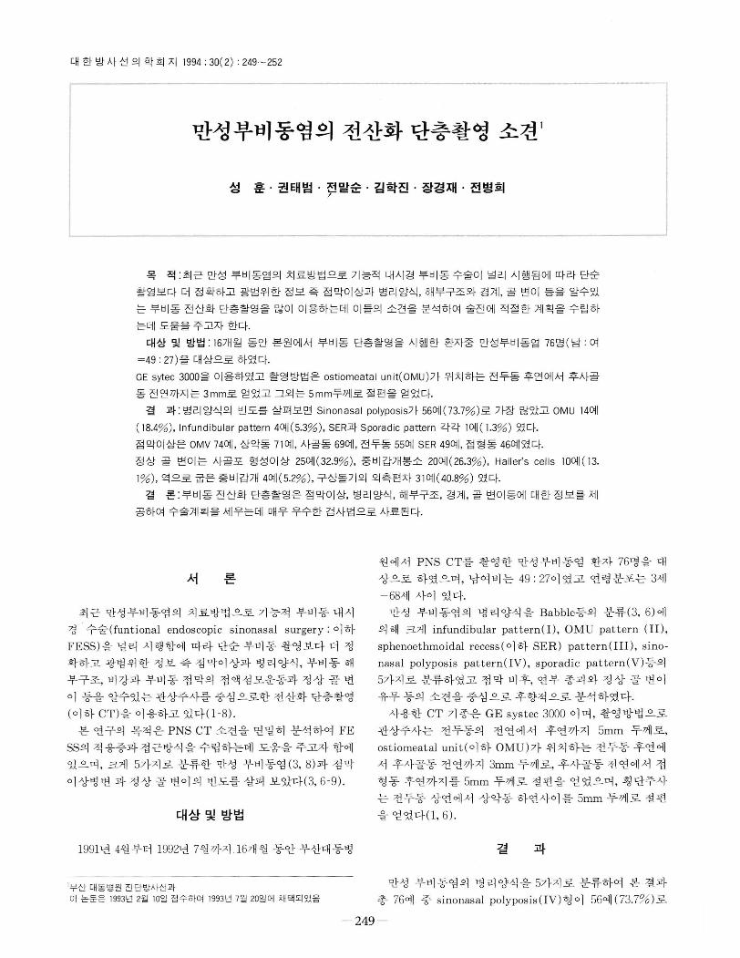
류하여 보고 한바 있는데, infundibular (I)형은 inferola
teral orbit가 외 측 경 계 를, 구상돌기 가 내 측경 계 를 사골포
와 반월열공(비atus semilunaris)이 상측경계를, 상악동의
소공(ostium) 이 하측경 계 를 형 성 하는 Infundibulum에 폐
쇄가 있어 동측 상악동염이 동반되는 경우이다(Fig. 1) ( 6 -
8).OMU(II)형은 한쪽 중비도 (middle meatus)가 막혀 동
측 전두동, 상악동, 전 과 중사골동의 전체 (complete type)
흑 일부(incomplete type) 에 부비강염이 초래된 경우이다
(Fig.2)( 6 -8).
Fig . 1. Inlundibular pattern(t) 01 inllammatory sinonasal disease
on right. Note opacilication 01 right inlundibulm(arrow) with com.
plicated concha bullosa(arrows) on left
Fig. 2. OMU pattern( lI) 01 inllammatory sinonasal disease on
right Note complete opacilication 01 right OMU(arrow) with both
concha bullosa(long arrow) and deviation 01 nasal spetum
n u 건
성 훈 외 · 만성부비동엽의 전산화 단층촬영
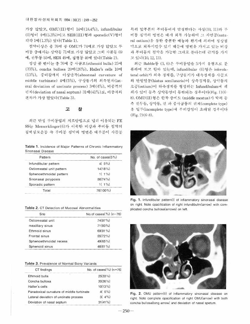
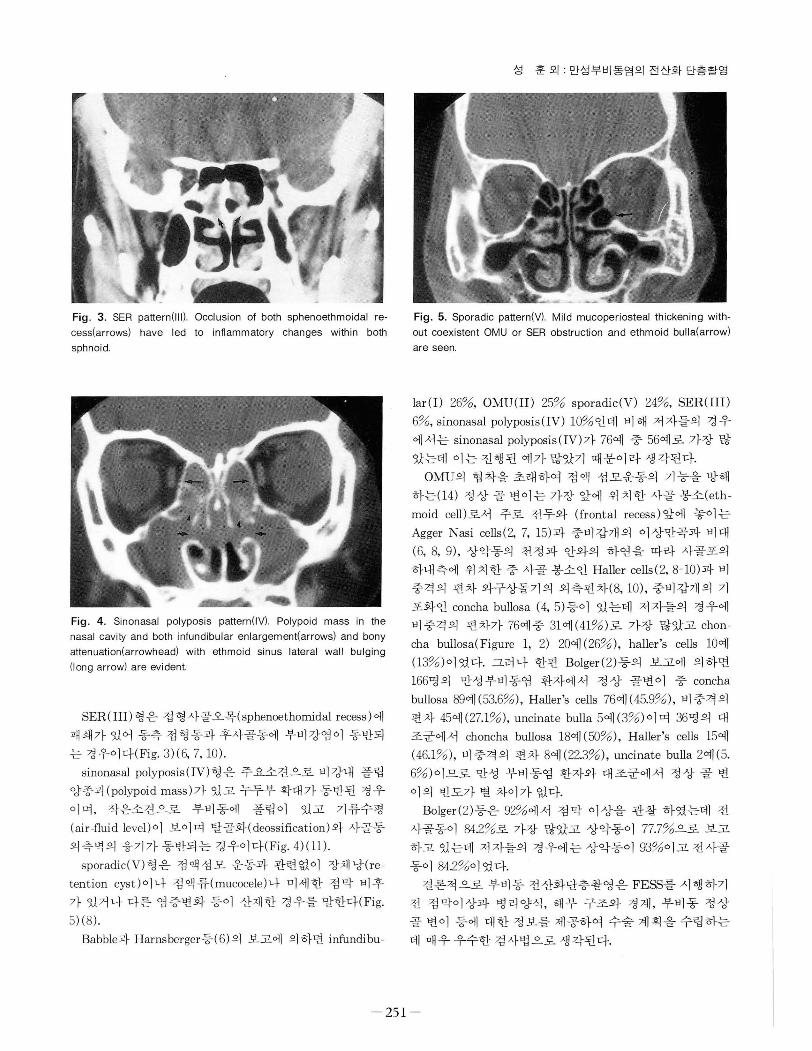
Fig. 3. SER pattern (1 11). Occlusion 01 both sphenoethmoidal re- Fig. 5. Sporadic pattern(V). Mild mucoperiosteal thickening with cess(arrows) have led to inllammatory changes within both out coexistent OMU or SER obstruction and ethmoid bulla(arrow) sphnoid. are seen.
Fig . 4 . Sinonasal polyposis pattern(IV). Polypoid mass in the nasal cavity and both inlundibular enlargement(arrows) and bony attenuation(arrowhead) with ethmoid sinus lateral wall bulging (l ong arrow) are evident
SER(III) 형 은 접 형 사골오목(sphenoethomidal recess) 에
패쇄가있어 동측 접형동과후사골동에 부비강염이 동반되
는 경우이다( Fig. 3)(6, 7, 10).
sinonasal polyposis (IV)행은 주요소견으로 비강내 폴립
양종괴 (polypoid mass)가 있고 누두부 확대가 동반된 경우
이며, 작은소견으로 부비동에 폴립이 있고 기류수평
(ai r -flui d level) 이 보이 며 탈골화( deossification) 와 사골동
외측벽의 융기가 동반되는 경우이다(Fig. 4)( 11).
sporadic( V)형은 점액섬모 운동과 관련없이 장체낭(re
tention cyst)이나 점액류(mucocele)나 미세한 점막 비후
가 있거나 다른 염증변화 등이 산재한 경우를 말한다(Fig.
5) (8).
Babble과 Harnsberger등( 6) 의 보고에 의 하면 infundibu
lar (I) 26%, OMU( II) 25% sporadic(V) 24% , SER(III)
6%, sinonasal polyposis(IV) 10%인데 비해 저자들의 경우
에서는 sinonasal polyposis (IV)가 76예 중 56예로 가장 많
았는데 이는 진행된 예가 많았기 때문이라 생각된다.
OMU의 협착을 초래하여 점액 섬모운동의 기능을 방해
하는(14) 정상 골 변이는 가장 앞에 위치한 사골 봉소(eth
moid cell )로서 주로 전두와 (frontal recess) 앞에 놓이는
Agger Nasi cells(2, 7, 15 )과 중비갑개의 이상만곡과 비대
(6, 8, 9) , 상악동의 천정과 안와의 하연을 따라 사골포의
하내측에 위치한 중 사골 봉소인 Haller cells(2, 8 - 10)과 비
중격의 편차 와구상돌기의 외측편차(8, 10) , 중비갑개의 기
포화인 concha bullosa (4, 5)등이 있는데 저자들의 경우에
비중격의 편차가 76예중 31예 (41% )로 가장 많았고 chon
cha bullosa(Figure 1, 2) 20예 (26% ), haller’s cells 10예
(13% )이였다. 그러나 한편 Bolger( 2)등의 보고에 의하면
166명 의 만성 부비동염 환자에서 정 상 골변이 중 concha
bullosa 89예 (53.6%) , H aller’s cells 76예 (45.9% ) , 비중격의
편차 45예 (27.1% ), uncinate bulla 5예 ( 3%)이며 36명의 대
조군에서 choncha bullosa 18예 (50%) , H aller’s cells 15예
(46.1% ), 비중격의 편차 8예 (22.3%) , uncinate bulla 2예 (5.
6%)이므로 만성 부비동염 환자와 대조군에서 정상 골 변
이의 빈도가 별 차이가 없다.
Bolger(2)등은 92%에서 점막 이상을 관찰 하였는데 전
사골통이 84.2%로 가장 많았고 상악동이 77.7%으로 보고
하고 있는데 저자들의 경우에는상악동이 93%이고전사골
동이 84.2%이었다.
결론적으로 부비동 전산화단층촬영은 FESS를 시행하기
전 정막이상과 병리양식 , 해부 구조와 경계, 부비동 정상
골 변이 등에 대한 정보를 제공하여 수술계획을수립하는
데 매우우수한 검사법으로생각된다.
건

대 한 방사선 의 학회 지 1994; 30(2) : 249 - 252
*~ I그 -:-l -•- 헌
1. Chow JM , Malee MF. Radiologic assessment preoperative t。
endoscopic sinus surgery. Oto/aryng Clinics North Amer
1989; 22: 691-701
2. Bolger WE , Butzin CA, Parsons DS. Paranasal sinus bony
abatomic variations and mucosal abnormalit ies : CT analysis lor endoscopic sinus surgery. Laryngoscope 1991 ; 101 : 56-64
3. Babell RW, Hansberger HR , Sonkens J, et al. Recurring patterns 01 inllammatory sinonasal disease demonstrated on
screening sinus CT. AJNR 1992 ;13:903-912
4. Katsantonis P, Friedman WH , Sivore MC. The role 01
computed tomography in revision sinus surgery. Laryngo
scope 1990 ; 1 00 : 811 -81 6
5. Chakeres DW. Computed tomography 01 the ethmoid sinuses Oto/aryng Clinics North Amer 1985; 18 : 29-42
6. Babbel RW , Hansberger HR. A contemporary look at the
imaging issues 01 sinusitis : Sinonasal anatomy, physiology
and CT techniques. Seminars in Ultrasound, CT, MR 1991 ; 12 526-540
7. Hansberger HR , Babbel RW, Davis WL. The major obstructive
inllammatory patterns 01 the sinonasal region seen on screen
sinus computed tomography. Seminars in Ultrasound CT, MR, 1991 ; 12 : 541 -560
8. Scuderi AJ , Babbel RW, Harnsberger HR. et al. The sporadi c
pattern 01 inllammatory sinonasal disease including post-surgical changes. Seminars in Ultrasound, CT, MR. 1991 ; 12 575-591
9. Vogelzang PJ , Babbel RW , Hansberger HR. The nose and
nasal vaul t. Seminars in Ultrasound, CT, MR . 1991 ; 12 592-612
10. Zinreich SJ, Kennedy DW, Rosenbaum AE, et al. Paranasal
sinuses : CT imaging requirements lor endosc이)IC surgery Radio/ogy 1987 ; 163 : 769-775
11. Drutman J, Babbel RW, Hansberger HR , et al. Sinonasal
polyposis. Seminars in Ultrasound, CT, MR. 1991 ; 12 : 561-574 12. Kennedy DW, Zinreich SJ, Rosenbaum AE, et al. Functional
endoscopic sinus surgery : theory and diagnostic evaluation Arch oto/aryngol 1985 ; 111 : 576-582
13. Kennedy DW, Functional endoscopic sinus surgery. Arch
oto/aryngol 1985 ; 111 : 643-649
14. Stammberger H. Endoscopic endonasal surgery-concepts in
treatment 01 recurring rhinosinusitis 1 & 11 anatomic and pathophysiologic considerations. Oto/arygol Head Neck Sur
gery 1986 ; 94: 143-156
15. Zinreich SJ , Kennedy DW, Gayler BW. Computer tomography
01 nasal cavity and paranasal sinuses : an evaluation 01 anat。my lor endoscopic sinus surgery. Clear image 1988 ; 1 : 2-1 0
Journal of the Korean Radiological Society, 1994 ; 30 ( 2) : 249-252
CT Findings of the Chronic Sinonasal Inflammatory Disease
Hun Seong, M.D., Tae Beom Kweon, M.D. , Mal soon Cheon M.D., Hack Jin Kim, M.D. ,
Kyung Jae Jang, M.D. , Byung Hee Chun, M.D.
Department of Radiology , Daedong Hospital
Purpose; Recently, paranasal sinus(PNS) CT has increasingly been used because of the wide applications of
a functional endoscopic sinonasal surgery(FESS) as one of the therapeutic modalities of the chronic sinonasal
inflammatory disease.
Materials and Methods: We retrospectively analyzed PNS CT findings in 76 patients with chronic sinonasal
inflammatory disease who had undergone the PNS CT from April 1991 to July 1992
Results: There were 5 sinonasal patterns of inflammation; 4 cases of infundibular type(5.3 %) , 14 cases of
。stiomeatal unit(OMU) type(18%) , one case of sphenoethmoidal(SER) type(1 %) , 56 cases of sinonasal p이yposis
type(74%) , and one case of sporadic type(1 %)
The mucosal abnormality was seen in 74 OMU cases, 71 maxillary sinus cases, 69 ethmoidal sinus cases, 55
frontal sinus cases, 49 SER , and 46 sphenoidal sinus cases. The normal bony variant included ethmoid bulla(25
cases, 335), concha bullosa (20 cases 25 % ) , Haller cells(10 cases, 13%) , paradoxical curvature of middle turbi
nate(4 cases , 5%) , lateral deviation of uncinate process(3 cases, 4 % ), and deviation of nasal septum(31 cases,
41 %).
Conculusion: The PNS CT is an excellent imaging method providing detailed informations about the
mucosal abnormality, path이。gical pattern , the anatomical structure and landmark, and bony variants prior t。
an operation.
Index Words: Paranasal sinuses , CT
Paranasal sinuses , diseases
Address r eprint requests t o : Hun Seong, M.D., Depar t ment of Radiology, Daedong H ospitaJ. 530 -1, M yungr yoon-{;\ong, Dongr ae-gu, Pusan Korea. T eJ. (051) 554 - 1233
이 4 건