13 potassium physiology
TRANSCRIPT

c h a p t e r 13Potassium Physiology
Introduction����������������������������������������������������������������������������������������425Objectives��������������������������������������������������������������������������������������������426Introductory Case 13-1: Why did I become so weak?�����������������426
P A R T A PRINCIPLES OF PHYSIOLOGY�������������������������������������������������427General concepts for the movement of K+ across
cell membranes���������������������������������������������������������������������� 428Driving force to shift K+ across cell membranes���������������������������428Pathways for the movement of K+ across cell membranes �������� 429
P A R T B SHIFT OF POTASSIUM ACROSS CELL MEMBRANES�����430Altering the negative voltage in cells���������������������������������������� 430Metabolic acidosis and a shift of potassium across
cell membranes������������������������������������������������������������������������������434Physiology of a shift of K+ into cells:
A Paleolithic perspective�������������������������������������������������������� 436
P A R T C RENAL EXCRETION OF POTASSIUM�������������������������������������438Components of the excretion of K+ in the cortical
collecting duct������������������������������������������������������������������������ 438K+ secretion in the late cortical distal nephron������������������������ 439
K+ channels�������������������������������������������������������������������������������������440Generation of a lumen-negative voltage����������������������������������������441Mechanism of action of aldosterone���������������������������������������������442Aldosterone paradox�����������������������������������������������������������������������444
Flow rate in the late cortical distal nephron����������������������������� 446Reabsorption of K+ in the medullary collecting duct��������������� 447
P A R T D INTEGRATIVE PHYSIOLOGY����������������������������������������������������447Control of the excretion of K+: A Paleolithic analysis�������������� 447WNK kinase signal system�������������������������������������������������������� 450Medullary recycling of K+: Implications
for kidney stone formation���������������������������������������������������� 453Mechanisms to shift K+ into liver cells after a meal���������������� 454Discussion of Introductory Case 13-1���������������������������������������� 455Discussion of questions������������������������������������������������������������� 456
Introduction Regulation of total body potassium (K+) homeostasis is vital. Changes in the concentration of K+ in plasma (PK) are inversely related to changes in the negative voltage across cell membranes, and this in turn influences many essential functions in the body. On the
425

POTASSIUM426
one hand, a change in the negative voltage across cell membranes may have direct and dangerous consequences (e.g., altered cardiac impulse conduction causing cardiac arrhythmias). On the other hand, subtle changes in this negative voltage across cell membranes have important physiologic functions in an indirect way by alter-ing the concentration of ionized calcium in cells (e.g., the release of insulin from β-cells of the pancreas, contractility in vascular smooth muscle cells).
The vast majority of K+ in the body is located in cells where this cation balances the negative charge on intracellular anions—primarily organic phosphates. Specific ion channels for K+ in cell membranes permit K+ to enter or exit cells.
The body deals with an intake of K+ in two phases. First, the K+ load is stored temporarily in cells, which is essential for short-term K+ homeostasis. Second, with the usual intake of K+, this extra K+ must ultimately be excreted in the urine, but there may be a long lag time before this renal process can be carried out.
The major site where the renal excretion of K+ is regulated is in the late cortical distal nephron. Secretion of K+ in these nephron segments requires the generation of a lumen-negative voltage via the electrogenic reabsorption of Na+ and the presence of open K+ chan-nels in the luminal membranes of their principal cells.
To gain insights into the regulation of K+ homeostasis, it is very helpful to examine this process from a Paleolithic perspective. The Paleolithic diet had a large but episodic intake of K+, and this K+ was provided with sugar in berries. In addition, there was little NaCl in the Paleolithic diet, and this created a potential difficulty to be over-come—the need for a large enough distal delivery of Na+ to secrete K+. This has implications for the extrarenal K+ homeostasis and the regulation of the excretion of K+ by the kidney.
It is important to realize that hyperkalemia and hypokalemia are not specific diseases; rather, they result from many disorders with dif-ferent underlying pathophysiology. Therefore, an understanding of the physiology of K+ is critical for the diagnosis and design of specific therapy for patients with these “electrolyte symptoms.”
OBJECTIVES
n To illustrate the common strategy used to ensure that K+ will enter or leave a given compartment. This requires a driving force to cause K+ movement (a negative voltage in the area K+ must be retained) and channels with a high conductance for K+.
n To consider how the voltage across cell membranes is regulated and its implications for the control of a shift of K+ into cells.
n To illustrate how the kidneys adjust the rate of excretion of K+ to maintain balance for K+ and keep the PK in the normal range.
n To illustrate that examining this process from a Paleolithic per-spective provides insights into the control of K+ homeostasis.
ABBREVIATIONSPK and UK, concentrations of K+ in plasma and in urinePNa and UNa, concentrations of Na+ in plasma and in urinePCl and UCl, concentrations of Cl– in plasma and in urinePCreatinine and UCreatinine, concentra-tions of creatinine in plasma and in urinePOsm and UOsm, osmolality in plasma and in urine
Introductory case 13-1: Why dId I Become so Weak?
(Case discussed on page 454)A very fit, active, 27-year-old Caucasian woman was in excel-
lent health until 1 year ago. Her past medical history revealed mild asthma, for which she took a bronchodilator on an intermittent basis.

13 : POTASSIUM PHYSIOLOGY 427
P A R T APRINCIPLES OF PHYSIOLOGY
• Close to 98% of total body K+ is inside cells, where it balances the anionic valance inside cells.
• Although only 2% of total body K+ is in the extracellular fluid (ECF) compartment, changes in the concentration of K+ in this compartment are critical, because they reflect the changes in resting membrane potential (i.e., the voltage difference across cell membranes).
In the past year, she had three episodes of extreme weakness; each lasted up to 12 hours; she felt perfectly well between attacks. On more detailed questioning, she said that she had ingested a large amount of sugar before the first attack. Each subsequent attack, however, was not preceded by the use of a bronchodilator, performance of exercise, or the ingestion of a large amount of sugar or caffeinated beverages. She denied the use/abuse of diuretics or laxatives, bulimic symptoms, glue sniffing, substance abuse (no needle marks), or the ingestion of licorice or over-the-counter drugs. She was not overly concerned about her body weight. There was no family history of hypokalemia, hypertension, or paralytic episodes. In the emergency room on each occasion, she was clearly very concerned, but other than the paraly-sis, the only findings of note were tachycardia (130/min) and mild systolic hypertension with a wide pulse pressure (150/70 mm Hg). There were no signs of hyperthyroidism. Because of the very low PK (~2.1 mmol/L), an intravenous infusion of KCl was started, and, as on the other occasions, she recovered promptly with the administra-tion of relatively little K+. The laboratory data are provided below. While not shown, all tests of thyroid function were in the normal range and the tests for a pheochromocytoma were negative. On the last admission, her PInsulin was in the normal range and the C-peptide level in blood was not elevated. In addition, the levels of cortisol, renin, and aldosterone and the ratio of renin to aldosterone were all in the normal range.
Na+ mmol/L 141 Glucose mg/dL (mmol/L) 133 (7.4)K+ mmol/L 2.1 Creatinine mg/dL (μmol/L 0.9 (77)HCO3
– mmol/L 22 BUN (Urea) mg/dL (mmol/L) 10 (3.4)Arterial pH 7.38 Pco2 mm Hg 38Urine K+ mmol/L 8 Urine creatinine (g/L) (mmol/L) 0.8 (7)
Questions
What is the most likely basis for the repeated episodes of acute hypokalemia?
Was an adrenergic effect associated with the acute hypokalemia?After the laboratory results were examined, were there any clues to
suggest what the cause of acute hypokalemia might be?Would your treatment be different for her next attack?What tests should be performed in the emergency room if she
has another episode of acute hypokalemia to reveal the correct diagnosis?

POTASSIUM428
The concentration of K+ in cells is very high relative to its concen-tration in the ECF compartment, and it does not vary appreciably in most circumstances. K+ in cells balances the charge on intracellular anions; intracellular anions cannot exit from cells because they are macromolecules. Moreover, these anions are essential for cell func-tions (DNA, RNA, phospholipids, compounds for energy provision such as ATP and phosphocreatine). Hence, the vast majority of K+ is retained in cells. Only 2% of body K+ is in the ECF compartment. Changes in this concentration of K+ in the ECF compartment are, however, extremely important because the ratio of concentrations of K+ across cell membranes largely reflects changes in the magnitude of the negative voltage difference across cell membranes. The PK is determined on a minute-to-minute basis by the distribution of K+ between the intracellular fluid (ICF) and ECF compartments (acute internal K+ balance) and on a day-to-day basis by the renal excre-tion of K+ (long-term external K+ balance). With the usual intake of K+, the kidney excretes the majority of the K+ load close to noon-time (see margin note); therefore, there must be sensitive regulatory mechanisms to minimize transient changes in the PK before renal K+ excretion occurs.
GENERAL CONCEPTS FOR THE MOVEMENT OF K+ ACROSS CELL MEMBRANES
There are two requirements for movement of K+ across cell mem-branes. First, there must be sufficient electrochemical driving force across that membrane; and second, there must be open K+ channels in that membrane.
Driving force to shift K+ across cell membranes
• This driving force is a more negative voltage in the compart-ment where K+ will be located.
• The active pumping of cations by the Na-K-ATPase initiates the process to create this voltage because it is an electrogenic pump, as it causes 3 Na+ to exit and 2 K+ to enter cells.
There are two ways to generate a negative voltage across a cell membrane: import anions or export cations. The usual mechanism is to export Na+, in part because of its abundance (Fig. 13-1). The Na-K-ATPase is responsible for electrogenic exit of Na+, because it extrudes 3 Na+ and imports only 2 K+ into cells. Export of intracel-lular, macromolecular phosphate does not occur; therefore, the net result is the generation of a more negative intracellular voltage, which limits the exit of K+ from cells.
The same principle for the movement of K+ applies to principal cells in the late cortical distal nephron, but the details are dif-ferent. A transepithelial lumen-negative electrical voltage drives the secretion of K+. This is generated when Na+ is reabsorbed in an electrogenic fashion (i.e., more Na+ than its accompanying an-ions, usually Cl−). Reabsorption of Na+ occurs via the epithelial Na+ channel (ENaC); it is driven by the low concentration of Na+ and the negative voltage in principal cells (created by the Na- K-ATPase in their basolateral membrane; see Part C for more discussion).
DIURNAL VARIATION IN ThE EXCRETION OF K+
The bulk of K+ is excreted close to noontime. This does not seem to be related to a rise in the PAldosterone or the delivery of Na+ to the late cortical distal nephron. While the mechanism is still open to debate, it is possible that it is due to increased delivery of HCO3
– to the distal nephron during an increase in the alkaline tide.

13 : POTASSIUM PHYSIOLOGY 429
Pathways for the movement of K+ across cell membranes
• There are several different types of K+ channels that permit K+ to cross cell membranes.
• Some of these channels are regulated by voltage, others by ligands such as calcium ions, and yet others by metabolites such as ADP—these are called KATP channels.
• Movement of K+ through the specific K+ channels depends on the driving force, the number of K+ channels, whether they are in an open configuration, and how quickly K+ can move through them (called their conductance).
K+ channels are composed of a diverse family of membrane- spanning proteins that selectively conduct K+ across cell mem-branes. K+ channels have a pore that permits K+ to cross the cell membrane, a selectivity filter that specifies K+ as the ion spe-cies to move through the channel (see margin note), and a gating mechanism that serves to switch between open and closed channel conformations.
When K+ move out of cells (Fig. 13-2), there is an increase in the net negative voltage in cells. Because the concentration of K+ in cells is higher than the predicted value from its electrochemi-cal equilibrium, control of the open probability of K+ channels in cell membranes is critical to regulate the magnitude of cell voltage, which in turn influences many essential cell functions. As illustrated in Figure 13-2, regulation of K+
ATP channels influences the gating of calcium ion channels as a result of a change in the voltage in cells (more negative if K+
ATP channels are open—the converse is
BASIS FOR ThE SELECTIVITY OF ThE K+ ChANNEL FOR K+ • Na+ are much smaller than K+,
yet the K+ channels are specific for K+.
• Ions in solution have layers of water surrounding them; hence, we must think in terms of their hydrated size.
• The chemical structure of the K+ channel at its mouth strips off the water shells surrounding K+. Thus, K+ becomes smaller than Na+ and can pass through the channel, whereas Na+ cannot do so.
Na-K-ATPase
3 Na�
NEGATIVE
K�
K�
2 K�
�
Cell Membrane Lumen of the CCD
ENaC
Principalcell
Na�
CCD
Cl�K�
NEGATIVE
FIGURE 13-1 Concept for the movement of K+ across cell membranes. The circle on the left represents a cell membrane and the cylinder on the right rep-resents the CCD (see margin note). There is a much higher concentration of K+ in cells than in the ECF compartment owing to the negative voltage inside cells, which is generated in part because more Na+ are extruded than K+ are imported by the Na-K-ATPase. For the passive movement of K+, there must be open K+ channels in the cell membrane. To cause a re-distribution of K+ across cell membranes, there must be either a change in the magnitude of the negative voltage inside cells or in the number, open probability, and conductance of K+ channels (because the concentration of K+ in cells is higher than the predicted value from the electrochemical equilibrium). Similarly, K+ will enter the lumen of the CCD when it has a negative voltage and if there are open K+ channels in the luminal mem-brane of principal cells in the CCD.
ABBREVIATIONCCD, late cortical distal nephron including the late distal convoluted tubule, the connecting segment, and the cortical collecting ductENaC, epithelial Na+ channels

POTASSIUM430
also true). Clinical examples where control of K+ATP channels has
important effects on physiologic functions are discussed in the answers to Question 13-1.
From a renal perspective, secretion of K+ in principal cells of the late cortical distal nephron requires that open K+ channels (primarily ROMK) must be present in the luminal membrane of these cells to permit K+ to enter the luminal fluid.
QUESTION
(Discussion on page 456)13-1 Is the [K+]ATP channel regulated in fact by ATP?
P A R T BSHIFT OF POTASSIUM ACROSS CELL MEMBRANESALTERING THE NEGATIVE VOLTAGE IN CELLS
• K+ shift into cells when there is an increase in the negative volt-age in cells.
• The components of the system that regulates intracellular volt-age include the Na-K-ATPase, the Na+/H+ exchanger (NHE), the Cl−/HCO3
− anion exchanger (AE), and the open probabil-ity of K+
ATP channels in cell membranes.
There are three mechanisms to increase the negative voltage in cells via augmenting flux through the Na-K-ATPase.
Ca2+
Voltage-gatedCa2+ channel
CLOSED [K+]ATP CHANNELS OPEN [K+]ATP CHANNELS
Ca2+
Voltage-gatedCa2+ channel
K+LESSNEGATIVE
�21
[K+] ATP
channels
MORENEGATIVE
Openchannel
K+
[K+] ATP
channels
Closedchannel �
21
FIGURE 13-2 Regulatory roles for [K+]ATP channels in cell function. The goal of this process is to adjust the con-centration of ionized calcium in cells, because this regulates many important cellular events. Begin the analysis with the [K+]ATP channels in each setting. As shown on the left, when the [K+]ATP channels are closed, positive volt-age (K+) cannot exit from cells and the interior of the cell becomes less negative. As a result, the voltage-gated ionized calcium channels open, and the concentration of ionized calcium rises in that cell. In contrast, as shown on the right, when the [K+]ATP channels are opened, positive voltage (K+) can exit from cells and the interior of the cell becomes more negative. As a result, the concentration of ionized calcium falls in that cell.
ABBREVIATIONROMK, rat outer medullary K+
channels

1
13 : POTASSIUM PHYSIOLOGY 43Raise the intracellular concentration of Na+
• More Na+ is pumped out of cells by the Na-K-ATPase when the concentration of Na+ rises in cells. This leads to an increase in cell negative voltage only if the source of Na+ is Na+ that existed in the cell or Na+ that entered the cell in an electroneutral fashion.
The first and quickest mechanism to increase the flux of Na+
through the Na-K-ATPase is to raise the intracellular concentration of Na+, because the extracellular concentration of K+ is always high enough for maximal activity of the Na-K-ATpase. The impact of this increase in Na+ pumping on the net cell voltage, however, depends on whether the Na+ entry step into cells is electrogenic or electroneutral.
Electrogenic entry of Na+ into cells
The Na+ channel in cell membranes is normally closed by the usual magnitude of the negative intracellular voltage. If the Na+ channel in skeletal muscle cell membranes were to open (e.g., by nerve impulses that lead to the release of acetylcholine), the resting membrane potential quickly becomes less negative. In more detail, one cationic charge enters the cell per Na+ transported. Because only one third of a charge exits per Na+ pumped via the Na-K-ATPase (Fig. 13-3), this diminishes the degree of intracellular negative voltage. This promotes the entry of ionized calcium and thereby muscle contraction. There is also a net exit of K+ from cells through open K+ channels in the cell membrane and thus a rise in the [K+] in the ECF compartment.
Clinical implications
An abnormally large increase in the entry of Na+ via Na+ channels in cell membranes of skeletal muscle and its subsequent exit via the Na-K-ATPase diminishes the magnitude of the intracellular negative
3 Na+Na+
Na+
Na+
ATP
ADP
Na ion-specific channel
2 K+
1/3 +
H+
NHE 1
2
3
FIGURE 13-3 Effect of electroneutral versus electrogenic entry of Na+ on the negative voltage in cells. A more negative voltage in cells is generated by the Na-K-ATPase, providing that the source of Na+ pumped out is either Na+ that exist in cells (site 1) or Na+ that enter cells in an electroneutral fashion via the Na+/H+ exchanger (NHE; site 2). In contrast, if the source of Na+ pumped out of cells is Na+ that enters cells via the Na+-specific ion chan-nel (site 3), the voltage in cells becomes less negative (count the charges). Although flux of K+ through K+ channels may be limited to some degree by their open probability, a higher concentration of K+ in the ICF compartment due to ion pumping by the Na-K-ATPase drives the electrogenic exit of K+.

POTASSIUM432
voltage. This is the underlying pathophysiology of hyperkalemia in some patients with hyperkalemic periodic paralysis.
Electroneutral entry of Na+ into cells
• If Na+ enters the cell in an electroneutral fashion, its subsequent electrogenic exit via the Na-K-ATPase results in a more negative cell interior voltage and hence less net exit of K+ from cells.
This occurs when Na+ enter cells in exchange for H+ via the NHE (see Fig. 13-3). The NHE in cell membranes is normally inactive. This can be deduced from the fact that it is an electroneutral exchange and that the concentrations of its substrates (Na+ in the ECF compartment and H+ in the ICF compartment) are considerably higher than that of its products (Na+ in the ICF compartment and H+ in the ECF compartment) in steady state. There are two major activators of NHE: a sudden spike in the release of insulin and a higher concentration of H+ in the ICF compartment.
Role of insulin
• Insulin plays an important role to cause K+ uptake by the liver before it enters the systemic circulation.
One major physiologic setting where there is a need to shift K+ into cells and do so quickly is when there is a large dietary K+ intake (see margin note). Fruit and berries were the major sources of calories in the Paleolithic diet. Accordingly, this diet provided a large quantity of sugar (fructose and glucose), K+, and a family of organic anions (e.g., citrate) that are metabolized promptly to produce HCO3
− in the liver. To remove this K+ before it can enter the systemic circulation where it can be dangerous, the first line of defense is to shift K+ into liver cells. The trigger for this control mechanism is by the sugar content of the diet. When the concentration of glucose in plasma (PGlucose) rises, there is a sudden spike in the release of insulin from β-cells of the pancreas. This high concen-tration of insulin activates NHE in cell membranes of hepatocytes after a meal rich in K+. As a result, Na+ enters these cells in an electroneutral fashion. When this Na+ is pumped out of cells by the Na-K-ATPase (see Fig. 13-3), there is an export of positive voltage and the interior of the cells becomes more negative; this negative voltage keeps some extra K+ in cells (for more discussion of this topic, see Part D, page 453).
Clinical implications
Because insulin activates NHE, this hormone has been used clini-cally in the emergency treatment of patients with hyperkalemia. On the other hand, a lack of actions of insulin results in a shift of K+ out of cells and the development of hyperkalemia in patients with dia-betic ketoacidosis despite having a total body K+ deficit.
Activate preexisting Na-K-ATPase by hormones
• The second mechanism for increasing flux through Na-K-ATPase is also a rapid one; it involves activation of existing Na-K-ATPase units in the cell membrane by β2-adrenergic actions.
ABBREVIATIONPGlucose, concentration of glucose in plasma
MAJOR SETTIN�S �hERE ASETTIN�S �hERE A �hERE A RAPID ShIFT OF K+ INTO CELLS IS NEEDED • After the ingestion of a large
K+ load: The major hormone involved is insulin.
• After a vigorous sprint: The major hormones involved are β2-adren-ergics, which are released in the “fight-or-flight” response.

13 : POTASSIUM PHYSIOLOGY 433
β2-Adrenergic agonists activate the Na-K-ATPase via a cyclic AMP–dependent mechanism, which phosphorylates this ion pump and leads to the export of preexisting intracellular Na+ (Fig. 13-4). This mechanism is particularly important during vigorous exercise in the second major physiologic setting where there is a need to shift K+ into cells quickly. In the fight-or-flight response, the stimulus for muscle contraction is the entry of Na+ through its voltage-gated Na+ channel. This entry of positive voltage diminishes the cell interior negative volt-age (called depolarization). As a result, K+ are released from exercising muscles, and there is a danger of acute hyperkalemia (see margin note). To minimize this risk, the β2-adrenergic effect of adrenaline released in this setting is exerted on hepatocytes and possibly on resting muscle cells, which obligates them to take up much of this K+ and thereby pre-vent a dangerous rise in the PK (see the discussion of Question 13-2 for a discussion of the mechanisms involved).
If the release of adrenaline is especially large, its α-adrenergic effect would inhibit the release of insulin, even if the PGlucose is high. This has the advantage of preventing working muscle cells from consuming the most valuable brain fuel, glucose, from the circulation. On the other hand, this α-adrenergic effect may override the β2-adrenergic effect, permitting a more severe degree of hyperkalemia to develop (see margin note).
Clinical implications
It should not be surprising that an acute shift of K+ into cells causing hypokalemia is seen in conditions associated with a surge of catecholamines (e.g., patients with a subarachnoid hemorrhage, myocardial infarction, and/or an extreme degree of anxiety). On the other hand, β2-agonists may be used to shift K+ into cells in patients with an emergency associated with hyperkalemia. In states with a very low effective arterial blood volume, the large α-adrenergic response leads to hyperkalemia because of a shift of K+ out of cells despite large losses of K+ (e.g., patients with cholera). The shift of K+ out of cells is valuable to prevent a severe degree of hypokalemia because there is a large loss of K+ in diarrhea fluid.
Na�
H�
Na�
�
3 Na�
�2-adrenergics
Negative
ATP
ADP
2 K�
K�
1/3 �
3 Na�
H�
Insulin
Na�
NHE
Negative
ATP
ADP
2 K�
K�
1/3 ��
FIGURE 13-4 Role of the insulin and β2-adrenergics on the distribution of K+. The circle represents a cell membrane. The Na-K-ATPase generates the electrical driving force for net K+ entry into cells, providing that the source of the Na+ pumped out is either Na+ that exist in cells or Na+ that enter cells in an electroneutral fashion via the Na+/H+ exchanger (NHE). Two hormones that cause more Na+ pumping by the Na-K-ATPase are shown; one is insulin, which activates NHE, and the other is β2-adrenergics, which activate Na-K-ATPase by phosphorylation. The increase in negative voltage in cells diminishes the exit of K+ from cells via K+ channels.
BENEFIT OF ACUTE hYPERKALEMIAAn acute rise in the PK helps increase local blood flow in working skeletal muscles.
EFFECT OF AN ADRENER�IC SUR�E ON K+ DISTRIBUTION ACROSS CELL MEMBRANES • β2-Adrenergics activate the
Na-K-ATPase and thereby make voltage in cells more negative, which causes K+ to be retained in cells.
• α-Adrenergic actions inhibit the release of insulin, and thereby lead to the exit of K+ from cells. A very large adrenergic surge is needed to have the α-effect dominate.
MEChANISMS FOR ThE RELEASE OF K+ FROM MUSCLE CELLS DURIN� A SPRINT • A diminished magnitude of the
negative voltage in cells occurs when Na+ enter muscle cells via open Na+ channels.
• Intracellular alkalinization when phosphocreatine is hydrolyzed activates the extrusion of HCOHCO3
– and, secondarily, the exit of K+ (see Chapter 1, page 32, and Fig. 13-5, page 435).

POTASSIUM434
QUESTION
(Discussion on page 458)13-2 What mechanisms permit nonexercising cells to take up K+ during
a sprint?
Increase in the number of Na-K-ATPase units in cell membranes
• Thyroid hormone and insulin lead to the synthesis of more Na-K-ATPase units.
The third mechanism to augment flux of K+ via the Na-K-ATPase takes time to develop because it involves the synthesis of more Na-K-ATPase units and their insertion into cell membranes. Having more Na-K-ATPase units in the cell membrane of skeletal muscle cells is not important at rest, but it is very important during vigorous exercise. In this latter setting, there is a strong positive correlation between the activity of this ion pump, which is essential for recovery from cell depolarization, and the maximum ability for skeletal muscle to contract during vigorous exercise. Hyperthyroidism is also associ-ated with a higher content of Na-K-ATPase units in the membrane.
Clinical implications
A severe degree of hypokalemia due to a shift of K+ into cells is seen in Asian patients with the thyrotoxic subtype of hypokalemic peri-odic paralysis. These patients can be managed effectively during their attacks with the use of nonselective β-blockers and the administration of a small dose of KCl (see Chapter 14, page 478 for more details). It is also interesting to note that many of these patients have attacks of acute hypokalemia and paralysis after eating a large amount of carbo-hydrates. Perhaps the effect of high levels of insulin to activate NHE in addition to the effect of thyroid hormone to cause the synthesis of more Na-K-ATPase units may lead to the severe degree of hypokale-mia (see Part D for a more detailed discussion of this topic).
METABOLIC ACIDOSIS AND A SHIFT OF POTASSIUM ACROSS CELL MEMBRANES
The effect of an acid load on the PK depends on whether the anions accompanying the H+ can cross cell membranes in an electroneutral fashion (i.e., on the monocarboxylic acid transporter).
Acids that can be transported by the monocarboxylic acid transporter
• Monocarboxylic acids (e.g., l-lactic acid or ketoacids) enter cells in an electroneutral fashion. Therefore, they do not cause a change in cell voltage.
• l-lactic acid produced in enterocytes can contribute to an important shift of K+ into liver cells after meals because of the large influx of H+ into these cells, which can activate their NHE.

13 : POTASSIUM PHYSIOLOGY 435
For example, when l-lactic acid is formed in exercising skeletal muscles during a sprint, many of these H+ along with l-lactate anions are removed by entering cells that are not involved in the exercise via their monocarboxylic acid transporter; this minimizes the fall in the intracellular pH and the rise in the PK. Because this entry of H+ is electroneutral, it does not change the magnitude of the negative voltage in these cells and hence does not result directly in a shift of K+. Nevertheless, both the high concentration of H+ inside these cells (by activating the NHE) and the β2-adrenergic response to exercise (by activating the Na-K-ATPase) may cause a shift of K+ into these resting muscle cells, thus diminishing the degree of hyperkalemia caused by release of K+ from exercising muscles. The close proximity of NHE and the monocarboxylic acid transporter could lead to a much stronger activation of NHE by the rise in the local ICF concentration of H+ (see Part D, page 453 for more discussion).
Acids that cannot be transported by the monocarboxylic acid transporter
• These acids cause K+ to shift out of cells when much of their H+ load is titrated by HCO3
− in the ECF compartment; thus the Cl−/HCO3
− anion exchanger likely participates in this mechanism.
Inorganic acids (e.g., HCl) or nonmonocarboxyl organic acids (e.g., citric acid) cannot be transported by the monocarboxylic acid transporter. Moreover, their H+ cannot enter cells via NHE, because this exchanger can cause only the export of H+ from, and not the entry of H+ into, cells (Fig. 13-5). Hence, a different mechanism is needed to permit some of these H+ to be titrated by HCO3
− in the ICF compartment. The mechanism has two steps. First, with a low PHCO3, there is activation of the anion exchanger (the exact mechanism is not known) and an electroneutral shift of HCO3
− out of cells and Cl− into cells. Because this exchange of anions in a 1:1 stoichiometry
Na+
H+ H+
HCO3�
HCO3�
HCO3�
Cl�
Citrate3� � H+
Cl�Cl�
H2O
CO2
CO2
+
L-Lactate
H+
K+
K+
FIGURE 13-5 Shift of K+ out of cells in patients with metabolic acidosis. The circles represent cell membranes. Shown on the left are the transport mechanisms for the entry of H+ into and the exit of HCO3
− from cells. The two transport mechanisms for net gain of H+ in cells, the monocarboxylic acid transporter and the Cl−/HCO3
− anion exchanger, are shown above the horizontal dashed line. The Na+/H+ exchanger, which is activated by intra-cellular acidosis and catalyzes only the exit of H+ from cells, is shown below the horizontal dashed line. As shown on the right, when there is an H+ load with anions that cannot enter cells (e.g., citric acid), the fall in the PHCO3 is associated with more flux through the HCO3
−/Cl− anion exchanger, which accelerates the electroneutral exit of HCO3
− from, and the entry of Cl− into, cells. The subsequent exit of Cl− via Cl− channels decreases the net negative intracellular voltage, leading to the exit of K+ from cells via K+ channels.

POTASS436
IUMis electroneutral, it does not change the magnitude of the negative voltage in these cells. Second, as a result of the combination of the higher concentration of Cl− in cells, the negative intracellular voltage, and the presence of open Cl− ion channels in cell membranes, Cl− is forced out of cells in an electrogenic fashion, and therefore the volt-age in these cells becomes less negative and K+ exit.
Clinical implications
If hyperkalemia is present in a patient with metabolic acidosis owing to an increased production of a monocarboxylic organic acid, causes of hyperkalemia other than the acidemia should be sought (e.g., lack of insulin in patients with diabetic ketoacidosis, tissue injury, or a decreased availability of ATP to drive the Na-K-ATPase in patients with l-lactic acidosis due to hypoxia).
Although inorganic acidosis (addition of HCl) causes a shift of K+ out of cells, patients with chronic hyperchloremic metabolic acidosis may have a low PK because of excessive loss of K+ in the diarrhea fluid in patients with chronic diarrhea or in the urine (e.g., patients with renal tubular acidosis; see Chapter 4, page 96 for more discussion).
There are only small changes in the PK in patients with respiratory acid-base disorders because there is little movement of Na+ or Cl− across cell membranes and hence no change in the ICF voltage.
PHYSIOLOGY OF A SHIFT OF K+ INTO CELLS: A PALEOLITHIC PERSPECTIVE
The Paleolithic diet consisted primarily of fruit and berries; hence, it provided sugar, K+, and organic anions (potential HCO3
−). Therefore, it is not surprising that one of the stimuli to shift K+ into cells is insulin, which is released after carbohydrate intake.
There are three phases in the process of handling a load of K+ and HCO3
−, and this depends on its magnitude.
• Low intake of K+ and HCO3−: In this setting, the entire load of
K+ and HCO3− can be shifted into cells. The key in this process
is activation of NHE in cell membranes by insulin. • Somewhat larger intake of K+ and HCO3
−: In this setting, as more HCO3
− has accumulated in cells, intracellular alkaliniza-tion will inhibit NHE. As a result, some of the Na+ that exist in cells are exported via Na-K-ATPase, which is also activated by insulin. Accordingly, there is a net gain of some Na+ and HCO3
− in the ECF compartment. • Even larger intake of K+ and HCO3
−: In this setting, there is a problem causing a further deficit of intracellular Na+. As a result, some K+ cannot enter cells despite the continuing actions of insulin. The net result is a gain of K+ and HCO3
− in the ECF compartment.
Low intake of K+ and HCO3−
Let us examine the net removal of an arbitrary three units of KHCO3. The process begins when 3 Na+ enter and 3 H+ exit from cells on NHE, which is activated by insulin. H+ that exit from cells neutralize all the new HCO3
− in the ECF compartment. The source

13 : POTASSIUM PHYSIOLOGY 437
of H+ for NHE in cells is the conversion of CO2 + H2O to H+ + HCO3
−, catalyzed by the enzyme carbonic anhydrase; hence, there is a gain of HCO3
− in cells. When 3 Na+ are pumped out of cells by the Na-K-ATPase, 2 K+ enter, and there is a more negative voltage in cells, which draws the remaining 1 K+ into the ICF compartment. The net result is that all the added K+ and HCO3
− are now inside these cells.
A somewhat larger intake of K+ and HCO3−
When more HCO3− enter cells, the concentration of H+ falls, which
inactivates NHE. To shift more K+ into cells, some of the Na+ that existed in cells is exported via the Na-K-ATPase, which is also activated by insulin. As a result, there is a net gain of some Na+ and HCO3
− in the ECF compartment. There is one other component to the picture: This newly gained Na+ and HCO3
− must be retained temporarily in the body—that is, hidden from the kidney (Fig. 13-6). Once a lower limit for intracellular Na+ concentration is reached, the ability to cause K+ to enter cells is compromised. Hence, to avoid a significant degree of hyperkalemia, the excess K+ load must be excreted.
A much larger input of K+ and HCO3−
• This represents the diet of vegetarians and of our Paleolithic ancestors.
• Not all of the K+ and HCO3− can be shifted into cells; accord-
ingly, this excess K+ and HCO3− must be excreted in the urine.
The ion pumping by the Na-K-ATPase declines when the intracel-lular Na+ concentration falls. This, together with the low NHE activ-ity resulting from the intracellular alkaline pH, leads to a decreased capacity to transfer K+ and HCO3
– into cells. Accordingly, there is a rise in the PK and in the PHCO3. Both of these effects help the kidney excrete the K+ and the HCO3 loads.
Disappear
GAG
HCO3�
• Inhibitors?
• Stimulators?
CO2 � H2O
Na+
Exchange bound H+ with Na+
H+ • SO4�
H+ • SO4�
H+ • SO4�
1
2
FIGURE 13-6 “Hiding” Na+ and HCO3− in the extracellular fluid (ECF) com-
partment. After the consumption of a large load of K+ and potential HCO3−,
there is a net gain of Na+ and HCO3− in the ECF compartment. To prevent
the loss of Na+ (and HCO3−) in the urine in subjects consuming little NaCl,
the higher PHCO3 may draw H+ from a bound form (glycosaminoglycans [GAG]) in connective tissue (the structure on the right), leaving a net anionic charge, which attracts Na+ (keeps them in bound form), completing the process to “hide” NaHCO3 in the ECF compartment. There is uncertainty about what may stimulate and what may lead to reversal of this process.
The process described in this figure is a speculation, but it could help explain why our Paleolithic ances-tors did not waste Na+ in their urine. The absence of a natriuresis has been observed in the Yano-mamo Indians whose diet is also virtually devoid of NaCl but rich in K+ and organic anions (potential HCO3
−).

POTASSIUM438
Exit of K+ and HCO3− from cells after excretion begins
• The process of exit of K+ and HCO3− from cells should begin
once the kidneys begin to excrete the K+ and potential HCO3−
in the urine. • The mechanisms responsible for this shift of K+ and HCO3
− out of cells are not clear.
There is probably a process that results in the exit of K+ and HCO3−
from cells after the excretion of K+ is underway. Its first step is likely to be activation of the Cl−/HCO3
− anion exchanger in cell membranes where K+ and HCO3
− were stored. This anion exchanger is both elec-troneutral and normally held in an inactive form in cell membranes (see Fig. 13-5, page 435). When activated, there is an electroneutral exit of HCO3
− and entry of Cl− into these cells, which raises the concentra-tion of Cl– in the ICF compartment (see margin note). Because of the presence of open channels for Cl− in the cell membrane and a higher concentration of Cl− in cells, there is an electrogenic exit of Cl−, which diminishes the magnitude of the intracellular negative voltage. Hence, this electrical driving force that retains K+ in cells is diminished, and K+ exit from these cells down their concentration difference via open K+ channels in the cell membrane. The net effect of all of these steps is to cause the export of K+ and HCO3
− from cells.
P A R T CRENAL EXCRETION OF POTASSIUM
Most of the secretion of K+ occurs in the late distal convoluted tubule and the connecting segment; the cortical collecting duct (CCD) also participates in this process when the K+ load is large.
COMPONENTS OF THE EXCRETION OF K+ IN THE CORTICAL COLLECTING DUCT
• Although the usual intake of K+ in adults eating a typical Western diet is close to 1 mmol/kg body weight, the rate of excretion of K+ can rise or fall by close to a factor of five to match the intake of K+ with only a minor change in the PK. While most of this K+ intake occurs during the evening meal, its excretion typically occurs around noon the next day.
• The kidneys must be able to excrete a K+ load with any anion that may accompany it; the strategy used, however, may differ with individual anions.
Arguably the most important immediate function of the kidney is to excrete a large K+ load to avoid the development of hyperkalemia and the risk of a cardiac arrhythmia. Control of K+ secretion occurs primarily in the cortical distal nephon, which includes the late distal convoluted tubule, the connecting segment and the cortical collecting duct (see margin note).
FUNCTION/CONTROL ANALYSISIt appears that the intracellular alkalinization facilitates, but does not initiate, this exit of K+ and HCO3
−, because it is present before the excretion of K+ and HCO3
− begins. It is possible that a signal related to a transient fall in the arte-rial PK initiates this process.
ABBREVIATIONFor simplicity, we use the abbrevia-tion CCD for all of these nephron segments.

13 : POTASSIUM PHYSIOLOGY 439
Although there are two components that affect the rate of excretion of K+, the flow rate in the CCD and the net secretion of K+ by prin-cipal cells, which raises the luminal concentration of K+ ([K+]CCD; see equation), it is the latter that controls the rate of excretion of K+. Therefore, we focus on factors that contribute to the regulation of the [K+]CCD.
K excretion = flow rate [K ]+CCD
+CCD×
K+ SECRETION IN THE LATE CORTICAL DISTAL NEPHRON
• The net secretion of K+ destined for excretion occurs in the late cortical distal nephron.
Overview
Although a large quantity of K+ are filtered daily (720 mmol in an adult; 4 mmol/L × 180 L GFR/day), very little will be excreted, as five sixths is reabsorbed in the proximal convoluted tubule when a similar proportion of filtered Na+ and water are reabsorbed (i.e., the concentration of K+ remains equal to the PK). Therefore, 120 mmol of filtered K+ are delivered to the loop of Henle (4 mmol/L × 30 L/day). Although there is a large passive entry of K+ into the lumen of the loop of Henle via its ROMK, the concentration of K+ in the fluid entering the early distal convoluted tubule, as measured during micropuncture studies in the fed rat, is about 1.5 mmol/L. Hence, there is net reabsorption of K+ in the loop of Henle and about 40 mmol of K+ are delivered to the CCD, the nephron sites where K+ secretion occurs (see margin note).
Two elements are required for the process whereby K+ is secreted in the CCD: the presence of K+ channels in an open configuration in the luminal membranes for cells that perform this secretion (principal cells) and a lumen-negative voltage in these nephron seg-ments. This voltage is generated by reabsorbing more Na+ than Cl–. For this to occur, separate pathways for the reabsorption of Na+ and Cl– must be present in the CCD, and the reabsorption of Na+ must be by an electrogenic pathway (i.e., an epithelial Na+ chan-nel [ENaC]). The driving force for Na+ reabsorption is a higher concentration of Na+ in the lumen of the CCD than in principal cells (~10 to 15 mmol/L), and a negative cell interior voltage owing to the actions of the Na-K-ATPase at the basolateral membrane in these cells.
There are two other features of this process of generating lumen-negative voltage. First, since aldosterone is the major hormone that causes an increase in the number of open ENaC units in the luminal membrane of principal cells, these cells must have a receptor for aldos-terone in their cytosol. Second, these cells also must have a way to prevent cortisol from reaching this aldosterone receptor, as the level of cortisol in plasma is more than an order of magnitude higher than the PAldosterone and both of these adrenal steroids bind avidly to this receptor (see margin note).
There are two steps in the secretory process that leads to the net secretion of K+. We shall describe them beginning with a setting where there is a large intake of K+ following a prolonged period of little intake of K+, which caused a deficit of K+ and thereby
REABSORPTION OF K+ IN ThE LOOP OF hENLE • This occurs via the Na+, K+, 2 Cl–
cotransporter in the medullary and cortical thick ascending limbs.
• In micropuncture experiments in the rat, the [K+] in the luminal fluid at the earliest part of distal convoluted tubule is ~1.5 mmol/L. A reasonable estimate of the volume of filtrate delivered to the distal nephron in humans is ~27 L/day. Hence ~40 mmol of K+ are delivered to the late cortical distal nephron (1.5 mmol/L × 27 L/day).
• Therefore, there is net reabsorp-tion of ~80 mmol of K+ per day in the loop of Henle.
SPECIFICTY OF ThE MINERALOCORTICOID RECEPTOR FOR ALDOSTERONE • The way to make these receptors
have specificity for aldosterone is to prevent cortisol from being present in their cytosol.
• Cortisol is “destroyed” by a pair of enzymes called 11β-hydroxysteroid dehydrogenase (11β-HSDH), which converts it to cortisone, which does not bind this receptor.

POTASSIUM440
the removal of K+ channels from the luminal membrane of the CCD.
K+ channels
• The main channel for K+ secretion in the CCD is ROMK. • In terms of regulation of K+ secretion, one should think of
these channels as being absent or present because control of K+ secretion by modulation of the number of channels does not provide control with enough sensitivity.
ROMK channels
A family of ROMK channels permits the secretion of K+ in the CCD when there is a negative voltage in the lumen of these neph-ron segments. These channels recycle; by this we mean that they can be removed from the luminal membrane and stored in vesicles inside the cytoplasm of principal cells when the PK declines to the lower end of its normal range. This process involves the dephos-phorylation of ROMK. On the other hand, when the PK rises, this leads to a set of signals to phosphorylate ROMK channels, and this results in the insertion of open ROMK channels in the luminal membrane. One should consider ROMK channels as preventing K+ secretion when a deficit of K+ is imminent and giving “permission” for K+ secretion when the PK rises above 4.0 mmol/L (i.e., after eating a K+ load).
The phosphorylation/dephosphorylation system involves a com-plicated mixture of kinases and phosphatases including the WNK kinases (see margin note; see Part D), tyrosine kinases, and serum and glucocorticoid kinase. The details concerning their importance for the secretion of K+ is discussed on page 447.
Maxi-K+ channels
• The physiologic importance of maxi-K+ channels is not clear. On the one hand, they could be important when there are insufficient ROMK channels to permit high rates of excretion of K+ (e.g. while consuming a Paleolithic diet). On the other hand, if these channels are always present in the CCD, they obscure the importance of the control mechanisms related to the abundance of ROMK.
• If these channels are present in the medullary collecting duct, they cannot play an important role in K+ secretion, because this nephron segment lacks a lumen-negative voltage.
The medullary collecting ducts and the CCD in rats and mice have another K+ channel called maxi-K+ channels. Maxi-K+ channels are open when the flow rate in the collecting ducts is high; this is mediated by an increase in the intracellular concentration of ionized calcium concentration. Their possible function depends on their location.
Maxi-K+ channels in the cortical distal nephron. When present in the CCD, maxi-K+ channels have the same function as ROMK channels—they permit high rates of K+ secretion to occur. They may have been important in Paleolithic times when there was a very high
�NK KINASES• K is the single-letter symbol for
the amino acid lysine, which is important for catalytic actions of kinases (in these kinases, lysine is near but not in the active site).
• WNK stands for With No Lysine (i.e., K).

13 : POTASSIUM PHYSIOLOGY 441
intake of K+ providing that there were insufficient ROMK channels in this setting. Nevertheless, there is a problem with this putative role of maxi-K+ channels because one would lose the advantages described above for limiting the excretion of K+ by removing ROMK when there is a deficit of K+ if the same control does not apply to these channels. In addition, since maxi-K+ channels in the CCD are acti-vated by a high flow rate, this could increase renal K+ wasting unless they too are removed from the luminal membrane or inactivated in states with a high flow rate in the CCD. One other comment merits emphasis: Maxi-K+ channels are located in intercalated cells rather than in principal cells. Hence, this may pose a problem for coordi-nated regulation.
Maxi-K+ channels in the medullary collecting duct. In this loca-tion, maxi-K+ channels could not be involved in K+ secretion because this nephron segment lacks the appropriate lumen-negative electrical driving force.
There is one disease state where maxi-K+ channels may permit an excessive quantity of K+ to be excreted and cause hypokalemia (i.e., in patients with Bartter’s syndrome caused by a truncating mutation in the gene encoding ROMK that results in failure to have active ROMK units). Affected infants present initially with a severe degree of hyper-kalemia during the first month of life, because maxi-K+ channels have a developmental delay (similar to AQP2). After this time, these channels are present in the CCD. Hence, the clinical picture changes to one of hypokalemia.
Generation of a lumen-negative voltage
A lumen-negative voltage in the late cortical distal nephron is generated by reabsorbing more Na+ than Cl− in this nephron seg-ment (Fig. 13-7). The maximum concentration of K+ that can be achieved in the lumen of the CCD is influenced by several factors (see margin note).
Cl�Cl�
Na+ Na+Na+
K+K+
ATP
Principalcell
Principalcell
CCD CCDAldosteroneactivates ENaC
Aldosteroneactivates ENaC
� �
HCO3�
FIGURE 13-7 Electrogenic reabsorption of Na+ in the late cortical distal nephron (CCD). The barrel-shaped struc-tures represent the CCD, and the rectangles represent its principal cells. Na+ is reabsorbed via the epithelial Na+ channel (ENaC); this reabsorption is increased when aldosterone binds to its receptor in principal cells, leading to more open ENaC units in the luminal membrane of these cells (shaded enlarged circle). Net secretion of K+ occurs through ROMK channels. An example of electrogenic reabsorption of Na+ (presence of HCO3
‐ or an alka-line luminal pH causing a decrease in the apparent permeability for Cl‐ in the CCD) is shown on the right.
MAXIMUM VALUE OF ThE [K+]CCD• Because the osmolality of luminal
fluid is close to 300 mOsm/L and if all the luminal osmoles are K+ and an anion such as Cl−, the maximum value for the [K+]CCD would be 150 mmol/L.
• When half the osmoles are urea plus Na+ and Cl−, the maximum value for the [K+]CCD would be 75 mmol/L.
• Since it is more likely that 75% of the osmoles in the fluid traversing the terminal CCD are urea (due to urea recycling), the maximum concentration of Na+ plus K+ is 37.5 mmol/L (¼ × 300 mOsm/L, and half of these osmoles are cations). Therefore the maximum value for [K+]CCD is ~40 mmol/L.
• With a volume of 6 L/day delivered to the terminal CCD, K+ excretion can rise to 240 mmol/day without a rise in the flow rate in the CCD (6 L × ~40 mmol/L).

POTASSIUM442
Reabsorption of Na+ in the late cortical distal nephron
• Na+ is reabsorbed separately from Cl− in the late cortical distal nephron segments.
• One Na+ must be delivered and reabsorbed without its accom-panying anion (usually Cl−) for every K+ secreted in the late cortical distal nephron.
• Under most circumstances, the usual variations in the luminal Na+ concentration in the CCD do not regulate the net secre-tion of K+.
• There are more open ENaCs in the luminal membranes of principal cells when aldosterone acts.
The principal pathway for the reabsorption of Na+ in the CCD is via ENaC (Fig. 13-8). Aldosterone increases the number of open ENaC units in the luminal membrane of principal cells in the CCD. It also has other actions to increase flux through ENaC (see margin note).
Mechanism of action of aldosterone
The first step is the binding of aldosterone to its receptor in the cytoplasm of principal cells. The second step is entry of this hor-mone-receptor complex into the nucleus, where it acts as a tran-scription factor and induces the synthesis of new proteins, including SGK (Fig. 13-9). SGK increases the number of open ENaC units in the luminal membrane of principal cells (see margin note next to Fig. 13-9).
The concentration of cortisol in plasma is more than an order of magnitude higher than that of aldosterone and these two hormones bind to the aldosterone receptor with equal avidity. Nevertheless, cortisol does not exert mineralocorticoid actions because it is converted into an inactive form (cortisone) by the 11β-hydroxysteroid dehydrogenase 1 and 2 enzyme system (11β-HSDH) before it can reach this receptor (see Fig. 14-8, page 497 for more details).
Cl�
Na+Na+
K+
AldosteroneRecK+
Principal cell
��
�
12
3
CCD
FIGURE 13-8 Aldosterone and the secretion of K+ in the late cortical dis-tal nephron (CCD). The barrel-shaped structure represents the late cortical distal nephron, where secretion of K+ occurs; reabsorbing Na+ more than Cl‐ creates a lumen-negative voltage. The rectangle represents a principal cell. When there are more open epithelial Na+ channel (ENaC) units in the luminal membrane of principal cells (requires actions of aldosterone), more Na+ is reabsorbed than Cl−. Rec, aldosterone receptor.
ABBREVIATIONSGK, serum and glucocorticoid-regulated kinase
ALDOSTERONE AND PROTEOLYTIC CLEAVA�E OF ENaCAldosterone induces the production of “channel-activating serine prote-ases,” which activate ENaC units by proteolytic cleavage. This increases the open probability of the channels but not their expression at the cell surface.

13 : POTASSIUM PHYSIOLOGY 443
Reabsorption of Cl– in the late cortical distal nephron
• The components responsible for the reabsorption of Cl– in the cortical distal nephron are less clearly defined than those for Na+.
The major pathway for reabsorption of Cl− in the late cortical distal nephron is paracellular (Cl− moves between cells), and this pathway appears to be regulated. The concentration of HCO3
− and/or an alkaline luminal fluid pH seem to increase the quantity of K+ secreted in the CCD (see Fig. 13-7). Although the mechanism has not been fully validated, this may result from a decrease in the apparent permeability of Cl−.
The maximum capacity for the reabsorption of Cl− in these neph-ron segments is less than that for Na+, providing that aldosterone acts and causes the insertion of more open ENaC units in the luminal membranes of principal cells. Hence, there is more electrogenic reab-sorption of Na+ when there is a stimulus for the release of aldosterone (e.g., a low effective arterial blood volume where the stimulus is a high level of angiotensin II), providing that the delivery of Na+ and Cl− to the CCD is high. This may explain the renal K+ wasting and hypo-kalemia with pharmacologic and osmotic diuretics, as well as that caused by ligand binding to the calcium-sensing receptor in the loop of Henle. A similar pathophysiology occurs with inborn errors that affect the transporters for the reabsorption of Na+ in upstream neph-ron segments to the CCD (e.g., Bartter’s or Gitelman’s syndrome).
When a subject has a much larger intake of NaCl with the same 70 mmol of K+ plus organic anions, balance for Na+, Cl−, and K+ must be achieved in steady state. Focusing on the excretion of K+, this requires that 70 mmol more Na+ than Cl− must be reabsorbed each day. Nevertheless, the volume of fluid in the lumen of the CCD will be larger owing to the extra Na+ and Cl− in this fluid (Fig. 13-10). Hence, the voltage need in the lumen of the CCD is less negative as the concentra-tion of K+ is 28 mmol/L with a total volume of 2.5 L on the high-salt diet whereas it was 47 mmol/L in a volume of 1.5 L before the NaCl was ingested in the example in Figure 13-10. The lower PAldosterone while consuming more NaCl could mediate this response (less open ENaC).
Na+
�-PxYY
Proteasome
Amino acids
Principal cell
Ubiquitin
Nedd4-2
Nedd4-2Aldosterone
CCD
ATP SGK-3��
�
P
FIGURE 13-9 Model for the control of epithelial Na+ channel (ENaC) units in the cortical collecting duct (CCD). The barrel-shaped structure represents the CCD, and the rectangle represents a principal cell. The oval in the princi-pal cell’s luminal membrane is ENaC, with its α-subunit (the channel pore); the β- and γ-subunits with their P x YY motif face the interior of the cell. When Nedd4-2 binds to these latter subunits, ENaC are removed from the luminal membrane by endocytosis (see margin note). SGK-3 phosphor-ylates Nedd4-2, causing its inactivation and thereby increasing the number of open ENaC units in the luminal membrane of principal cells.
ALDOSTERONE, Nedd4-2, AND S�K• Nedd4-2 binds to subunits
of ENaC, and this causes the removal of ENaC from the lumi-nal membrane by endocytosis.
• Nedd4-2 ligates ubiquitin to ENaC, which results in its target-ing to the proteasome, where it is destroyed.
• Phosphorylation of Nedd4-2 by SGK causes its inactivation, leading to an increased number of open ENaC units in the lumi-nal membrane, and thereby, an increased Na+ transport.

POTASSIUM444
There are disease states that result in low [K+]CCD, a low rate of excretion of K+, and hyperkalemia because there are near-equal rates of Na+ and Cl− transport in the CCD. For example, the delivery of Na+ and Cl− to the CCD can be very low because their reabsorption is aug-mented in the distal convoluted tubule as a result of increased activity of the Na+, Cl− cotransporter (Gordon’s syndrome). Alternatively, Na+ and Cl− can be reabsorbed at comparable rates when the delivery of Na+ and Cl− is normal, but the reabsorption of Cl− in the CCD is increased (Cl− shunt disorder). A steady state is eventually achieved, but the price is the presence of a degree of hyperkalemia needed to stimulate the release of aldosterone and lead to more electrogenic reabsorption of Na+ in CCD by increasing the number of open ENaC in luminal membranes of principal cells.
Aldosterone paradox
• There are more open ENaC units in the luminal membranes of principal cells, which permits both NaCl conservation and K+ secretion. Thus, when aldosterone acts, there must be another signal to select either of these possible effects.
• We suggest that the secretagogue for the release of aldosterone (angiotensin II or a high PK) via modulating the delivery of HCO3
− to the CCD, and hence the apparent permeability for Cl− in these nephron segments could provide the signal to select the appropriate renal response once there are more open ENaC units in the luminal membrane of principal cells.
Aldosterone can be a NaCl-retaining or a kaliuretic hormone. The paradox derives from the fact that aldosterone is under the control of two separate stimuli, angiotensin II when there is a low
Na+
130
200 Cl�
150 Na�, 150 Cl�, 70 K�, 70 A�
Aldosteroneactivates ENaC
Lowaldosteroneso less open
ENaCis present
More Na�
delivery, sohigher volume,
and lower[K�]CCD
CCD
Na+K+
70
150 Cl�
2.5 L1.5 L
300 Na�, 300 Cl�, 70 K�, 70 A�UrineUrine
450 Na�, 380 Cl�, 70 A�350 Na�, 280 Cl�, 70 A�
Expanded effective arterial blood volumeNormal effective arterial blood volume
CCD
80
K+
70
FIGURE 13-10 Excretion of K+ while eating a typical Western diet and with a higher intake of NaCl. The barrel-shaped structure represents the nephron segments responsible for the secretion of K+. In both settings, there is an intake of 70 mmol of K+, and since each subject is in balance for K+, 70 mmol of K+ will be excreted. The values on the left represent the typical Western diet, which contains 150 mmol NaCl and 70 mmol K+ with 70 mEq of anions other than Cl– (organic anions, inorganic phosphate [H2PO4
–] and SO42– (summarized
as A–). There are 70 mmol more Na+ reabsorbed than Cl–. The values on the right represent events when the diet contains 300 mmol NaCl, but no other changes. Because of the expanded effective arterial blood volume, there are fewer Na+ and Cl– reabsorbed upstream and thus more Na+ and Cl– are delivered to the CCD. Since the lumi-nal osmolality is equal to the POsm because vasopressin acts, there is a larger volume traversing the CCD. Hence, while the same quantity of K+ is secreted into a larger volume, the [K+]CCD is lower in each liter (lower PAldosterone overall due to lower PRenin, but the PAldosterone is increased when dietary K+ is absorbed). The imaginary numbers for delivery of Na+ Cl–, A– are shown at the top of the CCD.

13 : POTASSIUM PHYSIOLOGY 445
effective arterial blood volume and a higher PK following a large intake of K+. Each of these stimuli for the release of aldosterone has an identi-cal effect on ENaC in the CCD—they lead to a larger number of open ENaC units in the luminal membrane of principal cells. Therefore, the kidney must “know” whether the appropriate response is to reabsorb more NaCl or to secrete more K+ when the PAldosterone is high.
An increased distal delivery of HCO3− and/or the rise in the pH of lumi-
nal fluid in the CCD may provide the signal to augment the secretion of K+ in the late cortical distal nephron. The net effect may be to decrease the reabsorption of Cl−, which would increase the magnitude of the luminal negative voltage and thereby promote the secretion of K+. Alternatively and/or in addition, the luminal alkalinization may increase the number of open luminal ROMK channels in these cortical distal nephron sites.
When the PK is high, the reabsorption of HCO3− in the proximal con-
voluted tubule is inhibited. Accordingly, the kaliuretic actions of aldos-terone are selected. In contrast, when there is a low effective arterial blood volume, angiotensin II, the other secretagogue for aldosterone, is released. Angiotensin II is a potent activator of the reabsorption of HCO3
− in the proximal convoluted tubule (see Chapter 7, page 202 for more discussion). The net result is an electroneutral reabsorption of Na+ and Cl−, with little kaliuresis (Fig. 13-11). Another reason for the low excretion of K+ in this setting is that Na+ cannot be reabsorbed faster than Cl− because the reabsorption of Na+ and Cl− is stimulated in upstream nephron segments by the low effective arterial blood vol-ume, which lowers their distal delivery and therefore the capacity to generate a lumen-negative voltage in the CCD.
Implications for hypertension
• This concept of the regulatory role of HCO3− in K+ excretion
provides insights into the pathophysiology of certain forms of “low renin” hypertension.
There is experimental and epidemiologic evidence to support the notion of a role of K+ depletion in the pathogenesis of hypertension. A possible explanation is that a deficiency of K+ leads to an acidified proximal cell pH, which increases the reabsorption of HCO3
− in the proximal tubule and diminishes its distal delivery. This has the advantage of diminishing an unwanted kaliuresis by promoting the electroneutral reabsorption of NaCl when ENaC is activated. As a result, the effective arterial blood volume may become expanded, and low-renin hypertension could ensue in patients whose blood pres-sure is more sensitive to their effective arterial blood volume than to angiotensin II levels. In experimental studies in which these patients were given KCl supplementation, a negative balance for NaCl and a drop in blood pressure was observed.
Clinical issues
• In a patient with hypokalemia, a higher than expected rate of K+ excretion (assessed by the UK/UCreatinine in a spot urine) implies a more negative luminal voltage and open luminal ROMK channels are present in the CCD.
A higher than expected rate of excretion of K+ in a patient with hypokalemia is caused by reabsorbing more Na+ than Cl− in the

POTASSIUM446
CCD; the converse is true in a patient with hyperkalemia and a lower than expected rate of excretion of K+. The task for the clinician is to explain why this excretion rate is abnormally high or low.
The indices that help in the differential diagnosis of the patho-physiology of the abnormal rate of electrogenic reabsorption of Na+ in the CCD are an assessment of the effective arterial blood volume and blood pressure as well as the ability to conserve Na+ and Cl− in response to a contracted effective arterial blood volume. Measuring the activity of renin (PRenin) and the level of aldosterone in plasma (PAldosterone) is helpful in this setting (see Chapter 14, page 474, and Chapter 15, page 527). One should also assess the flow rate in the late cortical distal nephtron (see margin note).
FLOW RATE IN THE LATE CORTICAL DISTAL NEPHRON
• The flow rate in the CCD does not regulate the rate of secretion of K+ (see margin note).
ENaC isactivated
Na+
Cl�
Electroneutral� save NaCl
Electrogenic� excrete K+
ENaC isactivated
Angiotensin II
NaHCO3
NaHCO3
HCO3
NaHCO3
NaHCO3
CCD
Cl�
Na+
CCD
Cl� K+
Na+
�
Hyperkalemia
�
�
�
Urine Little Na+,Cl� or K+
Urine Excrete K+ withCl� and/or HCO3
�
FIGURE 13-11 Selecting the NaCl-retaining versus the kaliuretic actions of aldosterone. The stylized drawings on the left represent a nephron, and the cylindrical structures on the right represent its late cortical distal nephron (CCD). When aldosterone acts, more open epithelial Na+ channel (ENaC) units are present in the luminal mem-brane of the CCD. The NaCl-retaining actions of aldosterone are shown above the line, and the kaliuretic actions of this hormone are shown below the line. High levels of angiotensin II and hyperkalemia influence the net effects of aldosterone by altering the reabsorption of HCO3
‐ in the proximal convoluted tubule and thereby the distal de-livery of HCO3
− to select the desired renal response. Angiotensin II diminishes the distal delivery of HCO3–, which
leads to retention of NaCl. Hyperkalemia leads to inhibition of NaHCO3 reabsorption in the proximal convoluted tubules. Hence, there is increased delivery of NaHCO3 to the CCD, which results in a kaliuresis.
ABBREVIATIONSPRenin, the activity or mass of renin in plasmaPAldosterone, the concentration of aldosterone in plasma
FLO� RATE IN ThE CCD • When vasopressin acts, the flow
rate in the CCD is determined by the number of osmoles in its lumen.
• The major osmole in the lumen of the CCD is urea. In fact, 75% of these osmoles are urea owing to urea recycling (see Chapter 9, page 291 for more details).

13 : POTASSIUM PHYSIOLOGY 447
The only setting where the flow rate in the CCD may be important is when there is a very large quantity of K+ to excrete. Nevertheless, it is difficult to determine whether this increase in K+ excretion results from a high flow rate in CCD per se or an increase in delivery of Na+ to the CCD (see the section Control of the Excretion of K+: A Paleolithic Analysis, page 447). A low flow rate, however, may raise the concentration of drugs that block ENaC in the luminal fluid in the CCD and lead to a decrease in the rate of excretion of K+ (see the discussion of Case 15-3, page 539).
REABSORPTION OF K+ IN THE MEDULLARY COLLECTING DUCT
K+ reabsorption occurs primarily in the medullary collecting duct (Fig. 13-12). It is stimulated by K+ depletion and the transporter is an H+/K+-ATPase (see Chapter 1, page 21, Fig. 1-12). This exchange is electroneutral and requires H+ acceptors in the luminal fluid (i.e., HCO3
− and/or NH3; see margin note; see Fig. 1-17, page 27).
P A R T DINTEGRATIVE PHYSIOLOGYCONTROL OF THE EXCRETION OF K+: A PALEOLITHIC ANALYSIS
• Virtually all our major control mechanisms developed in Paleolithic times; the important ones provided an advantage for survival.
• There have been no pressures in modern times that had enough control strength to override these control mechanisms.
LUMINAL ACCEPTORS FOR ThE h+/K+-ATPase• HCO3
− is present in the lumen if its reabsorption is not complete in upstream nephron segments (see Part D for more details) and/or if it is secreted into the lumen via an active Cl−/HCO3
− anion exchanger. If NH3 is to be the luminal H+ acceptor, it must be produced in the proximal con-voluted tubule and transferred across the medullary interstitial compartment to form NH4
+ in the luminal fluid of the medullary collecting duct.
K+
K+
K+
MCD
H+
ATP
H+
H+
CO2
CO2
HCO3−
HCO3−
HCO3−
HCO3−
NH3 NH4+
FIGURE 13-12 K+ reabsorption in the medullary collecting duct (MCD). The barrel-shaped structure represents the MCD. The reabsorption of K+ is mediated by an H+/K+-ATPase, which is electroneutral and requires H+ acceptors in the luminal fluid (i.e., HCO3
− and/or NH3). HCO3− is present
in the lumen if its reabsorption was not complete in upstream nephron segments. NH3 could act as an H+ acceptor to form NH4
+ (lower left).
• When there is a deficit of K+ and hypokalemia, there is an enhanced reabsorption of HCO3
− and an increased production of NH4
+ in the proximal convoluted tubule. As a result, NH3 rather than HCO3
− is the most likely H+ acceptor in the lumen of the col-lecting duct in this setting.

POTASSIUM448
The diet of our most ancient ancestors consisted mainly of fruit and berries, which is a diet that provides sugar, K+, and organic anions (potential HCO3
−), but little NaCl. In addition, this intake was prob-ably large and episodic. Hence, control mechanisms were required for disposal of a large dietary K+ load at times and for prevention of hypokalemia when dietary K+ was not available. In addition, the Paleolithic diet had very little NaCl, but 1 mmol of Na+ must be deliv-ered to the cortical distal nephron to secrete 1 mmol of K+. Therefore, there must be a mechanism to increase the delivery of Na+ to the CCD when there is a large intake of K+. Notwithstanding, it was essential to minimize the excretion of Na+ and Cl− to remain in balance. The components of this control system will be described subsequently.
A word of caution is needed in this regard. Most of our under-standing of how the excretion of K+ is regulated in vivo has come from experiments in rats because invasive procedures are required. Nevertheless, because of the large difference in intake of NaCl and K+ per kg body weight in rats and humans (see margin note), control mecha-nisms in rats may not resemble those in humans in Paleolithic times.
Control of K+ excretion in Paleolithic times
Prevent the excretion of K+ when little K+ is consumed
• When the PK is at the nadir of the normal range, ROMK chan-nels are removed from the luminal membrane of the CCD.
If the PK is at the nadir of the normal range, the excretion of K+ must be very low. This is largely the result of removing ROMK from the luminal membrane of principal cells in the CCD. Therefore, none of the usual factors, including high levels of aldosterone, the presence of HCO3
− in luminal fluid, a lumen-negative voltage, and a high dis-tal delivery of Na+, can increase the net secretion of K+ when ROMK are absent in the luminal membranes of principal cells.
In addition, there is an active reabsorption of K+ (and HCO3−) in
the medullary collecting duct via the H+/K+-ATPase in this setting.
Initiate the excretion of K+ when there is a large intake of K+
• When this Paleolithic diet is consumed, the large intake of K+ causes a rise in the PK and thereby ROMK channels are inserted into the luminal membrane of principal cells.
• It is important to recall that the families of organic anions, which accompanied the K+ in the diet (e.g., citrate), are usually converted to HCO3
− in the liver. Accordingly, there may be a role for HCO3
− in the control of K+ excretion in this setting.
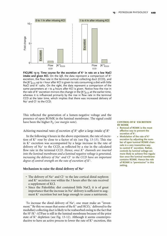
When open ROMK channels are present in the luminal membrane of principal cells, K+ are secreted if a lumen-negative voltage is gener-ated in the CCD. This requires the presence of open ENaC units in the luminal membrane and thereby the ability to reabsorb more Na+ than Cl−. When rats consuming a diet containing little NaCl and K+ salts were given a supplement of KCl, the PK rose somewhat and the calculated [K+]CCD rose to near-maximal levels very quickly. There was a modest initial increase in the rate of excretion of K+ that was caused, almost exclusively, by the rise in the [K+]CCD (Fig. 13-13).
COMPOSITION OF ThE DIET IN RATS CONSUMIN� LABORATORY ChO� • Rats that consume their usual
chow excrete 10 times more K+ than a 70-kg human while con-suming a typical Western diet on a per-kg per-day basis (700 vs. 70 mmol). It follows that the mecha-nisms to excrete an additional K+ load in the rat can reveal only how to raise a very large rate of K+ excretion to an even bigger one. They may not, however, indicate how to initiate a modest, episodic increment in K+ excretion while maintaining very low rates of excretion of K+ at other times.
• The need for a mechanism to increase the delivery of Na+ to the late cortical distal nephron may not be present in rats consuming their regular chow, because these rats consume almost four times more Na+ than humans eating a typical Western diet when expressed in per-kg weight.
-
-
13 : POTASSIUM PHYSIOLOGY 449
This reflected the generation of a lumen-negative voltage and the presence of open ROMK in the luminal membrane. The signal could have been the higher PK (see margin note).
Achieving maximal rates of excretion of K+ after a large intake of K+
In the following 4 hours in the above experiment, the rate of excre-tion of K+ rose by close to a factor of six (see Fig. 13-13). This rise in K+ excretion was accompanied by a large increase in the rate of delivery of Na+ to the CCD, as reflected by a rise in the calculated flow rate in the terminal CCD. Hence, once K+ channels are inserted into the luminal membrane and a luminal negative voltage is generated, increasing the delivery of Na+ and Cl‐ to the CCD have an important degree of control strength on the rate of excretion of K+.
Mechanism to raise the distal delivery of Na+
• The delivery of Na+ and Cl− to the late cortical distal nephron and K+ secretion rose within the 3 hours after the rats received a supplement of KCl.
• Since the Paleolithic diet contained little NaCl, it is of great importance that the increase in Na+ delivery is sufficient to aug-ment K+ excretion but not large enough to cause a natriuresis.
To increase the distal delivery of Na+, one must make an “invest-ment.” By this we mean that some of the K+ and HCO3
− delivered to the medullary collecting duct is likely to be reabsorbed owing to the fact that the H+/K+-ATPase is still in the luminal membrane because of the prior state of K+ depletion (see Fig. 13-12). Although it seems counterpro-ductive to have an active process to lower the rate of K+ excretion, this
CONTROL OF K+ EXCRETION BY ROMK• Removal of ROMK is the most
effective way to prevent the excretion of K+.
• Modulation of the rate of K+ excretion by adjusting the num-ber of open luminal ROMK channels is a very insensitive way to control K+ excretion. Rather, controls by luminal voltage are more likely to achieve fine regulation once the luminal membranecontains ROMK. Hence the role of ROMK is “permissive” in thissetting.
FlowCCD
[K+]CCD
10
5
0
Fol
d ch
ange
Exc
retio
n
FlowCCD
0 to 1 hr after infusing KCl 1 to 4 hr after infusing KCl
[K+]CCD
Exc
retio
n
FIGURE 13-13 Time course for the excretion of K+ in rats on a low NaCl intake and given KCl. On the left, the data represent a comparison of K+ excretion, the flow rate in the terminal cortical collecting duct (CCD), and the [K+]CCD up to 1 hour after KCl is given to rats consuming a diet with little NaCl and K+ salts. On the right, the data represent a comparison of the same parameters at 1 to 4 hours after KCl is given. Notice how the rise in the rate of K+ excretion mirrors the change in the [K+]CCD at the earlier time, whereas it is influenced primarily by the rise in flow rate in the terminal CCD at the later time, which implies that there was increased delivery of Na+ and Cl− to the CCD.

POTASSIUM450
causes a higher rate of K+ excretion owing to the following effects: The concentration of K+ rises in the medullary interstitial compartment, which depolarizes the basolateral membrane of cells of medullary thick ascending limb of the loop of Henle. Thus, there is a smaller intracel-lular negative voltage, which diminishes the exit of Cl−, and as a result, Cl− accumulates in these cells, and this decreases the net reabsorption of Na+ and Cl− in the medullary thick ascending limb (Fig. 13-14).
With time, as the rise in the PK is sustained, there are fewer luminal H+/K-ATPase units in the medullary collecting duct, so the rate of K+ reabsorption declines somewhat and the interstitial concentration of K+ is less elevated. Hence, the inhibition of reabsorption of NaCl in the medullary thick ascending limb is not as marked. As a result, there can be enough distal delivery of Na+ and Cl− to maintain high rates of K+ secretion in the CCD but not to induce a large natriuresis.
WNK KINASE SIGNAL SYSTEM
One way to think about the WNK kinase system is from the per-spective of what was required in Paleolithic times, when the diet contained very little NaCl. Moreover, there were relatively frequent periods when there was an obligatory and possibly large loss of
K+
Na+ 1 Cl2
Cl2
Cl2
LESSNEG
LESS NEG
2
3
4
5
1
Cl2
Reabsorbedvia NKCC-2
K+
H+
K+
K+
Cl2
FIGURE 13-14 Inhibition of the reabsorption of NaCl in the loop of Henle by a K+ load. The stylized structure represents a nephron. Shortly after K+ is ingested, there is a somewhat higher secretion of K+ in the cortical col-lecting duct (site 1). As a result, more K+ is delivered to the medullary CCD, where some of the K+ is reabsorbed by the H+/K+-ATPase, which is still ac-tive because of the prior K+ depletion (site 2). Recall that diet provides a family of organic anions that can be converted to HCO3
− and that a rise in PK diminishes the reabsorption of HCO3
− in the proximal convoluted tubule. As a result, an H+ acceptor is available when the H+/K+-ATPase is active. The higher concentration of K+ in the medullary interstitial compartment leads to a diminution in the negative voltage in cells of the medullary thick ascending limb (mTAL; site 3). This negative voltage in mTAL cells provides the driving force for the exit of Cl− from these cells. As a result of this dimin-ished driving force, the concentration of Cl− rises in cells of the mTAL (site 4). This higher intracellular concentration of Cl− diminishes reabsorption of NaCl via the Na+, K+, 2 Cl− cotransporter (NKCC-2), which then increases the delivery of Na+ to the late cortical distal nephron (site 5). The net result is that K+ reabsorption in the medullary collecting duct provides the signal (a high medullary interstitial [K+]) to increase the delivery of Na+ to the cortical distal nephron segments, thus leading to a large kaliuresis. NEG, negative.
PhYSIOLO�Y OF ThE �NK KINASES• While the WNK kinases par-
ticipate in the switch between the NaCl conservation and the kaliuretic effects described in this section, it is not certain whether they are the primary controllers or whether there are components to this regulatory system.
• In a control system, there must be a signal, a process to recog-nize the signal, the delivery of the message (perhaps the WNK kinases are operating at this step), and response elements, which include the early distal convoluted tubule for the con-servation of NaCl, and principal cells in the CCD for adjusting the secretion of K+.
• There is a potential parallel with cyclic AMP in the cascade for water conservation/excretion and the WNK kinase system in the shift between NaCl conserva-tion and K+ excretion. For the former, the signal, messenger, and response elements are more clearly defined.

13 : POTASSIUM PHYSIOLOGY 451
NaCl (i.e., loss of NaCl sweat when exercise was performed in a hot environment). Hence, the kidneys must be programmed to retain virtually all filtered Na+ and Cl− in this setting.
Interspersed with this low intake of NaCl were periods when there was an intermittent large intake of fruit and berries (foods rich in K+ and a family of organic anions that will be converted promptly into HCO3
− in the liver). At these times, there was a critical need to excrete K+ as quickly as possible. This will include mechanisms to decrease the reabsorption of Na+ and Cl− in upstream nephron segments to deliver the requisite quantity of Na+ for the secretion of K+ in the CCD, as well as the insertion of the needed elements of the K+ secretory system. Moreover, once there was no longer a threat of hyperkalemia, there was a need to reestablish the mechanisms to prevent the excretion of the increased delivery of Na+ and Cl− to the CCD. Hence, it is not surprising that a very complex regulatory system would be involved; it is in this context that we shall examine the WNK kinases (Fig. 13-15 and Table 13-1).
Period when retention of NaCl is required
• The WNK kinase that is most important for this effect is WNK kinase-3 (WNK-3).
WNK-3 is present in the cytoplasm of cells of the distal convoluted tubule. The expected effect of this kinase is to ensure that there will be an abundant quantity of the Na+, Cl− cotransporter in the luminal membrane of these cells (see Fig. 13-15). In addition, WNK-3 is also present in the medullary thick ascending limb of the loop of Henle, where it may contribute to stimulation of the reabsorption of Na+ and Cl− in this nephron segment.
Retain NaCl;do not excrete K�
Need to deliver Na�
to CCD to excrete K�Retain NaCl;
do not excrete K�
Less NaCl delivered to CCD Less NaCl delivered to CCDMore NaCl delivered to CCD
WNK3 WNK3
WNK4 WNK4
WNK1Insert
NCC units
WNK3
RemovesWNK4
InsertNCC unitsRemove
NCC units
NCC NCC NCC
� �
WNK4 �remove WNK3
Long WNK1 �reinsert WNK3
Response to low NaClin Paleolithic diet
DCT
DCT
DCT
FIGURE 13-15 Influence of the components of the WNK kinases on the reabsorption of Na+ and Cl− in the early distal convoluted tubule. The horizontal cylinders represent the lumen of the early distal convoluted tubule (DCT) and the rectangles below these cylinders represents cells of this nephron segment. As shown at far left, WNK-3 is present in their cytoplasm when little NaCl is consumed (i.e., the Paleolithic diet). As a result, more Na+, Cl− cotransporters (NCC) reside in the luminal membrane of these cells. When there is a large intake of K+ salts while little NaCl is consumed, WNK-3 is removed by WNK-4. NCC thus is internalized, and there is a larger de-livery of Na+ and Cl− to the CCD to facilitate the excretion of K+ (middle panel ). As shown in the right panel, NaCl must be retained when there is no longer a need for K+ secretion. Thus NCC must be reinserted in the early DCT. This is achieved by having WNK-1 remove WNK-4 and thus reestablish the effects of WNK3.

POTASSIUM452
Period when there is a need to excrete K+
• When there is a need to secrete a large quantity of K+, a different member of the WNK kinase family (WNK-4) has the task of removing the Na+, Cl− cotransporter from the luminal membrane of cells in the distal convoluted tubule (via removing the actions of WNK-3), while inserting K+ channels (ROMK) in the luminal membrane of principal cells in the CCD.
The actions of WNK kinase 4 (WNK-4) removes the effects of WNK-3 in the early distal convoluted tubule (see Fig. 13-15) and thereby promotes the delivery of Na+ and Cl− to the CCD. Since aldosterone is released in response to the higher PK, there will be more open ENaC units in the luminal membrane of principal cells and thereby more electrogenic reabsorption of Na+ in these nephron segments. WNK-4 leads to the insertion of ROMK into the luminal membrane of principal cells in the CCD. As a result, there will be an increased rate of secretion of K+ in the CCD.
Effects of stopping the intake of K+
• The effects of WNK-4 must be removed to reestablish the NaCl-retaining effect of WNK-3; this is accomplished by the long form of WNK-1 kinases (see margin notes).
The long form of WNK-1 inhibits WNK-4, which leads to higher levels of WNK-3 and thereby to the insertion of the Na+, Cl− cotransporter into the luminal membrane of cells in the early distal convoluted tubule. As a result, there is stimulation of NaCl reabsorp-tion in this nephron segment (see margin note). The short form of WNK-1 inhibits the effects of its long form. If the long and the short forms of WNK-1 are products of the same gene, this may provide a rapid way to modulate the system to enhance the excretion of K+ should the need arise.
TABLE 13-1 SYNOPSIS OF THE WNK KINASE SYSTEM AND THE REGULATION OF NCC AND ROMK
To facilitate understanding of this complex signaling system, we provide a function/control analysis based on the chang-ing needs in a Paleolithic setting when the intake of Na+ was low and the intake of K+ was very large but episodic. Hence, a control system is needed to conserve Na+, but also to allow for the excretion of K+ when faced with a large K+ load. Once the threat of a K+ load is dealt with, the signal to secrete K+ must be removed to reestablish the salt conservation mode. The basic premise of this signaling system is to enhance the electroneutral reabsorption of NaCl in the distal tubule to conserve salt, which also increases the delivery of NaCl to the CCD, where Na+ is reabsorbed in an electrogenic fashion and to insert ROMK into the luminal membrane of the CCD when there is a need to excrete K+.
FUNCTION CONTROL IN DISTAL NEPHRON SPECIFIC WNK KINASES
• Must minimize NaCl excretion when there is a low NaCl intake
• Increase the presence of NCC in the early distal convoluted tubule
• WNK-kinase-3 ensures that NCC is present in the DCT luminal membrane.
• Must excrete the large dietary intake of K+
• Remove NCC from DCT: deliver Na+ for electrogenic reabsorption of Na+ in CCD
• WNK-4 counters WNK-3: NCC is removed in the DCT and ROMK is inserted in CCD
• Conserve NaCl after K+ load is excreted
• Must reinsert NCC in the DCT and remove ROMK in the CCD
• Long form of WNK-1 removes WNK- 4, which allows WNK-3 to reinsert NCC into the DCT and to remove ROMK from the CCD.
LON� �NK-1 AND KS-�NK-1 • This interaction is complex, but
it is also important. Hence it is provided here.
• The WNK-1 gene produces two major products: a full-length or long WNK-1 and a truncated form that is expressed only in the kidney. It is called kidney-specific WNK-1 (KS-WNK-1).
• KS-WNK-1 inhibits the long form of WNK-1 and thus it reestab-lishes the effects of WNK-4 to inhibit WNK-3, which results in augmented secretion of K+.
• Dietary K+ loading increases the abundance of KS-WNK-1 relative to the long form of WNK-1, while K+ depletion has the opposite effect.

13 : POTASSIUM PHYSIOLOGY 453
K+
K+
MCD
H+
H+
CO2
CO2
K � HCO3�
HCO3�
FIGURE 13-16 Possible influence of increased H+/K+-ATPase flux on acid-base balance in the renal medulla. The cylindrical structure represents the medullary collecting duct (MCD). When more K+ are reabsorbed by the H+/K+-ATPase in the MCD, HCO3
– are added to the interstitial compartment. This alkalinization raises the concentration of CO3
2–, which may ultimately lead to local precipitation of Ca3(PO4)2 on the basolateral aspect of the thin limbs of the LOH and thereby be the initial solid phase that leads to deposi-tion of calcium oxalate precipitates.
Clinical implications
Mutations in genes encoding for WNK-1 and WNK-4 are associ-ated with the development of Gordon’s syndrome; this is discussed in more detail in Chapter 15, page 526.
MEDULLARY RECYCLING OF K+: IMPLICATIONS FOR KIDNEY STONE FORMATION
• When there is a large flux rate through the H+/K+-ATPase, this can lead to medullary alkalinization, which may increase the risk of developing calcium oxalate kidney stones.
The H+/K+-ATPase in Paleolithic times provided a means to increase the medullary interstitial K+ concentration and the distal delivery of Na+—a survival advantage when faced with a large K+ load. Nevertheless, modern diets provide much less K+. Accordingly, this transporter now has a different major function because it is needed to prevent K+ depletion rather than to generate the signal for the excretion of a large quantity of K+. This adaptive change, however, comes with a “price.” Increased reabsorption of K+ via the H+/K+-ATPase also adds HCO3
− to the interstitial compartment (Fig. 13-16). This alkalinization can lead to a rise in the concentration of carbonate and thereby the deposition of calcium carbonate, which may cause a local high pH and calcium concentration at its surface. Of interest, the initial solid phase in the formation of calcium oxalate stone may be a precipitate of calcium phosphate (apatite; Randall’s plaques) on the basolateral aspect of the ascending thin limbs of the loops of Henle. This precipitate subsequently erodes into the collecting duct and provides a nidus for deposition of calcium oxalate in patients with idiopathic hypercalciuria and calcium oxalate stones. To prevent medullary interstitial alkalinization, other processes that add H+ to this compartment are likely to be present (see margin note).
SPECULATIONOne candidate is the basolateral Cl–/HCO3
– anion exchanger in β-intercalated cells in the inner medullary collecting duct, the activ-ity of which might be modulated by the interstitial fluid pH. Lesions affecting this transporter or increasing the activity of H+/K+-ATPase (e.g., K+ depletion) might lead to medullary alkaliniza-tion and perhaps the development of calcium oxalate kidney stones.
�NK KINASES AND hYPERTENSION• K+ deprivation is associated with
an increased abundance of the long form of WNK-1 relative to the kidney-specific form (i.e., KS-WNK-1).
• The long WNK-1 activates serum/glucocorticoid-regulated kinase (SGK), which leads to increased abundance of ENaC in the lumi-nal membrane of principal cells in CCD.
• In addition, the long WNK-1, by releasing the inhibitory effect of WNK-4 on WNK-3, aug-ments NaCl reabsorption in the early distal convoluted tubule (i.e., there is now more NaCl cotransporter).
• Overall, these effects could help mediate the observed retention of NaCl and the prevalence of hypertension in subjects con-suming a low K+ diet. Another hypothesis to explain this obser-vation is described later in the section on aldosterone paradox (see page 444).

POTASSIUM454
MECHANISM TO SHIFT K+ INTO LIVER CELLS AFTER A MEAL
• K+ will shift into cells when there is an increased negative volt-age in their ICF compartment. This voltage is generated by an increase in ion flux through the Na-K-ATPase because it is an electrogenic pump (i.e., it exports 3 Na+ while importing only 2 K+).
• The combined actions of insulin and flux of l-lactic acid through the monocarboxylic acid transporter lead to activation the NHE and thereby the electroneutral uptake of Na+ into hepatocytes which drives flux through the Na-K-ATPase.
Our Paleolithic ancestors consumed a diet of fruit and berries that led to a large input of sugar, K+, and organic anions (potential HCO3
–; see margin note). If this K+ load were to enter the systemic circulation, it could lead to hyperkalemia and thereby a cardiac arrhythmia. Therefore, a mechanism is needed to shift this K+ into hepatocytes to minimize this risk.
The mechanism that is most likely to account for this shift of K+ is depicted in Figure 13-17, and it requires a stimulus for the release of insulin and a high local concentration of H+ in hepatocytes near the NHE. The flux through the Na-K-ATPase is increased owing to a higher concentration of Na+ in cells.
Stimuli for the release of insulin
This is easy to understand, as the absorption of sugar from the diet provides the stimulus to the β-cells of the pancreas to release insulin and deliver it at a high concentration to the liver (i.e., the insulin is
Na-K-ATPase
3 Na+
1/3�
2 K+L�
�
Insulin
L-Lactic acid
Na+Na+
NHE
MCT
H+ H+
FIGURE 13-17 Combined actions of insulin and l-lactic acid on a shift of K+ into hepatocytes. The circle represents the cell membrane of hepatocytes. When l-lactic acid enters hepatocytes on the monocarboxylic acid trans-porter (MCT), this leads to the release of H+ immediately adjacent to the NHE. When these H+ bind to the H+ modifier site of the NHE, this cation exchanger is activated, providing that insulin is present and binds to its receptor on the cell membrane. As a result, Na+ enter cells in exchange for H+ (an electroneutral exchange), and this higher concentration of Na+ in these cells drives the exit of Na+ through the Na-K-ATPase (an electrogenic export), which increases the negative voltage in these cells. Thus there is a shift of K+ into these cells.
PALEOLIThIC DIETARY K+ LOAD • One L of orange juice has 135 g
(750 mmol) of sugar and 50 mEq of K+ and of potential HCO3
–. • Since carbohydrates yield 4 kcal/g,
there are 540 kcal and 50 mEq of K+, or ~1 mmol K+ per 10 kcal.
• If the daily intake is ~3000 kcal, the intake of K+ and of HCO3
– would be ~250 mmol/day.

13 : POTASSIUM PHYSIOLOGY 455
added to the portal circulation so it is diluted in only one fifth of the plasma volume).
Source of l-lactic acid
To have a large delivery of l-lactic acid to the liver, this mono-carboxylic acid must be generated in the cells of the small intestine (see margin note). In addition, glycolysis produces l-lactic acid in these cells because this pathway is much faster than the rate of oxida-tion of pyruvate in the citric acid cycle, possible because the entry of glucose is so high and because these cells do a large amount of work. In more detail, the reabsorption of glucose occurs by a Na+-linked glucose transporter (SLGT1), which requires the export of Na+ by the Na-K-ATPase. This intestinal SLGT1 uses 2 Na+ per glucose, so there is an even larger generation of ADP, which stimulates intestinal glycolysis. In fact, there is a rise in the l-lactate concentration to the 3- to-4 mmol/L range in plasma in the portal vein in rats following a meal. The uptake of l-lactic acid into hepatocytes uniquely raises the local H+ concentration near the NHE at the inner aspect of the cell membrane. This together with high insulin levels leads to the uptake of K+ into liver cells (see Fig. 13-17).
DISCUSSION OF INTRODUCTORY CASE 13-1
Introductory case 13-1: Why DId I Become so Weak?
(Case presented on page 426)
What is the most likely basis for the repeated episodes of acute hypokalemia?
Since the time course was only a matter of hours, the excretion of K+ was low (UK/UCreatinine was ~1), there was no major acid-base disor-der, and the recovery was so rapid and required only a small infusion of KCl, the diagnostic category is an acute shift of K+ into cells.
Was an adrenergic effect associated with the acute hypokalemia?
It appears that the answer is yes, as the patient had tachycardia, systolic hypertension, and a wide pulse pressure on physical examina-tion on each episode. Notwithstanding, there was no obvious source of the adrenergic surge. In more detail, she denied an exogenous source of a long-acting β2-adrenergic (e.g., the intake of ampheta-mines), drugs with adrenergic actions (she had stopped using bron-chodilators), caffeine, or drugs that could inhibit cytochrome P450. There was no evidence of an endogenous disease that has a similar hormonal milieu (e.g., hyperthyroidism, pheochromocytoma). Therefore, the source of the surge of β2-adrenergics was still a mystery.
After the laboratory results were examined, were there any clues to suggest what the cause of acute hypokalemia might be?
There were two signs that could be associated with a shift of K+ into cells owing to the coordinated actions between the monocarboxy-lic acid transporter and NHE. First, there was a sign of an elevated Pl-lactate (i.e., the PHCO3 was 22 mmol/L [later the Pl-lactate was mea-sured and it was 3 mmol/L]). Second, the PGlucose was a little higher
�LUCOSE CONVERSION TO l-LACTIC ACIDIf all the ingested glucose were converted to l-lactic acid (which is extremely unlikely), the production of l-lactic acid from this glucose load would be 1500 mmol (750 mmol × 2).
POTASSIUM456
than expected at 133 mg/dL (7.4 mmol/L), and this is associated with a stimulus for the release of insulin. Hence, with the synergistic actions of the monocarboxylic acid transporter and NHE, this could be the basis for a shift of K+ into hepatocytes (see Fig. 13-17). Finally, the last clue is the presence of an adrenergic surge that must be explained. Perhaps the attacks were initiated by the release of insulin from an insulinoma, which caused a sustained period of hypogly-cemia and thereby an adrenergic surge. The metabolic consequence could be hypoglycemia, which is followed by both an increased pro-duction of glucose and more rapid stomach emptying. If the stomach contained sugar, the net result would be a higher PGlucose. One reason that the high PInsulin and the C-peptide in plasma were not observed is that the blood tests were drawn the next day after this association was considered.
Would your treatment be different for her next attack?
Based on the signs of an adrenergic surge on physical examination, the therapy of the next attack would include a nonselective β-blocker if the patient does not have evidence of bronchospasm.
What tests should be performed in the emergency room if she has another episode of acute hypokalemia to reveal the correct diagnosis?
The likely duration of the disorder is that its initiating cause may have been present for many hours earlier; hence, the blood that was drawn on admission may not reveal the factors responsible for the fall in the PK (Fig. 13-18). In addition, if an organ produces or removes a substance that influences a shift of K+ across cell membranes, the results in venous blood draining that organ may not reveal the etiol-ogy of the hypokalemia. In more detail, if there was a large uptake of glucose in muscle owing to the effects of insulin, the PGlucose may be considerably less than its concentration in arterial blood that was delivered to the kidneys.
Based on the above, the clues for the basis of the shift of K+ into cells may be present in the bladder urine—this urine should be saved and analyzed for glucose (i.e., to detect prior glucosuria), signs of a high prior PInsulin (i.e., a high excretion of C-peptide), signs of new glucose production during gluconeogenesis (a higher urea to cre-atinine ratio in the urine on admission as compared to subsequent values), and signs of the adrenergic response to the hypoglycemia (ketonuria, metabolic products that may indicate an adrenergic surge [e.g., urinary metanephrines or catecholamines]).
DISCUSSION OF QUESTIONS
13-1 Is the [K+]ATP channel regulated in fact by ATP?
• ADP acts as an “opener” of [K+]ATP channels in an indirect way.
• Ionized Ca2+ is a major signal for regulation of many intracel-lular processes.
The name of this channel is incorrect because K+ATP channels
are not regulated by the intracellular concentration of ATP, as the

GY 457
13 : POTASSIUM PHYSIOLOconcentration of ATP in cells never rises or falls sufficiently in health to modulate these channels. In fact, the prevailing concentration of ATP ensures that this channel is closed. Nevertheless, if one includes the effects of ADP, which opens this channel, under the usual ATP concentration, the channel may have a certain degree of open prob-ability. Thus, when the concentration of ADP rises, there is a higher degree of open probability of this channel; conversely, when the ADP concentration falls, this channel closes.
To understand what the change in voltage in cells is when the K+
ATP channel opens, recall that a cationic charge leaves the cell; hence, the voltage in cells becomes more negative, which may affect the gating of voltage-gated ion channels. The best example is the voltage-gated Ca2+ channel, which is closed when there is a more negative voltage in cells. Conversely, when fewer K+ leave the cell because the K+
ATP channel is closed, the voltage in cells becomes less negative. This less negative intracellular voltage opens the voltage-gated Ca2+ channel and thereby causes a higher intracellular ionized calcium concentration. By modulating the concentration of ion-ized Ca2+ in cells, many other functions are controlled because ionized Ca2+ is a major signal for regulation of many intracellular processes.
K+ATP channel and the release of insulin: Closing the K+
ATP channel by a fall in the concentration of ADP plays a critical role in the release of insulin from β-cells of the pancreas (Fig. 13-19). In more detail, when the PGlucose is high, more glucose enters β-cells because of their nonregulated glucose transporter (GLUT-2). Hence, some ADP is converted to ATP and the intracellular concentration of ADP falls, which causes the K+
ATP channels to close. The voltage in the ICF compartment thus becomes less
• Products insulin release• Adrenergic effects• New glucose effects
4.0
2.5
1.00 6
Time (hours)12
PK(m
mol
/L)
B
CA
FIGURE 13-18 Possible sequence of events in a patient with acute hypokalemia due to an insulinoma. The PK is shown on the y-axis, and the time course for the change in the PK is shown on the x-axis. The dashed lines represent the times when the PK is in the normal range. Very early on, there is a stimulus to cause a shift of K+ into hepatocytes and perhaps other cells (e.g., prolonged release of insulin from an insulinoma, a β2-adrenergic response to hypoglycemia, and possibly a high Pl-lactate; site A). Some time later (perhaps ~8 hr), the PInsulin falls, the PGlucose rises, and the β2-adrenergic hormone levels begin to decline. In this interval, a clue to the etiology of the acute hypokalemia may be in the urine (site B) including the C-peptide reflecting proinsulin, the presence of ketonuria as part of the adrenergic response to hypoglycemia, and evidence of protein conversion to glucose (high urea/creatinine in the urine as compared to later urine samples). When she arrives at the hospital (~10-hr time), clues may not be detected in the blood sample (site C).

4
PEC •
•
•
POTASSIUM58
negative, which increases the conductance of voltage-gated Ca2+ channels. The resultant rise in the intracellular ionized Ca2+ concentration provides the final signal for the release of insulin from β-cells.
K+ATP channel and vasodilatation: A similar logic can be used to deduce how the blood flow to exercising muscle may be con-trolled using intracellular Ca2+ as the signal (Fig. 13-20). During a sprint, l-lactic acid is released from exercising skel-etal muscle cells. When l-lactic acid enters vascular smooth muscle cells, the concentrations of H+ and l-lactate rise, which leads to opening of the K+
ATP channels independ-ent of the effect of ADP. As a result, K+ exit and the voltage in these cells becomes more negative. This causes closure of voltage-gated Ca2+ channels and less entry of extracellular cal-cium ions into cells and hence vasodilatation.
Clinical implications: Using a drug that closes K+ATP channels (e.g.,
sulfonylureas) can be a useful adjunct for therapy of patients with shock due to vasodilatation (e.g., septic shock).
13-2 What mechanisms permit nonexercising cells to take up K+ during a sprint?
During depolarization of exercising skeletal muscle cells, K+ is released into the spaces in the exterior of these muscles (called the t-tubules; see margin note). Nevertheless, a small quantity of this K+ that is released enters the circulation, and this raises the PK to close to 6.0 mmol/L. The cooperation of resting cells is needed to prevent much larger and dangerous rises in the PK. The mechanisms involved include activation of the Na-K-ATPase by the β2-adrenergic com-ponent of the fight-or-flight response, which pumps endogenous
K+ATP
Open
Closed(closes the
[K+]ATP)
High Ca2+
� releaseof insulin
Lessnegative
Glucose
Glucose
GLUT-2 Pyruvate ADP
Insulin
Ca2+
K+
5 4
3
�
1
2
FIGURE 13-19 Signal system to release insulin from beta cells of the pan-creas. The large oval represents a beta cell of the pancreas. When the PGlucose rises, the intracellular fluid (ICF)Glucose rises to the same extent because of the type of glucose transporter (GLUT-2; site 1) in these cells. The concentration of ADP falls when glucose is oxidized (site 2), and this permits KATP channels to be closed. The ICF compartment becomes less negative when K+
ATP channels close (site 3). This opens voltage-gated Ca2+ channels and raises the ionized Ca2+ concentration in the cytosol (site 4). A higher ionized Ca2+ level in these cells causes the release of insulin (site 5).
hOSPhOCREATINE AND ThE XIT OF K+ FROM MUSCLE ELLS DURIN� SPRNT The hydrolysis of phosphocrea-
tine yields an alkali load in cells (see Chapter 1, page 32 for more discussion).
The exit of HCO3– causes a fall in
the ICF negative voltage (see Fig. 13-5, page 435).
This fall in negative voltage leads to a shift of K+ out of cells.

59
13 : POTASSIUM PHYSIOLOGY 4Na+ out of cells and thereby creates a negative voltage inside cells. A second mechanism is the activation of the NHE by a high intracel-lular concentration of H+, which is the result of l-lactic acid uptake by the monocarboxylic acid transporter in nonexercising cells.
PROMOTE VASOCONSTRICTION
LESSNEGATIVE
Closechannel
�
�
2
1
K�
K�
KATP
ATP
‘A’
PROMOTE VASODILATION
MORENEGATIVE
Openchannel
�2
1
Ca2�
Ca2�
KATP
L�, H�
FIGURE 13-20 Vasoconstrictor tone in vascular smooth muscle cells. The circles represent vascular smooth muscle cells. When the intracellular fluid (ICF) has a less negative voltage because its K+
ATP channels are closed and actions of the hormones symbolized by the A (adrenaline, arginine vaso-pressin, and angiotensin II) cause the voltage-gated Ca2+ channel to be in a more open configuration, permitting a sustained rise in the intracellular ionized calcium concentration. Hence, vasoconstriction is the dominant response (shown on the left). In contrast, during a sprint, Kduring a sprint, K K+
ATP channels are open because of a high concentration of l-lactate anions (L−) and H+ (shown on the right). This leads to a more negative ICF voltage and closure of the voltage-gated Ca2+ channels.