實習醫學生臨床技能核心課程 - tmuh.org.tw · 實習醫學生臨床技能核心課程...
TRANSCRIPT
實習醫學生臨床技能核心課程 3.基礎腹部X-光影像的判讀
(Interpret an abdominal radiograph)
台北醫學大學 影像醫學部

教學目標
說明腹部x-光檢查的適應症及禁忌。
具備基礎放射學及腹部解剖學知識。
確認x光片病人姓名、檢查日期及x光片方向 (orientation)。
系統性的描述腹部X-光影像,並指出病灶之型態及特性。
判讀常見的腹部疾病x-光影像,並且列出鑑別診斷。
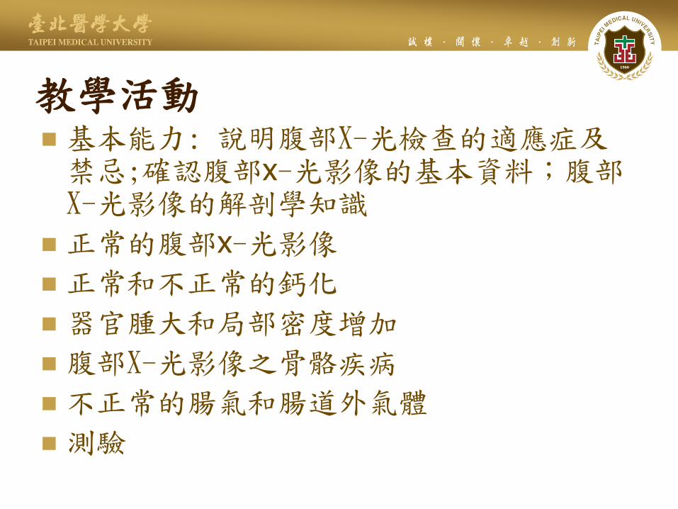
教學活動 基本能力: 說明腹部X-光檢查的適應症及禁忌;確認腹部x-光影像的基本資料;腹部X-光影像的解剖學知識
正常的腹部x-光影像
正常和不正常的鈣化
器官腫大和局部密度增加
腹部X-光影像之骨骼疾病
不正常的腸氣和腸道外氣體
測驗

腹部x-光檢查的適應症及禁忌
急腹症 (acute abdomen)
在急腹症之病患,腹部x-光檢查幾乎可用腹部電腦斷層影像取代,價值遠低於胸部x-光檢查
懷孕婦女因遊離輻射避免照射腹部x-光,視可否用其他替代性檢查取代
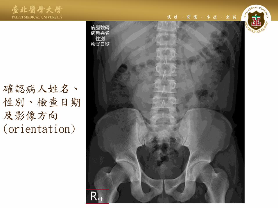
確認病人姓名、 性別、檢查日期 及影像方向(orientation)
病歷號碼
病患姓名
性別
檢查日期

腹部x-光檢查:supine abdomen(KUB) 範圍: 上緣—超過renal shadow 下緣--symphysis pubis
片子大小--
Descending colon gas with fecal materials
Gastric rugae Psoas shadows
Ascending colon with haustration
Rectal air
Renal shadow
Liver shadow
照相: AP view, 仰臥
左
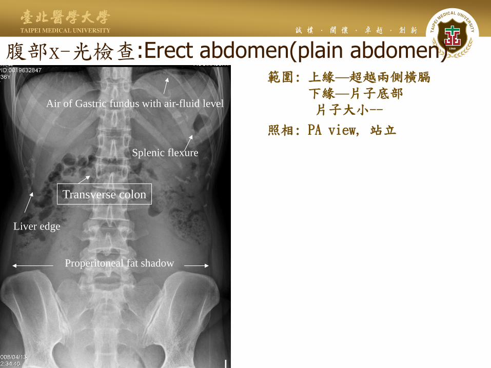
腹部x-光檢查:Erect abdomen(plain abdomen) 範圍: 上緣—超越兩側橫膈
下緣—片子底部 片子大小--
照相: PA view, 站立
Air of Gastric fundus with air-fluid level
Splenic flexure
Transverse colon
Liver edge
Properitoneal fat shadow
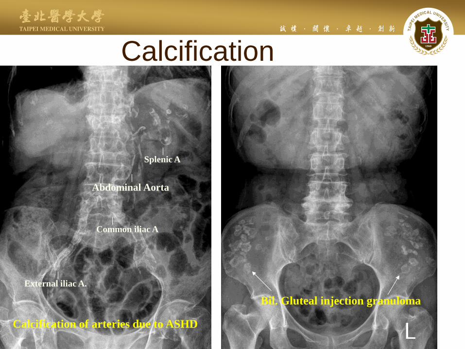
Calcification
Calcification of arteries due to ASHD
Splenic A
Abdominal Aorta
Common iliac A
External iliac A.
Bil. Gluteal injection granuloma
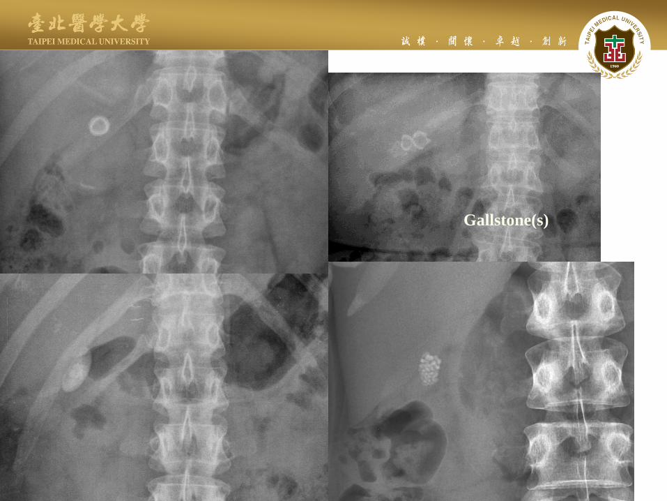
Gallstone(s)

Bladder stones
Bil. Renal stones with hydronephrosis
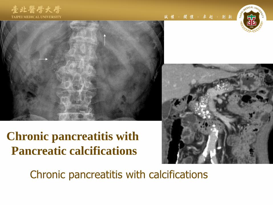
Chronic pancreatitis with calcifications
Chronic pancreatitis with
Pancreatic calcifications
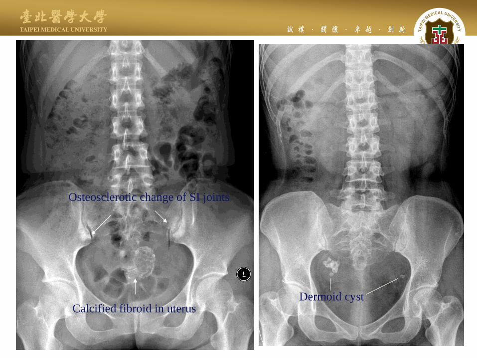
Calcified fibroid in uterus
Osteosclerotic change of SI joints
Dermoid cyst
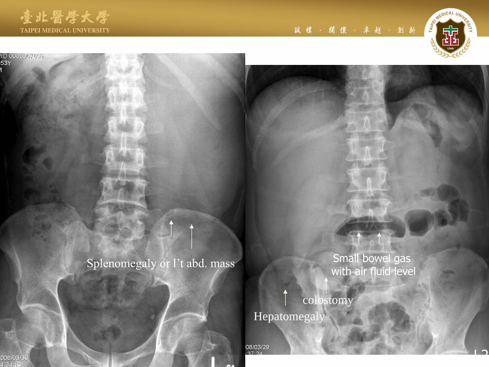
Hepatomegaly
Small bowel gas with air fluid level
colostomy
Splenomegaly or l’t abd. mass
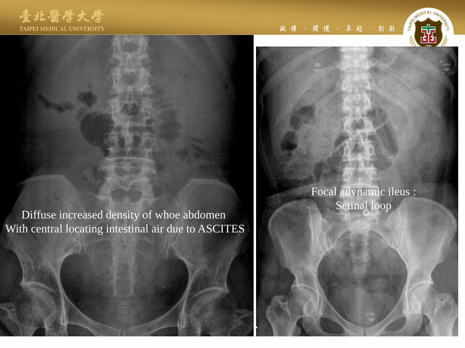
Diffuse increased density of whoe abdomen
With central locating intestinal air due to ASCITES
Focal adynamic ileus :
Setinal loop
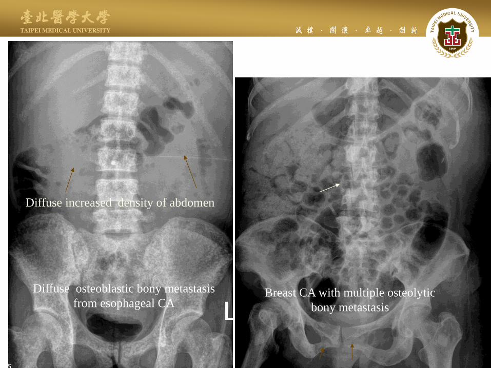
Diffuse osteoblastic bony metastasis
from esophageal CA Breast CA with multiple osteolytic
bony metastasis
Diffuse increased density of abdomen
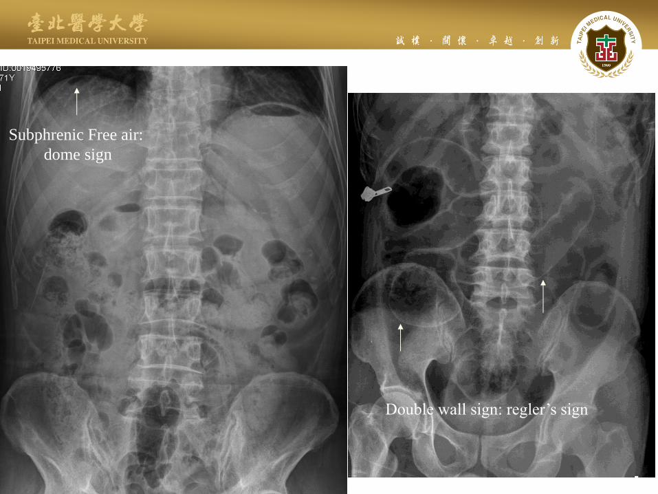
Subphrenic Free air:
dome sign
Double wall sign: regler’s sign

Disproportional dilatation of small bowel
Due to mechanical obstrution
Abnormal SBL>3cm
Wall thickness>3mm
Marked distended gastric air with
food residual due to
Gastric outlet obstruction
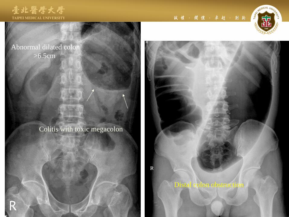
Colitis with toxic megacolon
Distal colon obstruction
Abnormal dilated colon
>6.5cm
Case demonstration on PACS

測驗
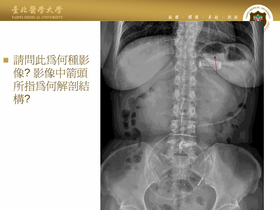
請問此為何種影像? 影像中箭頭所指為何解剖結構?
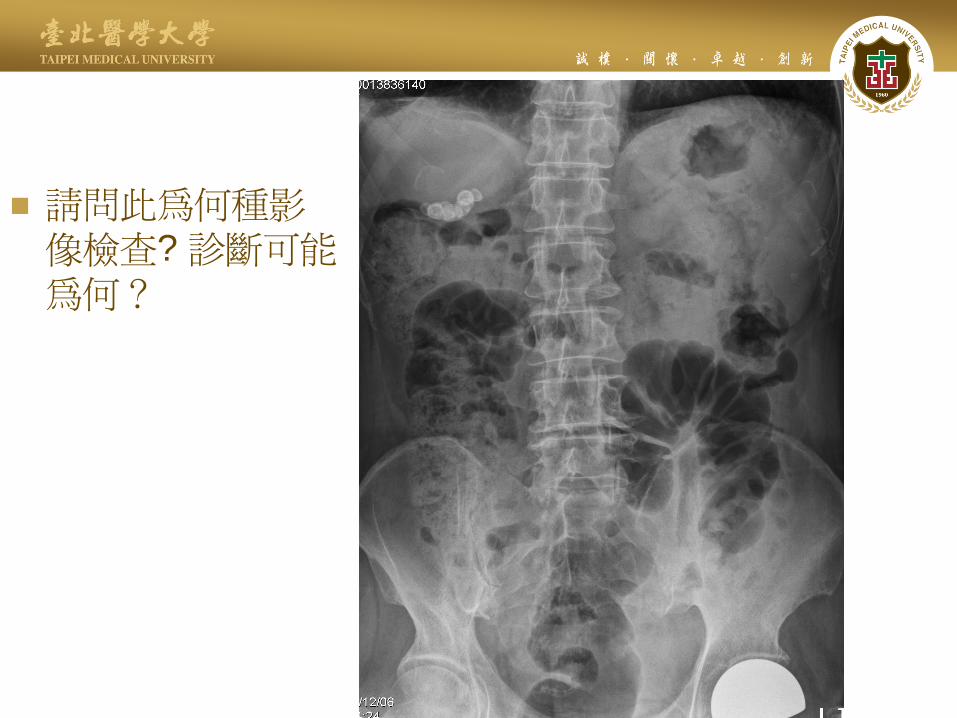
請問此為何種影像檢查? 診斷可能為何?
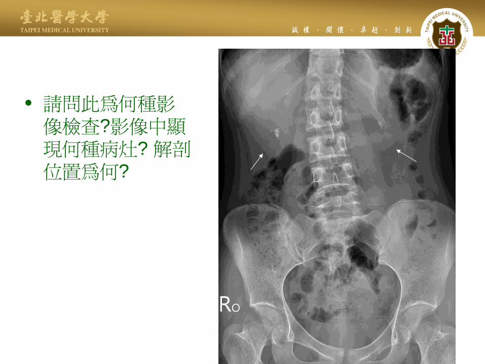
• 請問此為何種影像檢查?影像中顯現何種病灶? 解剖位置為何?

• 請問此為何種影像檢查?診斷可能為何?
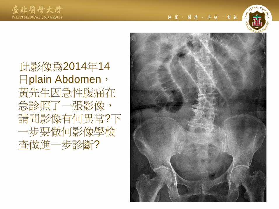
此影像為2014年14
日plain Abdomen,
黃先生因急性腹痛在急診照了一張影像,請問影像有何異常?下一步要做何影像學檢查做進一步診斷?
T2
WI

2
3
5 請問此為何種影像?
影像中數字所指為何解剖結構?
4
6
1