eport of institute for virus research department of viral oncology which consists of 4 laboratories....
TRANSCRIPT

Annual Report of the Institute for Virus Research Kyoto University Volume 56 2013
Annual Report of the Institute for Virus Research Kyoto University Volume 56 2013
ISSN 0075-7357ISSN 0075-7357
京都大学ウイルス研究所年報
AN
NU
AL R
EP
OR
T OF T
HE
I NS
TIT
UT
E FO
R V
IRU
S R
ES
EA
RC
H K
YO
TO
UN
IVE
RS
ITY V
OL
UM
E 56 2
01
3
Institute for Virus ResearchKyoto University

CHRONOLOGICAL TABLE
1956 April Institute for Virus Research, Kyoto University, was founded with two departments
(Pathology and Biophysics).
1956 April Scientific Lectures for the Public were presented commemorating the opening of the
Institute (the successive Memorial Lecture Series have been presented annually
hereafter).
1957 April Department of Biochemistry and Department of Serology and Immunology were
established.
1958 April Department of Prevention and Therapeutics was established.
1958 December "Advances in Virology", Vol. 1 (in Japanese) was published as collection of the
Memorial Lectures (the successive volumes were published annually hereafter until
1960).
1958 December "Annual Report of the Institute for Virus Research", Vol. 1, was published (the
successive volumes have been published annually hereafter).
1959 July Virus Diagnosis Center was established.
1961 October The 1st Symposium of the Institute for Virus Research was held under the auspices
of the Institute with the nationwide participants. The proceedings of the
Symposium were published as the first issue of the new series of "Advances in
Virology" in Japanese (the successive Symposia have been held and their
proceedings published annually hereafter).
1962 April Department of Tumor Virus was established.
1962 October Several staff members were appointed academic staff of the Graduate School of
Medicine, and students of the School were first admitted to the Institute.
1962 December Several staff members were appointed academic staff of the Graduate School of
Science, and students of the School were first admitted to the Institute.
1964 April Virus Diagnosis Center was renamed Virological Diagnosis Center.
1965 September Construction of the new building for the Institute commenced.
1967 March Construction of the new building was completed.
1968 April Department of Genetics was established.
1974 April Department of Molecular and Cellular Virology was established.
1977 April Department of Neurological Virus Disease was established as such that Visiting Staff
be appointed.
1978 April Animal Laboratory for Experimental Virus Infection was established.
1981 March Construction of extension of the main building was completed. Thus the main
building now constitutes five floors with a basement occupying the aggregate area of
5,410 ㎡. The major part (ca. 481 ㎡) of the extended area serves for researches
v
involving radioisotope labelling and in vitro DNA recombination experiments
requiring the P3 facilities.
1986 May The memorial events for the 30th anniversary of foundation of this Institute were
held on May 16-17.
1986 November Professor Yorio Hinuma was honoured as "Person of Cultural Merits
(Bunkakorosha)"
1987 May Department of Biophysics and Department of Tumor Virology were reorganized to
form Department of Viral Oncology which consists of 4 Laboratories.
1988 April Virological Diagnosis Center was reorganized to become Research Center for
Immunodeficiency Virus which consists of Laboratory for AIDS Immunology and
Laboratory of Viral Pathogenesis.
1989 April Department of Biochemistry and Department of Genetics were reorganized to form
Department of Genetics and Molecular Biology which consists of 3 Laboratories.
1990 March Construction of a new building was partly completed.
1990 April Department of Pathology and Department of Molecular and Cellular Virology were
reorganized to form Department of Cell Biology which consists of 3 Laboratories,
while Department of Serology and Immunology, Department of Prevention and
Therapeutics and Department of Neurological Virus Disease were reorganized to
form Department of Biological Responses which consists of 2 laboratories and one
for visiting staff.
1992 April Laboratory of Regulatory Information was established within the Department of
Cell Biology to host a visiting professor as well as a research group.
1993 December Construction of the new building which accommodates three laboratories from this
Institute as well as some from the Medical School and the Center for Molecular
Biology and Genetics of the University was completed.
1994 October Construction of a new animal facility with some laboratories was completed.
1998 April One stuff member was appointed academic staff of the Graduate School of
Pharmaceutical Sciences, and students of the school were first admitted to the
Institute.
1998 April Research Center for Immunodeficiency Virus was reorganized to become Research
Center for Acquired Immunodeficiency Syndrome.
1998 April Laboratory of Virus Control in Research Center for Immunodeficiency Virus was
established as such that Visiting Stuff be appointed.
1999 April Several staff members were appointed academic staff of the Graduate School of
Biostudies, and students of the school were first admitted to the Institute.
2002 April The Experimental Research Center for Infected Animals was abolished and the
Experimental Research Center for Infectious Diseases was established instead.
vi

2005 April Research Center for Emerging Virus was established. 2009 June The Institute commenced service as a Joint Usage / Research Center for fusion of
advanced technologies and innovative approaches to viral infections and life science.
2010 April Center for Acquired Immunodeficiency Syndrome Research was reorganized to
become Center for Human Retrovirus Research.
2010 April Research Center for Emerging Virus was reorganized to become Center for
Emerging Virus Research.
2013 October Laboratory of Evolutional Virology was established in Experimental Research
Center for Infectious Diseases.
vii

ORGANIZATION AND STAFF (as of December, 2013)
(Numerals in parentheses indicate year of association with the Institute)
Director Masao Matsuoka, M.D., D.Med.Sc. Deputy Director Yoshio Koyanagi, M.D., D.Med.Sc. Professors Emeriti Yoshimi Kawade, D.Sc. (1956-1988) Yorio Hinuma, M.D., D.Med.Sc. (1980-1988) Masao Hanaoka, M.D., D.Med.Sc. (1959-1989) Mutsuo Imai, D.Sc. (1965-1991) Takashi Yura, D.Sc. (1960-1993) Masakazu Hatanaka, M.D., D.Med.Sc. (1980-1995) Akinori Ishimoto,M.D.,D.Med.Sc.(1964-1968, 1978-2002) Yoshiaki Ito, M.D., D.Med.Sc. (1984-2002) Masanori Hayami, D.V.M., D.Agr. (1988-2006) Koreaki Ito, D.Sc. (1971-2007) Kunitada Shimotohno, Pharm.D. (1996-2007) Junji Yodoi, M.D., D.Med.Sc. (1989-2010) Department of Viral Oncology Laboratory of Gene Analysis Professor Yoshinori Akiyama, D.Sc. (1988) Associate Professor Hiroyuki Sakai, D.Med.Sc. (1996) Hiroyuki Mori, D.Sc. (1996) Assistant Professor Shin-ichi Yanagawa, D.Agr. (1986)
Laboratory of Cell Regulation Professor Masahiko Sugita, M.D., D.Med.Sc. (2004) Assistant Professor Daisuke Morita, D.Bio. (2013)
Laboratory of Tumor Biogenesis Professor Shin Yonehara, D.Sc. (1994) (concurrent) Assistant Professor Akira Murakami, D.Sc. (1979)
Laboratory of Human Tumor Viruses Professor Keizo Tomonaga, D.V.M., D.Vet.Med. (2011) Associate Professor Makoto Hijikata, D.Med.Sc. (1997) Assistant Professor Tomoyuki Honda, M.D., D.Med.Sc. (2011) Department of Genetics and Molecular Biology
Laboratory of Molecular Genetics Professor Takashi Fujita, D.Sc. (2005) Associate Professor Hiroki Kato, D.Med.Sc. (2010)
Laboratory of Biochemistry Professor Mutsuhito Ohno, D.Sc. (2001)
viii

Assistant Professor Makoto Kitabatake, D.Sc.(2004) Ichiro Taniguchi, D.Sc. (2007) Department of Biological Responses
Laboratory of Biological Protection Professor Koichi Ikuta, M.D., D.Med.Sc. (2002) Assistant Professor Keiko Takemoto, D.Sc. (1992) Shizue Tani-ichi, D.Health Sc. (2007) Takahiro Hara, D. Bio. (2008) Technical Staff Satsuki Kitano (Konaka) (2004)
Laboratory of Infection and Prevention
Professor Osamu Takeuchi, M.D.,Ph.D. (2012) Associate Professor Hiroshi Masutani, M.D., D.Med.Sc. (1992) Assistant Professor Takashi Mino, D.Eng. (2012)
Bioresponse Regulation Laboratory
Visiting Professor Yoshihiro Kawaoka , D.V.M., D.Med.Sc. (2010) Visiting Assistant Professor Hironori Yoshiyama , D.Med.Sc. (2013)
Department of Cell Biology
Laboratory of Subcellular Biogenesis Professor Fumiko Toyoshima, D.Sc. (2008) Assistant Professor Shigeru Matsumura, D.Bio. (2008)
Laboratory of Growth Regulation
Professor Ryoichiro Kageyama, M.D., D.Med.Sc. (1997) Associate Professor Toshiyuki Ohtsuka, M.D., D.Med.Sc. (2000) Associate Professor (Hakubi) Itaru Imayoshi, D.Bio.(2008) Assistant Professor Taeko Kobayashi, D.Sc. (2005) Assistant Professor (Hakubi) Tomoko Tateya , M.D.,D.Med.Sc.(2008)
Laboratory of Signal Transduction
Associate Professor Takayuki Miyazawa, D.V.M., D.Vet.Med. (2005)
Laboratory of Regulatory Information Visiting Professor Susumu Tonegawa, Ph.D, D.Sc. (1992)
Center for Human Retrovirus Research
Laboratory of Viral Pathogenesis Professor Yoshio Koyanagi, M.D., D.Med.Sc. (2004) Assistant Professor Hirotaka Ebina, D.Med.Sc. (2009)
Kei Sato, D.Med.Sc. (2012) Laboratory of Virus Control
Professor Masao Matsuoka, M.D., D.Med.Sc. (1999) Lecturer Jun-ichirou Yasunaga, M.D., D.Med.Sc. (2010) Assistant Professor Kazuya Shimura, D.Med.Sc. (2011) Technical Staff Junko Tanabe (2006)
ix

Laboratory of Viral Immunology Visiting Professor Hiroaki Mitsuya, M.D.,Ph.D. (2012)
Experimental Research Center for Infectious Diseases
Laboratory of Mouse Model Associate Professor Makoto Tachibana, D.Agr. (1998)
Laboratory of Primate Model
Professor Tatsuhiko Igarashi, D.V.M., D.Med.Sc. (2007) Associate Professor Tomoyuki Miura, D.V.M., D.Agr. (1988) Assistant Professor Takayuki Hishiki, D.Bio. (2013)
Laboratory of Virus Evolution Professor HirofumiAkari, D.V.M., D.Vet.Sc. (2013) (concurrent) Associate Professor Takayuki Miyazawa, D.V.M., D.Vet.Med. (2005) Assistant Professor (Spe.*) Amane Kogure, D.Med.Sc. (2013) Takeshi Yoshida, D.Med.Sc. (2013) (concurrent)
Technical Specialist Hitoshi Miyachi (2006) Technical Staff Ai Dantsuka (2011)
Setsuo Asahi (2013) Center for Emerging Virus Research
Head・Professor Yoshio Koyanagi, M.D., D.Med.Sc. (2010) Assistant Professor (Spe.*) Yosuke Yamaoka, Pharm.D. (2012) Akiko Makino, D.V.M.Ph.D. (2012) Yohei Hizukuri, D.Sc. (2013)
Lecturers (part time) Makoto Miyata Sokichi Matsumoto Takeshi Ichinohe Koji Yasutomo Tamotsu Yoshimori Shosei Yoshida Tatsuya Saito Yasumasa Iwatani Hiroshi Kimura Hirofumi Arakawa Jiro Yasuda Research Fellows Eiji Ishii (Lab. of Gene Analysis) Kan Fujino (Lab. of Human Tumor Viruses) Yusuke Matsumoto (Lab. of Human Tumor Viruses) Yuya Hirai (Lab. of Human Tumor Viruses) Yuichi Abe (Lab. of Human Tumor Viruses) Yuki Kaname (Lab. of Molecular Genetics) Ryota Ouda (Lab. of Molecular Genetics) Asako Mclosky (Lab. of Biochemistry)
x

Tomoko Imamura (Lab. of Infection and Prevention) Daisuke Ori (Lab. of Infection and Prevention) Atsuko Wakabayashi (Lab. of Infection and Prevention) Akihiro Isomura (Lab. of Growth Regulation) Yukiko Harima (Lab. of Growth Regulation) Tsuyoshi Hirashima (Lab. of Growth Regulation) Rokusuke Yoshikawa (Lab. of Signal Transduction) Junpei Terakawa (Lab. of Signal Transduction) Matouskoba Magda (Lab. of Signal Transduction) Junko Takeuchi (Lab. of Viral Pathogenesis) Tomoko Kobayashi (Lab. of Viral Pathogenesis) Guangyong Ma (Lab. of Virus Control) Kenji Sugata (Lab. of Virus Control) Library
Committee Chairman Hiroyuki Mori Administration Office
Chief Officer Katsumi Sakamoto (2013) General Affairs Hiroyuki Matsunaga (2011)
Kazue Hattori (2013) Note *Spe. : Program-Specific
xi

DEPARTMENT OF VIRAL ONCOLOGY LABORATORY OF GENE ANALYSIS I. First Group Members
Professor Yoshinori Akiyama Associate Professor Hiroyuki Mori Assistant Professor (Spe.) Yohei Hizukuri (Center for Emerging Virus Research) Research fellow Eiji Ishii Graduate Student Tokuya Hattori Yasushi Daimon Ryoji Miyazaki Narimasa Hasimoto Koichiro Akiyama Kazuya Mito Chigusa Masui Shinya Mizuno Akira Mukuno
Introduction
The research projects carried out in this group are concerned with post-translational events in the expression of genetic information. Specifically, processes of protein translation, protein translocation across and integration into the membrane, membrane protein proteolysis and extracytoplasmic stress responses are investigated by combined molecular genetic, biochemical biophysical and structural approaches. Topics
Protease homolog BepA (YfgC) promotes assembly and degradation of β-barrel membrane proteins in Escherichia coli: S. NARITA1, C. MASUI, T. SUZUKI2, N. DOHMAE2, and Y. AKIYAMA. (1University of Morioka, IVR, 2RIKEN)
The cell envelope of gram-negative bacteria is composed of two layers of biological membranes, the outer membrane (OM) and the inner (cytoplasmic) membrane (IM). Outer
−3−

membrane proteins (OMPs) span the OM with amphipathic, antiparallel β-strands that form a barrel structure. Gram-negative bacteria are equipped with quality-control systems for the OM that sense and cope with defective biogenesis of its components. Accumulation of misfolded outer membrane proteins (OMPs) in Escherichia coli leads to activation of σE. Disruption of bepA (formerly yfgC), a σE-regulated gene encoding a putative periplasmic metalloprotease, sensitizes cells to multiple drugs, suggesting that it may be involved in maintaining OM integrity. However, the specific function of BepA has been unclear. We showed that BepA enhances biogenesis of LptD, an essential OMP involved in OM transport and assembly of lipopolysaccharide, by promoting rearrangement of intramolecular disulfide bonds of LptD. In addition, BepA possesses protease activity and is responsible for the degradation of incorrectly folded LptD. In the absence of periplasmic chaperone SurA, BepA also promotes degradation of BamA, the central OMP subunit of the β-barrel assembly machinery (BAM) complex. Interestingly, defective oxidative folding of LptD caused by bepA disruption was partially suppressed by the expression of protease-active site mutants of BepA, suggesting that BepA functions independently of its protease activity. We also showed that BepA exhibits genetic and physical interactions with components of the BAM complex. These findings raised the possibility that BepA maintains the integrity of the OM both by promoting assembly of OMPs and by proteolytically eliminating OMPs when their correct assembly is compromised1).
1) Narita, S.-i., et al. (2013) Protease homolog BepA (YfgC) promotes assembly and degradation of β-barrel membrane
proteins in Escherichia coli. Proc. Natl. Acad. Sci. USA 110, E3612–E3621
Membrane targeting-mediated regulation of Escherichia coli heat shock transcription factor, 32: B. LIM1, R. MIYAZAKI, S. NEHER1, D.A. SIEGELE2, K. ITO3, P. WALTER1, Y. AKIYAMA, T. YURA3, and C.A. GROSS1 (1University of California, San Francisco, 2Texas A&M University, 3Kyoto Sangyo University)
Heat shock response is a major homeostatic mechanism for controlling the state of protein folding and degradation in all organisms. Expression of heat shock genes in E. coli is both under positive control by 32, a transcription factor dedicated to the heat shock response, and under negative feedback control (inactivation/degradation of 32) by stress-inducible molecular chaperones (DnaK/J-GrpE, GroEL/S). 32 is extremely unstable in vivo and is degraded by membrane-localized protease FtsH. Chaperones contribute to rapid degradation of 32 in vivo, whereas its degradation in vitro is very slow and not enhanced by chaperones. It is possible that some other factors are involved in the degradation of 32 in vivo. In collaboration with Drs. Takashi Yura and Carol A. Gross 1), we identified the co-translational protein targeting machinery, comprised of the Signal Recognition Particle (SRP) and the SRP Receptor (SR; FtsY), as a regulator of 32. We showed that SRP directly binds to 32,
−4−

that a population of 32 is present in the membrane fraction and that both the SRP-dependent machinery and the 32 region 2.1 involved in the feedback control are important for the localization of 32. The regulatory defects in heat shock response circuitry caused by mutations of either the
32 region 2.1 or the co-translational targeting system are circumvented by artificially tethering 32 to the membrane. We proposed that SRP-dependent membrane localization is a critical step in the control circuitry that governs the activity and stability of 32.
1) Lim, B., et al. (2013) Heat shock transcription factor 32 co-opts the signal recognition particle to regulate protein
homeostasis in E. coli. PLoS Biology 11, e1001735
Molecular mechanisms of the enhancement of protein export by the membrane protein complex SecDF: K. MITO, A. MUKUNO, Y. MACHIDA, Y. AKIYAMA and H. MORI
The SecYEG translocon and the SecA ATPase cooperate to facilitate protein export across the bacterial cytoplasmic membrane. In addition to these essential core components, SecDF, a complex containing two membrane-integrated Sec factors, play important roles in efficient protein export in vivo. We determined the crystal structure of SecDF from Thermus thermophilus at 3.3 Å resolution and proposed a working hypothesis based on structure-instructed biochemical and biophysical studies1). According to the model, SecDF forms a complex with SecYEG translocon, captures a substrate polypeptide emerging from the translocon by its P1 (the first periplasmic) domain and undergoes conformational changes using the PMF (proton motive force) to facilitate forward movement of the polypeptide. However, modes of interactions between SecDF and Sec-related factors including SecYEG and a substrate polypeptide remain largely unknown. To gain information on this issue, we performed systematic site-directed in vivo photo-cross-linking analysis2) targeted to E. coli SecD. Based on the Thermus thermophilus SecDF structure, we designed E. coli SecD mutations so as to introduce a photo reactive amino acid, p-benzoyl phenylalanine (pBPA) on the molecular surface of the protein. We constructed more than 80 SecD derivatives and carried out the cross-linking experiments with them. So far, we identified several E.coli SecD residues that are in close proximity to SecF, periplasmic chaperones or a substrate protein. In addition, possible intramolecular crosslinkings within the SecD P1 domain were detected. Interestingly, the amounts of some of the SecD P1-SecF intermolecular and the SecD P1 intramolecular crosslinked products were dramatically decreased, when cells expressing these SecD derivatives were treated with CCCP (a protonophore) prior to the UV-crosslinking to collapse PMF across the membrane. These results nicely fit our model that the proton conductance of SecDF somehow couples with the movement of the P1 domain.
−5−

1) Tsukazaki, T. et al. (2011) Structure and function of a membrane component SecDF that enhances protein export.
Nature 474, 235-238.
2) Chin, J. W. et al. (2002) Addition of a photocrosslinking amino acid to the genetic code of Escherichia coli. Proc.
Natl. Acad. Sci. USA. 99, 11020-11024.
List of Publications Lim, B., Miyazaki, R.a, Neher, S.a, Siegele, D.A., Ito, K., Walter, P., Akiyama, Y.*, Yura, Y.*, and Gross, C.A.* (2013) Heat shock transcription factor 32 co-opts the signal recognition particle to regulate protein homeostasis in E. coli. PLoS Biology 11, e1001735. a These authors contributed equally to this work. *Corresponding author Narita, S.-i., Masui, C., Suzuki, T., Dohmae, N., and Akiyama, Y. (2013) Protease homolog BepA (YfgC) promotes assembly and degradation of β-barrel membrane proteins in Escherichia coli. Proc. Natl. Acad. Sci. USA 110, E3612-E3621. Ishii, E., Eguchi, Y., and Utsumi, R. (2013) Mechanism of activation of PhoQ/PhoP two-component signal transduction by SafA, an auxiliary protein of PhoQ histidine kinase in Escherichia coli. Biosci. Biotechnol. Biochem. 77, 814-819. Kroos, L., and Akiyama, Y. (2013) Biochemical and structural insights into intramembrane metalloprotease mechanisms. Biochim. Biopys. Acta 1828, 2873-2885. Ha, Y., Akiyama, Y., and Xue, Y. (2013) Structure and mechanism of rhomboid protease. J. Biol. Chem. 288, 15430-15436. Ogura, T., Okuno, T., Suno, R., and Akiyama, Y. (2013) FtsH Protease. Handbook of Proteolytic Enzymes 3rd ed. (ed. Rawlings, N. D. and Salvesen, G.) Elsevier Ltd. pp685-692. Hizukuri, Y., Ito, K., and Akiyama, Y. (2013) RseP Peptidase. Handbook of Proteolytic Enzymes 3rd ed. (ed. Rawlings, N. D. and Salvesen, G.) Elsevier Ltd. pp1546-1550. Akiyama, Y., and Ito, K. (2013) HtpX Peptidase. Handbook of Proteolytic Enzymes 3rd ed. (ed. Rawlings, N. D. and Salvesen, G.) Elsevier Ltd. pp683-685. 森 博幸、塚崎智也(2013)細菌のタンパク質分泌を促進する膜タンパク質 SecDF の構造
−6−

と機能 化学と生物 51、28-35. 江口陽子、加藤明宣、石井英治、内海龍太郎(2013)コネクターがつなぐ細菌情報伝達ネ
ットワーク 化学と生物 51、241-249. Mori, H., Tsukazaki. T., Machida, Y., Mito, K., Nureki, O., Ito, K., and Akiyama, Y.: Structure and function of SecDF, a membrane integrated protein translocation enhancing factor. 第 86 回日本細
菌学会総会ワークショップ W7「細菌構造研究の進展開:分泌装置、細胞骨格、運動装置、
細菌表層の構造体を中心に」、幕張、2013 年 3 月 19 日 成田新一郎、秋山芳展:大腸菌 バレル型タンパク質の品質管理に関わるプロテアーゼホ
モログ BepA(YfgC) の機能解析、2012 年度国立遺伝学研究所研究会「単細胞システムの構
築とその維持機構の研究」、三島、2013 年 3 月 28-29 日 森 博幸:ビブリオ菌 SecDF パラログの発現制御機構、2012 年度国立遺伝学研究所研究会
「単細胞システムの構築とその維持機構の研究」、三島、2013 年 3 月 28-29 日 檜作洋平、禾 晃和、小田 隆、田畑早苗、川上-田村恵子、佐藤 衛、高木淳一、秋山芳展:
大腸菌膜内切断プロテアーゼ RseP の基質認識における PDZ ドメインの役割、第 86 回日本
生化学会大会、シンポジウム「非常識なプロテアーゼ反応:膜内部でのタンパク質切断」、
横浜、2013 年 9 月 11-13 日 森 博幸、三登一八、町田裕紀子、塚崎智也、伊藤維昭、秋山芳展:タンパク質膜透過促進
因子 SecDF の構造と機能、第 51 回日本生物物理学会年会シンポジウム「バーグ教授記念
講演と踊る運動超分子マシナリー」、京都、2013 年 10 月 28-30 日 秋山芳展:膜内部でのタンパク質切断による表層ストレス応答制御機構、京都大学微生物
科学寄付研究部門主催第二回シンポジウム「微生物科学研究の多様性と新展開」、京都、2013年 11 月 8 日 成田新一郎、鈴木健裕、堂前 直、秋山芳展:大腸菌 βバレル型膜タンパク質の生合成に関
わる BepA (YfgC) の機能解析、日本農芸化学会 2013 年度(平成 25 年度)大会、仙台、2013年 3 月 27 日 檜作洋平、小田 隆、田畑早苗、川上-田村恵子、佐藤 衛、高木淳一、禾 晃和、秋山芳展:
膜内切断プロテアーゼ RseP のタンデム PDZ ドメインが介する切断基質選別機構、第 10 回
−7−

21 世紀大腸菌研究会、伊豆、2013 年 6 月 20-21 日 三登一八、町田裕紀子、塚崎智也、伊藤維昭、秋山芳展、森 博幸:部位特異的 in vivo 光架橋法によるタンパク質膜透過促進因子 SecDF の基質結合部位の探索、第 10 回 21 世紀大
腸菌研究会、伊豆、2013 年 6 月 20-21 日 秋山光市郎、禾 晃和、秋山芳展:S2P ファミリー膜内切断プロテアーゼ RseP の「膜内部
に挿入した ヘアピン領域」の機能、第 10 回 21 世紀大腸菌研究会、伊豆、2013 年 6 月 20-21日 舛井千草、成田新一郎、秋山芳展 :部位特異的 in vivo 光架橋法による大腸菌ペリプラズ
ムタンパク質 BepA(YfgC)の近接因子探索、第 10 回 21 世紀大腸菌研究会、伊豆、2013 年
6 月 20-21 日 Nogi, T., Hizukuri, Y., Oda, T., Tabata, S., Tamura-Kawakami, K., Sato, M., Takagi, J., and Akiyama, Y.: Structural analysis of the PDZ tandem fragment of the bacterial intramembrane-cleaving protease RseP. International Conference on Structural Genomics 2013, Sapporo, Japan, 29 July -21 August 2013. 宮﨑亮次、由良 隆、森 博幸、秋山芳展:大腸菌熱ショック転写因子 32 の膜への targetingを介した機能制御機構、第 86 回日本生化学会大会、横浜、2013 年 9 月 11-13 日 成田新一郎、舛井千草、鈴木健裕、堂前 直、秋山芳展:大腸菌プロテアーゼ BepA は外膜
タンパク質の組立と分解を促進する、日本農芸化学会東北支部第 148 会大会、盛岡、2013年 10 月 26 日 檜作洋平、小田 隆、田畑早苗、川上-田村恵子、佐藤 衛、高木淳一、禾 晃和、秋山芳展:
Substrate discrimination mechanism by a PDZ tandem in the intramembrane protease RseP that regulates extracytoplasmic stress response. 第 51 回日本生物物理学会年会、京都、2013 年 10月 28 日 宮﨑亮次:大腸菌熱ショック転写因子 32 の膜への targeting を介した機能制御機構、京都大
学微生物科学寄付研究部門主催第二回シンポジウム「微生物科学研究の多様性と新展開」、
京都、2013 年 11 月 8 日 秋山光市郎:大腸菌の S2P ファミリー膜内切断プロテアーゼ RseP の「膜内部に挿入した
ヘアピン領域」の機能、京都大学微生物科学寄付研究部門主催第二回シンポジウム「微生
−8−

物科学研究の多様性と新展開」、京都、2013 年 11 月 8 日 檜作洋平、小田 隆、田畑早苗、川上-田村恵子、佐藤 衛、高木淳一、禾 晃和、秋山芳展:
立体構造解析に基づく大腸菌膜内切断プロテアーゼ RseP のタンデム PDZ ドメインによる
切断基質選別機構モデル、第 36 回日本分子生物学会年会、神戸、2013 年 12 月 3 日 II. Second Group Members
Associate Professor Hiroyuki Sakai Assistant Professor Shin-ichi Yanagawa Research Fellow Naoko Kajitani
Topics
Analysis of Keratin-Associated Protein 13-Induced Activation of Canonical Wnt Signaling Pathway in vivo: S. YANAGAWA
I found that Keratin associated protein (Krtap) 13 binds to cytoplasmic portion of LRP6, a co-receptor for Wnt. Surprisingly, Krtap13 overexpression markedly stimulates Wnt signaling. Krtap13 was found to induce co-clustering of LRP6 and Dvls, thereby inhibiting Axin mediated b-catenin destruction complex that leads to activation of Wn signaling. To analyze effect of ectopic overexpression of Krtap13 in vivo, I generated a Krtap13-trans-gene (Krtap13-Tg) consisting of CAG-promoter, loxp-polyA-loxp cassette, and 3XFLAG-tagged human Krtap13 cDNA and transgenic mouse lines carrying this Tg were established. This Krtap13-Tg can express Krtap13 only after Cre-induced recombination of Tg. By crossing these Krtap13-Tg mice with another transgenic mice that express Cre in a tissue-specific way, I generated a mice system that allowed tissue specific overexpression of Krtap13. Twenty-five% of mice born from crossing between Krtap13-Tg mice and CAG-Cre-Tg mice developed Lymphoma/ Leukemia 1~1.5 year after birth. Lymphoma/ Leukemia cell typing analyses using FACS are underway.
Identification of Novel Function of Human Papillomavirus E4: N. KAJITANI and H. SAKAI
−9−

HPV infection begins in the basal cells of the epithelium, and as these cells divide, differentiate, and migrate toward the surface of the epithelium, the virus is able to complete its life cycle. The viral life cycle depends on the differentiation of the epithelium, but how the life cycle is controlled is not well understood. It is interesting that although viral oncoproteins cause the increase of cellular proliferation and/or transformation, terminally cellular differentiation of epithelium is required for completion of the viral life cycle. The expression of E1^E4 occurs in the upper layers of the HPV-infected epithelium, coordinating with the onset of viral genome amplification and the expression of viral late genes. It is known that E1^E4 disrupts the keratin networks. It is also known that E1^E4 induces G2/M cell cycle arrest. But it is yet to be known well about the details of E1^E4. To investigate novel functions of E1^E4, we performed yeast two-hybrid assays and got several candidate proteins as which interacts with E1^E4. As the results, it is suggested that E1^E4 associates with the Aggresome compartment that is one of cellular inclusion body systems. In the future, we will ascertain the function of E1^E4 and its involvement in the viral life cycle.
Analysis of CAF formation mechanism using HPV positive cells: H. SAKAI and N. KAJITANI
In many reports, the importance of the interaction between the cancer stem cells and the microenvironments has been indicated. In the previous studies, it was suggested that HPV E6, E7, c-Myc, and H-ras were the key factors for the establishment of the cancer stem cell in the cervical cancer. These factors might alter the microenvironment to be favorable for cancer development. To examine the effect of the cancer cells in fostering the cancer-associated fibroblasts (CAFs), HPV-positive cancer cells, SiHa, HeLa, and Caski, were applied to the organotypic raft culture, and the effects on the fibroblasts were analyzed by gene-expression profiling. The expressions of CD44 and α-SMA were used as the markers for the CAF induction. In another experiment, the fibroblasts expressing an oncogene, myc, src, or ras were used as the transformed fibroblasts, and normal HFKs or HeLa cells were overlaid on these cells. The effect of TGF produced by CAFs on the EMT of normal and HPV-positive keratinocytes was also examined. These inter-cellular communications might be important for the progression of the cervical cancer. List of Publications Ma, G., Yasunaga, J.-I., Fan, J., Yanagawa, S.-I., Matsuoka, M. (2013). HTLV-1bZIP factor dysregulates the Wnt pathways to support proliferation and migration of adult T-cell leukemia cells. Oncogene, 32, 4222-4230.
−10−

Kajitani N., Satsuka A., Yoshida S. and Sakai H. (2013). HPV18 E1^E4 is assembled into aggresome-like compartment and involved in sequestration of viral oncoproteins. Frontiers in Virology 4: article 251. 柳川伸一 (2013)20 年前の Wnt 研究の現場はどうだったか 細胞工学 32、406.
梶谷直子、酒井博幸:HPV E1^E4 はアグリソームを形成しウイルス因子のタンパク量制御
に関与する、第 72 回日本癌学会学術総会、横浜、2013 年 10 月 3-5 日 梶谷直子、酒井博幸:Human papillomavirus (HPV) E1^E4 は aggresome を形成する、第 61回日本ウイルス学会学術集会、神戸、2013 年 11 月 10-12 日
−11−

DEPARTMENT OF VIRAL ONCOLOGY LABORATORY OF CELL REGULATION Members
Professor Masahiko Sugita Assistant Professor Daisuke Morita Graduate Student Yuki Hattori
Yukie Yamamoto Satoru Murata Hiromi Tashiro Ayumi Miyamoto Takeharu Watanabe
Introduction
Presentation of protein-derived peptide antigens (Ags) by major histocompatibility complex (MHC)-encoded class I and class II molecules has been a central dogma in modern immunology. Peptide Ags bound to MHC molecules are recognized by T cells bearing T-cell receptors (TCRs). However, the paradigm that Ag-specific T cell activation only involved recognition of peptide Ags turned out to be incorrect. Some T cells bearing TCRs recognize lipid Ags in a group 1 CD1 (CD1a, -b, and -c)-dependent manner. Thus, the Ag-specific adaptive immune system comprises two separate pathways, one directed against peptide Ags (mediated by MHC molecules) and the other directed against lipid Ags (mediated by group 1 CD1 molecules). These two pathways function cooperatively to achieve highest levels of Ag-specific host defense.
Both MHC and CD1 pathways are equally important; however, only several laboratories,
including ours, focus on the latter pathway. This is primarily due to the fact that mice and rats that are highly useful for immunological studies have deleted genes for group 1 CD1 family, and thus, lack the lipid recognition system that is comparable to that in humans. Therefore, we have developed three distinct but complementary animal models; namely, human CD1 transgenic (Tg) mice, guinea pigs, and rhesus monkeys. The human CD1A genome-Tg mice were established, in which immature thymocytes and epidermal Langerhans cells specifically expressed human CD1a proteins. Alternatively, we found guinea pigs invaluable for lipid immunity research as the animals have evolved the CD1 system that is comparable with that in humans. Finally, our laboratory has made efforts to utilize non-human primates, such as rhesus macaques. As described below, rhesus monkeys have now been analyzed extensively in our laboratory for lipid immunity to mycobacteria and retroviruses, resulting in identification of lipid-based vaccine candidates against tuberculosis
−12−

and discovery of viral lipopeptide-specific cytotoxic T lymphocyte (CTL) responses. Topics
Lipid-specific immunity in tuberculosis: A. MIYAMOTO, D. MORITA, Y. HATTORI, T. Nakamura1, T. igarashi2, H. HARASHIMA1 and M. SUGITA (1Hokkaido Univ., 2Laboratory of Primate Model, IVR.)
Mycobacteria, such as Mycobacterium tuberculosis, possess highly lipid-rich cell walls that
are critical not simply for their acid-fast properties but also their survival and replication. The cell wall contains mycolic acids (MAs), an -alkyl- -hydroxy fatty acid with extremely long carbon chains (~C80), which are densely aligned in covalent association with the underlying arabinogalactan sugar layer. Arabinogalactan-linked MAs extend outward and interact non-covalently with carbon chains of the surface exposed mycolyl glycolipids, such as trehalose 6,6’-dimycolate (TDM), thereby forming the hydrophobic cell wall architecture that is unique to mycobacteria. Although this is a well accepted model, we reasoned that this could not represent pathogenic mycobacteria surviving within the host. TDM is an essential component of the Freund's adjuvant and a potent ligand for innate immunity receptors, such as the C-type lectin Mincle; therefore, pathogenic mycobacteria should have evolved an evasive maneuver to down-regulate TDM expression within the host in order to escape from the host innate immunity. Indeed, upon entry into the host, pathogenic mycobacteria produce glucose monomycolate (GMM), a glucosylated species of MAs, by utilizing host-derived glucose as a competitive substrate for mycolyltransferases that primarily catalyze the final step of TDM synthesis when glucose is absent (J. Biol. Chem. 283: 28835-28841, 2008). Thus, GMM is a marker lipid for pathogenic mycobacteria sustaining active metabolism within the host. Strikingly, our acquired immune system is equipped with CD1-restricted T cells that recognize GMM and thus, is able to precisely monitor live infection with pathogenic mycobacteria.
We have obtained evidence for the delayed-type hypersensitivity (DTH) or type IV allergy
to GMM both in guinea pigs and in rhesus monkeys that is associated with the up-regulated production of host-protective cytokines, such as IFN- and TNF- (J. Biol. Chem. 286: 16800-16806, 2011; Infect. Immun. 81: 311-316, 2013). More importantly, our recent studies have indicated that an intracutaneous vaccination of guinea pigs with GMM alone is able to confer host resistance to mycobacterial infections (unpublished), raising the possibility that GMM could be utilized as a new type of lipid-based vaccines against human tuberculosis. We have completed the formulation of GMM/adjuvant vaccines that are capable of inducing GMM-specific T cell
−13−

responses efficiently in rhesus monkeys and thus, are potentially applicable to humans (unpublished).
Lipopeptide-specific immunity in AIDS: D. MORITA, Y. YAMAMOTO, H. TASHIRO, J. SUZUKI1, N. MORI2, T. IGARASHI3, and M. SUGITA (1Primate Research Inst., Kyoto Univ., 2Graduate Sch. Agriculture, Kyoto Univ., 3Laboratory of Primate Model, IVR,)
By taking full advantage of IVR’s superb monkey research environments and by fostering
intra-institutional collaborations with Prof. Igarashi’s laboratory and inter-institutional collaboration with the Primate Research Institute, we are encouraged to address a naive question as to how lipid immunity functions in host defense against viral infections as viruses do not express their own lipids. Given that some of the viral proteins require modification with host-derived fatty acids for their critical function, we hypothesized that the host immunity might be able to detect lipidated viral proteins (lipoproteins). Indeed, we found that rhesus macaque monkeys infected with the simian immunodeficiency virus (SIV) mounted cytotoxic T lymphocyte responses to N-myristoylated SIV Nef 5-mer lipopeptide (C14nef5) (J. Immunol. 187: 608-612, 2011). Functional studies with C14nef5-derived structural analogues revealed that the putative lipopetide Ag-presenting molecule might have two separate Ag-binding sites, one for interaction with a C14 saturated acyl chain and the other for anchorage of the C-terminal serine residues (J. Virol. 87: 482-488, 2013). We are now successful in determining the molecular identity of the lipopeptide Ag-presenting molecule (unpublished). List of Publications Morita, D., Yamamoto, Y., Suzuki, J., Mori, N., Igarashi, T., Sugita, M. (2013). Molecular Requirements for T Cell Recognition of N-Myristoylated Peptides Derived from the Simian Immunodeficiency Virus Nef Protein. J Virol. 87, 482-488. Morita, D., Hattori, Y., Nakamura, T., Igarashi, T., Harashima, H., Sugita, M. (2013). Major T cell response to a mycolyl glycolipid is mediated by CD1c molecules in rhesus macaques. Infect Immun. 81, 311-316. Morita, D., Miyamoto, A., Hattori, Y., Komori, T., Nakamura, T., Igarashi, T., Harashima, H., Sugita, M. (2013). Th1-skewed tissue responses to a mycolyl glycolipid in mycobacteria-infected rhesus macaques. Biochem Biophys Res Commun. 441, 108-113.
−14−

一瀬大志、杉田昌彦(2013)ミコール酸糖脂質の生化学と免疫学 医学のあゆみ 246、 479-483. 杉田昌彦:CD1 と獲得免疫、第 63 回日本アレルギー学会秋季学術大会、東京、2013 年 11月 28-30 日 森田大輔、杉田昌彦:Lipopeptide Ag presentation by MHC class I molecules: evidence from SIV-infected monkeys. 第 42 回日本免疫学会学術集会、千葉、2013 年 12 月 11-13 日
−15−

DEPARTMENT OF VIRAL ONCOLOGY LABORATORY OF TUMOR BIOGENESIS Members
Professor Shin Yonehara Assistant Professor Akira Murakami
Introduction
Apoptosis, or programmed cell death, plays an important role in many biological processes, including embryogenesis, development of immune system, maintenance of tissue homeostasis, and elimination of virus-infected and tumor cells. We found cell surface Fas antigen (Fas), which can directly mediate apoptosis-inducing signals into cells by stimulation with agonistic anti-Fas mAbs or Fas ligand. Our main research project is to understand the intracellular signal transduction mechanism of cell death including apoptosis and caspase-independent novel types of cell death, and the biological significance/physiological role of cell death and cell death-regulating molecules. Investigations of molecular mechanisms and physiological roles of cell death are important for a better understanding of mammalian immune system, embryogenesis and tumorigenesis. Topics
Identification of a novel type 2 innate immunocyte with ability to enhance IgE production: A. FUKUOKA, S. FUTATSUGI-YUMIKURA, S. TAKAHASHI, H. KAZAMA, T. IYODA, T. YOSHIMOTO, K. INABA, K. NAKAHISHI and S. YONEHARA
Fas (CD95), a member of the TNF receptor super family, mediates apoptosis-inducing
signals in its expressing cells, especially in self-reactive cells. We recently reported that Fas-/- mice with a BALB/c background (BALB/c Fas-/- mice) developed blepharitis with allergic inflammation that was accompanied by hyper IgE production. Here, we found a novel type of immunocyte in the spleen of BALB/c Fas-/- mice, which enhanced the production of IgE by B cells in the presence of IL-4 and CD40 signaling in vitro. The immunocyte did not express lineage markers, but expressed Thy-1 and Sca-1 just like recently identified type 2 innate lymphoid cells, such as natural helper (NH) cells and nuocytes. However, they did not express c-Kit, IL-7R and IL-33R (T1/ST2), important markers of type 2 innate lymphoid cells. Instead, our identified Lin-Thy-1+Sca-1+ cells
−16−

expressed IL-18R, and secreted Th2 cytokines when co-cultured with B cells or stimulated with IL-18 and IL-2. Moreover, we found essentially the same type of cells in BALB/c wild-type mice as in BALB/c Fas-/- mice, which expressed Fas and enhanced IgE production in contact with B cells in vitro. Collectively, the newly identified Lin-Thy-1+Sca-1+ cell, which we designated a F-NH cell (Fas-expressing natural helper cell), is a novel type 2 innate immunocyte with activity to enhance IgE production from B cells with the help of IL-4 and CD40 signaling. F-NH cells may play an important role in the development of chronic allergic inflammation.
FLASH interacts with the LSD1/CoREST1/HDAC1 complex: W.F. KUANG, M. KIRIYAMA, M. SAITO, K.K. LEE, F. ISHIKAWA and S. YONEHARA
FLICE-associated huge protein (FLASH) /CASP8AP2 is a multifunctional protein that has
been linked to transcriptional control, S phase progression, and histone pre-mRNA processing. To further understand the functions of FLASH, we used mass spectrometry to identify the proteins co-immunoprecipitated with Flag-tagged FLASH and identified histone lysine-specific demethylase 1 (LSD1) and corepressor of REST 1 (CoREST1), both of which are members of the LSD1/CoREST1/HDAC corepressor complex. The interaction domain of FLASH with LSD1 or CoREST1 was mapped to the central region of FLASH. Although LSD1 and CoREST1 interacted with each other, both could directly and independently bind to FLASH. The downregulation of either LSD1 or CoREST1 expression did not interfere with the interaction of each molecule with FLASH. Both LSD1 and CoREST1 formed nuclear foci that co-localized with FLASH in vivo. The overexpression of FLASH resulted in an increase in dimethyl histone H3 K4 (H3K4) levels, whereas the methylation levels of trimethyl H3K4, dimethyl H3K9, and trimethyl H3K9 remained unchanged. These results indicate that FLASH may modulate histone methylation by interacting with the LSD1/CoREST1 complex.
IFN-γ triggers RIP1/RIP3-mediated programmed necrosis in human and mouse monocyte-derived cell lines: Y. Mori and S. YONEHARA
Stimulation of death receptor family members such as TNF receptor or Fas (CD95/Apo-1)
generally induces apoptosis; however, necrotic cell death, called necroptosis, can be induced when the activity of caspase-8 is impaired. Recently, programmed necrosis was reported to be induced by treatment with not only TNF-α or FasL but also LPS, poly(I:C) or etoposide through activation of receptor-interacting protein kinase 1 (RIP1) and RIP3. We also found that interferon-γ (IFN-γ) induces RIP3-dependent programmed necrosis in caspase-8 KO MEFs. However, it remains unclear whether IFN-γ can induce programmed necrosis in other types of cells. Here, we show that human monocytic leukemia-derived U937 cells and mouse macrophage-derived RAW264.7 cells are
−17−

susceptible to IFN-γ-induced cell death in the presence of zVAD-fmk, a pan-caspase inhibitor. The IFN-γ-induced necrotic cell death was not accompanied with the feature of apoptosis; cleavage of caspase-3 and PARP, and pre-exposure of phosphatidylserine to the cell surface. In the same manner as TNF-α-induced necrosis, IFN-γ-induced cell death was inhibited by pretreatment with necrostatin-1, an inhibitor of RIP1 kinase. On the other hand, inhibition of protein synthesis by co-treatment with cycloheximide notably inhibited IFN-γ-induced cell death, but significantly enhanced TNF-α-induced apoptosis and necrosis. RAW264.7 cells expressing a specific shRNA to RIP3 or MLKL were fully protected from IFN-γ-induced cell death. In contrast to wild-type RIP3, kinase mutant or RHIM mutant RIP3 failed to induce necrosis. All the results indicate that the IFN-γ-induced cell death is a novel type of programmed cell death mediated by new gene expression and RIP1/RIP3-MLKL signaling.
A role of Wnt signals in the differentiation of mouse ES cells: A. MURAKAMI
ES cells are maintained in an undifferentiated state or are induced to differentiated cells under various culture conditions. Many signaling pathways or factors have been identified to be involved in those processes. Among them, we are currently interested in the Wnt signaling pathway, which is likely to contribute to both processes. An activation of the Wnt signal keeps ES cells in undifferentiated state. On the other hand, there are some Wnt signals that are involved in an induction of differentiation.
In ES cells, an expression of several members of the Wnt family was detected, such as Wnt1, Wnt3, Wnt3a, Wnt6, Wnt8a and Wnt10b. Among them, Wnt3 and Wnt8a seem to be involved in mesoderm induction through an activation of Brachyury expression, one of key factors for the mesoderm induction. Their expression patterns during the differentiation were similar to that of Brachyury, and knock down of either gene expression specifically inhibited the mesoderm induction. Analysis of a precise mechanism by which the Wnt signals contribute to the mesoderm induction process is underway. List of Publications Fukuoka, A., Futatsugi-Yumikura, S., Takahashi, S., Kazama, H., Iyoda, T., Yoshimoto, T., Inaba,
K., Nakanishi, K., and Yonehara, S. (2013). Identification of a novel type 2 innate immunocyte with ability to enhance IgE production. Int Immunol 25, 373-382. Takahashi, S., Futatsugi-Yumikura, S., Fukuoka, A., Yoshimoto, T., Nakanishi, K., and Yonehara, S. (2013). Fas deficiency in mice with the Balb/c background induces blepharitis with allergic
−18−

inflammation and hyper-IgE production in conjunction with severe autoimmune disease. Int Immunol 25, 287-293. Nakanishi, Y., Seno, H., Fukuoka, A., Ueo, T., Yamaga, Y., Maruno, T., Nakanishi, N., Kanda, K., Komekado, H., Kawada, M., Isomura, A., Kawada, K., Sakai, Y., Yanagita, M., Kageyama, R., Kawaguchi, Y., Taketo, M.M., Yonehara, S., and Chiba, T. (2013). Dclk1 distinguishes between tumor and normal stem cells in the intestine. Nat Genet 45, 98-103. Uchiyama, R., Yonehara, S., and Tsutsui, H. (2013). Fas-mediated inflammatory response in Listeria monocytogenes infection. J Immunol 190, 4245-4254. 米原 伸(2013)【企画】細胞死 Update:基礎から臨床までを俯瞰して 医学のあゆみ 第1土曜特集 246. 米原 伸(2013)はじめに、細胞死 Update:基礎から臨床までを俯瞰して 医学のあゆみ 第1土曜特集 246、353. 福岡あゆみ、米原 伸(2013)Fasで除去される新規2型免疫細胞とIgE抗体産生、細胞死 Update:基礎から臨床までを俯瞰して 医学のあゆみ 第1土曜特集 246、411-415. 米原 伸:【特別講演】Fasとcaspase-8の新しい生理・病理機能、第22回 日本Cell Death学会 学術集会、京都市、2013年 7月19-20日 米原 伸:がん細胞特異的機能分子FLASH/Casp8ap2の機能および レチノイン酸シグナル
を調節する新しい分子機構について、平成25年度 第2回芝蘭会産学情報交流会、京都市、
2013年11月8日
Shota Sakaguchi, Shunsuke Kuroki, and Shin Yonehara: Analysis of the molecular mechanism of a novel type of programmed necrosis induce by interferon-γ. The 11th International Student Seminar, Kyoto, 5-6 March 2013. 森 勇貴、米原 伸:IFN-γはヒトおよびマウス単球由来細胞株にプログラムされたネクロー
シスを引き起こす、第36回日本分子生物学会年会、神戸市、2013年 12月3-6日
−19−

DEPARTMENT OF VIRAL ONCOLOGY LABORATORY OF HUMAN TUMOR VIRUSES I. First Group Members
Professor Assistant Professor Project Assistant Professor Research Fellow Graduate Student Assistant Clerk
Keizo Tomonaga Tomoyuki Honda Akiko Makiko Kan Fujino Yuya Hirai Yusuke Matsumoto Kozue Sofuku Shoko Nakamura Shohei Kojima Kazumi Wakaki
Introduction
The researches carried out in this group are focused on RNA viruses, especially negative strand RNA viruses replicating in the cell nucleus, such as bornavirus and influenza virus. All our projects aim to understand the fundamental mechanisms of the replication and pathogenesis of the viruses. In current researches we are investigating the replication and persistent mechanism of the bornavirus in the cell nucleus. The understanding the biological significance of the endogenous element of bornavirus nucleoprotein (EBLN) in mammalian genomes is one of the main focuses of bornavirus researches. Topics
1) Comprehensive analysis of endogenous bornavirus-like elements in eukaryote genomes: K. TOMONAGA.
Bornaviruses are the only animal RNA viruses that establish a persistent infection in their
host cell nucleus. Studies of bornaviruses have provided unique information about viral replication strategies and virus–host interactions. Although bornaviruses do not integrate into the host genome during their replication cycle, we and others have recently reported that there are DNA sequences
−20−

derived from the mRNAs of ancient bornaviruses in the genomes of vertebrates, including humans, and these have been designated endogenous borna-like (EBL) elements. Therefore, bornaviruses have been interacting with their hosts as driving forces in the evolution of host genomes in a previously unexpected way. Studies of EBL elements have provided new models for virology, evolutionary biology and general cell biology. In this review, we summarize the data on EBL elements including what we have newly identified in eukaryotes genomes, and discuss the biological significance of EBL elements, with a focus on EBL nucleoprotein elements in mammalian genomes. Surprisingly, EBL elements were detected in the genomes of invertebrates, suggesting that the host range of bornaviruses may be much wider than previously thought. We also review our new data on non-retroviral integration of Borna disease virus.
2) Inhibition of BDV replication by an endogenous bornavirus element in ground squirrel genome: K. FUJINO, M. HORIE, T. HONDA and K. TOMONAGA. Animal genomes contain endogenous viral sequences, such as endogenous retroviruses.
Recently, we and others discovered that non-retroviral viruses (NRV) have also been endogenized in many vertebrate genomes. Bornavirus belongs to the Mononegavirales and have left endogenous elements, called EBLN, in the genomes of many mammals. The striking features of EBLN are that they hold relatively long open reading frame and show high sequence homology to the nucleoprotein (N) of current bornaviruses. Furthermore, it has been known that some EBLNs are transcribed as mRNA. These features of EBLNs provide us to speculate that EBLNs might have functions in the cells as adopted genes. The EBLN in the thirteen-lined ground squirrel (TLS) genome is the one of the most intriguing EBLNs, because the TLS EBLN exhibits 77% sequence identity to bornavirus N. To analyze the possible function of TLS EBLN, we revived TLS EBLN from the ground squirrel genomes and investigated the roles of the revived protein in Borna disease virus (BDV) replication. Interestingly, TLS EBLN co-localized with the viral factory of BDV in nucleus. In addition, TLS EBLN appeared to affect BDV polymerase activity by being incorporated into the viral ribonucleoprotein. Moreover, cell lines stably expressing TLS EBLN showed the resistance to BDV infection. Our results suggested that TLS EBLN may also have potential to inhibit the infection of related exogenous viruses.
3) Interaction between viral RNP and host factors in the nucleus: T. HONDA, S. KOJIMA, S. NAKAMURA, A. MAKINO and K. TOMONAGA. BDV, a nonsegmented, negative-strand RNA virus, is characterized highly neurotropic and
noncytopathic infection. BDV has several unique features. The most striking feature of BDV is that it establishes a long-lasting persistent infection in the cell nucleus without overt cytopathic effects.
−21−

This characteristic makes BDV the only animal RNA virus capable of intranuclear parasitism. Therefore, the study of BDV allows us to uncover previously unknown interactions between RNA virus and host factors. Recently, we demonstrated that BDV ribonucleoprotein (RNP) interacts directly with a host chromatin-binding protein, high mobility group box protein 1 (HMGB1), which influences BDV replication and persistent infection. To further investigate the role of HMGB1 in BDV persistence, we isolated HMGB1-binding proteins (HBPs) from the nucleus of BDV-infected cells. We identified that HBP-1, one of HBPs, associated with HMGB1 and BDV RNP in the nucleus. Knockdown of HBP-1 enhanced BDV replication, suggesting that HBP-1 represses BDV replication. Furthermore, we demonstrated that knockdown of HBP-1 decreased the formation of BDV RNP speckles in the cytosol. These results suggest that HBP-1 might translocate BDV RNP into the cytosol, resulting in the repression of BDV replication. Our data may provide a novel mechanism for regulation of RNA virus replication in the nucleus. List of Publications Sassa Y, Horie M, Fujino K, Nishiura N, Okazaki S, Furuya T, Nagai M, Omatsu T, Kojima A, Mizugami M, Ueda K, Iki H, Ebisawa K, Tomonaga K and Mizutani T. (2013). Molecular epidemiology of avian bornavirus from pet birds in Japan. Virus Genes 47, 173-177. Horie M, Kobayashi Y, Suzuki Y and Tomonaga K. (2013). Comprehensive analysis of bornavirus-like elements in eukaryote genomes. Philos. Trans. R. Soc. Lond.-B Biol. Sci. 368, 20120499. Honda T and Tomonaga K. (2013). Nucleocytoplasmic trafficking of viral molecules in Borna disease virus infection. Viruses 5, 1978-1990. Honda T and Tomonaga K. (2013). Molecular chaperons: Cell surface receptors for viruses. P293-307. In Brian Henderson (ed.), Moonlighting Cell Stress Proteins in Microbial Infection. Springer Publishing Co., New York. 朝長啓造(2013)RNAウイルスの内在化と感染記憶 「RNAに隠されたメッセージと新た
な役割」実験医学 増刊号、塩見春彦、稲田利文、泊 幸秀、廣瀬哲郎編 羊土社 31、107-114. 朝長啓造(2013)RNAウイルスの核内持続感染機構 医学のあゆみ 246、978-980.
−22−

Honda T., Makino A., Fujino K., Sofuku K., Nakamura S. and Tomonaga K.: Identification of host factors interacting Borna disease virus ribonucleoprotein in the nucleus. The 15th International Conference of Negative Strand Viruses. Granada, Spain, 16-21 June 2013. Nakamura S., Horie M., Yasugi M., Okuzaki D., Makino A., Honda T. and Tomonaga K.: The regulation and functional significance of GALNT3 expression during influenza A virus infection. The 15th International Conference of Negative Strand Viruses. Granada, Spain, 16-21 June 2013. Makino A., Hirai Y., Sofuku K., Nakamura S., Kojima S., Honda T. and Tomonaga K.: Inhibition of NF-kB activation by peptide derived from nucleoprotein of Borna disease virus. The 15th International Conference of Negative Strand Viruses. Granada, Spain, 16-21 June 2013. Fujino K., Horie M., Honda T. and Tomonaga K.: Inhibition of Borna disease virus replication by an endogenous bornavirus element in ground squirrel genome. The 15th International Conference of Negative Strand Viruses. Granada, Spain, 16-21 June 2013. Honda, T., Kojima, S., Tomonaga, K.: Endogenous viral elements: a novel restriction non-coding RNA of Borna disease virus infection, 2013 RiboClub Program, Orford Canada, September 22-25, 2013. Tomonaga K.: Identification of a novel HMGB1-binding protein: implication for the intranuclear sensing of viral RNP. The 2nd Meeting on RNA and Biofunctions-ASIA Study RNA Biofunctions and Viruses. Fukuoka 9-11 January 2013. 朝長啓造:私たちのゲノムに潜むウイルスたち-敵か味方か?-、東京大学医科学研究所
ラブラボ2013、東京、2013年7月23日 Tomonaga, K.: Persistent infection of bornavirus reveals possible mechanism of intranuclear sensing of RNA virus infection, The 12th Awaji International Forum on Infection and Immunity, Awaji Hyogo, September 10-13, 2013. Tomonaga, K.: Endogenous bornavirus-like elements: cellular co-option and impact on host evolution, The 1st Kyoto International Symposium on Virus-Host Coevolution, Kyoto, November 7, 2013. Tomonaga, K.: Cellular co-option of endogenous bornavirus-like elements in mammalian hosts. シンポジウム「転移因子:ゲノム進化の推進者」、第36回日本分子生物学会年次会、神戸、2013
−23−

年12月3-6日 本田知之:宿主細胞による核内ウイルスRNPの認識機構の解明、2nd Negative Strand Virus-Japan. 沖縄、2013年1月14-16日 中村祥子:A型インフルエンザウイルス感染によるムチン型糖転移酵素GALNT3の発現制御
機序と意義の解析、2nd Negative Strand Virus-Japan. 沖縄、2013年1月14-16日 牧野晶子、藤野 寛、惣福 梢、中村祥子、伊藤睦美、本田知之、新矢恭子、河岡義裕、朝
長啓造:ウイルスタンパク質内のTLRシグナル伝達阻害ペプチドの探索と評価、第6回日本
ボルナウイルス研究会、東京府中、2013年3月14日 松永秀典、朝長啓造:Radioligand assay を用いた人抗トリボルナウイルス抗体の測定、第6回日本ボルナウイルス研究会、東京府中、2013年3月14日 藤野 寛、堀江真行、本田知之、大東卓史、松本祐介、朝長啓造:ジュウサンセンジリスゲ
ノムより復元した内在性ボルナウイルスによるボルナ病ウイルスの感染阻害、第6回日本ボ
ルナウイルス研究会、東京府中、2013年3月14日 本田知之、牧野晶子、藤野 寛、惣福 梢、中村祥子、朝長啓造:ボルナ病ウイルス由来宿
主非コードRNA の機能解析、第6回日本ボルナウイルス研究会、東京府中、2013年3月14日 Matsunaga H, Fukumori A, Mori K, Kimura R, Tomonaga K and Takeda M.: Ribavirin reduced treatment-resistant symptoms in two bornavirus-seropositive patients. 11th World Congress of Biological Psychiatry.Kyoto, 23-27 June 2013. 朝長啓造、小嶋将平、本田知之:内在性RNAウイルス由来lncRNAの発現と機能解析、第15回日本RNA学会年会、松山、2013年7月24-26日 Sofuku, K., Honda, T. and Tomonaga, K: Epigenetic regulation of the expression of an endogenous bornavirus element in the human genome, The 12th Awaji International Forum on Infection and Immunity, Awaji Hyogo, September 10-13, 2013. Honda, T., Sofuku, K., Ohtaki, N. and Tomonaga, K: A cooperative role of the mutations within the polymerase gene and the leader sequence of Borna disease virus in viral transcription activity and viral factory formation, The 12th Awaji International Forum on Infection and Immunity, Awaji Hyogo, September 10-13, 2013.
−24−

堀江真行、小林由紀、本田知之、赤坂卓美、Marcel Mueller、Victor Corman、鈴木善幸、Martin Schwemmle、朝長啓造:コウモリゲノムに内在するモノネガウイルス RNA 依存性 RNA ポリ
メラーゼ様配列に関する研究、第 156 回日本獣医学会学術集会、岐阜、2013 年 9 月 20-22日 牧野晶子、藤野 寛、平井悠哉、惣福 梢、中村祥子、小嶋将平、本田知之、朝長啓造:ボ
ルナ病ウイルス N タンパク質のアンキリン様配列を介した NF-κB 活性化阻害、第 61 回日
本ウイルス学会学術集会、神戸、2013 年 11 月 10-12 日 惣福 梢、本田知之、朝長啓造:ボルナ病ウイルスタンパク質と DNA ダメージ修復因子の
相互作用、第 61 回日本ウイルス学会学術集会、神戸、2013 年 11 月 10-12 日 本田知之、小嶋将平、牧野晶子、藤野 寛、惣福 梢、朝長啓造:内在性ボルナ病ウイルス
様エレメントによるボルナ病ウイルスの新しい制御機構の解明、第 61 回日本ウイルス学会
学術集会、神戸、2013 年 11 月 10-12 日 平井悠哉、本田知之、朝長啓造:ボルナウイルス感染細胞における特異的核内構造体 vSPOTの構造形成メカニズムの解明、第 61 回日本ウイルス学会学術集会、神戸、2013 年 11 月 10-12日 藤野 寛、堀江真行、本田知之、朝長啓造:ジリスゲノムより復元した内在性ボルナウイル
スはボルナ病ウイルスの感染を阻害する、第 61 回日本ウイルス学会学術集会、神戸、2013年 11 月 10-12 日 小嶋将平、本田知之、惣福 梢、牧野晶子、朝長啓造:ボルナ病ウイルス複製における温度
感受性の分子機構の解析、第 61 回日本ウイルス学会学術集会、神戸、2013 年 11 月 10-12 日 中村祥子、堀江真行、安木真世、大道寺智、久野 敦、奥崎大介、牧野晶子、本田知之、成
松 久、中屋隆明、朝長啓造:A 型インフルエンザウイルス感染におけるムチン型糖転移酵
素 GALNT3 の機能解析、第 61 回日本ウイルス学会学術集会、神戸、2013 年 11 月 10-12 日 中村祥子、堀江真行、安木真世、大道寺智、久野 敦、奥崎大介、牧野晶子、本田知之、成
松 久、中屋隆明、朝長啓造:A型インフルエンザウイルス感染におけるムチン型糖転移酵
素GALNT3の機能解析、第36回日本分子生物学会年次会、神戸、2013年12月3-6日 本田知之、朝長啓造:RNA ウイルスとLINE-1との相互作用解析、第36回日本分子生物学
会年次会、神戸、2013年12月3-6日
−25−

II. Second Group Members
Associate Professor Makoto Hijikata Research Fellow Yuichi Abe Graduate Student Yoji Tsugawa
Yuichi Akahori Hitomi Okamura Hikari Hasegawa
Assistant Eri Toyama Introduction
The researches carried out in this group are focused on hepatitis viruses, hepatitis B virus and hepatitis C virus, which cause chronic liver diseases, such as chronic hepatitis, liver cirrhosis, and hepatocellular carcinoma. Our projects aim to reveal the lifecycles of these viruses, the interaction between human hepatocytes and these viruses at the molecular level. Development of novel anti-viral strategies based on our research outcomes is also intended. Understanding the pathogenesis of liver cancer and the liver differentiation using hepatic stem cells are also with a scope of our research purposes. Topics
1) Thromboxane A2 synthase inhibitors prevent production of infectious hepatitis C virus in thromboxane A2 receptor independent manner: Y. Abe, H. Hasegawa, M. IMAMURA, N. HIRAGA, T. WAKITA, K. SHIMOTOHNO, K. CHAYAMA, M. HIJIKATA
Previously, we developed the three-dimensional (3D) cell culture system, using human
immortalized hepatocytes (HuS-E/2 cells), supporting the lifecycle of blood-borne hepatitis C virus (bbHCV). In this culture system, infectious HCV were produced only from 3D-cultured HuS-E/2 cells. Thus, comparing gene expression profiles between 2D- and 3D-cultured HuS-E/2 cells, we have identified a novel host factor, thromboxane A2 synthase (TXAS) that plays an important role in infectious HCV particle production.
To study the functional role of TXAS, we examined the effects of agonist and antagonist of TXA2 receptor (TP). However, TP agonist and antagonist did not affect infectious HCV production
−26−

at all. And the TP mRNA expression was not observed in the cell lines used for recombinant HCV (HCVcc) production, and liver tissue from HCV-infected mice. In addition, we found that the TP-dependent signaling is deficient in cell lines used for HCVcc production. All these data suggested that TXAS inhibitor prevent production of infectious hepatitis C virus in TXA2 receptor independent manner.
We previously observed that TXAS inhibitor blocked HCV expansion in drug treated chimeric mice with human liver at early stage of post-infection, but its effect waned over time, suggesting the probable appearance of drug resistant mutant HCV. Therefore, we performed the secondary infection experiments using sera from those mice, and found that the no suppressive effect of TXAS inhibitor on the expansion of secondary inoculated HCV, indicating the appearance of TXAS inhibitor resistant strains. So, we are now studying the characteristic feature of TXAS inhibitor resistant strains to reveal the molecular mechanism of infectious HCV production related with TXAS.
2) Type I and type III Interferons play anti-viral roles cooperatively in human hepatocytes to prevent early infection spread of viruses: Y. Tsugawa, H. Kato, T. FUJITA, K. SHIMOTOHNO, M. HIJIKATA
Hepatitis C virus (HCV) is known to infect human liver and cause the hepatitis, but the
interferon (IFN) response, a first-line defense against viral infection, of virus-infected hepatocytes has not been defined clearly yet. Previously, we have reported that IFN- constitutively produced in human in human hepatocytes plays a role of priming antiviral response. We also observed rapid induction of type III IFN in the cells immediately after RNA viral infection. However, we did not know how type III IFN interacts with type I IFN in terms of antiviral innate immune system. In this study, we investigated the interaction between type I and type III IFN responses in human hepatocytes at early phase of viral infection. Using immortalized human hepatocytes (HuS-E/2 cells), which preserve intact innate immunity, we examined the roles of those signals by silencing the signal from each IFN receptor. As the results, we found that only when both IFN receptor signalings were repressed, the clear reduction of the gene expression induced by virus infection, including IFN- , IFN- , IFN- 3, IRF-7, and RIG-I genes was caused. The anti-RNA viral effects of those IFN were also assessed similarly. Simultaneous inhibition of those signals resulted in enhanced viral replication efficiently at 9 hrs post-infection, although the single inhibition showed lesser effect. These results suggest that type I and type III IFN signals in combination operate in human hepatocytes to prevent early infection spread of viruses including HCV. Further studies will help to understand the functional roles of these signals in anti-viral responses of human hepatocytes.
3) Construction of Hepatitis C Virus Infected Cell-Labeling System: Y. AKAHORI, Y.
−27−

KUSHIMA, H. OKAMURA, M. HIJIKATA Hepatitis C Virus (HCV) is the major causative agent of hepatocellular carcinoma in Japan.
HCV cell-culture systems have been developed by using human hepatoma-derived HuH-7 cells and recombinant HCV genomes and utilized in the study of HCV lifecycle. However, the mechanism of its persistent infection and the role of HCV in the hepatocellular carcinogenesis remain unclear. In order to study these issues, we aimed to build a system to label and isolate the cells persistently infected with HCV.
This system was built up with the reporter gene to label the infected cells and "sensor" protein to detect the HCV NS3/4A protease activity. The "sensor" comprising a transcriptional activator (TA) was designed to localize on the intracellular membrane through its trans-membrane domain (TMD). After HCV infection, TA domain is cleaved off from the "sensor" by NS3/4A protease. The released TA enters into nucleus and induces the reporter gene expression via the TA specific transcriptional promoter. Thus far, the "sensor" was constructed by using the HTLV-1 TAX or GAL4-VP16, the peptide sequence of IPS-1, and a part of the HCV NS2, as domains of TA, the target of NS3/4A protease, and TMD, respectively. As the reporter gene, the EGFP or Gaussia luciferase (GLuc) gene was used. When the Huh-7.5 cells transduced with the system were infected with recombinant HCV, GLuc activity in the culture supernatant was increased significantly. Furthermore, this activity was reduced by the addition of anti-CD81 antibody that inhibits HCV infection. List of Publications Kuroki M, Ariumi Y., Hijikata M., Ikeda M., Dansako H., Wakita T., Shimotohno K., Kato N. (2013). PML tumor suppressor protein is required for HCV production, Biochem. Biophys. Res. Commun. 430, 592-597. Abe Y., Aly H.H., Hiraga N., Imamura M., Wakita T., Shimotohno K., Chayama K., Hijikata M. (2013). Thromboxane A2 synthase inhibitors prevent production of infectious hepatitis C Virus in mice with humanized livers, Gastroenterology, 145, 3, 658-667. Abe Y., Aly H.H., Hiraga N., Imamura M., Wakita T., Shimotohno K., Chayama K., Hijikata M.: Thromboxane A2 Synthase Inhibitors Prevent Production of Infectious Hepatitis C Virus. 20th International symposium on hepatitis C virus and related viruses. Melbourne, Australia, Oct 6-10, 2013.
−28−
Tsugawa Y, Kato, H. Fujita T., Shimotohno K., Hijikata M.: Type I and type III interferon play anti-viral roles cooperatively in human hepatocytes to prevent early infection spread of viruses. 20th International symposium on hepatitis C virus and related viruses. Melbourne, Australia, Oct 6-10, 2013. 土方 誠:C 型肝炎治療薬の新たな分子標的の探索、第 13 回肝疾患フォーラム学術集会 基
調講演、大阪、2013 年 11 月 9 日
阿部雄一、アリ・ハッサン・フセイン、平賀伸彦、今村道雄、脇田隆字、下遠野邦忠、茶
山一彰、土方 誠:トロンボキサン A2 合成酵素は感染性 HCV 粒子形成を制御する、第 9 回
広島肝臓プロジェクト研究センターシンポジウム、広島、2013 年 6 月 29 日
津川陽司、加藤博己、藤田尚志、下遠野邦忠、土方 誠:ヒト肝細胞の抗ウイルス初期応答
においてⅠ型およびⅢ型インターフェロンは協調的に作用する、第 61 回日本ウイルス学会
学術集会、神戸、2013 年 11 月 10-12 日
阿部雄一、長谷川輝、アリ・ハッサン・フセイン、平賀伸彦、今村道雄、脇田隆字、下遠
野邦忠、茶山一彰、土方 誠:トロンボキサン A2 合成酵素は C 型肝炎ウイルスの感染性粒
子形成を制御する、第 36 回日本分子生物学会年会、神戸、2013 年 12 月 3-6 日
赤堀祐一、岡村 瞳、久島透嘉、土方 誠:C型肝炎ウイルス感染検出培養細胞系の開発、
第36回日本分子生物学会年会、神戸、2013年12月3-6日
−29−

DEPARTMENT OF GENETICS AND MOLECULAR BIOLOGY LABORATORY OF MOLECULAR GENETICS Members
Professor Takashi Fujita Associate Professor Hiroki Kato Assistant Professor Kiyohiro Takahasi
Amane Kogure Research Fellow Yuki Kaname JSPS Fellow Ryouta Ouda Technical Fellow Ryo Narita
Masahide Funabiki Graduate Student Seigyoku Go
Ji-Seung Yoo Chen-Seng Ng Wan-Ling Yao
Madoka Shinji Shintaro Yamada Sayoko Okajima Tomoki Atsumori Saki Okumura Ryo Yamaue Mai Wakimoto Takara Hajyake Sotaro Ikeda Tim-Wai Sha Amanda Horton
Technical Assistant Emi Hirano Etsu Murakami
Secretary Satomi Koshiba Introduction
We have been studying on antiviral innate immunity, particularly on the mechanism of type I interferon (IFN) gene regulation. 10 years ago, we identified viral RNA sensors, collectively termed as RIG-I-Like receptor. We have been focusing on the mechanism how RLR recognizes non-self RNA from self RNA. We also try to understand how viral replication within the cells is
−30−

sensed and triggers signals to activate antiviral program, by live cell imaging. We study different viruses including Polio, Influenza A, SFTS, hepatitis B viruses in the context of antiviral immune responses of the host. Topics
Functional Characterization of Domains of IPS-1 Using an Inducible Oligomerization System: TAKAMATSU, S., ONOGUCHI, K., ONOMOTO, K., NARITA, R., TAKAHASI, K., ISHIDATE, F., FUJIWARA, T.K., YONEYAMA, M., KATO, H. AND FUJITA, T.
The innate immune system recognizes viral nucleic acids and stimulates cellular antiviral
responses. Intracellular detection of viral RNA is mediated by the Retinoic acid inducible gene (RIG)-I Like Receptor (RLR), leading to production of type I interferon (IFN) and pro-inflammatory cytokines. Once cells are infected with a virus, RIG-I and MDA5 bind to viral RNA and undergo conformational change to transmit a signal through direct interaction with downstream CARD-containing adaptor protein, IFN-β promoter stimulator-1 (IPS-1, also referred as MAVS/VISA/Cardif). IPS-1 is composed of N-terminal Caspase Activation and Recruitment Domain (CARD), proline-rich domain, intermediate domain, and C-terminal transmembrane (TM) domain. The TM domain of IPS-1 anchors it to the mitochondrial outer membrane. It has been hypothesized that activated RLR triggers the accumulation of IPS-1, which forms oligomer as a scaffold for downstream signal proteins. However, the exact mechanisms of IPS-1-mediated signaling remain controversial. In this study, to reveal the details of IPS-1 signaling, we used an artificial oligomerization system to induce oligomerization of IPS-1 in cells. Artificial oligomerization of IPS-1 activated antiviral signaling without a viral infection. Using this system, we investigated the domain-requirement of IPS-1 for its signaling. We discovered that artificial oligomerization of IPS-1 could overcome the requirement of CARD and the TM domain. Moreover, from deletion- and point-mutant analyses, the C-terminal Tumor necrosis factor Receptor-Associated Factor (TRAF) binding motif of IPS-1 (aa. 453–460) present in the intermediate domain is critical for downstream signal transduction. Our results suggest that IPS-1 oligomerization is essential for the formation of a multiprotein signaling complex and enables downstream activation of transcription factors, Interferon Regulatory Factor 3 (IRF3) and Nuclear Factor- B (NF- B), leading to type I IFN and pro-inflammatory cytokine production.
Virus-induced expression of retinoic acid inducible gene-I and melanoma differentiation-associated gene 5 in the cochlear sensory epithelium: HAYASHI, Y.,
−31−

ONOMOTO, K., NARITA, R., YONEYAMA, M., KATO, H., NAKAGAWA, T., ITO, J., TAURA, A. AND FUJITA, T.
The inner ear has been regarded as an immunoprivileged site because of isolation by the
blood-labyrinthine barrier. Several reports have indicated the existence of immune cells in the inner ear, but there are no reports showing immunocompetence of the cochlear tissue. In this report, we examined the potential involvement of retinoic acid inducible gene-I (RIG-I) and melanoma differentiation-associated gene 5 (MDA5), which are critical for initiating antiviral innate immune responses. We found that RIG-I and MDA5 are expressed in the mouse cochlear sensory epithelium, including Hensen’s and Claudius’ cells. Ex vivo viral infection using Theiler’s murine encephalomyelitis virus revealed that the virus replicates in these cells and that protein levels of RIG-I and MDA5 are up-regulated. Furthermore, the critical antiviral transcription factor, interferon (IFN) regulatory factor-3, is activated in the infected cells as judged by its nuclear translocation and the accumulation of type I IFN transcripts. These results strongly suggest that RIG-I and MDA5 participate in innate antiviral responses in cochlear tissue.
Encephalomyocarditis Virus Disrupts Stress Granules, the Critical Platform for Triggering Antiviral Innate Immune Responses: NG, C-S., JOGI, M., YOO, J-S., ONOMOTO, K., KOIKE, S., IWASAKI, T., YONEYAMA, M., KATO, H. AND FUJITA, T.
In response to stress, cells induce ribonucleoprotein aggregates, termed stress granules
(SGs). SGs are transient loci containing translation-stalled mRNA, which is eventually degraded or recycled for translation. Infection of some viruses including influenza A virus with a deletion of non-structural protein 1 (IAV NS1) induces SG-like protein aggregates. Previously, we showed that IAV NS1-induced SGs are required for efficient induction of type I interferon (IFN). Here, we investigated SG formation by different viruses using GFP-tagged Ras-GAP SH3 domain binding protein-1 (GFP-G3BP1) as an SG probe. HeLa cells stably expressing GFP-G3BP1 were infected with different viruses and GFP fluorescence was monitored live with time-lapse microscopy. SG formation by different viruses was classified into 4 different patterns: no SG formation, stable SG formation, transient SG formation and alternate SG formation. We focused on EMCV infection, which exhibited transient SG formation. We found that EMCV disrupts SGs by cleavage of G3BP1 at late stages of infection (>8 h) through a similar mechanism to that by poliovirus. Expression of a G3BP1 mutant, which is resistant to the cleavage, conferred persistent formation of SG as well as an enhanced induction of IFN and other cytokines at late stages of infection. Additionally, knockdown of endogenous G3BP1 blocked SG formation with attenuated induction of IFN and potentiated viral replication. Taken together, our findings suggest a critical role of SG as an antiviral
−32−

platform and shed light on one of the mechanisms by which a virus interferes with host stress and subsequent antiviral responses.
List of Publications Nitta, S., Sakamoto, N., Nakagawa, M., Kakinuma, S., Mishima, K., Kusano-Kitazume, A., Kiyohashi, K., Murakawa, M., Nishimura-Sakurai, Y., Azuma, S., Tasaka-Fujita, M., Asahina, Y., Yoneyama, M., Fujita, T., Watanabe, M. (2013). Hepatitis C virus NS4B protein targets STING and abrogates RIG-I-mediated type-I interferon-dependent innate immunity. Hepatology. 57, 46-58. Takamatsu, S., Onoguchi, K., Onomoto, K., Narita, R., Takahasi, K., Ishidate, F., Fujiwara, TK., Yoneyama, M., Kato, H., Fujita, T. (2013). Functional Characterization of Domains of IPS-1 Using an Inducible Oligomerization System. PLoS One. 2013;8(1):e53578. doi: 10.1371/journal.pone.0053578. Epub 2013 Jan 7. Hayashi, Y., Onomoto, K., Narita, R., Yoneyama, M., Kato, H., Nakagawa, T., Ito, J., Taura, A., Fujita, T. (2013). Virus-induced expression of retinoic acid inducible gene-I and melanoma differentiation-associated gene 5 in the cochlear sensory epithelium. Microbes and Infection. 15, 592-598. Ng, C-S., Jogi, M., Yoo, J-S., Onomoto, K., Koike, S., Iwasaki, T., Yoneyama, M., Kato, H., Fujita T. (2013). Encephalomyocarditis Virus Disrupts Stress Granules, the Critical Platform for Triggering Antiviral Innate Immune Responses. Journal of Virology 87, 9511-9522. Fung, G., Ng, C-S., Zhang, J., Shi, J., Wong, J., Piesik, P., Han, L., Chu, F., Jagdeo, J., Jan, E., Fujita, T., Luo, H. (2013). Production of a Dominant-Negative Fragment Due to G3BP1 Cleavage Contributes to the Disruption of Mitochondria-Associated Protective Stress Granules during CVB3 Infection. PLoS ONE 8(11): e79546. doi:10.1371/journal.pone.0079546. Ouda, R., Fujita, T.: Antiviral MicroRNA, Ed. Shibasaki M. et al, Chembiomolecular Science: At the Frontier of Chemistry and Biology, Springer 2013.
Fujita T.: Size-dependent sensing of viral RNA during antiviral responses. Positive strand RNA Viruses Keystone Symposia, Boston USA, 28 April - 3 May 2013.
−33−

藤田尚志:ウイルス感染によるストレス応答と抗ウイルス自然免疫応答の活性化、第86回日本生化学会大会シンポジウム、横浜、2013年9月13日
−34−

DEPARTMENT OF GENETICS AND MOLECULAR BIOLOGY LABORATORY OF BIOCHEMISTRY Members
Professor Mutsuhito Ohno Assistant Professor Makoto Kitabatake
Ichiro Taniguchi Research Fellow Asako Mc Closkey Graduate Student Tomoko Sakata
Toshihiko Takeiwa Hiroto Izumi Koudai Sano
Introduction
In eukaryotic cells, many genes are separated by introns into multiple exons that should be joined together. In addition, the cell itself is separated by the nuclear envelope into two major compartments, the nucleus and the cytoplasm. These two types of separations necessitate specific gene expression mechanisms such as RNA splicing and nuclear transport. Prof. Mutsuhito OHNO’s laboratory is studying various aspects of eukaryotic gene expression with great emphasis on “RNA” as a key molecule. In addition, Kitabatake’s subgroup is focusing on quality control mechanisms of eukaryotic ribosome particles. Topics
1) RNA distribution in the cell:
1-1) p54nrb/ NonO and PSF promote U snRNA nuclear export by accelerating its export complex assembly The assembly of spliceosomal U snRNPs in metazoans requires nuclear export of U
snRNA precursors. Four factors, nuclear cap-binding complex (CBC), phosphorylated adaptor for RNA export (PHAX), the export receptor CRM1, and RanGTP, gather at the m7G-cap-proximal region and form the U snRNA export complex. Here we show that the multi-functional RNA-binding proteins p54nrb/NonO and PSF are U snRNA export stimulatory factors. These proteins, likely as a heterodimer, accelerate the recruitment of PHAX, and subsequently CRM1 and
−35−

Ran onto the RNA substrates in vitro, which mediates efficient U snRNA export in vivo. Our results reveal a new layer of regulation for U snRNA export and, hence, spliceosomal U snRNP biogenesis.
1-2) Conflicts between HIV-specific factors and the host factors in the viral RNA export Intron-containing mRNA precursors are normally not exported to the cytoplasm but are
retained in the nucleus until they are spliced to generate mature mRNAs. However, the AIDS virus, HIV-1 facilitates export of unspliced or partially-spliced viral RNAs by the action of the Rev protein that are encoded in the viral genome. This is achieved by switching the export pathways from normal cellular mRNA export (cellular type) pathway to the NES-dependent (virus type) pathway. In this research project, we attempt to understand the molecular mechanisms how the conflicts between the two export pathways are solved. To know that, we constructed a model RNA that mimicked partially-spliced viral RNA and microinjected it to the nucleus of Xenopus oocytes to examine its export pathway. We confirmed Rev changed the export pathway of the model RNA from cellular type to virus type. Moreover, we found Rev defines the export pathway not only by committing the virus type but also inhibiting the cellular type. That is to say, it is possible that Rev associates the cellular export factors on an identical RNA and the association may solve the conflicts between the two export pathway. We are focusing on physical and functional association between Rev and cellular export factors to understand the molecular mechanisms in detail.
1-3) hDbr1 is a nucleocytoplasmic shuttling protein with a protein phosphatase-like motif essential for debranching
In higher eukaryotes most genes contain multiple introns. Introns are excised from
pre-mRNAs by splicing and eventually degraded in the nucleus. It is likely that rapid intron turnover in the nucleus is important in higher eukaryotes, but this pathway is poorly understood. In order to gain insights into this pathway, we analyzed the human lariat RNA debranching enzyme1 (hDbr1) protein that catalyzes debranching of lariat-intron RNAs. Transfection experiments demonstrate that hDbr1 is localized in a nucleoplasm of HeLa cells through a bipartite type nuclear localization signal near carboxyl-terminus. The conserved GNHE motif, originally identified in protein phosphatase protein family, is critical for hDbr1 to dissolve lariat structure in vitro. Furthermore, heterokaryon experiments show that hDbr1 is a nucleocytoplasmic shuttling protein, suggesting novel role(s) of hDbr1 in the cytoplasm.
1-4) Identification of a novel component C2ORF3 in the lariat-intron complex
To identify novel factors involved in the post-splicing intron turnover pathway, we
−36−

performed immunoprecipitation with known post-splicing factors, hPrp43 and TFIP11. As an interacting factor, we identified C2ORF3 protein by mass spectrometry. We found that C2ORF3 protein is present in the previously characterized Intron Large (IL) complex with an excised lariat intron. In vitro splicing using C2ORF3-depleted nuclear extracts showed significant repression of splicing, suggesting that C2ORF3 protein is required for pre-mRNA splicing through its presumable role in efficient intron turnover. Interestingly, C2ORF3 protein is localized in both the nucleoplasm and nucleoli, which suggests a potential function in rRNA processing.
2) rRNA quality control mechanisms:
How the eukaryotic cells deal with non-functional RNA molecules that were either mutated or damaged? We are searching for novel RNA quality control mechanisms in mammalian and yeast cells by mainly focusing on ribosomal RNAs.
Quality control mechanisms operate in various steps of ribosomal biogenesis to ensure the production of functional ribosome particles. It was previously reported that mature ribosome particles containing nonfunctional mutant rRNAs are also recognized and selectively removed by a cellular quality control system (nonfunctional rRNA decay; NRD). Here, we show that the NRD of 25S rRNA requires a ubiquitin E3 ligase component Rtt101p and its associated protein Mms1p, previously identified as factors involved in DNA repair. We revealed that a group of proteins associated with nonfunctional ribosome particles are ubiquitinated in a Rtt101-Mms1-dependent manner. 25S NRD was disrupted when ubiquitination was inhibited by the overexpression of modified ubiquitin molecules, demonstrating a direct role for ubiquitin in this pathway. These results uncovered an unexpected connection between DNA repair and the quality control of rRNAs. Our findings support a model in which responses to DNA and rRNA damages are triggerd by a common ubiquitin ligase complex, during genotoxic stress harmful to both molecules.
Although we have clarified that 25S NRD requires an E3 ubiquitin ligase complex, which is involved in ribosomal ubiquitination, the degradation process of nonfunctional ribosomes remained unknown. Using genetic screening, we further identified two ubiquitin-binding complexes, the Cdc48–Npl4–Ufd1 complex (Cdc48 complex) and the proteasome, as the factors involved in 25S NRD. We show that the nonfunctional 60S subunit is dissociated from the 40S subunit in a Cdc48 complex-dependent manner, before it is attacked by the proteasome. When we examined the nonfunctional 60S subunits that accumulated under proteasome-depleted conditions, the majority of mutant 25S rRNAs retained their full length at a single-nucleotide resolution. This indicates that the proteasome is an essential factor triggering rRNA degradation. We further showed that ribosomal ubiquitination can be stimulated solely by the suppression of the proteasome, suggesting that ubiquitin–proteasome-dependent RNA degradation occurs in broader situations, including in general rRNA turnover.
−37−

List of Publications Kataoka N, Hagiwara M and Ohno M (2013). hDbr1 is a nucleocytoplasmic shuttling protein with a protein phosphatase-like motif essential for debranching activity. Sci. Rep. 3:1090. マクロースキー亜紗子、谷口一郎、大野睦人(2013)RNA の長さに隠された秘密 実験医学増刊
号 Vol.31 No.7、135-143. Ohno M, McCloskey A, Taniguchi I:HnRNP C tetramer measures RNA length to classify RNA polymerase II transcripts for export. 第 86 回 日本生化学学会、横浜、2013 年 9 月 11-13 日 竹岩俊彦、大野睦人:Exportin-5 が SRP RNA の核外輸送に果たす役割、平成 25 年度京都
大学ウイルス研究所学術交流会、京都、2013 年 12 月 19 日 Ohno M, McCloskey A:RNAi-mediated depletion of hnRNP C revealed a novel surveillance mechanism of proper mRNP formation. RNA Sciences in Cell and Developmental Biology. Kobe, 7 December 2013. Ohno M, McCloskey A:Inhibition of mRNA export through dispersion of nucleoporins. 第 36 回
日本分子生物学会、神戸、2013 年 12 月 3-6 日 北畠 真、佐野広大、坂田知子、大野睦人:真核生物リボソーム RNA の機能を検査し、不
良品を分解する共通のメカニズム、第 36 回日本分子生物学会、神戸、2013 年 12 月 3-6 日 Taniguchi I, Mabuchi N and Ohno M: HIV-1 Rev protein specifies the viral RNA export pathway by suppressing TAP/NXF1 recruitment. THE36TH Annual MEETING OF THE MOLECULAR BIOLOGY SOCIETY OF JAPAN. Kobe, 3-6 December 2013. Takeiwa T and Ohno M : Role of exportin-5 in nuclear export of SRP RNA. THE36TH Annual MEETING OF THE MOLECULAR BIOLOGY SOCIETY OF JAPAN. Kobe, 3-6 December 2013. 北畠 真、佐野広大、坂田知子、大野睦人:真核生物リボソームの品質管理、第 2回RIBOSOME MEETING、府中、2013 年 3 月 28-29 日 Ohno M:Classical low-throughput analyses on RNA export from the nucleus. The2th RNA and Biofunctions-ASIA Study. Fukuoka, 10 January 2013.
−38−

DEPARTMENT OF BIOLOGICAL RESPONSES LABORATORY OF BIOLOGICAL PROTECTION Members
Professor Koichi Ikuta Assistant Professor Shizue Tani-ichi
Takahiro Hara Graduate Student Akifumi Abe
Soichiro Shitara Guangwei Cui
Akihiro Shimba Introduction
Our laboratory has made two major achievements. First, we have found that fetal and adult hematopoietic stem cells have different developmental potential to differentiate into lymphocytes. Second, we have demonstrated that interleukin-7 (IL-7) controls DNA recombination of lymphocyte antigen receptor genes by changing chromatin structure. Both of them are related with fundamental questions in medicine and biology.
Based on these findings, we are now pursuing research on development and regulation of the immune system, focusing on the following questions: (1) function of IL-7 receptor (IL-7R) in immune system; (2) control mechanism of lymphocyte antigen receptor genes by IL-7; (3) regulation of immune response by IL-7R expression; and (4) distribution and function of IL-7-producing cells in lymphoid organs. Topics
The interleukin-7 receptor controls development and maturation of late stages of thymocyte subpopulations: S. TANI-ICHI, A. SHIMBA, K. WAGATSUMA, H. MIYACHI1, S. KITANO1, K. IMAI, T. HARA and K. IKUTA (1Reproductive Engineering Team, Institute for Virus Research, Kyoto University)
IL-7 is a cytokine essential for T lymphocyte development and homeostasis. However,
little is known about the roles of IL-7 receptor -chain (IL-7R ) in late stages of T cell development. To address this question, we established IL-7R -floxed mice and crossed them with CD4-Cre transgenic mice. Resultant IL-7R conditional knockout (IL-7RcKO) mice exhibited
−39−

marked reduction in CD8 single positive (SP) T cells, regulatory T cells (Tregs) and natural killer T (NKT) cells in thymus. The proportion and proliferation of both mature CD4SP and CD8SP thymocytes were decreased without affecting Runx expression. In addition, expression of the glucocorticoid-induced TNF receptor (GITR) was reduced in CD4SP and CD8SP thymocytes, and expression of CD5 was decreased in CD8SP thymocytes. IL-7RcKO mice also showed impaired Treg and NKT cell proliferation and inhibition of NKT cell maturation. Bcl-2 expression was reduced in CD4SP and CD8SP thymocytes but not in Tregs and NKT cells, and introduction of a Bcl-2 transgene rescued frequency and CD5 expression of CD8SP thymocytes. Furthermore, IL-7RcKO mice exhibited greatly increased numbers of B cells and, to a lesser extent, of T and dendritic cells in thymus. Overall, this study demonstrates that IL-7R differentially controls development and maturation of thymocyte subpopulations in late developmental stages and suggests that IL-7R expression on T cells suppresses development of other cell lineages in thymus.
Lymphocyte-stromal cell interaction induces IL-7 expression by interferon regulatory factors: M. SEKAI, S. TANI-ICHI, M. YONEYAMA1, T. FUJITA1, T. KINA2, and K. IKUTA (1Laboratory of Molecular Genetics, Institute for Virus Research, Kyoto University, 2Department of Immunology, Institute for Frontier Medical Sciences, Kyoto University)
The interaction between lymphocytes and stromal cells plays important roles in
coordinated development of early lymphocytes. IL-7 is an essential cytokine for early lymphocyte development produced by stromal cells in the thymus and bone marrow. Although IL-7 is induced by interaction of early lymphocytes and stromal cells, its molecular basis is still unknown. To address this question, we employed co-culture system with an IL-7-dependent pre-B cell line, DW34, and a thymic stromal cell line, TSt-4. Co-culture with DW34 cells enhanced the levels of IL-7 transcripts in TSt-4 cells. Interestingly, the co-culture also induced transcripts of IFN- and IFN- but not of IFN- . In addition, exogenous IFN- stimulation increased the levels of IL-7 transcripts in TSt-4 cells. Next, to elucidate the molecular mechanism of IL-7 induction, we analyzed the IL-7 promoter activity by reporter assay. The IL-7 promoter showed specific transcriptional activity in TSt-4 cells. An interferon-stimulated response element (ISRE) in the IL-7 promoter was essential for the induction of IL-7 transcription by both co-culture and IFN- stimulation. Finally, overexpression of wild-type and dominant-negative forms of interferon regulatory factors (IRFs) activated and repressed, respectively, the IL-7 promoter in TSt-4 cells. Collectively, these results suggested that IRFs activated by lymphocyte adhesion induce IL-7 transcription through ISRE in stromal cells and that type I IFNs may be involved in the activation
−40−

of IRFs. Thus, this study implied a physiological function of the IFN/IRF signal during lymphocyte development.
Interleukin-7 produced by thymic epithelial cells plays a major role in the development of thymocytes and TCR + intraepithelial lymphocytes: S. SHITARA, T. HARA, B. LIANG, K. WAGATSUMA, S. ZUKLYS1, G. A. HOLLÄNDER1, H. NAKASE2, T. CHIBA2, S. TANI-ICHI and K. IKUTA (1Pediatric Immunology, Center for Biomedicine and the University Children's Hospital of Basel, 2Department of Gastroenterology and Hepatology, Graduate School of Medicine, Kyoto University)
IL-7 is a cytokine essential for T cell development and survival. However, the local
function of IL-7 produced by thymic epithelial cells (TECs) is poorly understood. To address this question, we generated IL-7-floxed mice and crossed them with FoxN1-Cre mice to establish knockout mice conditionally deficient for the expression of IL-7 by TECs. We found that and T cells were significantly reduced in the thymus of IL-7f/f FoxN1-Cre mice. Proportion of mature single positive thymocytes was increased. In lymph nodes and spleen, the numbers of T cells were partially restored in IL-7f/f FoxN1-Cre mice. In addition, T cells were absent from fetal thymus and epidermis of IL-7f/f FoxN1-Cre mice. Furthermore, TCR + intraepithelial lymphocytes (IELs) were significantly decreased in the small intestine of IL-7f/f FoxN1-Cre mice. To evaluate the function of IL-7 produced in the intestine, we crossed the IL-7f/f mice with villin-Cre mice to obtain the mice deficient in IL-7 production from intestinal epithelial cells. We observed that and IELs of IL-7f/f villin-Cre mice were comparable to control mice. Collectively, our results suggest that TEC-derived IL-7 plays a major role in proliferation, survival and maturation of thymocytes and is indispensable for T cell development. This study also demonstrates that IL-7 produced in the thymus is essential for the development of IELs and indicates the thymic origin of IELs. List of Publications Tani-ichi, S., Shimba, A., Wagatsuma, K., Miyachi, H., Kitano, S., Imai, K., Hara, T., and Ikuta, K. (2013). The interleukin-7 receptor controls development and maturation of late stages of thymocyte subpopulations. Proc. Natl. Acad. Sci. USA. 110, 612-617. Sekai, M., Tani-ichi, S., Yoneyama, M., Fujita, T., Kina, T., and Ikuta, K. (2013). Lymphocyte-stromal cell interaction induces IL-7 expression by interferon regulatory factors. Mol. Immunol. 54, 378-385.
−41−

Shitara, S., Hara, T., Liang, B., Wagatsuma, K., Zuklys, S., Holländer, G. A., Nakase, H., Chiba, T., Tani-ichi, S., and Ikuta, K. (2013). Interleukin-7 produced by thymic epithelial cells plays a major role in the development of thymocytes and TCR + intraepithelial lymphocytes. J. Immunol. 190, 6173-6179. Lee, J. K., Won, C., Yi, E. H., Seok, S. H., Kim, M. H., Kim, S. J., Chung, M. H., Lee, H. G., Ikuta, K., and Ye, S. K. (2013). Signal transducer and activator of transcription 3 (Stat3) contributes to T-cell homeostasis by regulating pro-survival Bcl-2 family genes. Immunol. 140, 288-300. Hanazawa, A., Hayashizaki, K., Shinoda, K., Yagita, H., Okumura, K., Löhning, M., Hara, T., Tani-ichi, S., Ikuta, K., Eckes, B., Radbruch, A., Tokoyoda, K., and Nakayama, T. (2013). CD49b-dependent establishment of T helper cell memory. Immunol. Cell Biol. 91, 524-531. Tsuneto, M., Tokoyoda, K., Kajikhina, E., Hauser, A. E., Hara, T., Tani-ichi, S., Ikuta, K., and Melchers, F. (2013). B cell progenitors and precursors change their microenvironment in fetal liver during early development. Stem Cells 31, 2800-2812. 130419. 原 崇裕、生田宏一(2013) IL-7 産生細胞のリンパ組織内分布 医学のあゆみ 246、1055-1056.
生田宏一、原 崇裕、谷一靖江(2013)肝臓 IL-7 の新たな機能 感染・炎症・免疫 43、333-335.
Shimba, A., Tani-ichi, S., and Ikuta, K.: Differential role of IL-7 and IL-2 receptor signals in development and maintenance of lymphocytes revealed by IL-7R /IL-2R chimera knock-in mouse. The 11th International Student Seminar, Kyoto, 5-6 March 2013. Wagatsuma, K., Tani-ichi, S., Liang, B., Shitara, S., Miyachi, H., Kitano, S., Kimura, H., Hara, T., and Ikuta, K.: STAT5 motifs in the T cell receptor J promoters control the local chromatin accessibility and rearrangements of the J gene segments. The 11th International Student Seminar, Kyoto, 5-6 March 2013. Shitara, S., Hara, T., Liang, B., Wagatsuma, K., Tani-ichi, S., and Ikuta, K.: Interleukin-7 produced by thymic epithelial cells plays a major role in the development of thymocytes and TCR + intraepithelial lymphocytes. The 11th International Student Seminar, Kyoto, 5-6 March 2013.
−42−

Ikuta, K., Cui, G., Shitara, S., Liang, B., Tani-ichi, S., and Hara, T.: Visualization of the immune microenvironment by reporter mice. The 6th International Workshop of Kyoto T Cell Conference 2013, Kyoto, 7 June 2013. Hara, T., Cui, G., Shitara, S., Tani-ichi, S. and Ikuta, K.: Visualization and characterization of IL-7- and IL-15-expressing cells in vivo. The 6th International Workshop of Kyoto T Cell Conference 2013, Kyoto, 4-5 June 2013. Wagatsuma, K., Tani-ichi, S., Shimba, A., Liang, B., Shitara, S., Hara, T., and Ikuta, K.: Differential roles of J promoters and E enhancers in controlling chromatin accessibility of the TCR locus by STAT5. The 6th International Workshop of Kyoto T Cell Conference 2013, Kyoto, 4-5 June 2013. Hara, T. and Ikuta, K.: Distribution and function of IL-7-expressing cells in the intestinal tissues. The 8th International Symposium of the Institute Network, Kyoto, 27 June 2013. Ikuta, K., Cui, G., Shitara, S., Liang, B., Tani-ichi, S., and Hara, T.: Visualizing the immune microenvironment by reporter mice. The 20th East Asia Joint Symposium on Global Medical Science for the 22th Century, Tokyo, 7 November 2013. Cui, G., Hara, T., Simmons, S., Wagatsuma, K., Tani-ichi, S., Ishii, M., and Ikuta, K.: Identification and characterization of the IL-15-expressing cells in vivo. The 20th East Asia Joint Symposium on Global Medical Science for the 22th Century, Tokyo, 7 November 2013. Hara, T. and Ikuta, K.: Distribution and function of IL-7-expressing cells in large intestine. The 4th Workshop on Synthetic Immunology, Kyoto, 16 November 2013. 谷一靖江、我妻慶祐、生田宏一:Role of a TCR enhancer, E 4, in differentiation and function of T cell subsets、第 42 回日本免疫学会学術集会、千葉、2013 年 12 月 11 日 原 崇裕、崔 広為、設楽宗一朗、生田宏一:Distribution and function of IL-7-expressing cells in intestines、第 42 回日本免疫学会学術集会、千葉、2013 年 12 月 11 日 中村真隆、柴田健輔、山田久方、生田宏一、吉開泰信:Notch-RBP-J -IL-7R pathway plays a pivotal role in the maintenance of IL-17-producing T cells、第 42 回日本免疫学会学術
集会、千葉、2013 年 12 月 11 日
−43−

西嶋 仁、森本純子、毛利安宏、西川裕美子、生田宏一、松本 満:Requirement of Aire expression within thymic medulla but not cortex for establishing self-tolerance、第 42 回日本免疫
学会学術集会、千葉、2013 年 12 月 11 日 崔 広為:Identification and characterization of IL-15-expressing cells in vivo、平成 25 年度京都
大学ウイルス研究所学術交流会、京都、2013 年 12 月 19 日
−44−

DEPARTMENT OF BIOLOGICAL RESPONSES LABORATORY OF INFECTION AND PREVENTION I. First Group Members
Professor Osamu Takeuchi Assistant Professor Takashi Mino Office Administrator Yoko Asahira Postdoctoral Fellow Atsuko Wakabayashi
Daisuke Ori Tomoko Imamura Special Research Student Sarang Tartey Graduate Student Masaki Abe Miya Haruna Hiroto Wakabayashi Research Student Masanori Yoshinaga Visiting Scientist Takuya Uehata
Introduction
The research projects carried out in this group are aiming to uncover the molecular mechanisms of the regulation of inflammation in innate immunity. Since inflammation is mediated by the production of proinflammatory cytokines, we are studying the cytokine gene expression at the transcriptional and posttranscriptional levels. Topics
Regulatory mechanism for the Regnase-1-mediated mRNA decay in innate immunity and its post-transcriptional regulation: T. MINO, A. FUKAO1, T. IMAMURA, M. HARUNA, T. UEHATA, M. YOSHINAGA, T. FUJIWARA1 and O. TAKEUCHI 1Laboratory of Molecular Health Sciences, Graduate School of Pharmaceutical Sciences, Nagoya City University) Gene expression in response to inflammatory stimuli is controlled at the transcriptional and
post-transcriptional mechanisms in immune cells. Post-transcriptional regulation that modifies
−45−

mRNA stability and translation provides rapid and flexible control of gene expression. Control of mRNA stability is mediated by a set of RNA binding proteins including Tristetraprolin, Roquin and Regnase-1. Roquin (RING finger and CCCH zinc finger protein) prevents development of autoimmunity in mice by destabilizing the mRNA such as inducible T cell costimulator (Icos) and tumor necrosis factor (Tnf). We have previously identified an RNase Regnase-1 (also known as Zc3h12a, Mcpip1) degrades mRNAs such as Interleukin-6 (Il6) and Regnase-1 itself, and plays a critical role in preventing autoimmunity in mice. In the present study, we demonstrate that Regnase-1 is essential for preventing aberrant effector CD4+ T cell generation cell-autonomously by generating Regnase-1 conditional allele. Moreover, in T cells, Regnase-1 regulates the mRNAs of a set of genes, including c-Rel, Ox40 and Il2, through cleavage of their 3 -UTRs. Interestingly, T cell receptor (TCR) stimulation leads to cleavage of Regnase-1 at R111 by Malt1/paracaspase, freeing T cells from Regnase-1-mediated suppression. We found that Malt1 protease activity is critical for controlling mRNA stability of these genes. Taken together, these results indicate that dynamic control of Regnase-1 expression in T cells is critical for controlling T cell activation.
Furthermore, we investigated Regnase-1 target mRNA structures, and found that stem-loop motives present in immune-related mRNAs are shared by Regnase-1 and Roquin for recognition by RNA-immunoprecipitation sequencing in HeLa cells. Whereas Roquin localizes to stress granules (SGs) and processing bodies (PBs), which is cytoplasmic foci now known to be involved in the posttranscriptional processing of mRNAs, Regnase-1 localizes to cytoplasm and endoplasmic reticulum (ER), but not to PBs and SGs, and colocalizes with ribosome. Regnase-1-mediated target mRNA cleavage requires active transcription of the RNAs. Knockdown of Regnase-1 increases mRNA localized on polysome, whereas knockdown of Roquin increased mRNA localized on ribonucleoprotein complex (RNPs), suggesting that Regnase-1 and Roquin destabilizes translationally active and inactive mRNA, respectively. Regnase-1 is physically associated with the ribosome and translation termination is required for the Regnase-1-mediated mRNA decay. Thus, Regnase-1 and Roquin proteins act as spatial regulators of mRNA stability by recognizing a common stem-loop RNA degradation motifs.
Role of Akirin2 in Toll-like receptor-mediated cytokine gene expression in macrophages: S. TARTEY, D. ORI, A. WAKABAYASHI, M. ABE, H. WAKABAYASHI and O. TAKEUCHI The Toll-like receptor (TLR) signaling leads to the activation of a set of transcription
factors including NF- B and AP-1 for inducing proinflammatory cytokine genes. In addition, transcription of inflammatory genes in innate immune cells is coordinately regulated by transcription factors and chromatin modifiers. However, it remains unclear how microbial sensing
−46−

initiates chromatin remodeling. Here we show that Akirin2, an evolutionarily conserved nuclear protein, bridges NF-κB and the chromatin remodeling SWI/SNF complex by interacting with BRG1 Associated Factor 60 (BAF60) proteins as well as IκB-ζ, which forms a complex with the NF-κBp50 subunit. These interactions are essential for Toll-like receptor-, RIG-I- and Listeria-mediated expression of proinflammatory genes including Il6 and Il12b in macrophages. Consistently, effective clearance of Listeria infection required Akirin2. Akirin2 (IκB-ζ) recruitment to the Il6 promoter upon LPS stimulation was found to depend on IκB-ζ (Akirin2), where it regulates chromatin remodeling. BAF60 proteins were also essential for the induction of Il6 in response to LPS stimulation. Collectively, the IκB-ζ-Akirin2-BAF60 complex physically links the NF-κB and SWI/SNF complexes in innate immune cell activation. List of Publications Sandig, H., Jobbings, C.E., Roldan, N.G., Whittingham-Dowd, J., Orinska, Z., Takeuchi, O., Akira, S., and Bulfone-Paus, S. (2013). IL-33 causes selective mast cell tolerance to bacterial cell wall products by inducing IRAK1 degradation. Eur. J. Immunol. 43, 979-988. Ori, D., Kato, H., Sanjo, H., Tartey, S., Mino, T., Akira, S., and Takeuchi, O. (2013). Essential Roles of K63-Linked Polyubiquitin-Binding Proteins TAB2 and TAB3 in B Cell Activation via MAPKs. J. Immunol. 190, 4037-4045. Satoh, T., Kidoya, H., Naito, H., Yamamoto, M., Takemura, N., Nakagawa, K., Yoshioka, Y., Morii, E., Takakura, N., Takeuchi, O., and Akira, S. (2013). Critical role of Trib1 in differentiation of tissue-resident M2-like macrophages. Nature 495, 524-528. Fukasaka, M., Ori, D., Kawagoe, T., Uematsu, S., Maruyama, K., Okazaki, T., Kozaki, T., Imamura, T., Tartey, S., Mino, T., Satoh, T., Akira, S., and Takeuchi, O. (2013). Critical Role of AZI2 in GM-CSF-Induced Dendritic Cell Differentiation. J. Immunol. 190, 5702-5711. Uehata, T., Iwasaki, H., Vandenbon, Matsushita, K., Cuellar, E.H., Kuniyoshi, K., Satoh, T., Mino, T., Suzuki, Y., Standley, D.M., Tsujimura, T., Rakugi, H., Isaka, Y., Takeuchi, O., and Akira, S. (2013). Malt1-Induced Cleavage of Regnase-1 in CD4+ Helper T Cells Regulates Immune Activation. Cell 153, 1036-1049. Masuda, K., Ripley, B., Nishimura, R., Mino, T., Takeuchi, O., Kiyonari, H., Shioi, G., and Kishimoto, T. (2013). Arid5a controls IL-6 mRNA stability, which contributes to elevation of IL-6
−47−

level in vivo. Proc. Natl. Acad. Sci. USA 110, 9409-9414. Kawashima, T., Kosaka, A., Yan, H., Guo, Z., Uchiyama, R., Fukui, R., Kaneko, D., Kumagai, Y., You, D.J., Carreras, J., Uematsu, S., Jang, M.H., Takeuchi, O., Kaisho, T., Akira, S., Miyake, K., Tsutsui, H., Saito, T., Nishimura, I., and Tsuji, N.M. (2013). Double-Stranded RNA of Intestinal Commensal but Not Pathogenic Bacteria Triggers Production of Protective Interferon-β. Immunity 38, 1187-1197. Lee, H., Komano, J., Saitoh, Y., Yamaoka, S., Kozaki, T., Misawa, T., Takahama, M., Satoh, T., Takeuchi, O., Yamamoto, N., Matsuura, Y., Saitoh, T., and Akira, S. (2013). Zinc-finger antiviral protein mediates retinoic acid inducible gene I-like receptor-independent antiviral response to murine leukemia virus. Proc. Natl. Acad. Sci. USA 110, 12379-12384. Maruyama, K., Uematsu, S., Kondo, T., Takeuchi, O., Martino, M.M., Kawasaki, T., and Akira, S. (2013). Strawberry notch homologue 2 regulates osteoclast fusion by enhancing the expression of DC-STAMP. J. Exp. Med. 210, 1947-1960. Ma, J., Bang, B.R., Lu, J., Eun, S.Y., Otsuka, M., Croft, M., Tobias, P., Han, J., Takeuchi, O., Akira, S., Karin, M., Yagita, H., and Kang, Y.J. (2013). The TNF family member 4-1BBL sustains inflammation by interacting with TLR signaling components during late-phase activation. Sci. Signal. 6, ra87. Mino, T., and Takeuchi, O. (2013). Post-transcriptional Regulation of Cytokine mRNA Controls the Initiation and Resolution of Inflammation. Biotechnology & Genetic Engineering Reviews 29, 49-60. 織 大祐、竹内 理(2013)自然免疫と炎症制御の分子機構 医薬ジャーナル 49、71-76. Takeuchi, O.: Posttranscriptional control of immune responses. Weizmann Institute of Science, Department of Immunology, Department seminar, April 9, 2013. Takeuchi, O.: Posttranscriptional control of inflammation by an RNase, Regnase-1. France - Japan Joint Forum “Frontiers in Innate Immunity” Strasbourg, France, 5-8 June 2013. Takeuchi, O.: Regnase-1-mediated posttranscriptional regulation of inflammation. Fall International Convention of The Pharmaceutical Society of Korea, Osong, Korea, 17 October 2013.
−48−

Tartey, S., Matsushita, K., Akira, S., and Takeuchi, O.: Akirin2 as a Conserved Nuclear Factor Involved in NF-кB Dependent Gene Expression in Inflammation. 78th Cold Spring Harbor Symposium on Quantitative Biology, Immunity & Tolerance, New York, USA, 29 June-3 July 2013. Takeuchi, O.: Posttranscriptional control of inflammation by an RNase, Regnase-1. Ochanomizu atherosclerosis forum, Tokyo, 23 February 2013. Takeuchi, O.: Molecular mechanisms of immune cell regulation by an RNase, Regnase-1. The 86th Annual Meeting of the Japanese Biochemical Society. Yokohama, 13 September 2013. 竹内 理:RNA分解酵素Regnase-1による炎症制御機構、生化学若い研究者の会 近畿支部
秋のセミナー、 2013年10月27日 竹内 理:炎症制御の分子メカニズム―mRNA分解の役割を中心に―、日本薬学会東海支部
特別講演会、2013年11月25日 竹内 理:Regnase-1 による炎症制御機構の解析、第 36 回日本分子生物学会年会、神戸市、
2013 年 12 月 3 日 三野享史:自然免疫応答の転写後調節におけるRegnase-1 の作用機構、日本化学会北海道
支部 平成25年度室蘭・苫小牧地区化学講演会、苫小牧市、 2013年12月6日 Takeuchi, O.: Posttranscriptional regulation in chronic inflammation Annual Meeting of Japanese Society for Immunology. Makuhari, 11-13 December 2013. Takeuchi, O.: Critical role of an RNase, Regnase-1, in the control of inflammation. The 2nd Meeting of RNA and Biofunctions-Asia Study RNA biofunctions and Viruses, Fukuoka, 9-11 January 2013. Mino, T., and Takeuchi, O.: Analysis of mechanisms suppressing chronic inflammation via posttranscriptional regulation in innate immunity. JST-CREST International Symposium on Frontiers in Immunology and Inflammation: From Molecules to Disease, No. CCI-28. Tokyo, Japan, 12-13 February 2013. Tartey, S., Matsushita, K., Akira, S., and Takeuchi, O.: Akirin2 as a Conserved Nuclear Factor Involved in NF-кB Dependent Gene Expression in Inflammation. 11th International Student Seminar, Kyoto University, Kyoto, Japan, 4-9 March 2013.
−49−

Ori, D., Kato, H., Sanjo, H., Tartey, S., Mino, T., Akira, S., and Takeuchi, O.: Essential roles of K63-linked polyubiquitin binding proteins, TAB2 and TAB3, in B cell activation via MAPKs. 11th International Student Seminar, Kyoto, Japan, 4-9 March 2013. Uehata, T., Takeuchi, O., and Akira, S.: Malt1-induced cleavage of Regnase-1 in CD4 helper T cells regulates immune activation. The 6th International Workwhop of Kyoto T Cell Conference 2013, Session 11, T cell activation and effector functions, Kyoto, Japan, 3-7 June 2013. Ori, D., Kato, H., Sanjo, H., Tartey, S., Mino, T., Akira, S., and Takeuchi, O.: Essential Roles of K63-Linked Polyubiquitin-Binding Proteins, TAB2 and TAB3 in B Cell Activation via MAPKs. The 12th Awaji International Forum on Infection and Immunity, No. P-79 (S5-6), Awaji, Japan. 10-13 September 2013. Tartey, S., Akira, S., and Takeuchi, O.: Akirin2 is essential for inducing inflammatory genes in macrophages by bridging IκB-ζ and the chromatin remodeling complex. 20th East Asia Joint Symposium, Global Medical Science for the 22nd Century, Tokyo, Japan, 6-8 November 2013. Tartey, S., Akira, S., and Takeuchi, O.: Akirin2 is indispensable for T cell development and maintenance of T cell homeostasis. 42nd Annual Meeting of the Japanese Society for Immunology, Chiba, Japan, 11-13 December 2013. Uehata, T., Takeuchi, O., and Akira, S.: Regnase-1 regulates TCR-mediated T cell activation. 42nd Annual Meeting of the Japanese Society for Immunology, Chiba, Japan, 11-13 December 2013.
−50−

II. Second Group Members
Associate Professor Hiroshi Masutani Research Student Cristiane Lumi Hirata
Introduction
The research projects carried out in this group are studies on α-arrestin family proteins including thioredoxin binding protein-2 (TBP-2) also referred as thioredoxin interacting protein (Txnip) or Vitamin D3 up-regulated protein 1 (VDUP1). TBP-2 has attracted much attention as a multifunctional regulator in cancer suppression, metabolism, vascular stress, as well as immune response and inflammation. TBP-2 also acts as a negative regulator of thioredoxin. The other subject is redox signaling and host defense mechanism against oxidative stress. Thioredoxin is a key component of redox regulation and plays a protective role in various diseases, associated with oxidative stress and inflammation. In collaboration with Okayama University, thioredoxin has been reported to ameliorate influenza virus-induced acute lung injury in mice. In collaboration with Tohoku University, thioredoxin has been reported to attenuate early graft loss after intraportal islet transplantation in mice. Meanwhile, a member of the system in endoplasmic reticulum, Transmembrane Thioredoxin-Related Protein (TMX) has been reported to play an important protective role against inflammatory liver injury. Topics
Development of redox modulating approaches against cancer and diabetes by Thioredoxin binding protein-2 (TBP-2)/Txnip: H. MASUTANI and C. L. HIRATA TBP-2/Txnip-/- mice are much more susceptible to carcinogenesis than wild-type mice,
indicating a role for TBP-2 in cancer suppression. TBP-2 regulates TGF-beta signaling and Epithelial-Mesenchymal transition. We here further investigated the mechanism by analyzing the interaction with NEDD family ubiquitin ligases such as Smurf1/2 and proteomics approaches. Meanwhile, TBP-2/Txnip plays essential roles in metabolic energy control. We previously showed that TBP-2/Txnip is a critical molecule in fasting response and that disruption of TBP-2/Txnip dramatically improves hyperglycemia, by enhanced insulin sensitivity with activated IRS-1/Akt signaling in skeletal muscle and augmented glucose-induced insulin secretion in beta cells. Based on the double face nature of TBP-2/Txnip, approaches against cancer and metabolic disorders are to
−51−

be designed. We showed that several reagents cause the change of TBP-2/Txnip expression, revealing novel aspects of the role of TBP-2/Txnip in biostress responses. List of Publications Asami, K., Inagaki, A., Imura, T., Sekiguchi, S., Fujimori, K., Masutani, H., Yodoi, J., Satomi, S., Ohuchi, N., and Goto, M. (2013). Thioredoxin-1 attenuates early graft loss after intraportal islet transplantation in mice. PLoS One 8, e70259. Matsuo, Y., Irie, K., Kiyonari, H., Okuyama, H., Nakamura, H., Son, A., Lopez-Ramos, D.A., Tian, H., Oka, S., Okawa, K., Kizaka-Kondoh, S., Masutani, H., and Yodoi, J. (2013). The protective role of the transmembrane thioredoxin-related protein TMX in inflammatory liver injury. Antioxid Redox Signal 18, 1263-1272. Ogata, F.T., Batista, W.L., Sartori, A., Gesteira, T.F., Masutani, H., Arai, R.J., Yodoi, J., Stern, A., and Monteiro, H.P. (2013). Nitrosative/Oxidative Stress Conditions Regulate Thioredoxin-Interacting Protein (TXNIP) Expression and Thioredoxin-1 (TRX-1) Nuclear Localization. PLoS One 8, e84588. Yashiro, M., Tsukahara, H., Matsukawa, A., Yamada, M., Fujii, Y., Nagaoka, Y., Tsuge, M., Yamashita, N., Ito, T., Masutani, H., Yodoi, J., and Morishima, T. (2013). Redox-active protein thioredoxin-1 administration ameliorates influenza A virus (H1N1)-induced acute lung injury in mice. Crit Care Med 41, 171-181.
−52−

DEPARTMENT OF CELL BIOLOGY LABORATORY OF SUBCELLULAR BIOGENESIS Members
Professor Fumiko Toyoshima Assistant Professor Shigeru Matsumura Lab Manager Nami Kohashi Yoko Ohta Graduate Student Mayumi Hamasaki Keisuke Ikawa Sayaka Iwano Riki Ishibashi Megumi Ikeda Ryo Ichijo Mitsuko Fukuhara Kanae Mori Saori Yoneda
Introduction
Oriented cell division plays an essential role in asymmetric cell division, tissue morphogenesis and organogenesis. Our group seeks to explore the molecular mechanisms underlying the determination of cell division axis in culture cells and in mouse tissues. Our research is focused on the following subjects: 1, Mechanisms for orientated cell division in culture cells and skin basal cells. 2, Symmetric and asymmetric cell division of ES cells. 3, Metabolism and cell division. Topics
Inhibition of endocytic vesicle fusion during mitosis: K. IKAWA, A. SATOU, M. FUKUHARA, S. MATSUMURA, N. SUGIYAMA, H. GOTO, M. FUKUDA, M. INAGAKI, Y. ISHIHAMA, F. TOYOSHIMA
Endocytic vesicle fusion is inhibited during mitosis, but the molecular pathways that
mediate the inhibition remain unclear. Here we uncovered an essential role of polo-like kinase 1
−53−

(Plk1) in this mechanism. Phosphoproteomic analysis revealed that Plk1 phosphorylates the intermediate filament protein vimentin on Ser459, which is dispensable for its filament formation, but is necessary for the inhibition of endocytic vesicle fusion in mitosis. Furthermore, this mechanism is required for integrin trafficking toward the cleavage furrow during cytokinesis. Our results thus identify a novel mechanism for fusion inhibition in mitosis and implicate its role in vesicle trafficking after anaphase onset. List of Publications Ikawa, K.,Satou, A., Fukuhara, A., Matsumura, S., Sugiyama, N., Goto, H., Fukuda, M.,Inagaki, M.,*Ishihama, Y., and Toyoshima, F. (2014). Inhibition of endocytic vesicle fusion byPlk1-mediated phosphorylation of vimentin during mitosis. Cell Cycle 13, 126-137. 松村 繁、豊島文子(2013)細胞の非対称分裂から見る細胞社会 実験医学増刊 31、114-121. 豊島文子:細胞の分裂の向きを決めるメカニズム~培養細胞から皮膚組織へのアプローチ
~、花王株式会社生物化学研究所、先端研究講演会、栃木、2013 年 2 月 6 日
豊島文子:細胞分裂軸を決めるシグナル伝達ネットワーク、愛知県がんセンター特別招聘
セミナー、愛知、2013 年 3 月 14 日
Fumiko Toyoshima: Pregnenolone Associates with Mitotic Spindles and Functions in Centriole Cohesion. The 25th CDB Meeting “Cilia and Centrosomes: from Fertilization to Cancer” Kobe, 17-18 June, 2013.
Fumiko Toyoshima: A novel role of pregnenolone in centriol cohesion. 第 65 回日本細胞生物学
会、愛知、2013 年 6 月 19-21 日
豊島文子:ステロイドによる細胞分裂制御、第 32 回分子病理学研究会「吉野シンポジウム」、
奈良、2013 年 7 月 20-21 日
豊島文子:細胞分裂の軸回転と生き物の形づくり、IGRE グリーン自然科学レクチャー、名
古屋、2013 年 7 月 26 日
豊島文子: 細胞分裂期の膜と骨格を制御する新たなリン酸化シグナル伝達、第 86 回日本生
−54−

化学会大会シンポジウム、横浜、2013 年 9 月 11-13 日
濱崎眞弓、松村 繁、豊島文子:プレグネノロンは中心小体エンゲージメントを制御する、
第 65 回日本細胞生物学会、愛知、2013 年 6 月 19-21 日
松村 繁、豊島文子:間期細胞形態と細胞分裂軸方向を繋ぐシグナル伝達機構の解析、第
65 回日本細胞生物学会、愛知、2013 年 6 月 19-21 日
Sayaka Iwano, Shigeru Matsumura, Ayaka Satou, Masaki Wakabayashi,Yasushi Ishihama and Fumiko Toyoshima:PCTK1 Regulates Spindle Orientation via PKA Regulatory Subunit, KAP0 第 65 回日本細胞生物学会、愛知、2013 年 6 月 19-21 日
一條 遼、松村 繁、豊島文子:妊娠期における皮膚基底細胞の増殖制御機構の解析、第 65回日本細胞生物学会、愛知、2013 年 6 月 19-21 日
池田 愛、前川桃子、豊島文子:分化誘導後に ES 細胞が未分化状態を維持する機構の解析、
日本細胞生物学会、愛知、2013 年 6 月 19-21 日
Sayaka Iwano, Shigeru Matsumura, Ayaka Satou, Masaki Wakabayashi,Yasushi Ishihama and Fumiko Toyoshima: Characterization of PCTK1 as a Novel Regulator for Oriented Cell Division, 第 8 回研究所ネットワーク国際シンポジウム、京都、2013 年 6 月 27-28 日
井川敬介、佐藤綾香、松村 繁、杉山直幸、後藤英仁、福田光則、稲垣昌樹、石濱 泰、豊
島文子:Plk1 は分裂期における vimentin のリン酸化を介して初期エンドソームの fusion を
阻害する、第 86 回日本生化学会大会、横浜、2013 年 9 月 11-13 日
濱崎眞弓、松村 繁、豊島文子:Pregnenolone Associates with Mitotic Spindles and Functions in Centriole Cohesion、第 86 回日本生化学会大会、横浜、2013 年 9 月 11-13 日
石橋理基、前川桃子、豊島文子:マウス ES 細胞の中胚葉と内胚葉への分化制御機構の解
析、第 36 回日本分子生物学会、神戸、2013 年 12 月 3-6 日
Sayaka Iwano, Shigeru Matsumura, Ayaka Satou, Masaki Wakabayashi,Yasushi Ishihama and Fumiko Toyoshima:PCTK1 Regulates Spindle Orientation by phosphorylating PKA Regulatory Subunit KAP0, 第 36 回日本分子生物学会、神戸、2013 年 12 月 3-6 日
−55−

一條 遼、松村 繁、豊島文子:妊娠期における皮膚基底細胞の増殖制御機構の解析、第 36回日本分子生物学会、神戸、2013 年 12 月 3-6 日
池田 愛、前川桃子、豊島文子:分化環境下での多能性維持を促進する転写因子の探索、第
36 回日本分子生物学会、神戸、2013 年 12 月 3-6 日
−56−
DEPARTMENT OF CELL BIOLOGY LABORATORY OF GROWTH REGULATION Members
Professor Ryoichiro Kageyama Associate Professor Toshiyuki Ohtsuka Assistant Professor Taeko Kobayashi Hakubi Associate Professor Itaru Imayoshi Hakubi Assistant Professor Tomoko Tateya iCeMS Assistant Professor Hiromi Shimojo Research Fellow Akihiro Isomura
Yukiko Harima Takahiko Matsuda
Tsuyoshi Hirashima Graduate Student Naoki Watanabe
Mitsushige Ando Shama Ratiram Bansod Kumiko Kobayashi Kyogo Kawaguchi Yuki Maeda Anna Araki
Chihiro Masumoto Introduction
The research interest of this laboratory is to understand the molecular mechanism of cell differentiation and organogenesis. Particularly, we are interested in basic helix-loop-helix (bHLH) transcription factors that regulate various developmental processes including neural development and somite formation. We are characterizing the functions of bHLH genes by misexpressing the genes with virus vectors and electroporation (gain-of-function study) and by generating knock-out mice (loss-of-function study). We previously showed that bHLH proneural genes such as Ascl1 (also called Mash1) and Math3 promote neuronal versus glial fate determination, whereas the bHLH repressor genes Hes1 and Hes5 regulate maintenance of neural stem cells by repressing proneural gene expression. These results indicate that the balance between bHLH proneural and bHLH repressor genes is important for a choice favoring neuronal differentiation or neural stem cell proliferation. It was recently shown however that the proneural gene Ascl1 not only promotes neuronal fate determination but also induces proliferation of neural stem cells. In addition, it was
−57−

found that the bHLH gene Olig2 regulates both oligodendrocyte fate determination and neural stem cell proliferation. Our group found that Hes1 promotes astrocyte formation as well as neural stem cell proliferation. Thus, each bHLH fate determination factor has contradictory functions, neural stem cell proliferation and specific cell fate determination, but the detailed mechanism of how these bHLH factors regulate such contradictory functions is unknown.
We found that in neural stem cells, Hes1 expression oscillates by negative feedback with a period of about 2-3 hours. Hes1 oscillations drive cyclic expression of the proneural genes Ascl1 and Neurog2 and the Notch ligand gene Deltalike1 (Dll1). In contrast, the expression of Ascl1, Neurog2 and Dll1 is sustained (non-oscillatory) in postmitotic differentiating neurons. We also found that although Hes1 expression oscillates in neural stem cells, it becomes up-regulated and sustained during astrocyte formation. Similarly, the bHLH factor Olig2 expression oscillates in neural stem cells but becomes up-regulated and sustained during oligodendrocyte formation. Our results therefore suggest that the multipotency is a state of oscillatory expression of multiple fate determination factors, while the fate determination is a process of dominant expression of a selected single bHLH factor, which represses the other fate determination factors. We further showed by a new optogenetics approach that sustained expression of the proneural gene Ascl1 induces neuronal fate determination, whereas oscillatory expression of Ascl1 activates proliferation of neural stem cells. Thus, the expression dynamics are very important for a choice between neural stem cell proliferation and specific cell fate determination. Topics
1) Oscillatory control of factors determining multipotency and fate in mouse neural progenitors: I. IMAYOSHI, A. ISOMURA, Y. HARIMA, K. KAWAGUCHI, H. KORI, H. MIYACHI, T. FUJIWARA, F. ISHIDATE and R. KAGEYAMA
The basic helix-loop-helix transcription factors Ascl1/Mash1, Hes1, and Olig2 regulate
fate choice of neurons, astrocytes, and oligodendrocytes, respectively. These same factors are coexpressed by neural progenitor cells. Here, we found by time-lapse imaging that these factors are expressed in an oscillatory manner by mouse neural progenitor cells. In each differentiation lineage, one of the factors becomes dominant. We used optogenetics to control expression of Ascl1 and found that, although sustained Ascl1 expression promotes neuronal fate determination, oscillatory Ascl1 expression maintains proliferating neural progenitor cells. Thus, the multipotent state correlates with oscillatory expression of several fate-determination factors, whereas the differentiated state correlates with sustained expression of a single factor.
−58−

2) Hedgehog signaling regulates prosensory cell properties during the basal-to-apical wave of hair cell differentiation in the mammalian cochlea: T. TATEYA, I. IMAYOSHI, I. TATEYA, K. HAMAGUCHI, H. TORII, J. ITO and R. KAGEYAMA
Mechanosensory hair cells and supporting cells develop from common precursors located
in the prosensory domain of the developing cochlear epithelium. Prosensory cell differentiation into hair cells or supporting cells proceeds from the basal to the apical region of the cochleae, but the mechanism and significance of this basal-to-apical wave of differentiation remain to be elucidated. Here, we investigated the role of Hedgehog (Hh) signaling in cochlear development by examining the effects of up- and downregulation of Hh signaling in vivo. The Hh effector smoothened (Smo) was genetically activated or inactivated specifically in the developing cochlear epithelium after prosensory domain formation. Cochleae expressing a constitutively active allele of Smo showed only one row of inner hair cells with no outer hair cells (OHCs); abnormal undifferentiated prosensory-like cells were present in the lateral compartment instead of OHCs and their adjacent supporting cells. This suggests that Hh signaling inhibits prosensory cell differentiation into hair cells or supporting cells and maintains their properties as prosensory cells. Conversely, in cochlea with the Smo conditional knockout (Smo CKO), hair cell differentiation was preferentially accelerated in the apical region. Smo CKO mice survived after birth, and exhibited hair cell disarrangement in the apical region, a decrease in hair cell number, and hearing impairment. These results indicate that Hh signaling delays hair cell and supporting cell differentiation in the apical region, which forms the basal-to-apical wave of development, and is required for the proper differentiation, arrangement and survival of hair cells and for hearing ability.
3) Control of Hes7 expression by Tbx6, the Wnt pathway and the chemical Gsk3 inhibitor LiCl in the mouse segmentation clock: A. GONZALEZ, I. MANOSALVA, T. LIU and R. KAGEYAMA
The mouse segmentation is established from somites, which are iteratively induced every
two hours from the presomitic mesoderm (PSM) by a system known as the segmentation clock. A crucial component of the segmentation clock is the gene Hes7, which is regulated by the Notch and Fgf/Mapk pathways, but its relation to other pathways is unknown. In addition, chemical alteration of the Wnt pathway changes the segmentation clock period but the mechanism is unclear.To clarify these questions, we have carried out Hes7 promoter analysis in transgenic mouse embryos and have identified an essential 400 bp region, which contains binding sites of Tbx6 and the Wnt signaling effector Lef1. We have found that the Hes7 promoter is activated by Tbx6, and normal activity of the Hes7 promoter in the mouse PSM requires Tbx6 binding sites. Our results demonstrate that Wnt pathway molecules activate the Hes7 promoter cooperatively with Tbx6 in cell culture and are
−59−

necessary for its proper expression in the mouse PSM. Furthermore, it is shown that the chemical Gsk3 inhibitor LiCl lengthens the oscillatory period of Hes7 promoter activity.Our data suggest that Tbx6 and the Wnt pathway cooperatively regulate proper Hes7 expression. Furthermore, proper Hes7 promoter activity and expression is important for the normal pace of oscillation.
4) Hes1 in the somatic cells of the murine ovary is necessary for oocyte survival and maturation: I. MANOSALVA, A. GONZALEZ and R. KAGEYAMA
The Notch pathway plays an important role in ovary development in invertebrates like
Drosophila. However its role for the mammalian ovary is unclear. Mammalian Hes genes encode transcriptional factors that mediate many of the activities of the Notch pathway. Here, we have studied the function of Hes1 during embryonic development of the mouse ovary. We find that Hes1 protein is present in somatic cells and oocyte cytoplasm and decreases between E15.5 and P0. Conventional Hes1 knock-out (KO), Hes1 conditional KO in the ovarian somatic, and chemical inhibition of Notch signaling decrease the total number, size and maturation of oocytes and increase the number of pregranulosa cells at P0. These defects correlate with abnormal proliferation and enhanced apoptosis. Expression of the proapoptotic gene Inhbb is increased, while the levels of the antiapoptotic and oocyte maturation marker Kit are decreased in the Hes1 KO ovaries. Conversely, overactivation of the Notch pathway in ovarian somatic cells increases the number of mature oocytes and decreases the number of pregranulosa cells. Fertility is also reduced by either Hes1 deletion or Notch pathway overactivation. In conclusion, our data suggest that the Notch-Hes1 pathway regulates ovarian somatic cell development, which is necessary for oocyte survival and maturation.
5) Oscillatory links of Fgf signaling and Hes7 in the segmentation clock: Y. HARIMA and R. KAGEYAMA
Somitogenesis is controlled by the segmentation clock, where the oscillatory expression of
cyclic genes such as Hes7 leads to the periodic expression of Mesp2, a master gene for somite formation. Fgf signaling induces the oscillatory expression of Hes7 while Hes7 drives coupled oscillations in Fgf and Notch signaling, which inhibits and activates Mesp2 expression, respectively. Because of different oscillatory dynamics, oscillation in Fgf signaling dissociates from oscillation in Notch signaling in S-1, a prospective somite region, where Notch signaling induces Mesp2 expression when Fgf signaling becomes off. Thus, oscillation in Fgf signaling regulates the timing of Mesp2 expression and the pace of somitogenesis. In addition, Fgf signaling was found to be a primary target for hypoxia, which causes phenotypic variations of heterozygous mutations in Hes7 or Mesp2, suggesting gene-environment interaction through this signaling.
−60−

List of Publications Imayoshi, I., Isomura, A., Harima, Y., Kawaguchi, K., Kori, H., Miyachi, H., Fujiwara, T.K., Ishidate, F., and Kageyama, R. (2013) Oscillatory control of determination factors for multipotency versus fate choice mouse neural progenitors. Science 342, 1203-1208. Harima, Y., Takashima, Y., Ueda, Y., Ohtsuka, T., and Kageyama, R. (2013) Accelerating the tempo of the segmentation clock by reducing the number of introns in the Hes7 gene. Cell Reports 3, 1-7. Tateya, T., Imayoshi, I., Tateya, I., Hamaguchi, K., Torii, H., Ito, J., and Kageyama, R. (2013) Hedgehog signaling regulates prosensory cell properties during the basal-to-apical wave of hair cell differentiation in the mammalian cochlea. Development 140, 3848-3857. Manosalva, I., González, A., and Kageyama, R. (2013) Hes1 in the somatic cells of the murine ovary is necessary for oocyte survival and maturation. Dev. Biol. 375, 140-151. González, A., Manosalva, I., Liu, T., and Kageyama, R. (2013) Control of the Hes7 expression by Tbx6, the Wnt pathway and the chemical Gsk3 inhibitor LiCl in the mouse segmentation clock. PLoS ONE, 8, e53323. Imayoshi, I., Tabuchi, S., Hirano, K., Sakamoto, M., Kitano, S., Miyachi, H., Yamanaka, A., and Kageyama, R. (2013) Light-induced silencing of neural activity in Rosa26 knock-in mice conditionally expressing the microbial halorhodopsin eNpHR2.0. Neurosci. Res. 75, 53-58. Harima, Y., and Kageyama, R. (2013) Oscillatory links of Fgf signaling and Hes7 in the segmentation clock. Curr. Opin. Genet. Dev. 23, 484-490. Imayoshi, I., Shimojo, H., Sakamoto, M., Ohtsuka, T., and Kageyama, R. (2013) Genetic visualization of notch signaling in mammalian neurogenesis. Cell. Mol. Life Sci. 70, 2045-2057. Shimojo, H., Maeda, Y., Ohtsuka, T., and Kageyama, R. (2013) Dynamic Notch signaling in neural progenitor cells. In Cortical Development (Eds: R. Kageyama and T. Yamamori) Springer, pp. 1-17. Kitagawa, M., Hojo, M., Imayoshi, I., Goto, M., Ando, M., Ohtsuka, T., Kageyama, R., and Miyamoto, S. (2013) Hes1 and Hes5 regulate vascularremodeling and arterial specification of endothelial cells in brain vascular development. Mech Dev. 130, 458-466.
−61−

Sparrow, D.B., Faqeih, E.A., Sallout, B., Alswaid, A., Ababneh, F., Al-Sayed, M., Rukban, H., Eyaid, W.M., Kageyama, R., Ellard, S., Turnpenny, P.D., and Dunwoodie, S.L. (2013) Mutation of HES7 in a large extended family with spondylocostal dysostosis and dextrocardia with situs inversus. Am. J. Med. Genet.161, 2244-2249. Benraiss, A., Toner, M.J., Xu, Q., Bruel-Jungerman, E., Rogers, E.H., Wang, F., Economides, A.N., Davidson, B.L., Kageyama, R., Nedergaard, M., and Goldman, S.A. (2013) Mobilization of endogenous progenitor cells regenerates functionally-integrated medium spiny striopallidal projection neurons and delays disease progression in an transgenic model of Huntington’s disease. Cell Stem Cell 12, 787-799. Nakashima, N., Ishii, T.M., Bessho, Y., Kageyama, R., Yamada, S., Adelman, J.P., and Ohmori, H. (2013) Hyperpolarisation-activated cyclic nucleotide-gated channels regulate the spontaneous firing rate of olfactory receptor neurons and affect glomerular formation in mice. J. Physiol. 591, 1749-1769. Jacob, J., Kong, J., Moore, S., Milton, C., Sasai, N., Gonzalez-Quevedo, R., Terriente, J., Imayoshi, I., Kageyama, R., Wilkinson, D.G., Novitch, B.G., and Briscoe, J. (2013) Retinoid signalling specifies distinct neuronal identities through graded expression of the transcription factor, Ascl1. Curr. Biol. 23, 412-418. Ninomiya, S., Esumi, S., Ohta, K., Fukuda, T., Ito, T., Imayoshi, I., Kageyama, R., Ikeda, T., Itohara, S., and Tamamaki, N. (2013) Amygdala kindling induces nestin expression in the leptomeninges of the neocortex. Neurosci. Res. 75, 121-129. Guiu, J., Shimizu, R., D’Altri, T., Fraser, S., Hatakeyama, J., Bresnick, E., Kageyama, R., Dzierzak, E., Yamamoto, M., Espinosa, L., and Bigas, A. (2013) Hes repressors are essential regulators of hematopoietic stem cell development downstream of Notch signaling. J. Exp. Med. 210, 71-84. Nakanishi, Y., Seno, H., Fukuoka, A., Ueo, T., Yamada, Y., Maruno, T., Nakanishi, N., Kanda, K., Komekado, H., Kawada, M., Isomura, A., Kawada, K., Sakai, Y., Yanagita, M., Kageyama, R., Kawaguchi, Y., Taketo, M.M., Yonehara, S., and Chiba, T. (2013) Dclk1 distinguishes between tumor and normal stem cells in the intestine. Nature Genet. 45, 98-103. Kageyama, R.: “Ultradian oscillations in somitogenesis and neurogenesis”, Dynamics of Stem Cell Decisions, Copenhagen, Denmark, 28-30 August 2013.
−62−

Kageyama, R.: Oscillatory gene expression in somitogenesis and neurogenesis. Mouse Molecular Genetics, Cambridge, UK, 18-21 September 2013. Kageyama, R.: Dynamic control of neural determination genes in multipotency and fate choice. 5th International Stem Cell Meeting. Jerusalem, Israel, 8-9 October 2013. Kageyama, R.: Dynamic control of neural determination genes in multipotency and fate choice. Cold Spring Harbor Asia/International Society for Stem Cell Research Joint Meeting on Stem Cells in Science and Medicine, Suzhou, China, 14-17 October 2013. Kageyama, R.: Oscillatory gene expression with ultradian rhythmes in somitogenesis and neurogenesis. National Taiwan University & Kyoto University Symposium, Taipei, 19-20 December 2013. Ohtsuka, T. and Kageyama, R.: Overexpression of Hes1 leads to decelerated timing of cortical neurogenesis and expansion of neural stem cell reservoir in postnatal brain. Jerusalem, Israel, 8-9 October 2013. Imayoshi, I. and Kageyama, R.: Oscillatory expression of bHLH transcriptional factors in neural stem cells. Jerusalem, Israel, 8-9 October 2013. Kageyama, R.: Functional significance of adult neurogenesis in the olfactory bulb. International Symposium on “Sensory Systems and Neural Circuits” Celebrating the 22nd Anniversary of Odorant-Receptor Gene Discovery. 東京、2013 年 2 月 11-12 日 影山龍一郎:短周期遺伝子発現振動の動作原理と意義、東北大学脳センターシンポジウム、 仙台、2013 年 3 月 7 日 影山龍一郎:大人の脳で新たに生まれる神経細胞とその不思議な役割、第 8 回京都大学附
置研究所・センターシンポジウム、札幌、2013 年 3 月 16 日 Kageyama, R.: Ultradian oscillations in somitogenesis and neurogenesis. The 10th NIBB-EMBL Symposium. 岡崎、2013 年 3 月 17-19 日 Imayoshi, I.: Oscillatory Expression of bHLH Transcriptional Factors in Neural Stem Cells. Neuro2013. 京都、2013 年 6 月 20-23 日
−63−

Kageyama, R.: Ultradian oscillations in somitogenesis and neurogenesis. The 8th International Symposium of the Institute Network. 京都、2013 年 6 月 27-28 日 影山龍一郎:体節形成と遺伝子発現振動、第 14 回運動器科学研究会、東京、2013 年 9 月
13-14 日 Imayoshi, I.: Oscillatory expression of bHLH transcriptional factors in neural stem cells. Neurogenesis 2013 in Matsushima. 松島、2013 年 10 月 16-18 日 Kageyama, R.: Oscillatory control of determination factors for multipotency versus fate choice in mouse neural progenitors. OIST Symposium on Gradients and Signalling. 沖縄、2013 年 11 月
11-15 日 Kageyama, R.: Oscillatory control of determination factors for multipotency versus fate choice in mouse neural progenitors. International Symposium “Neocortical Organization 2”. 岡崎、2013 年
11 月 22 日 影山龍一郎:多分化能と運命決定における神経分化決定遺伝子のダイナミックな制御、第
36 回日本分子生物学会年会、神戸、2013 年 12 月 3-6 日 下條博美、播磨有希子、前田勇樹、大塚俊之、宮地 均、影山龍一郎:神経発生過程における遺
伝子発現ダイナミクスによる神経分化制御機構の解明、第 36 回日本分子生物学会年会、神戸、2013 年 12 月 3-6 日 Imayoshi, I.: Continuous postnatal neurogenesis contributes to formation and maintenance of the functional olfactory bulb neural circuits. International Symposium on “Sensory Systems and Neural Circuits” Celebrating the 22nd Anniversary of Odorant-Receptor Gene Discovery. 東京、2013 年 2月 11-12 日
Harima, Y., Takashima, Y., Ueda, Y., Ohtsuka, T., Kageyama, R.: Accelerated tempo of the segmentation clock by reducing the number of introns in the Hes7 gene. CDB Symposium 2013 “Making of Vertebrate”. 神戸、2013 年 3 月 4-6 日 Harima, Y., Takashima, Y., Ueda, Y., Ohtsuka, T., Kageyama, R.: Accelerated tempo of the segmentation clock by reducing the number of introns in the Hes7 gene. The 46th Annual Meeting for the Japanese Society of Developmental Biologists. 松江、2013 年 5 月 29-31 日
−64−

Harima, Y., Takashima, Y., Ueda, Y., Ohtsuka, T., Kageyama, R.: Accelerated tempo of the segmentation clock by reducing the number of introns in the Hes7 gene. The 20th East Asia Joint Symposium. 東京、2013 年 11 月 5-8 日
−65−

DEPARTMENT OF CELL BIOLOGY LABORATORY OF SIGNAL TRANSDUCTION Members
Associate Professor Takayuki Miyazawa
Introduction
Our research objective is to understand the pathogenesis of animal retroviruses, functions of endogenous retroviruses and potential iatrogenic risks by infection of endogenous retroviruses in xenotransplantation and vaccination. We are currently studying simian retroviruses, feline endogenous retroviruses, bovine endogenous retroviruses and koala retroviruses. Topics
1) Fematrin-1 is involved in fetomaternal cell-to-cell fusion in Bovinae placenta and has contributed to diversity of ruminant placentation: Y. NAKAYA, K. KOSHI, S. NAKAGAWA, K. HASHIZUME and T. MIYAZAWA
During placentation, mammals employ different strategies for nourishing and supporting
fetuses. Members of the Bovidae family, consisting of cloven-hoofed ruminants, utilize multiple maternal attachment points on the placenta, known as cotyledons, and hybrid cells, named trinucleate cells or syncytial plaques, made up of a fusion of fetal trophoblasts and maternal endometrial cells to provide essential hormones and maintain long gestation periods. These hybrid cells are unique to the Bovidae, as fetomaternal borders are clearly separated by syncytiotrophoblasts or epithelial cells in the placenta of other mammals. Recently, it was reported that Syncytin-Rum1 was inserted into ruminant genomes, including cattle and sheep, and was possibly involved in fetomaternal cell-to-cell fusion in both species. However, Syncytin-Rum1 alone is insufficient to explain the morphological diversity of the fetomaternal hybrids between Bovinae and Caprinae (i.e., trinucleate cells in Bovinae and syncytial plaques in Caprinae). Here we report that the bovine endogenous retrovirus K1 (BERV-K1) envelope, which we term Fematrin-1, was specifically expressed in binucleated trophoblasts throughout gestation in cattle and induced fusion with bovine endometrial cells in vitro at a significantly higher level than Syncytin-Rum1 under physiological conditions. Fematrin-1 was found to be integrated into intron 18 of FAT tumor suppressor homolog 2 (FAT2) about 18.3 to 25.4 million years ago and has been subject to purifying selection through the evolution of Bovinae. Phylogenetically, Fematrin-1 is distinct from Syncytin
−66−

genes found in other mammalian species that form syncytiotrophoblasts. Our results suggest that the newly acquired endogenous retroelement has contributed to generating placentation diversity through ruminant evolution.
2) Identification of a novel subgroup of Koala retrovirus from Koalas in Japanese zoos : T. SHOJIMA, R. YOSHIKAWA, S. HOSHINO, S. SHIMODE, S. NAKAGAWA, T. OHATA, R. NAKAOKA, and T. MIYAZAWA.
We identified a new subgroup of koala retrovirus (KoRV), named KoRV-J, which utilizes
thiamine transport protein 1 as a receptor instead of the Pit-1 receptor used by KoRV (KoRV-A). By subgroup-specific PCR, KoRV-J and KoRV-A were detected in 67.5 and 100% of koalas originating from koalas from northern Australia, respectively. Altogether, our results indicate that the invasion of the koala population by KoRV-J may have occurred more recently than invasion by KoRV-A. KoRV-J isolate (strain OJ-4) has three 37bp tandem repeats named DR-2. We also found that the promoter activity of the KoRV-J strain OJ-4 is stronger than that of original KoRV-A, suggesting that KoRV-J may replicate more efficiently than KoRV-A.
3) Construction and characterization of an infectious molecular clone of Koala retrovirus : T. SHOJIMA, S. HOSHINO, M. ABE, J. YASUDA, H. SHOGEN, T. KOBAYASHI, and T. MIYAZAWA
Koala retrovirus (KoRV) is a gammaretrovirus that is currently endogenizing into koalas.
Studies on KoRV infection have been hampered by the lack of a replication-competent molecular clone. In this study, we constructed an infectious molecular clone, termed plasmid pKoRV522, of a KoRV isolate (strain Aki) from a koala reared in a Japanese zoo. The virus KoRV522, derived from pKoRV522, grew efficiently in human embryonic kidney (HEK293T) cells, attaining 10(6) focus-forming units/ml. Several mutations in the Gag (L domain) and Env regions reported to be involved in reduction in viral infection/production in vitro are found in pKoRV522, yet KoRV522 replicated well, suggesting that any effects of these mutations are limited. Indeed, a reporter virus pseudotyped with pKoRV522 Env was found to infect human, feline, and mink cell lines efficiently. Analyses of KoRV L-domain mutants showed that an additional PPXY sequence, PPPY, in Gag plays a critical role in KoRV budding. it was demonstrated that WWP2 or WWP2-like E3 ubiquitin ligases possessing the WW domain closely related to WWP2 and Vps4A/B are involved in KoRV budding. These data suggest that KoRV Gag recruits the cellular endosomal sorting complex required for transport machinery through interaction of the PPPY L-domain with the WW domain(s) of WWP2 and that progeny virions are released from cells by utilizing the multivesicular body sorting pathway. Altogether, our results demonstrate the construction and characterization of
−67−

the first infectious molecular clone of KoRV. The infectious clone reported here will be useful for elucidating the mechanism of endogenization of the virus in koalas and screening for antiretroviral drugs for KoRV-infected koalas.
4) Characterization of feline ASCT1 and ASCT2 as RD-114 virus receptor : S. SHIMODE, R NAKAOKA, H. SHOGEN, and T. MIYAZAWA
RD-114 virus is a replication-competent feline endogenous retrovirus (ERV). RD-114 virus
had been thought to be xenotropic; however, recent findings indicate that RD-114 virus is polytropic and can infect and grow efficiently in feline cells. Receptor(s) for RD-114 virus has not been identified and characterized in cats. In this study, we confirmed that two feline sodium-dependent neutral amino acid transporters (ASCTs), fASCT1 and fASCT2, function as RD-114 virus receptors. By chimeric analyses of feline and murine ASCTs, we revealed that extracellular loop 2 of both fASCT1 and fASCT2 determines the susceptibility to RD-114 virus. Further, we revealed ubiquitous expression of these genes, consistent with the general metabolic role of the ASCT molecules. Our study indicates that RD-114 virus may reinfect tissues and cells in cats, once the virus is activated. Implications of the involvement of RD-114 virus in feline oncogenesis are also discussed.
5) A new approach to establish a cell line with reduced risk of endogenous retroviruses : A. FUKUMA, R. YOSHIKAWA, T. MIYAZAWA and J. YASUDA
Endogenous retroviruses (ERVs) are integrated as DNA proviruses in the genomes of all
mammalian species. Several ERVs are replication-competent and produced as fully infectious viruses from host cell. Thus, live-attenuated vaccines and biological substances have been prepared using the cell lines which may produce ERV. Indeed, we recently reported that several commercial live-attenuated vaccines for pets were contaminated with the infectious feline endogenous retrovirus, RD-114. In this study, to establish a cell line for vaccine manufacture with reduced risk of ERVs, we generated a cell line stably expressing human tetherin (Teth-CRFK cells). The release of infectious ERV from Teth-CRFK cells was suppressed to undetectable levels, while the production of parvovirus in Teth-CRFK cells was similar to that in parental CRFK cells. These observations suggest that Teth-CRFK cells will be useful as a cell line for the manufacture of live-attenuated vaccines or biological substances with reduced risk of ERV.
−68−

List of Publications Togami, H., Shimura, K. Okamoto, M., Yoshikawa, R., Miyazawa, T., and Matsuoka, M.(2013). Comprehensive in vitro analysis of simian retrovirus type 4 susceptibility to antiretroviral agents. J. Virol. 87, 4322-4329. Nakagawa, S., Bai, H., Sakurai, T., Nakaya, Y., Konno, T., Miyazawa, T., Gojobori, T., and Imakawa, K. (2013). Dynamic evolution of endogenous retrovirus-derived genes expressed in bovine conceptuses during the period of placentation. Genome Biol. Evol. 5, 296-306. Fukuma, A., Yoshikawa, R., Miyazawa, T., and Yasuda, J. (2013). A new approach to establish a cell line with reduced risk of endogenous retroviruses. PLoS One 8, e61530. Shojima, T., Hoshino, S., Abe, M., Yasuda, J., Shogen, H., Kobayashi, T., and Miyazawa, T.* (2013). Construction and characterization of an infectious molecular clone of koala retrovirus. J. Virol. 87, 5081-5088. Shimode, S., Nakaoka, R., Shogen, H., and Miyazawa, T.* (2013). Characterization of feline ASCT1 and ASCT2 as RD-114 virus receptor. J. Gen. Virol. 94, 1608-1612. Shimode, S., Nakaoka, R., Hoshino, S., Abe, M., Shogen, H., Yasuda, J., and Miyazawa, T.* (2013). Identification of cellular factors required for the budding of koala retrovirus. Microbiol. Immunol. 57, 543-546. Shojima, T., Yoshikawa, R., Hoshino, S., Shimode, S., Nakagawa, S., Ohata, T., Nakaoka, R., and Miyazawa, T.* (2013). Identification of a novel subgroup of koala retrovirus from koalas in Japanese zoos. J. Virol. 87, 9943-9948. Nakaya, Y., Koshi, K., Nakagawa, S., Hashizume, K., and Miyazawa, T.* (2013). Fematrin-1 is involved in fetomaternal cell-to-cell fusion in Bovinae placenta and has contributed to diversity of ruminant placentation. J. Virol. (Epub ahead of print) Shimode, S., Nakagawa, S., Yoshikawa, R., Shojima, T., and Miyazawa, T.* (2013). Heterogeneity of koala retrovirus isolates. FEBS Lett. (Epub ahead of print) Yoshikawa, R., Shimode, S., Sakaguchi, S., and Miyazawa, T.* (2013). Contamination of live attenuated vaccines with an infectious feline endogenous retrovirus (RD-114 virus). Arch. Virol. (Epub
−69−
ahead of print) Imakawa, K., Yasuda, J., Kobayashi, T., and Miyazawa, T. (2013). Changes in gene expression associated with conceptus implantation to the maternal endometrium. J. Mamm. Ova. Res. 30, 2-10. 宮沢孝幸*(2013)新しいタイプのコアラレトロウイルスの発見とその意味 東獣ジャーナ
ル 559、24-25. 宮沢孝幸*(2013)FIV 感染ネコへのエール:君はきっと FIV に打ち克つ(前編)NJK 144、28-33. 宮沢孝幸*(2013)FIV 感染ネコへのエール:君はきっと FIV に打ち克つ(後編)NJK 145、31-34. 宮沢孝幸*(2013)猫白血病ウイルスとコアラレトロウイルスの類似性 日本獣医畜産新報 66、677-682.
Miyazawa, T., Nakaya, Y., Shimode, S., Sakaguchi, S., Yoshikawa R., and Nakaoka R.: Multiple invasions of KoRV-related gammaretroviruses in the koala genome. 25th Workshop on Retroviral Pathogenesis, Reykjavik, Iceland, 7-10 August 2013. Nakaya, Y., Koshi, K., Nakagawa, S., Nakaya T., Hashizume, K., and Miyazawa, T.: Fematrin-1 contributed to generating the diversity of ruminant placentation. 25th Workshop on Retroviral Pathogenesis, Reykjavik, Iceland, 7-10 August 2013. Yoshikawa, R., Okamoto, M., Miura, T., Hirai, H., and Miyazawa, T.: Identification of a causative agent of thrombocytopenia in Japanese monkeys (Macaca fuscata) as simian retrovirus type 4. 25th Workshop on Retroviral Pathogenesis, Reykjavik, Iceland, 7-10 August 2013. Takayuki Miyazawa.: Exogenous and endogenous betaretroviruses; implications in the evolution of mammals. 1st Kyoto International Symposium on Virus-Host Coevolution , Kyoto, Japan, 7 November 2013. 吉川禄助、下出紗弓、中岡里江、坂口翔一、宮沢孝幸:ニホンザル由来サルフォーミーウ
イルスの全塩基配列決定と感染クローンの作製、第 155 回日本獣医学会学術集会、東京、
2013 年 3 月 28-30 日
−70−

下出紗弓、吉川禄助、仲屋友喜、中岡里江、小林 剛、宮沢孝幸:ネコにおける RD-114 ウ
イルス受容体の同定および機能解析、第 155 回日本獣医学会学術集会、東京、2013 年 3 月
28-30 日 今川和彦、安田二朗、宮沢孝幸:生殖研究の新展開:ウシ受胎率の向上に向けて、第 155回日本獣医学会学術集会、東京、2013 年 3 月 28-30 日 吉川禄助、坂口翔一、宮沢孝幸:ニホンザルからの感染性フォーミーウイルスの分離と全
塩基配列の決定、第 19 回日本野生動物医学会、京都、2013 年 8 月 29 日-9 月 1 日 宮沢孝幸、星野重樹、仲屋友喜、吉川禄助、下出紗弓、坂口翔一、中岡里江:コアラレト
ロウイルス関連ガンマレトロウイルスはコアラのゲノムに複数回侵入している、第 19 回日
本野生動物医学会、京都、2013 年 8 月 29 日-9 月 1 日 坂口翔一、吉川禄助、岡本宗裕、明里宏文、齊藤 暁、佐藤英次、安永純一郎、三浦智行、
宮沢孝幸:感染性分子クローン由来ウイルスを用いた日本サル血小板減少症の病態解析、
第 19 回日本野生動物医学会、京都、2013 年 8 月 29 日-9 月 1 日 中岡里江、吉川禄助、宮沢孝幸:新規コアラレトロウイルスの受容体同定と病原性、第 19回日本野生動物医学会、京都、2013 年 8 月 29 日-9 月 1 日 仲屋友喜、中屋隆明、宮沢孝幸:宿主による内在性レトロウイルスの取捨選択、第 15 回日
本進化学会、つくば、2013 年 8 月 28-31 日 吉川禄助、吉田友教、中川 草、宮沢孝幸:レトロウイルスの内在化機序解明に向けて、第
15 回日本進化学会、つくば、2013 年 8 月 28-31 日 下出紗弓、中川 草、宮沢孝幸:霊長類における内在性レトロウイルス由来胎盤形成遺伝子
の進化、第 15 回日本進化学会、つくば、2013 年 8 月 28-31 日 仲屋友喜、中屋隆明、宮沢孝幸:ウシ内在性レトロウイルス K2 エンベロープ蛋白質の開
裂不全機構の解析、第 156 回日本獣医学会学術集会、岐阜、2013 年 9 月 20-22 日 坂口翔一、吉川禄助、岡本宗裕、明里宏文、齊藤 暁、佐藤英次、安永純一郎、三浦智行、
宮沢孝幸:感染性分子クローン由来サルレトロウイルス 4 型実験感染ニホンザルにおける
ウイルスの体内動態、第 156 回日本獣医学会学術集会、岐阜、2013 年 9 月 20-22 日
−71−

中岡里江、吉川禄助、宮沢孝幸:コアラレトロウイルスサブタイプ J(KoRV-J)の受容体
の同定、第 156 回日本獣医学会学術集会、岐阜、2013 年 9 月 20-22 日 佐藤英次、鈴木樹理、渡邉朗野、兼子明久、吉川禄助、吉田友教、山中淳史、齋藤 暁、齋
藤波子、坂口翔一、明里宏文、宮沢孝幸、岡本宗裕:ニホンザルにおけるサルレトロウイ
ルス 4 型の臓器特異性の調査、第 156 回日本獣医学会学術集会、岐阜、2013 年 9 月 20-22日 安田二朗、吉川禄助、宮沢孝幸、福間藍子:抗ウイルス細胞因子 Tetherin を利用した安全
なワクチン製造用細胞株の樹立、第 156 回日本獣医学会学術集会、岐阜、2013 年 9 月 20-22日 佐藤英次、兼子明久、齋藤 暁、山中淳史、鈴木樹理、渡邉朗野、吉田友教、吉川禄助、齋
藤波子、牧野瀬恵美子、道家由美子、塩澤裕子、安江美雪、森本真弓、宮沢孝幸、明里宏
文、岡本宗裕:サルレトロウイルス 4 型持続感染ニホンザルにおける免疫学的役割の解明、
第 61 回日本ウイルス学会学術集会、神戸、2013 年 11 月 10-12 日 仲屋友喜、中屋隆明、宮沢孝幸:レトロウイルス RNA 核外輸送機構における内在化の影響、
第 61 回日本ウイルス学会学術集会、神戸、2013 年 11 月 10-12 日 仲屋友喜、中屋隆明、宮沢孝幸:レトロウイルス RNA 核外輸送活性と内在化の関係性、
第 36 回日本分子生物学会年会、神戸、2013 年 12 月 3-6 日 宮沢孝幸:古代レトロウイルスとほ乳類の進化第 36 回日本分子生物学会年会、神戸、2013年 12 月 3-6 日
−72−

CENTER FOR HUMAN RETROVIRUS RESEARCH LABORATORY OF VIRAL PATHOGENESIS Members
Professor Yoshio Koyanagi Assistant Professor Hirotaka Ebina
Kei Sato Post Doc Junko Shibata Takeuchi
Tomoko Kobayashi Graduate Student Yuka Kanemura
Yuichi Kimura Technical assistant Naoko Misawa
Introduction
We have been focusing on basic researches of human viruses, including human immunodeficiency virus type 1 (HIV-1), herpes simplex virus type 1 (HSV-1), and other human viruses. The goal of our researches is to elucidate the molecular mechanisms of viral pathogenesis and to find the strategy for treatment. We generated a humanized mouse model that NOG mice are transplanted with human heamatopoietic stem cells (HSCs). Using this model, we have a series of projects aimed at elucidating how accessory HIV-1 proteins (Vpr, Vif, and Vpu) contribute to viral replication and its pathogenesis in vivo. Viral replication was completely attenuated in humanized mice infected with vif-deficient condition (Sato et al. JV 2010). In contrast, profound attenuation of vpr-deficient HIV-1 replication was found preferentially in regulatory CD4+ T cells (Tregs). We have other series of research projects by using cell-cultured base systems and reported novel observation. We generated a genome-editing strategy for HIV provirus using clustered regularly interspaced palindromic repeats (CRISPR)/Cas9 system and found TAR region of HIV-1 LTR is a proper target. SAMHD1 is a potential innate anti-viral player that suppresses the replication of a wide range of DNA viruses including HSV-1, as observed in HIV, in non-dividing myeloid cells. We performed an experimental-mathematical investigation for replication property of enterovirus 71 (EV71) and also other human viruses. Topics
HIV-1 infection in humanized mice: K. SATO, MISAWA and Y. KOYANAGI
−73−

The precise role of Vpr during HIV-1 infection and its contribution to the development of AIDS remain unclear. Previous reports have shown that Vpr has the ability to cause G2 cell cycle arrest and apoptosis in HIV-1-infected cells in vitro. In addition, vpr is highly conserved in transmitted/founder HIV-1s and in all primate lentiviruses, which are evolutionarily related to HIV-1. Although these findings suggest an important role of Vpr in HIV-1 pathogenesis, its direct evidence in vivo has not been shown. By using humanized mouse model, we demonstrated that Vpr causes G2 cell cycle arrest and apoptosis predominantly in proliferating MKI67+ CCR5+ CD4+ T cells, which mainly consist of Tregs, resulting in Treg depletion and enhanced virus production during acute infection. The Vpr-dependent enhancement of virus replication and Treg depletion is observed infected with CCR5-tropic but not CXCR4-tropic HIV-1, suggesting that these effects are dependent on the coreceptor usage by HIV-1. Immune activation was observed in wild-type (WT) not in vpr-deficient HIV-1-infected humanized mice. When humanized mice were treated with denileukin diftitox (DD), to deplete Tregs, DD-treated humanized mice showed massive activation/proliferation of memory T cells compared to the untreated group. This activation/proliferation enhanced CCR5 expression in memory CD4+ T cells and rendered them accelerating susceptible to HIV-1. These results suggest that Vpr takes advantage of proliferating CCR5+ CD4+ T cells for enhancing viremia of HIV-1. Because Tregs exist in a higher cycling state than other T cell subsets, Tregs appear to be more vulnerable to exploitation by Vpr during acute HIV-1 infection (Sato et al. PLOS Pathog., 2013).
HIV-1 evolution: K. SATO, J. S. TAKEUCHI, T. KOBAYASHI, Y. KIMURA, N. MISAWA and Y. KOYANAGI
Primate lentiviruses evolved through the acquisition of antagonists against intrinsic host
restriction factors, such APOBEC3G and tetherin. We found that SIV Nef derived from old world monkey antagonizes own tetherin, suggesting that some SIV Nef share a common ancestor with other SIV Nef. More importantly, molecular phylogenetic analyses reveal that tetherins of some genus of old world monkeys are under positive selection, which is presumably accelerated by primate lentiviruses.
Genome-Editing for HIV cure: H. EBINA, N. MISAWA, Y. KANEMURA and Y. KOYANAGI
Latent infection occurs when the HIV-1 provirus becomes transcriptionally inactive,
resulting in a latent reservoir that has become the main obstacle in preventing viral eradication from HIV-1 infected individuals. While highly active anti-retroviral therapy (HARRT) has dramatically decreased mortality from HIV-1 infection, there is currently no effective strategy to target the latent
−74−

form of HIV-1 proviruses. We validated the HIV-1 proviral-editing strategy using the latest genome-editing technology, CRISPR/Cas9 system. We have demonstrated the efficacy of the CRISPR/Cas9 system for HIV-1 provirus editing as follows. (1) The CRISPR/Cas9 system targeted for the TAR region of HIV-1 LTR drastically repressed HIV-1 expression. (2) The CRISPR/Cas9 system was available in T cells. (3) This HIV-1 LTR-targeting CRISPR/Cas9 system was strongly effective against latently integrated HIV-1 proviruses. (4) The CRISPR/Cas9 system targeting the LTR permitted excision of HIV-1 provirus (Ebina et al. Sci. Rep. 2013). We chose the TAR region of HIV LTR as the target of genome editing because the region is absolutely imperative for efficient elongation of HIV RNA. Therefore, the TAR region may be one of the best targets for HIV-1 proviral editing. Proviral editing can be an alternative strategy for HIV therapy for cure.
HSV-1 Restriction in Non-Dividing Myeloid cells: P. GEE, Y. KANEMURA, N. KASAI, H. EBINA and Y. KOYANAGI
Various viral pathogens such as HIV-1 and HSV-1 infect terminally-differentiated/non-dividing macrophages during the course of viral pathogenesis. Unlike dividing cells, non-dividing cells lack chromosomal DNA replication, do not enter the cell cycle, and harbor very low levels of cellular dNTPs, which are substrates of viral DNA polymerases. A series of recent studies revealed that the host protein SAMHD1 is dNTP triphosphohydrolase, which contributes to the poor dNTP abundance in non-dividing myeloid cells, and restricts proviral DNA synthesis of HIV-1 and other lentiviruses in macrophages, dendritic cells, and resting T cells. We found that SAMHD1 also controls the replication of large dsDNA viruses, HSV-1 and vaccinia virus (collaboration with Dr. Baek Kim), in primary human monocyte-derive dendritic cells (Hollenbaugh and Gee et al. PLOS Pathog. 2013). Our study suggests that SAMHD1 is a potential innate anti-viral player that suppresses the replication of a wide range of DNA viruses, as well as retroviruses, which infect non-dividing myeloid cells.
Experimental-Mathematical Investigation of Viruses: M. FUKUHARA, T. KOBAYASHI, J. S. TAKEUCHI, K. SATO, S. IWAMI and Y. KOYANAGI
We created a novel mathematical model of viral replication that is incorporated from
experimental data and then found that virus productivity and transmissibility but not the cytotoxicity are drastically different among viral strains and can be associated with their epidemiological backgrounds in the case of EV71. The synergistic experimental-mathematical strategy is a powerful tool and will be used to quantitatively investigate the dynamics of virus infections not normally accessible by conventional experimental strategies (Fukuhara et al. J. Virol., 2013). We also have another project of investigation of HIV-1 replication under the pressure of
−75−
APOBEC3G or APOBEC3F either deaminase-dependent or independent condition. List of Publications Sato, K.*, Misawa, N., Iwami, S., Satou, Y., Matsuoka, M., Ishizaka, Y., Ito, M., Aihara, K., An, D.S., Koyanagi, Y*.(2013). HIV-1 Vpr accelerates viral replication during acute infection by exploitation of proliferating CD4+ T cells in vivo. PLOS Pathog. 9,e1003812. Ebina, H.*, Misawa, N., Kanemura, Y., Koyanagi, Y. (2013) Harnessing the CRISPR/Cas9 system to disrupt latent HIV-1 provirus. Sci. Rep. 3, 2510. Yoshida, T., Koyanagi, Y., Strebel, K.* (2013). Functional antagonism of rhesus macaque and chimpanzee BST-2 by HIV-1 vpu is mediated by cytoplasmic domain interactions. J Virol., 87,13825-13836. Hollenbaugh, J.A., Gee, P., Baker, J., Daly, M.B., Amie, S., Kasai, N., Kanemura, Y., Ward, B.M., Koyanagi, Y.*, Kim, B.* Host factor SAMHD1 restricts DNA viruses in non-dividing myeloid cells. PLOS Pathog. 9, e1003481, 2013. Ogawa, Y., Kawamura, T.*, Matsuzawa, T., Aoki, R., Gee, P., Yamashita, A., Moriishi, K., Yamasaki, K., Koyanagi, Y., Blauvelt, A., Shimada, S. Antimicrobial peptide LL-37 produced by HSV-2-infected keratinocytes enhances HIV infection of Langerhans cells. Cell Host Microbe 13, 77-86, 2013. Matsuzawa, T., Kawamura, T. *, Ogawa, Y., Takahashi, M., Aoki, R., Moriishi, K., Koyanagi, Y., Gatanaga, H., Blauvelt, A., Shimada, S. Oral administration of the CCR5 inhibitor, maraviroc, blocks HIV ex vivo infection of Langerhans cells within epithelium. J. Invest. Dermatol. 133, 2803-2805, 2013. Fukuhara, M., Iwami, S., Sato, K.*, Nishimura, Y., Shimizu, H., Aihara, K., Koyanagi, Y. Quantification of the dynamics of enterovirus 71 infection by experimental-mathematical investigation. J. Virol. 87, 701-705, 2013. Takahashi, N., Nomura, T., Takahara, Y., Yamamoto, H., Shiino, T., Takeda, A., Inoue, M., Iida, A., Hara, H., Shu, T., Hasegawa, M., Sakawaki, H., Miura, T., Igarashi, T., Koyanagi, Y., Naruse, T.K., Kimura, A., Matano, T.* A novel protective MHC-I haplotype not associated with dominant
−76−

Gag-specific CD8(+) T-cell responses in SIVmac239 infection of Burmese rhesus macaques. PLOS One 8, e5430, 2013.
Sato, K.: Dynamics of HIV-1 Infection in Humanized Mouse Model. 熊本大学エイズ学研究セン
ターWYIS セミナー、Kumamoto, 2013 年 7 月 10 日 佐藤 佳:エイズウイルス研究の最前線:これまでの研究とこれからの研究、京都薬科大学
セミナー、京都、2013 年 11 月 28 日 Sato, K., Iwami, S., Koyanagi, Y.: Quantification system of HIV replication dynamics in vivo. 1st Annual q-bio Meeting, Honolulu, USA, 18-21 February 2013. Sato, K., Misawa, N., Satou, Y., Matsuoka, M., Ito, M., Koyanagi, Y.: HIV-1 Vpr accelerates viral replication by exploiting regulatory CD4+ T cells in humanized mice. 20th Conference on Retroviruses and Opportunistic Infections, Atlanta, USA, 3-6 March 2013. Sato, K., Shibata, J., Izumi, T., Misawa, N., Kobayashi, T., Kimura, Y., Ito, M., Pathak, K.V., Koyanagi, Y.: Differential Impact of HIV-1 G-to-A Hypermutation Induced by APOBEC3G and APOBEC3F in vivo. Retroviruses Meeting Cold Spring Harbor, New York, USA. 20-25 May 2013. 佐藤 佳: Distinct impact of HIV-1 G-to-A hypermutation induced by APOBEC3G and APOBEC3F in humanized mice. 第 15 回白馬シンポジウム、名古屋、2013 年 7 月 20 日 竹内(柴田)潤子、Perche, B., Migraine, J., Mercier-Delarue, S., Ponscarme, D., Simon, F., Clavel, F.,: Labrosse B. High level of susceptibility to human TRIM5 conferred by HIV-2 capsid sequences. 第 15 回白馬シンポジウム、名古屋、2013 年 7 月 20 日 Koyanagi, Y.: Intrinsic cellular defenses against retroviruses and DNA viruses. The 12th Awaji International Forum on Infection and Immunity (AIFII 12), Awaji, Japan. 10-13 September 2013. Sato, K., Takeuchi, J.S., Misawa, N., Izumi, T., Kobayashi, T., Kimura, Y., Iwami, S., Takaori-Kondo, A., Hu W.-S., Aihara, K., Ito, M., An, D.S., Pathak, K.V., Koyanagi, Y.: Restriction and diversification of HIV-1 mediated by APOBEC3-induced G-to-A mutation in humanized mouse model. 4th International Workshop on Humanized mice, Seoul, Korea. 30 September - 2 October 2013.
−77−

Kobayashi, T., Sato, K., Misawa, N., Yoshikawa, R., Shibata, J., Kanemura, Y., Fukuhara, M., Okamoto, M., Miyazawa, T., Yasunaga, J., Matsuoka, M., Aihara, K., An, D.S., Ito, M., and Koyanagi, Y.: Assessment of the pathogenic potential of simian retrovirus type 4 in humanized mice model, 4th International Workshop on Humanized Mice, Seoul, Korea, 1 October 2013. 蝦名博貴:ゲノム編集技術による潜伏 HIV プロウイルスの制御と除去、第3回ゲノム編集研究会、 広島、2013 年 10 月 26 日 Koyanagi, Y.: Strategy of disrupting latent form of HIV-1 proviral DNA, 14th Kumamoto AIDS Seminar, Japan. 28-30 October 2013. Takeuchi, J. S., Sato, K., Iwami, S., Misawa, N., Kobayashi, T., Aihara, K., Koyanagi, Y.: Quantification of HIV cell-free and cell-to-cell transmissions based on experimental-mathematical investigations, 14th Kumamoto AIDS Seminar, Kumamoto, 30 October 2013. Koyanagi, Y. Misawa N., Sato K., Ebina H.: HIV strategy for acceleration of viral replication in vivo and eradication approach of HIV proviral DNA, Japan-Russia International Workshop, Kyoto, Japan. 30-31 October 2013. 小柳義夫、Gee Peter, 金村優香:核酸代謝酵素 SAMHD1 による HSV-1 複製の抑制、第 60回日本ウイルス学会学術集会、神戸、2013 年 11 月 10 日 佐藤 佳、竹内(柴田)潤子、三沢尚子、泉 泰輔、小林朋子、木村雄一、岩見真吾、高折
晃史、Hu, W.-S., 合原一幸、伊藤 守、An, D.S., Pathak, V.K., 小柳義夫:生体内 HIV-1 増殖
過程における APOBEC3G, APOBEC3F 依存的 G A 変異のウイルス学的意義の解明、第 61回日本ウイルス学会学術集会、神戸、2013 年 11 月 10 日 竹内(柴田)潤子、佐藤 佳、岩見真吾、三沢尚子、小林朋子、合原一幸、小柳義夫:HIV-1感染における Cell-free 感染と Cell-to-cell 感染の定量的解析、第 61 回日本ウイルス学会学術
集会、神戸、2013 年 11 月 10 日 金村優香、Gee Peter、蝦名博貴、Yoo Ji Seung, 藤田尚志、小柳義夫:I 型 IFN 発現制御に
おける SAMHD1 の新規機能の解析、第 61 回日本ウイルス学会学術集会、神戸、2013 年 11月 10 日 Takeuchi, J. S., Sato, K., Iwami, S., Kobayashi, T., Aihara, K., Koyanagi, Y. :Quantification of HIV cell-free and cell-to-cell transmissions based on experimental-mathematical investigations,
−78−

The 3rd International Symposium on Innovative Mathematical Modelling, Tokyo, 12 November 2013. Kobayashi, T., Koizumi, Y., Misawa, N., Takeuchi, J. S., Aihara, K., Koyanagi, Y., Iwami, S., Sato, K.: Quantification of G-to-A Mutation-dependent and –independent Inhibition of HIV-1 Replication Mediated by APOBEC 3G/3F Based on Experimental-Mathematical Investigation, The 3rd International Symposium on Innovative Mathematical Modelling, Tokyo, 12 November 2013. Iwami, S., Takeuchi, J. S., Sato, K., Aihara, K.: Quantification of cell-to-cell infection in cell culture system, The 3rd International Symposium on Innovative Mathematical Modelling, Tokyo, 14 November 2013. Nishimura, S.-I., Sato, K., Iwami, S., Aihara, K.: A Theoretical Model of CD4 T cell Migration and Cell-to-Cell Transmission of HIV Virus in Lymph Nodes, The 3rd International Symposium on Innovative Mathematical Modelling, Tokyo, 14 November 2013. Sato, K., Takeuchi, J. S., Izumi, T., Misawa, N., Iwami, S., Kobayashi, T., Kimura, Y., Pathak, V. K., Aihara, K., Koyanagi, Y.: Differential Impact of HIV-1 G-to-A Hypermutation Induced by APOBEC3G and APOBEC3F in vivo, The 3rd International Symposium on Innovative Mathematical Modelling, Tokyo, 15 November 2013. 竹内(柴田)潤子、佐藤 佳、三沢尚子、泉 泰輔、小林朋子、木村雄一、岩見真吾、高折
晃史、Hu, W.-S., 合原一幸、伊藤 守、An, D.S., Pathak, V.K., 小柳義夫:HIV-1 感染ヒト化
マウスモデルを用いた APOBEC3G/F の機能解析、第 27 回日本エイズ学会学術集会・総会、
熊本、2013 年 11 月 20 日 蝦名博貴、三沢尚子、金村優香、小柳義夫:ゲノム編集技術による潜伏 HIV プロウイルス
の制御と除去、第 27 回日本エイズ学会学術集会、熊本、2013 年 11 月 20 日 佐藤 佳:テトラスパニンタンパク質のウイルス粒子への取り込みによる HIV 感染性の制
御、第 36 回日本分子生物学会年会、神戸、2013 年 12 月 3 日 Ebina, H., Misawa, N., Kanemura, Y., Koyanagi, Y.: Development of the CRISPR/Cas9 system editing for latent HIV-1 provirus, 第 36 回日本分子生物学会年会、神戸、2013 年 12 月 3 日 金村優香、Peter Gee, 蝦名博貴、Yoo Ji Seung, 藤田尚志、小柳義夫: Novel function of SAMHD1 to involve in regulation of type I interferon induction. 第 36回日本分子生物学会年会、
−79−

神戸、2013 年 12 月 4 日
−80−

CENTER FOR HUMAN RETROVIRUS RESEARCH LABORATORY OF VIRUS CONTROL Members
Professor Masao Matsuoka Lecturer Jun-ichirou Yasunaga Assistant Professor Kazuya Shimura Laboratory Technician Junko Tanabe Research Fellow Paola Miyazato
Kenji Sugata Guangyong Ma Azusa Tanaka-Nakanishi
Graduate Student Michi Miura Yuichi Mitobe Naoki Sono Keiko Yasuma Akihiro Kawatsuki Yu Mitagami Mohamed Mohamed Mahgoub Mohamed Ahmed Rie Furuta Haruka Kinosada Hiroto Murayama
Introduction
Both human T-cell leukemia virus type 1 (HTLV-1) and human immunodeficiency virus (HIV) are pathogenic human retroviruses. HTLV-1 promotes clonal proliferation of CD4+ T cells, which leads to adult T-cell leukemia (ATL), while HIV decreases CD4+ T cells resulting in onset of acquired immunodeficiency syndrome (AIDS). Our research objectives are to clarify the molecular mechanisms of virus-induced diseases, and to develop novel therapeutic strategies through research of these human retroviruses. Topics
Roles of HTLV-1 bZIP factor (HBZ) in pathogenesis by HTLV-1: J. YASUNAGA, P. MIYAZATO, K. SUGATA, G. MA, Y. MITOBE, Y. MITAGAMI, H. KINOSADA and
−81−

M. MATSUOKA.
Human T-cell leukemia virus type 1 (HTLV-1) causes a neoplastic disease, adult T-cell leukemia (ATL), and inflammatory diseases. HTLV-1 encodes regulatory genes (tax and rex) and several accessory genes, including p30, p12, p13 and HTLV-1 bZIP factor (HBZ). Tax and HBZ are thought to play important roles in HTLV-1-induced pathogenesis. Tax expression is frequently silenced in ATL cells while the HBZ gene transcription is detected in all of the ATL cell lines and primary ATL cases, indicating that HBZ is critical for ATL leukemogenesis. We have reported that HBZ-transgenic (Tg) mice develop T-cell lymphomas and systemic inflammatory diseases, such as dermatitis and alveolitis. This indicates that HBZ is closely linked with both T-cell lymphoma and inflammatory diseases. Immunological analyses showed that regulatory T cells (Tregs) were increased in HBZ-Tg. Interestingly, the suppressive function of Tregs from HBZ-Tg was impaired compared with non-Tg littermates, suggesting that HBZ expression increases dysfunctional Tregs resulting in chronic inflammation and malignant transformation in vivo. Recently, we have reported that HBZ enhances the generation of inducible Treg (iTreg) cells, which is likely converted to Foxp3- T cells producing IFN-γ, in vivo. HBZ-mediated proinflammatory phenotype of CD4+ T cells is likely implicated in the pathogenesis of HTLV-1-associated inflammation. We are now investigating about the association between inflammation and oncogenesis induced by HBZ using this mouse model.
Molecular functions of HBZ in ATL leukemogenesis: A. TANAKA-NAKANISHI, G. MA, P. MIYAZATO, Y. MITOBE, N. SONO, K. YASUMA, A. KAWATSUKI, M. MOHAMED, R. FURUTA, J. YASUNAGA and M. MATSUOKA.
HBZ and Tax have opposite effects on various signaling pathways. Tax activates both the
classical and alternative NF- B pathways, whereas HBZ specifically suppresses the classical NF- B pathway by targeting p65. Tax suppresses TGF- signaling through inhibition of Smad proteins, although HBZ can form a complex with Smad2/3 and p300 to activate the transcription of TGF- -responsive genes, such as Foxp3. Regarding Wnt/ -catenin (canonical Wnt) pathway, Tax activates it by forming complex with DAPLE and DVL, while HBZ suppresses the signaling by interacting with the transcription factors, TCF-1/LEF-1. On the other hand, HBZ up-regulates a noncanonical Wnt ligand, Wnt5a, and supports proliferation and migration of ATL cells, suggesting one of the mechanisms of HBZ-mediated oncogenesis. Recently, we have reported that HBZ suppresses two major apoptotic pathways, Bim-mediated and Fas-mediated pathways, by attenuating the function of FoxO3a. Since Tax is also known to inhibit Fas-mediated apoptosis, it is suggested that HTLV-1-infected cells have redundant means of escaping from apoptosis. We identified other cellular targets of HBZ and Tax by yeast two-hybrid screening and the
−82−

transcriptional profiling. We are analyzing their significances in ATL leukemogenesis.
Development of new therapeutic strategies for HTLV-1 infection using the nonhuman primate model: M. MIURA, K. SUGATA, P. MIYAZATO, J. TANABE, J. YASUNAGA, and M. MATSUOKA.
Simian T-cell leukemia virus type 1 (STLV-1) is closely related to HTLV-1. We found that
approximately 60% of Japanese macaques are naturally infected with STLV-1, and the dynamics of STLV-1-infected cells in the monkeys are quite similar to that of HTLV-1-infected cells in the human carriers. STLV-1 mainly infects CD4+ T-cells, and induces the clonal proliferation of infected cells. T-cell lymphoma was developed in an STLV-1-infected Japanese macaque. STLV-1 Tax and STLV-1 bZIP factor (SBZ) have the same molecular functions of HTLV-1 Tax and HBZ, respectively; Tax of HTLV-1 and STLV-1 activate NF- B, CREB, AP-1, NFAT, and Wnt pathways, whereas HBZ and SBZ inhibit them. In addition, HBZ and SBZ activate TGF- signaling and Tax of both viruses suppress it. A humanized anti-CCR4 monoclonal antibody, mogamulizumab, is now clinically used for the treatment of refractory ATL. Proviral load of STLV-1 in the infected Japanese macaques was drastically decreased by mogamulizumab, suggesting that this antibody has a preventive effect against development of HTLV-1-associated diseases. These observations indicate that STLV-1-infected Japanese macaque is a highly valuable animal model to study the association between viral pathogenesis and immune response in HTLV-1 carriers, and to develop new antiviral treatments. We are trying to establish novel therapeutic strategies using this animal model.
Development of novel small-molecule anti-viral drugs: H. MURAYAMA, K. SHIMURA, and M. MATSUOKA
Current anti-HIV therapy consisting of several classes of anti-HIV drugs potently
suppresses HIV-1 replication, and reduction of the viral load to undetectable levels has been sucsesfully achieved. However, HIV-infected individuals need to continue a life-long treatment to prevent the onset of AIDS. Hence, the emergence of drug-resistant viruses is a serious problem. The most effective way to deal with this kind of obstacles is to suppress the drug-resistant variants using anti-HIV drugs with novel modes of action. Until now, we have screened more than 30,000 compounds to identify unique lead anti-HIV drugs. We identified several compounds with anti-HIV activities, and among them, we focused on one small molecle compound, 3,4-dihydro-2H,6H-pyrimido[1,2-c][1,3]benzothiazin-6-imine (PD 404182). Although PD 404182 did not show such a strong anti-HIV activity, its mode of action seemed different to pre-existing anti-HIV drugs. From the results of several analyses, we revealed that PD 404182 does not target entry, reverse transcription, or integration, but it inhibits HIV in initial phases of the viral life cycle.
−83−

Moreover, PD 404182 preferentially abolishes the infectivity of HIV, but anti-HIV activity is also observed by the pre-treatment of target cells. Interestingly, PD 404182 shows anti-viral activity not only against HIV but also other enveloped viruses, including murine leukemia virus, influenza virus, and herpes simplex virus. Collectively, PD 404182 possesses a broad-spectrum anti-viral activity by affecting factor(s) in the viral envelope.
Comparison of the potency of antiviral agents in cell-free infection versus cell-to-cell transmission of HIV: K. SHIMURA and M. MATSUOKA
HIV infects target cells mainly by two pathways; cell-free infection, in which HIV virions
present in the extracellular environment infect target cells, and cell-to-cell transmission, in which HIV is transmitted from infected to uninfected cells through cellular gag or virological synapses. Although it has been reported that the infection efficiency of HIV is higher in cell-to-cell than in cell-free infection pathway, the impact of HIV infection pathways on the potency of anti-HIV drugs is poorly understood. In order to analyse the effect of infection routes on drug susceptibility, we first establised an assay system, in which HIV-1 carrying the blue flurescent protein (BFP) gene was employed for precise quantification of HIV-1-infected cells, and a cell-labeling dye was used for the discrimination of donor and target cells. Using this system, we evaluated the anti-HIV-1 activity of several kinds of anti-HIV drugs targeting adsorption, entry, fusion, reverse transcription, and integration steps. We observed weak anti-HIV activity of all tested drugs under the cell-to-cell pathway compared to the cell-free pathway with several-fold ranges. Among the tested agents, HIV integrase inhibitors showed most significant changes. Interestingly, BFP intensity was weak in the presence of integrase inhibitors compared to other classes of anti-HIV drugs under cell-to-cell transmission. It is well known that integrase inhibitors block the integration of proviral DNA into the host genome, and simultaneously, increase the formation of circularized HIV DNA, which is a marker of deficient integration. It has been reported that weak expression of viral genes can be observed from circularized HIV DNA. These observations indicate that, in addition to the transfer of a high copy number of provirus, weak expression of viral product from circularized HIV DNA by integrase inhibitors is another feature of the cell-to-cell HIV infection pathway. List of Publications
Zhao T, Coutts A, Xu L, Yu J, Ohshima K, and Matsuoka M. (2013). HTLV-1 bZIP factor supports proliferation of adult T cell leukemia cells through suppression of C/EBP signaling. Retrovirology 10, 159.
−84−

Miura M, Yasunaga J-I, Tanabe J, Sugata K, Zhao T, Ma G, Miyazato P, Ohshima K, Kaneko A, Watanabe A, Saito A, Akari H and Matsuoka M. (2013). Characterization of simian T-cell leukemia virus type 1 in naturally infected Japanese macaques as a model of HTLV-1 infection. Retrovirology 10, 118.
Yamamoto-Taguchi N, Satou Y, Miyazato P, Ohshima K, Nakagawa M, Katagiri K, Kinashi T, and Matsuoka M. (2013). HTLV-1 bZIP factor induces inflammation through labile Foxp3 expression. PLoS Pathogens. 9, e1003630.
Mizuhara T, Kato T, Hirai A, Kurihara H, Shimada Y, Taniguchi M, Maeta H, Togami H, Shimura K, Matsuoka M, Okazaki S, Takeuchi T, Ohno H, Oishi S, Fujii N. (2013). Structure-activity relationship study of phenylpyrazole derivatives as a novel class of anti-HIV agents. Bioorg. Med. Chem. Lett. 23, 4557-4561.
Shimane K, Kawaji K, Miyamoto F, Oishi S, Watanabe K, Sakagami Y, Fujii N, Shimura K, Matsuoka M, Kaku M, Sarafianos SG, Kodama EN. (2013). HIV-1 resistance mechanism to an electrostatically constrained peptide fusion inhibitor that is active against T-20-resistant strains. Antimicrob. Agents Chemother. 57, 4035-4038.
Togami H, Shimura K, Okamoto M, Yoshikawa R, Miyazawa T and Matsuoka M. (2013). Comprehensive in vitro analysis of simian retrovirus 4 susceptibility to antiretroviral agents. J. Virol. 87, 4322-4329.
Izumi K, Kawaji K, Miyamoto F, Shimane K, Shimura K, Sakagami Y, Hattori T, Watanabe K, Oishi S, Fujii N, Matsuoka M, Kaku M, Sarafianos SG, Kodama EN. (2013). Mechanism of resistance to S138A substituted enfuvirtide and its application to peptide design. Int. J. Biochem. Cell Biol. 45, 908-915.
Ma G, Yasunaga J-I, Fan J, Yanagawa S-I, Matsuoka M. (2013). HTLV-1 bZIP factor dysregulates the Wnt pathways to support proliferation and migration of adult T-cell leukemia cells. Oncogene 32, 4222-4230.
Matsuoka M and Yasunaga J-I. (2013). Human T-cell leukemia virus type 1: replication, proliferation and propagation by Tax and HTLV-1 bZIP factor. Curr. Opin. Virol. 3, 1-8.
Satou Y and Matsuoka M. (2013). Virological and immunogical mechanisms in the pathogenesis of human T-cell leukemia virus type 1. Rev. Med. Virol. 23, 269-280.
−85−

Fujii M and Matsuoka M. (2013). Human T-cell leukemia virus type 1 and 2. Fields Virology, 6th edition, Lippincott Williams & Wilkins, 1474-1501.
Masao Matsuoka, et al. (2012). IARC MONOGRAPHS/BIOLOGICAL AGENTS vol.100B/A Review of Human Carcinogens: IARC (International Agency for Research on Cancer) World Health Organization. Masao Matsuoka: State of the Art Lecture and Session Summary: 16th International Conference on Human Retrovirology: HTLV and Related Viruses 2013, Holiday Inn Montreal-Midtown, Montreal, Canada, 26-30 June 2013. Masao Matsuoka: Molecular Pathogenesis by HTLV-1 bZIP Factor: The 15th Annual International Meeting of the Institute of Human Virology, Moscow, Russia, 8-12 September 2013. Masao Matsuoka: How human T-cell leukemia virus type 1 induces diseases: FRONTIERS OF RETROVIROLOGY, Churchill College, Cambridge University, UK, 16-18 September 2013. Masao Matsuoka: HTLV-1 bZIP factor determines cell tropism: The 3rd French Japanese Cancer Research Workshop, Hotel Mercure Toulouse Compans Caffarelli, 20-23 November 2013. Michi Miura, Junko Tanabe, Kenji Sugata, Tiejun Zhao, Guangyong Ma, Paola Miyazato, Jun-ichirou Yasunaga, Masao Matsuoka: STLV-1 infected Japanese macaque as a model of HTLV-1 infection: 16th International Conference on Human Retrovirology: HTLV and Related Viruses 2013, Holiday Inn Montreal-Midtown, Montreal, Canada, 26-30 June 2013. Azusa Tanaka-Nakanishi, Jun-ichirou Yasunaga, Ken Takai, Masao Matsuoka: Molecular mechanisms of apoptosis suppression by HTLV-1 bZIP factor in HTLV-1 infected cells: 16th International Conference on Human Retrovirology: HTLV and Related Viruses 2013, Holiday Inn Montreal-Midtown, Montreal, Canada, 26-30 June 2013. Guangyong Ma, Jun-ichirou Yasunaga, Jun fan, Shin-ichi Yanagawa, Masao Matsuoka: HTLV-1 mediated dysregulation of the Wnt pathways: Roles of Tax and HBZ: 16th International Conference on Human Retrovirology: HTLV and Related Viruses 2013, Holiday Inn Montreal-Midtown, Montreal, Canada, 26-30 June 2013. Akihiro Kawatsuki, Jun-ichirou Yasunaga, Masao Matsuoka: HTLV-1 bZIP factor suppresses c-Fos
−86−

transcription and impairs T cell activation: 16th International Conference on Human Retrovirology: HTLV and Related Viruses 2013, Holiday Inn Montreal-Midtown, Montreal, Canada, 26-30 June 2013. 松岡雅雄:ヒト T 細胞白血病ウイルス 1 型による炎症と発がんの連環、H24 年度がん研究
分野の特性等を踏まえた支援活動公開シンポジウム、東京、2013 年 1 月 29-30 日
松岡雅雄:ヒト T 細胞白血病ウイルス 1 型感染症研究と動物モデル、H24 年度個体レベル
でのがん研究支援活動ワークショップ、滋賀、2013 年 2 月 6-7 日
松岡雅雄:ヒト T 細胞白血病ウイルス 1 型の生き残り戦略と病原性、桜山学術セミナー、
名古屋、2013 年 4 月 26 日
Masao Matsuoka: How Human T-cell Leukemia Virus Type 1 induces leukemia: The 4th JSH International Symposium 2013, YAMATOYA-HONTEN, Ehime, Japan, 24-25 May 2013.
松岡雅雄:ヒト T 細胞白血病ウイルス 1 型の病原性発現機構と治療戦略、第 17 回日本がん
分子標的治療学会学術集会ランチョンセミナー1、京都、2013 年 6 月 13 日
松岡雅雄:ヒト T 細胞白血病ウイルス 1 型の病原性発現機構、第 37 回阿蘇シンポジウム、
熊本、2013 年 8 月 2-3 日 松岡雅雄、安永純一朗:ヒト T 細胞白血病ウイルス 1 型による発がん、Tax と HBZ の拮抗
と協調:第 61 回日本ウイルス学会学術集会、兵庫、2013 年 11 月 10-12 日 三浦未知:HTLV-1 感染の理解に向けたサル T 細胞白血病ウイルス 1 型感染ニホンザルの
解析、H24 年度個体レベルでのがん研究支援活動ワークショップ、滋賀、2013 年 2 月 6-7日
菅田謙治:HTLV-1 bZIP factor は IFN-γ産生を抑制し、細胞性免疫を障害する、H24 年度個
体レベルでのがん研究支援活動ワークショップ、滋賀、2013 年 2 月 6-7 日
安永純一朗:ヒト T 細胞白血病ウイルス 1 型による病原性発現メカニズム、第 4 回ナノバ
イオ創薬研究シンポジウム、京都、2013 年 3 月 9 日
安永純一朗:HTLV-1 がコードする二つのがん遺伝子 tax と HTLV-1 bZIP factor、第 15 回白
馬シンポジウム in 名古屋、愛知、2013 年 7 月 19-20 日
−87−

田中 梓、安永純一朗、高井 健、松岡雅雄:HTLV-1 bZIP factor (HBZ)は転写因子 FoxO3aの機能を阻害することによりアポトーシスを抑制する、第 6 回 HTLV-1 研究会・シンポジ
ウム、東京、2013 年 8 月 25-26 日
Miyazato Paola、佐藤賢文、山口智之、大島孝一、大倉永也、中川正法、坂口志文、松岡雅
雄:HTLV-1 impairs the function of regulatory T cells、第 6 回 HTLV-1 研究会・シンポジウム、
東京、2013 年 8 月 25-26 日 田中 梓、安永純一朗、松岡雅雄:HTLV-1 bZIP factor suppresses intrinsic and extrinsic apoptotic pathways by targeting FoxO3a、第 72 回日本癌学会学術総会、横浜、2013 年 10 月 3-5 日 三浦未知、趙 鉄軍、馬 広勇、安永純一朗、松岡雅雄:Simian T-cell leukemia virus type 1-infected Japanese Macaques as a model for HTLV-1 research、第 72 回日本癌学会学術総会、
横浜、2013 年 10 月 3-5 日 Jun-ichirou Yasunaga, Guangyong Ma, Jun Fan, Shin-ichi Yanagawa, Masao Matsuoka: Perturbation of the Wnt pathway by HTLV-1 is importantant in viral replication and cell proliferation: 第 75 回日本血液学会学術集会、札幌、2013 年 10 月 11-13 日 菅田謙治、安永純一朗、三浦未知、明里宏文、小柳義夫、小原道法、松岡雅雄:組換えウ
イルスを用いた抗 HTLV-1 ワクチンの作製と Macaque 属での応用、第 61 回日本ウイルス学
会学術集会、神戸、2013 年 11 月 10-12 日 園 直希、馬 広勇、萩屋啓太、安永純一朗、松岡雅雄:FBXL11 は HTLV-1 bZIP factor とTax の機能を共に増強し ATL 細胞の増殖を促進する、第 61 回日本ウイルス学会学術集会、
神戸、2013 年 11 月 10-12 日
紀ノ定明香、安永純一朗、伊豫田智典、稲葉カヨ、松岡雅雄:HBZ による CD4 陽性 T 細
胞増殖促進の免疫学的機序、第 61 回日本ウイルス学会学術集会、神戸、2013 年 11 月 10-12日
志村和也、大石真也、藤井信孝、松岡雅雄:抗ウイルス薬感受性に対する HIV 感染経路の
影響、第 27 回日本エイズ学会学術集会、熊本、2013 年 11 月 20-22 日
−88−

EXPERIMENTAL RESEARCH CENTER FOR INFECTIOUS DISEASES LABORATORY OF MOUSE MODEL Members
Associate Professor Makoto Tachibana Research Assistant Shunsuke Kuroki
Mika Akiyoshi Technical Assistant Masae Hiraiwa Graduate Student Mayuko Inoue
Koh Ideguchi Shoko Baba Introduction
Our research object is to understand the molecular mechanisms that contribute to epigenetic gene regulation in mammals. To address this issue, we are analyzing biological functions of histone modification and the responsible enzyme, mainly using gene-modifying techniques in mice. Topics
JMJD1C, a JmjC domain-containing protein, is required for long-term maintenance of male germ cells : S. KUROKI, M. AKIYOSHI and M. TACHIBANA
The JmjC domain-containing proteins are a class of enzymes responsible for histone
demethylation. Previous studies revealed that a JmjC domain-containing protein, KDM3A, possesses intrinsic demethylase activity towards lysine 9 of histone H3 and plays essential roles in spermiogenesis. In contrast, the biological roles of JMJD1C, a KDM3A homolog in mice, are largely unknown. Here we present the crucial role of JMJD1C in male gametogenesis. Jmjd1c-deficient males became infertile, due to the progressive reduction of germ cells after 3-months of age. Importantly, Jmjd1c-deficient testes frequently contained abnormal tubules lacking developmentally immature germ cells. The JMJD1C is most abundantly expressed in undifferentiated spermatogonia in mouse testis. The numbers of ZBTB16-positive spermatogonia and apoptotic germ cells in Jmjd1c-deficient testes decreased and increased in aging-dependent manners, respectively. Our studies demonstrated that JMJD1C contributes to the long-term maintenance of the male germ line.
−89−
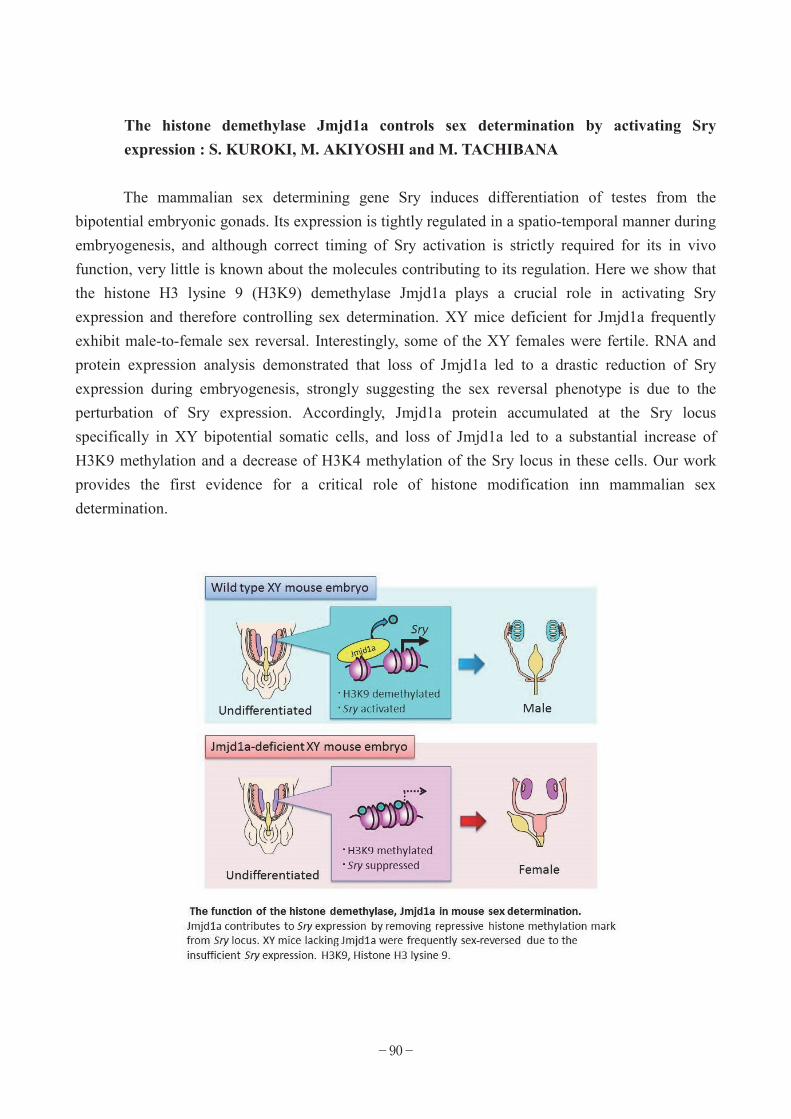
The histone demethylase Jmjd1a controls sex determination by activating Sry expression : S. KUROKI, M. AKIYOSHI and M. TACHIBANA
The mammalian sex determining gene Sry induces differentiation of testes from the
bipotential embryonic gonads. Its expression is tightly regulated in a spatio-temporal manner during embryogenesis, and although correct timing of Sry activation is strictly required for its in vivo function, very little is known about the molecules contributing to its regulation. Here we show that the histone H3 lysine 9 (H3K9) demethylase Jmjd1a plays a crucial role in activating Sry expression and therefore controlling sex determination. XY mice deficient for Jmjd1a frequently exhibit male-to-female sex reversal. Interestingly, some of the XY females were fertile. RNA and protein expression analysis demonstrated that loss of Jmjd1a led to a drastic reduction of Sry expression during embryogenesis, strongly suggesting the sex reversal phenotype is due to the perturbation of Sry expression. Accordingly, Jmjd1a protein accumulated at the Sry locus specifically in XY bipotential somatic cells, and loss of Jmjd1a led to a substantial increase of H3K9 methylation and a decrease of H3K4 methylation of the Sry locus in these cells. Our work provides the first evidence for a critical role of histone modification inn mammalian sex determination.
−90−

List of Publications Balemans, M.C., Kasri, N.N., Kopanitsa, M.V., Afinowi, N.O., Ramakers, G., Peters, T.A., Beynon, A.J., Janssen, S.M., van Summeren, R.C., Eeftens, J.M., et al. (2013). Hippocampal dysfunction in the Euchromatin histone methyltransferase 1 heterozygous knockout mouse model for Kleefstra syndrome. Human molecular genetics 22, 852-866. Deguchi, K., Nagamatsu, G., Miyachi, H., Kato, Y., Morita, S., Kimura, H., Kitano, S., Hatada, I., Saga, Y., Tachibana, M., et al. (2013). Posttranscriptional Regulation of Histone Lysine Methyltransferase GLP in Embryonic Male Mouse Germ Cells. Biology of reproduction 88, 36. Fujimoto, Y., Tanaka, S.S., Yamaguchi, Y.L., Kobayashi, H., Kuroki, S., Tachibana, M., Shinomura, M., Kanai, Y., Morohashi, K., Kawakami, K., et al. (2013). Homeoproteins six1 and six4 regulate male sex determination and mouse gonadal development. Dev Cell 26, 416-430. Inagawa, M., Nakajima, K., Makino, T., Ogawa, S., Kojima, M., Ito, S., Ikenishi, A., Hayashi, T., Schwartz, R.J., Nakamura, K., et al. (2013). Histone H3 lysine 9 methyltransferases, G9a and GLP are essential for cardiac morphogenesis. Mechanisms of development. Kuroki, S., Akiyoshi, M., Tokura, M., Miyachi, H., Nakai, Y., Kimura, H., Shinkai, Y., and Tachibana, M. (2013a). JMJD1C, a JmjC Domain-Containing Protein, Is Required for Long-Term Maintenance of Male Germ Cells in Mice. Biology of reproduction. Kuroki, S., Matoba, S., Akiyoshi, M., Matsumura, Y., Miyachi, H., Mise, N., Abe, K., Ogura, A., Wilhelm, D., Koopman, P., et al. (2013b). Epigenetic regulation of mouse sex determination by the histone demethylase Jmjd1a. Science 341, 1106-1109. Muramatsu, D., Singh, P.B., Kimura, H., Tachibana, M., and Shinkai, Y. (2013). Pericentric Heterochromatin Generated by HP1 Protein Interaction-defective Histone Methyltransferase Suv39h1. The Journal of biological chemistry 288, 25285-25296. Ohno, R., Nakayama, M., Naruse, C., Okashita, N., Takano, O., Tachibana, M., Asano, M., Saitou, M., and Seki, Y. (2013). A replication-dependent passive mechanism modulates DNA demethylation in mouse primordial germ cells. Development 140, 2892-2903. 立花 誠:エピジェネティック因子によるほ乳類の性決定の制御、第 60 回日本実験動物学会
−91−

総会、つくば、2013 年 5 月
−92−

EXPERIMENTAL RESEARCH CENTERFOR INFECTIOUS DISEASES LABORATORY OF PRIMATE MODEL Members
Professor Tatsuhiko Igarashi Associate Professor Tomoyuki Miura Assistant Professor Takayuki Hishiki Technical Assistant Hiromi Mori
Momoko Tsuji Graduate Student Hiroyuki Otsuki
Fumihiro Kato Ryosuke Hyuga
Yuji Watanabe Yuki Ishida Yuriko Nishiyama
Mai Yoneda Daiki Hara
Introduction
We are interested in the pathogenesis of virus infection and wish to contribute to development of better prophylactic and therapeutic interventions against viral diseases. We are conducting research concerning HIV-1 infection, employing non-human primate model, and dengue viruses. Some of the scientific achievements we published in 2012 are briefly summarized below. Topics
Generation of novel chimeric simian-human immunodeficiency virus (SHIV) strains through intracellular homologous recombination
SHIV, which carries tat, rev, vpu and env genes derived from HIV-1 in the backbone of SIV
genome, has been a useful virus in the non-human primate model for AIDS. The demand for SHIV strains exhibiting a variety of antigenicity of Env has been increasing further since the identifications of the newer generations of “broadly-neutralizing antibodies” to evaluate their “breadth” of neutralization in context of in vivo infection. However, there are only a handful of SHIV strains available in the field. We, who have been generating SHIV strains for more than 20
−93−

years, know that generation of a SHIV strain is time-consuming and often ends up with production of a non-infectious virus. We attribute the difficulty to the following two strategies employed for the generation of the virus; 1) utilization of a single infectious molecular clone of HIV-1 as the representative of a given strain, which consists of viral “swarm”, and 2) recombination of the HIV-1 genome with that of SIV at restriction sites already present or newly introduced, which are often at the interfaces of the genes. As per the first strategy, it is not known whether a particular molecular clone employed is appropriate as a part of chimeric virus and functional in monkey cells. Concerning the second strategy, there are circulating recombinant forms of HIV-1 strains, naturally occuring chimeric viruses, whose breakpoints are not at the end of certain genes, suggesting that breakpoints based on the restriction sites existing or newly-introduced may not be adequate for a given combination of particular viruses, in this case, SIV and HIV-1. To circumnavigate the above-mentioned uncertainties, we tried to generate SHIV strains through intracellular homologous recombination, a mechanism to repair damage made in double stranded DNA in the cell. We prepared three segments of genes to cover the entire SHIV genome by PCR. For the SIV backbone, we prepared two gene segments from the existing SHIV KS661 plasmid as template. For the HIV-1 gene segment, complementary DNA prepared from the genomic RNA extracted from HIV-1 particles was utilized as template. The three gene segments were co-transfected to human T cell line. To our surprise, recombinant virus emerged in two weeks, compared to months-long efforts of the conventional method for generating recombinant viruses. Employing this method, we generated two strains of novel SHIV, SHIV 97ZA012 and SHIV MNA. SHIV 97ZA012 carries Env derived from HIV-1 97ZA012 strain, a clinical isolate belonging to clade C, a group of viruses responsible for more than 50% of infections worldwide. SHIV MNA carries Env derived from HIV-1 MNA strain, which exhibits neutralization resistant phenotype by conventional neutralization assay but sensitive in the presence of small molecule CD4 mimic. Experimental infection of these SHIVs showed that they were infectious to monkeys. These viruses will be useful to investigate antibody-based control of HIV-1 infection in non-human primate model. We reported our achievements in Virology and Journal of General Virology, respectively.
No viral evolution in the lymph nodes of SIV-infected rhesus macaques during combined antiretroviral therapy
Combined antiretroviral therapy (cART), a combinational administration of three or more anti-HIV-1 drugs, has transformed HIV-1 infection to manageable medical condition. Despite enormous benefit of cART, it is not the ideal therapeutic intervention yet. Even after clinically excellent viral suppression for years, relapse of viremia is commonly observed once the therapy is interrupted, suggesting persistence of virus even during cART. Since a life-long taking of cART causes eventual emergence of resistant mutant virus and adverse effects of the drugs, current goal of
−94−

therapeutic intervention against HIV-1 infection is “functional cure”, that is, life-long control of viremia under detection limit without drug administration. To make this materialize, it is necessary to reveal where and how virus persists during cART.
The last year we reported the establishment of long-term cART in SIV-infected rhesus macaques. Despite of clinically successful suppression of viral burdens in the circulation for one year, we detected expression of viral RNA in lymphatic tissues.
We wondered how the viral RNA was expressed. There are two hypotheses concerning maintenance of virus during cART. One is that virus replication persists even during cART in “viral sanctuary”, where anti-viral compounds somehow do not reach. The other is that virus is frozen as provirus integrated in the chromosome of the infected cells prior to the initiation of cART and viral RNA is occasionally expressed from it.
Lentivirus, including HIV-1 and SIV, possesses error-prone reverse transcriptase. It is established that these viruses accumulate mutations with time at a constant rate due to the enzyme. We reasoned that we could tell whether the virus was replicating during the cART we conducted by analyzing accumulation of the mutations in the viral genome. We sequenced env gene, which is known to accumulate more mutations than any other viral genes. Genetic analyses revealed that env gene expressed in the lymphatic tissues after one year of cART had accumulated little mutations, and was literally indistinguishable to those expressed immediately prior to the initiation of cART. This suggests that there is virtually no virus replication during cART. This implies that the elimination of cells harboring provirus or permanent suppression of viral gene expression from provirus is essential to achieve functional cure. We reported this result in Journal of Virology.
Natural Infection of cynomolgus monkeys with dengue virus occurs in epidemic cycles in the Philippines
Dengue virus, a member of flavivirus, is the causative agent of dengue fever, dengue
hemorrhagic fever and dengue shock syndrome. It is an arthropod-borne virus and transmitted to humans by mosquito bites. Besides “epidemic dengue”, which circulates in human population in the tropic regions via mosquitos, there is another cycle of dengue virus infection which is maintained between non-human primate and canopy-dwelling mosquitos, called “sylvatic dengue”. It is believed that epidemic dengue “spilled” from sylvatic cycle by changing tropism to mosquito species in the past. It is known that epidemic and sylvatic dengue viruses are phylogenetically distinct. While there have been a couple of reports concerning sylvatic dengue virus infection in humans, there is only an anecdotal description of “spill back”, infection of non-human primate with epidemic dengue. To gain better understanding on non-human primate in the natural history of dengue virus, we conducted serological and genetic survey of cynomolgus macaques housed in a monkey breeding facility in the Philippines, one of the endemic countries of dengue.
−95−

Out of 100 plasma samples of the animals, we detected 35 of them contained antibodies reactive to dengue virus antigen by ELISA. Neutralization test confirmed 8 of 35 samples harbored dengue virus-specific antibodies. Since virus isolation from these positive samples was unsuccessful, we alternatively conducted amplification of viral gene segments by PCR. We successfully amplified a segment of NS1 gene from two samples and E gene from one. Phylogenetic analysis revealed that these genes belonged to epidemic dengue virus, not sylvatic one. The result indicates that non-human primate could serve as the reservoir of epidemic dengue virus. For better surveillance of dengue epidemic, further investigation of non-human primate will be benefitial. List of Publications Hashimoto, C., Narumi, T., Otsuki, H., Hirota, Y., Arai, H., Yoshimura, K., Harada, S., Ohashi, N., Nomura, W., Miura, T., Igarashi, T., Matsushita, S., and Tamamura, H. (2013) A CD4 mimic as an HIV entry inhibitor: Pharmacokinetics. Bioorg. Med. Chem. 21, 7884-9. Morita, D., Miyamoto, A., Hattori, Y., Komori, T., Nakamura, T., Igarashi, T., Harashima, H., and Sugita, M. (2013) Th1-skewed tissue responses to a mycolyl glycolipid in mycobacteria-infected rhesus macaques. Biochem. Biophys. Res. Commun. 441, 108-13. Morita, D., Yamamoto, Y., Suzuki, J., Mori, N., Igarashi, T., and Sugita, M. (2013) Molecular requirements for T cell recognition of N-myristoylated peptides derived from the simian immunodeficiency virus Nef protein. J. Virol. 87, 482-488. Morita, D., Hattori, Y., Nakamura, T., Igarashi, T., Harashima, H. and Sugita, M. (2013) Major T cell response to a mycolyl glycolipid is mediated by CD1c molecules in rhesus macaque monkeys. Infect. Immun. 81, 311-316. Fujita, Y., Otsuki, H., Watanabe, Y., Yasui, M., Kobayashi, T., Miura, T., and Igarashi, T. (2013) Generation of a replication-competent chimeric simian-human immunodeficiency virus carrying env from subtype C clinical isolate through intracellular homologous recombination. Virology 436, 100-111. Takahashi, N., Nomura, T., Takahara, Y., Yamamoto, H., Shiino, T., Takeda, A., Inoue, M., Iida, A., Hara, H., Shu, T., Hasegawa, M., Sakawaki, H., Miura, T., Igarashi, T., Koyanagi, Y., Naruse, T.K., Kimura, A., and Matano, T. (2013) A novel protective MHC-I haplotype not associated with
−96−

dominant Gag-specific CD8+ T-cell responses in SIVmac239 infection of Burmese rhesus macaques. PLoS ONE 8, e54300. Oue, M., Sakabe, S., Horiike, M., Yasui, M., Miura, T., and Igarashi, T. (2013) No viral evolution in the lymph nodes of SIV-infected rhesus macaques during combined antiretroviral therapy. J. Virol. 87, 4789-4793. Kato, F., Ishida, Y., Kawagishi, T., Kobayashi, T., Hishiki, T., Miura, T., and Igarashi, T. (2013) Natural Infection of cynomolgus monkeys with dengue virus occurs in epidemic cycles in the Philippines. J. Gen. Virol. 94, 2202-2207. Nomaguchi, M., Yokoyama, M., Kono, K., Nakayama, E.E., Shioda, T., Doi, N., Fujiwara, S., Saito, A., Akari, H., Miyakawa, K., Ryo, A., Ode, H., Iwatani, Y., Miura, T., Igarashi, T., Sato, H., and Adachi, A. (2013) Generation of rhesus macaque-tropic HIV-1 clones that are resistant to major anti-HIV-1 restriction factors. J. Virol. 87, 11447-11461. Otsuki, H., Hishiki, T., Miura, T., Hashimoto, C., Narumi, T., Tamamura, H., Yoshimura, K., Matsushita, S., and Igarashi, T. (2013) Generation of a replication-competent simian–human immunodeficiency virus, the neutralisation sensitivity of which can be enhanced in the presence of a small molecule CD4 mimic. J Gen Virol. 2013 Sep 11. [Epub ahead of print] PubMed PMID: 24026672. Matsuyama-Murata, M., Inaba, K., Horiuchi, R., Fukazawa, Y., Ibuki, K., Hayami, M., and Miura, T.(2013) Genetic similarity of circulating and small intestinal virus at the end stage of acute pathogenic simian-human immunodeficiency virus infection. Front. Microbiol. 4, 204.
Saito A, Matsuoka K, Ode H, Otsuki H, Yoshida T, Iwatani Y, Sugiura W, Matano, T, Miura, T, Akari H.: A novel HIV-1mt encoding CCR5-tropic Env established persistent infection in Cynomolgus macaques. 2014 Conference on Retroviruses and Opportunistic Infections, Boston, 3-6 March 2014.
米田 舞、大附寛幸、一瀬裕太郎、松田健太、松下修三、五十嵐樹彦、三浦智行:新規 CCR5指向性 SHIV のサルへの順化と中和抵抗性の解析、第 155 回日本獣医学会、東京、2013 年
3 月 28-30 日
齊藤 暁、大附寛幸、東濃篤徳、鈴木紗織、松田健太、高橋尚史、岩谷靖雅、杉浦 亙、保
−97−
富康宏、俣野哲朗、三浦智行、明里宏文:ウイルスの標的細胞指向性はサル指向性 HIV-1の増殖に影響するか?、第 156 回日本獣医学会学術集会、岐阜、2013 年 9 月 20-22 日
加藤文博:抗デングウイルス活性を有する化合物の探索と性状解析、第20回トガ・フラビ・
ペスチウイルス研究会、神戸、2013年11月9日
加藤文博、小林 剛、三浦智行、五十嵐樹彦、日紫喜隆行:分泌型ルシフェラーゼ遺伝子を
有するデングウイルス1型レプリコンの構築、第61回日本ウイルス学会学術集会、神戸、2013年11月10-12日
日紫喜隆行、Han Qi ス学会、下遠野邦忠、五十嵐樹彦、鈴木陽一、山本直樹:ISGylation (ISG15-conjugation)によるデングウイルスの複製制御機構、第 61 回日本ウイルス学会学術
集会、神戸、2013 年 11 月 10-12 日
石田裕樹、加藤文博、川岸崇裕、小林 剛、日紫喜隆行、三浦智行、五十嵐樹彦:フィリピ
ンカニクイザルにおけるデングウイルス自然感染、第 61 回日本ウイルス学会学術集会、神
戸、2013 年 11 月 10-12 日
大附寛幸、五十嵐樹彦、三浦智行:CCR5 指向性サブタイプ C エンベローを持つサル指向
性 HIV-1 のブタオザルにおける複製、第 61 回日本ウイルス学会学術交流会、神戸、2013年 11 月 10-12 日
齊藤 暁、大附寛幸、東濃篤徳、鈴木紗織、松田健太、高橋尚史、岩谷靖雅、杉浦 亙、野
間口雅子、足立昭夫、保富康宏、俣野哲朗、三浦智行、明里宏文:CCR5 指向性を示す新
規サル指向性 HIV-1 はサル個体に持続感染する、第 61 回日本ウイルス学会学術交流会、神
戸、2013 年 11 月 10-12 日
大附寛幸、丸田泰広、橋本知恵、鳴海哲夫、廣田雄樹、原田恵嘉、三浦智行、吉村和久、
玉村啓和、松下修三、五十嵐樹彦:抗 V3 抗体および低分子 CD4 ミミック曝露後投与によ
るアカゲザルでの SHIV 複製抑制、第 27 回日本エイズ学会学術集会、熊本、2013 年 11 月
20-22 日
齊藤 暁、大附寛幸、東濃篤徳、鈴木紗織、松田健太、高橋尚史、岩谷靖雅、杉浦 亙、野
間口雅子、足立昭夫、保富康宏、俣野哲朗、三浦智行、明里宏文:CCR5 指向性を示す新
規サル指向性 HIV-1 はサル個体に持続感染する、第 27 回日本エイズ学会学術集会、熊本、
2013 年 11 月 20-22 日
−98−

米田 舞、大附寛幸、松下修三、五十嵐樹彦、三浦智行:新規CCR5指向性かつ中和抵抗性
SHIV分子クローンの作製及び解析、第27回日本エイズ学会学術集会、熊本、2013年11月20-22日
−99−

CENTER FOR EMERGING VIRUS RESEARCH I. First Group Members
Assistant Professor (Spe.) Yohei Hizukuri
Introduction The cell surface of bacteria is exposed to a variety of stresses caused by fluctuations of environmental conditions. The extracytoplasmic stress response (ESR) plays key roles to cope with these cell surface stresses and are important for bacterial survival strategy. ESR is thought to be one of the major systems of virulent bacteria to resist host defense systems triggered by a bacterial infection or invasion. Therefore it is of great importance to understand the whole picture of ESR from a medical viewpoint. This research group focuses on and aims to clarify the mechanism and physiological roles of the E-dependent ESR, one of the major ESR pathways in Escherichia coli. Topics
A structure-based model of substrate-discrimination by a tandem PDZ domain of an Escherichia coli intramembrane-cleaving protease RseP: Y. HIZUKURI, T. ODA1, S. TABATA2, K. TAMURA-KAWAKAMI2, R. Oi1, M. SATO1, J. TAKAGI2, T. NOGI1
and Y. AKIYAMA (1Yokohama City Univ., 2Osaka Univ.) During the E extracytoplasmic stress response in Escherichia coli, cell envelope stresses such as accumulation of malfolded outer membrane proteins (OMPs) or lipopolysaccharides (LPSs) triggers sequential cleavages of RseA, a membrane-spanning anti- E protein that inhibits E activity. The stress cues activate a membrane-anchored protease DegS to cleave RseA, which is followed by the second cleavage by an intramembrane-cleaving protease RseP, leading to eventual activation of
E. RseP cleaves RseA only after DegS truncates the periplasmic part of RseA. Our previous works suggested that this two-step proteolysis of RseA is controlled by tandemly-arranged two PDZ domains (PDZ-tandem) in the periplasmic region of RseP.
We determined the crystal structure of the PDZ tandem from an RseP orthologue of a hyperthermophilic bacterium Aquifex aeolicus and revealed that the two putative ligand-binding grooves constitute a single pocket-like structure. We built a model of the structure and disposition of the E. coli PDZ tandem, which suggest that the PDZ-pocket presumably lies just above the active
−100−

center sequestrated within the membrane. Complete removal of the PDZ tandem from E. coli RseP led to the deregulated cleavage of RseA independent of prior truncation by DegS in vivo. From these and other results we propose that the PDZ tandem serves as a size-exclusion filter to discriminate between the intact and truncated forms of RseA.1)
1) Hizukuri, Y., et al. (2014) A structure-based model of substrate discrimination by a noncanonical PDZ tandem in the
intramembrane-cleaving protease RseP. Structure, 22, 326-336
List of Publications Hizukuri, Y., Ito, K., and Akiyama, Y. (2013) RseP Peptidase. Handbook of Proteolytic Enzymes 3rd ed. (ed. Rawlings, N. D. and Salvesen, G.) Elsevier Ltd. pp1546-1550. 檜作洋平、禾 晃和、小田 隆、田畑早苗、川上-田村恵子、佐藤 衛、高木淳一、秋山芳展:
大腸菌膜内切断プロテアーゼ RseP の基質認識における PDZ ドメインの役割、第 86 回日本
生化学会大会、シンポジウム「非常識なプロテアーゼ反応:膜内部でのタンパク質切断」、
横浜、2013 年 9 月 11-13 日 檜作洋平、小田 隆、田畑早苗、川上-田村恵子、佐藤 衛、高木淳一、禾 晃和、秋山芳展:
膜内切断プロテアーゼ RseP のタンデム PDZ ドメインが介する切断基質選別機構、第 10 回 21 世紀大腸菌研究会、伊豆、2013 年 6 月 20-21 日 Nogi, T., Hizukuri, Y., Oda, T., Tabata, S., Tamura-Kawakami, K., Sato, M., Takagi, J., and Akiyama, Y.: Structural analysis of the PDZ tandem fragment of the bacterial intramembrane-cleaving protease RseP. International Conference on Structural Genomics 2013, Sapporo, Japan, 29 July - 21 August 2013. 檜作洋平、小田 隆、田畑早苗、川上-田村恵子、佐藤 衛、高木淳一、禾 晃和、秋山芳展:
Substrate discrimination mechanism by a PDZ tandem in the intramembrane protease RseP that regulates extracytoplasmic stress response, 第 51 回日本生物物理学会年会、京都、2013 年 10月 28 日 檜作洋平、小田 隆、田畑早苗、川上-田村恵子、佐藤 衛、高木淳一、禾 晃和、秋山芳展:
立体構造解析に基づく大腸菌膜内切断プロテアーゼ RseP のタンデム PDZ ドメインによる
切断基質選別機構モデル、第 36 回日本分子生物学会年会、神戸、2013 年 12 月 3 日
−101−

II. Second Group Members
Assistant Professor (Spe. ) Akiko Makino Introduction
Borna disease virus (BDV), which has broad host range in vertebrates, causes persistent infection in nucleus that can lead to neural disorder in horses and sheep. Constitutive activation of NF-κB suppresses the BDV growth, however, NF-κB pathway is not activated in the acute infection of BDV. Topics
Cross-talk between BDV infection and NF-κB pathway: A. MAKINO, K. FUJINO, K. SOFUKU, S. NAKAMURA, T. HONDA, K. TOMONAGA
Here, to elucidate mechanism for the inhibition of NF-κB activation by BDV infection, we
evaluated cross-talk between BDV infection and NF-κB pathway. In THP1-CD14 cells, which has SEAP gene as a NF-κB reporter, BDV infection did not increase SEAP activity from 12 hour to 2 weeks post infection, compared with TLR ligand stimulation. This result is consistent with previous report and suggests BDV has gene(s) that inhibits the activation of NF-κB. To discover common motifs between the amino acid sequences of BDV genes and NF-κB family, we performed Multiple EN for Motif Elicitation analysis and found that nucleoprotein of BDV (BDV-N) and NF-κB1, which is one of the NF-κB family and has a transcription factor activity by processing of precursor p105 form, possess a common ankyrin-like motif.
Pre-treatment with the ankyrin-like peptide of BDV-N suppressed the SEAP activation by the stimulation with TLR ligands, while control peptide had no effect on it. NF-κB1 is phosphorylated by signal transduction and its C-terminal region containing ankyrin repeat undergoes selective degradation by proteasome pathway. Immunoprecipitation assay showed BDV-N was co-precipitated with NF-κB1. Also BDV-N and BDV-N peptide suppressed NF-kB1 degradation by 20S proteasome. These results indicate BDV inhibits NF-κB activation via BDV-N, resulting in its benefit for BDV persistent infection.
−102−

List of Publications
Akiko Makino, Kan Fujino, Kozue Sofuku, Shoko Nakamura, Tomoyuki Honda, Keizo Tomonaga.: Inhibition of NF-κB activation by peptide derived from nucleoprotein of Borna disease virus. 15th
International Negative Strand Virus Meeting, Granada, Spain, 16-21 June 2013. 牧野晶子、藤野 寛、惣福 梢、中村祥子、伊藤睦美、本田知之、新矢恭子、河岡義裕、朝
長啓造:ウイルスタンパク質内の TLR シグナル伝達阻害ペプチドの探索と評価、第六回ボ
ルナウイルス研究会、東京、2013 年 3 月 14 日 牧野晶子、藤野 寛、惣福 梢、中村祥子、本田知之、朝長啓造:ボルナ病ウイルス N タン
パク質のアンキリン様配列を介した NF-κ B 活性化阻害、第 61 回日本ウイルス学会、神戸、
2013 年 11 月 10-12 日
−103−
REPRODUCTIVE ENGINEERING TEAM Members
Technical Specialist Hitoshi Miyachi Technical Staff Satsuki Kitano (Konaka)
Introduction Reproductive engineering team is a support unit for generating transgenic mouse (Tg) and
knockout mouse (KO) under the animal committee of our institute. We also perform cryopreservation of mouse fertilized eggs. Current staffs are Kitano and Miyachi. Results of last three years are as follows.
1) Freezing embryos 2011 117 strains 25,130 embryos 2012 140 strains 32,836 embryos 2013 198 strains 52,272 embryos
2) Transgenic mouse production with cloned DNAs
No of constructs No of embryos No of transgenic injected pups obtained 2011 81 29,031 227(0.8%) 2012 77 31,452 176(0.6%) 2013 71 27,924 78(0.3%)
3) Production of chimeric mouse No of ES clones No of embryos No of coatcolor injected chimera obtained 2011 107 5,828 324(5.5%) 2012 63 5,145 192(3.7%) 2013 70 5,510 129(2.3%)
−104−

List of Publications Deguchi, K., Nagamatsu, G., Miyachi, H., Kato, Y., Morita, S., Kimura, H., Kitano, S., Hatada, I., Saga, Y., Tachibana, M., and Shinkai, Y. (2013). Posttranscriptional regulation of histone lysine methyltransferase GLP in embryonic male mouse germ cells. Biol. Reprod. 88, 36. Huong le, T., Kobayashi, M., Nakata, M., Shioi, G., Miyachi, H., Honjo, T., and Nagaoka, H. (2013). In vivo analysis of Aicda gene regulation: a critical balance between upstream enhancers and intronic silencers governs appropriate expression. PLoS One 8, e61433. Imayoshi, I., Isomura, A., Harima, Y., Kawaguchi, K., Kori, H., Miyachi, H., Fujiwara, T., Ishidate, F., and Kageyama, R. (2013). Oscillatory control of factors determining multipotency and fate in mouse neural progenitors. Science 342, 1203-1208. Kuroki, S., Akiyoshi, M., Tokura, M., Miyachi, H., Nakai, Y., Kimura, H., Shinkai, Y., and Tachibana, M. (2013). JMJD1C, a JmjC Domain-Containing Protein, Is Required for Long-Term Maintenance of Male Germ Cells in Mice. Biol. Reprod. 89, 93. Kuroki, S., Matoba, S., Akiyoshi, M., Matsumura, Y., Miyachi, H., Mise, N., Abe, K., Ogura, A., Wilhelm, D., Koopman, P., et al. (2013). Epigenetic regulation of mouse sex determination by the histone demethylase Jmjd1a. Science 341, 1106-1109. Tani-Ichi, S., Shimba, A., Wagatsuma, K., Miyachi, H., Kitano, S., Imai, K., Hara, T., and Ikuta, K. (2013). Interleukin-7 receptor controls development and maturation of late stages of thymocyte subpopulations. Proc. Natl. Acad. Sci. U. S. A. 110, 612-617.
−105−

COMPUTER NETWORK OF INSTITUTE FOR VIRUS RESEARCH
Assistant Professor Keiko Takemoto
Introduction Institute for Virus Research LAN system (IVR-LAN) has administrated by the network
committee consisted of four staffs (Prof. Toyoshima, Prof. Akiyama, Associate Prof. Mori and Instructor Takemoto). IVR-LAN service has covered for researchers of some medical departments as well as IVR, and the primary purpose of IVR-LAN is to offer accessibility to the Internet in support of their studies. IVR-LAN has provided a variety of network services, including E-Mail, WEB-mail, WWW, File-sharing, online reservation of seminar rooms, SSH and all Outgoing TCP services except for P2P. Main services are working on Sun Sparc platform with Solaris 10 and DELL with Linux.
This year we installed plone server which enabled us to maintain our local web site by other staffs and allowed our institute members to upload documents on a board portal and hold paperless meeting with tablet devices.
However IVR-LAN has adequately equipped, we must have a responsibility for sending/getting data. A few accidents have occurred in this year. IVR-LAN users need to get certifications of training of e-learning course which is provided by Institute for Information Management and Communication of Kyoto university.
In addition to the administration of network, Takemoto have studied the epigenetic regulation of mouse endogenous retroviruses during cell differentiation. Conditional knockdown mice of histone methyltransferase ESET, and/or DNA de novo methyltransferase Dnmt1 have been analyzed with RNA-seq and ChIP-seq.
List of Publications 竹本経緯子:H3K9 メチル化による内在性レトロウイルスの制御、NGS 現場の会第 3 回研
究会、神戸、2013 年 9 月 4-5 日 井上真悠子、黒田貴雄、加藤雅紀、竹本経緯子、駒井 妙、眞貝洋一、水谷健一:Prdm8 は
発生期大脳皮質の多極性形態期を調節する、第 36 回日本分子生物学会年会、神戸、2013年 12 月 3-6 日
−106−

INTERNATIONAL SYMPOSIUM
Our Institute held “1st Kyoto International Symposium on Virus-Host
Coevolution” on November 07, 2013 at the Shiran kaikan Inamori Hall, Kyoto.
Program
Opening Remarks: Prof. Masao Matsuoka, Institute for Virus Research, Kyoto University
Session 1: Emerging Viral Diseases and the Hosts
Dr. Takayuki Miyazawa, Institute for Virus Research, Kyoto University, “Exogenous and Endogenous Betaretroviruses: Implications in the Evolution of Mammals” Dr. Shigeru Morikawa , National Institute of Infectious Diseases, Japan, “Canine Distemper Virus Crosses the Species Barrier to Macaque Monkeys” Prof. Ayato Takada , Research Center for Zoonosis Control, Hokkaido University, “Ecology of Avian Influenza Viruses: Topics from Surveillance in Asia and Africa”
Keynote Lectures:
Prof. Robin Weiss, Wohl Viron Centre, University College London, “Virus-Host Coevolution and Cross-Species Infection” Prof. Fumitoshi Ishino, Medical Research Institute, Tokyo Medical and Dental University, “Mammalian Evolution Promoted by LTR-Retrotransposon-Derived Genes”
Session 2: Virus Infection and the Host Factors
Dr. Welkin Jonson, Biology Department, Boston College, “Genetic Barriers to Cross-Species Transmission and Emergence of Primate Lentiviruses”
−107−

Dr. Massimo Pizzato, Centre for Integrative Biology, University of Trento, “Searching for Host Determinants of Lentivirus and Gammaretrovirus Infection” Prof. Keizo Tomonaga, Institute for Virus Research, Kyoto University, “Endogenous Bornavirus-Like Elements: Cellular Co-option and Impact on Host Evolution”
Closing Remarks:
Prof. Hirohisa Hirai, Director, Primate Research Institute, Kyoto University
−108−

SEMINARS OF THE INSTITUTE FOR VIRUS RESEARCH
Twenty-one seminars were held at the Institute for Virus Research under the auspices of the Institute in 2013. Eleven lectures were from abroad and ten others were from Japan. January 23 Dr. Becca Asquith, Imperial College London, UK. “What Constitutes a
Protective HLA Class I Genotype in HTLV-1 Infection?” February 7 Dr. Rob J. De Boer, Utrecht University, the Netherlands. “Analysing
immune cell migration” February 13 Dr. Naoya Ohara, Okayama University, Japan. “Mechanism of the
drug-resistance of Mycobacterium tuberculosis against
para-aminosalicylic acid (PAS) and the application”
February 26 Dr. Luis Menéndez-Arias, CSIC, Spain. “Complex mutational patterns
associated with resistance to HIV-1 reverse transcriptase inhibitors” March 22 Dr. Jun Hatakeyama, Kumamoto University, Japan. “Neuron maintains
temporarily adhesion belt to regulate neuronal differentiation through
Notch signaling”
March 27 Dr. Koichiro Uryu, RIKEN Advanced Science Institute, Japan.
“Synchronization dynamics of migrating cells” April 17 Dr. Keiji Hirota, Osaka University, Japan. “Function and plasticity of
inflammatory T helper cells” May 15 Dr. Hirofumi Arakawa, National Cancer Center, Japan. “Mechanism
of mitochondrial quality control and Cancer ~ Novel tumor suppressor
function of p53~”
−109−

May 17 Dr. Akatsuki Kimura, National Institute of Genetics, Japan. “How cell
changes its interior depending on its size - a study on mitotic spindles
in the C. elegans embryo”
June 5 Dr. Rongge Yang, WIV of CAS, China. “Molecular epidemiological
study of HIV spread in China” July 19 Dr. Makoto Miyata, Osaka City University, Japan. “Mycoplasma
gliding”
July 22 Dr. Sokichi Matsumoto, Osaka City University, Japan. “Elimination of
tuberculosis by investigating regulatory mechanism of dormancy and
proliferation of Mycobacterium tuberculosis”
August 26 Dr. Daron M. Standley, Osaka University, Japan. “3D Molecular
Modeling of antibodies and protein-RNA interactions” September 9 Dr. Alexander Hoffmann, UCSD, US. “Combinatorial and Dynamical
Codes to specify Immune Responses” September 9 Dr. Dong Sung An, UCLA, US. “Developing hematopoietic stem cell based gene therapy strategies for treating HIV infected patients” October 8 Dr. Seiichi Uchida, Kyushu University, Japan. “Bioimage-informatics
– automatic quantification and knowledge discovery for bioimages” October 28 Dr. Charles R M Bangham, Imperial College London, UK. “Clonality,
latency and integration of HTLV-1 in vivo” November 8 Dr.Tinxiao Riu, IMP, Austria. “Anatomical and functional dissection
of fruitless positive courtship circuit” November 18 Dr. Thirumala-Devi Kanneganti, St. Jude Children’s Research
Hospital, US. “Role of NLRs in inflammation and disease”
−110−

December 17 Dr. Keith Willison, Imperial College London, UK. “Quantitative single cell and single molecule proteomics for clinical studies”
December 24 Dr. Yoshiaki Ito, National University of Singapore, “RUNX3: Is it a
partner of p53 ?”
−111−

がんウイルス研究部門 Department of Viral Oncology がん遺伝子研究分野 Laboratory of Gene Analysis
Ⅰ. First Group
教授 秋山芳展
准教授 森博幸
特定助教 檜作洋平(附属新興ウイルス研究センター)
特定研究員 石井英治
大学院生 服部徳哉
大門康志
宮﨑亮次
橋本成祐
秋山光市郎
舛井千草
三登一八
水野慎也
椋野翠
本年度は、理学研究科大学院生(M1)として水野慎也さん、椋野 翠さんが、博士研究
員として石井英治さんが新たに加わり、一方、共同研究者の成田新一郎特定助教が盛岡大
学へと転出しました。また、博士研究員(日本学術振興会特別研究員)の檜作洋平さんが
附属新興ウイルス研究センター特定助教に就任し、引き続き密接な協力の下に研究を行っ
ています。
1)ペリプラズムプロテアーゼ BepA の TPR ドメインの機能解析及び部位特異的 in vivo 光
架橋法による近接因子探索
大腸菌を初めとするグラム陰性細菌の細胞質は、内膜と外膜の二重膜に包まれており、
これら二つの膜の間には親水的なペリプラズム空間が存在します。内膜が一般的なリン脂
質二重層であるのに対して、外膜は内葉のリン脂質と外葉のリポ多糖から構成されます。
外膜はグラム陰性細菌の生存に必須な構造体であり、その機能を正常に保持する事は生存
のために重要です。そのため大腸菌は、外膜機能の障害につながる外的環境の変化に対し
て、複数の表層ストレス応答機構を備えることで対応しています。その中でも σE 経路は、
−115−

大腸菌の生存に唯一必須な表層ストレス応答経路であり、これまでに 114 の遺伝子が、転
写因子 σE による制御を受ける標的として同定されています。この σE によって転写誘導さ
れる遺伝子の一つに、bepA (yfgC)があります。これまでに、bepA を欠失する大腸菌変異株
が様々な抗生物質に対して感受性を示すことが複数の研究室によって報告されています。
BepA は亜鉛メタロプロテアーゼの典型的な活性部位モチーフ H136EXXH を持ち、M48
ファミリーに属するペリプラズムプロテアーゼです。近年、当研究室によって、BepA がリ
ポ多糖の輸送に重要な外膜タンパク質 LptD のバイオジェネシスを促進し、またそれに失敗
した LptD の分解を行っていること、さらに外膜タンパク質のアセンブリーに関わる BAM
複合体と相互作用していることなど、BepA が外膜タンパク質のバイオジェネシスや品質管
理に働いていることを示唆する結果が報告されました。しかし、BepA のどの領域が、どの
ような様式で BAM 複合体や LptD と相互作用しているのか、また、別の基質は存在するの
かなどは明らかになっていません。そこで、BepA の C 末端領域には保存された TPR モチ
ーフが 4 個存在することに注目しました。TPR モチーフはタンパク質間相互作用の足場と
して働くことが知られています。BepA における役割は不明ですが、BepA もこのモチーフ
を介して、上記タンパク質等と相互作用している可能性が考えられます。そこで本研究で
は、BepA の TPR ドメインの機能解析を行うとともに、部位特異的 in vivo 光架橋法を用い
て TPR ドメインに近接する因子の探索を行いました。
TPR モチーフを段階的に欠失した
BepA 変異体を作製し、bepA 欠失株の
薬剤感受性に対する相補活性を指標
として機能解析を行いました(図 1)。
その結果、TPR モチーフ欠失変異体は
全て弱い相補活性を示しましたが、そ
の相補活性は、野生型 BepA に比べる
と非常に低いものでした。また、BepA
のプロテアーゼ活性部位モチーフ変異体(E137Q)を発現させると大腸菌野生株が薬剤感
受性を示すのに対し、TPR モチーフを欠失した E137Q 変異体の発現では、薬剤感受性を示
しませんでした。以上の結果から、TPR モチーフは BepA が正常に機能するために重要で
あることが示唆されました。
−116−
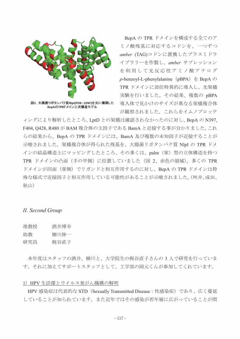
BepA の TPR ドメインを構成する全てのア
ミノ酸残基に対応するコドンを、一つずつ
amber (TAG)コドンに置換したプラスミドラ
イブラリーを作製し、amber サプレッション
を利用して光反応性アミノ酸アナログ
p-benzoyl-L-phenylalanine(pBPA)を BepA の
TPR ドメインに部位特異的に導入し、光架橋
実験を行いました。その結果、複数の pBPA
導入体で見かけのサイズが異なる架橋複合体
が観察されました。これらをイムノブロッテ
ィングにより解析したところ、LptD との架橋は確認されなかったのに対し、BepA の N397,
F404, Q428, R480 が BAM 複合体の主因子である BamA と近接する事が分かりました。これ
らの結果から、BepA の TPR ドメインには、BamA 及び複数の未知因子が近接することが
示唆されました。架橋複合体が得られた残基を、大腸菌リポタンパク質 NlpI の TPR ドメ
インの結晶構造上にマッピングしたところ、その多くは、palm(掌)型の立体構造を持つ
TPR ドメインの凸面(手の甲側)に位置していました(図 2、赤色の領域)。多くの TPR
ドメインが凹面(掌側)でリガンドと相互作用するのに対し、BepA の TPR ドメインは特
殊な様式で近接因子と相互作用している可能性があることが示唆されました。(舛井、成田、
秋山)
II. Second Group
准教授 酒井博幸
助教 柳川伸一 研究員 梶谷直子
本年度はスタッフの酒井、柳川と、大学院生の梶谷直子さんの 3 人で研究を行っていま
す。それに加えてサポートスタッフとして、工学部の岡元くんが参加してくれています。
1)HPV 生活環とウイルス発がん機構の解明
HPV 感染症は代表的な STD(Sexually Transmitted Disease:性感染症)であり、広く蔓延
していることが知られています。また近年ではその感染が若年層に広がっていることが問
−117−

題となっています。HPV感染は発がんと関連することが知られていて、特に子宮頚癌では,
ほとんどの発症例で HPV の感染が確認されており,HPV 感染が子宮頚癌発症の主要なリ
スクファクターであると考えられています。HPV 感染によるがん化を防ぐためには,HPV
の感染・複製を抑制することが効果的であると考えられます。しかし HPV の複製・遺伝子
発現の制御は,上皮細胞の分化に強く依存していて,通常の組織培養では HPV の生活環を
再現できないので,これまでその制御機構はほとんど分かっていませんでした。私たちは
既に皮膚モデル培養系を用いて,HPV の複製を組織培養下で再現しています。この系をさ
らに発展させるべく,独自の HPV レプリコンを構築し,効率よく HPV の複製・遺伝子発
現機構を解析する手法を検討しています。また一方で、機能のよく分かっていないウイル
ス制御遺伝子,特に E4 と E5 に注目し,それらの生物活性の同定を行っています。これら
の遺伝子機能から、ウイルス複製の調節機構を探り、抗ウイルス剤開発の標的を見出した
いと考えています。(梶谷、川手、酒井)
2)LRP6 結合蛋白 Krtap13 による Wnt シグナル伝達経路活性化のマウス個体での解析
柳川は、Wnt の Co-receptor である一回膜貫通型蛋白 LRP6 の細胞質ドメインに結合する
蛋白として、Keratin associated protein 13 (Krtap13)を、見いだした。
Wnt 非存在下、Krtap13 を強制発現させるだけで、Wnt 経路の著しい活性化が生じた。
Krtap13 は、細胞膜上で、LRP6 および Dvl と共に凝集体を形成するが、それは、通常の
Wnt 経路を活性化の際に生じる LRP6 Signalosome の代わりをしていると考えられた。
Krtap13 による Wnt 経路の活性化が与える影響を、in vivo で解析する為、公汎な組織で
の発現が可能な CAG プロモーターとヒト Krtap13 の cDNA の間に lox-polyA-lox 配列を挿
入した Trans gene (Tg)を作成した。この Tg マウスと種々の Cre マウスを交配する事より、
recombinant Tg (R-Tg)生じさせ、組織特異的に Krtap13 高発現させる事が出来た。CAG-Cre
マウスとの交配によっては、R ウスとを持つマウスは、5%しか誕生しなかったが、そのう
ちの 25%のマウスは、24 39 週齢を経ると、悪性リンパ腫/白血病を発症した。名古屋大
学 生体反応病理学の豊国先生に協力頂き解析中である。
(柳川)
−118−

がんウイルス研究部門 Department of Viral Oncology 細胞制御研究分野 Laboratory of Cell Regulation
教授 杉田昌彦
助教 森田大輔
大学院生 服部祐季
山本侑枝
邑田悟
田代寛実
宮本歩己
渡邊丈治
「脂質免疫」の研究は難しい。当たり前のことだが脂質は水に溶けない(それが定義だ)。
とりわけ私達が扱っている結核菌脂質の多くは、ミコール酸という C80 を越える長鎖脂肪
酸を含んだ脂質であり、水系溶媒に馴染むはずがない。そのまま T 細胞培養系に投与して
も、T 細胞抗原受容体と巡り会う前にメディウムにはじかれてしまう。ましてやどうやっ
て生体に投与し、効率的に T 細胞応答を誘導するのか、試行錯誤を繰り返すことになる。
幸い共同研究者に恵まれ、ようやくこの点は解決できるようになってきた。しかし脂質抗
原と水溶性アジュバントをどううまく一緒に投与するか、まだ工夫が必要である。もう一
つの大きなチャレンジは実験動物の開拓である。私達の目標は、言うまでもなくヒトの免
疫病態の理解と制御であるが、免疫系が個体レベルで機能する生体システムである以上、
実験動物の利用を避けては通れない。幸か不幸か(?)、免疫解析に有用なマウスとラット
は、ヒト脂質免疫の基盤となるグループ 1CD1 分子がない。2004 年 9 月のウイルス研着任
を契機に、ヒト培養細胞の解析研究から動物個体を用いた研究への展開を図り、ヒト CD1
トランスジェニックマウス、モルモット、アカゲザルと段階を踏みながら、着実に(いや
苦労に苦労を重ねながら)実験系を確立し、脂質免疫の研究を進めてきた。市販品がない
ため、未だにモルモットサイトカインに対するモノクローナル抗体を作製して ELISA シス
テムを立ち上げたり、個体差の大きいアカゲザル MHC 遺伝子の基礎解析を行ったりしてい
る段階だが、これらの実験動物のそれぞれの長所を生かし、結果を比較し合いながら脂質
免疫の実態を理解しつつある。その過程で、たとえばヒトとアカゲザルの脂質免疫系にお
いて、私が予想もしなかった違いを認識することもある(Infect. Immun. 81: 311-316,
2013)。ラボミーティングで最初にその結果を見たとき、そんなことあり得ない、と口走っ
た自分が恥ずかしい。つまらぬ常識や固定観念ほど新たな発見の障害となることを今更な
−119−

がら実感した次第である。
ラボの掃除と基盤整備に 1年半程を要し、新しく大学院生を受け入れ始めたのが 2006 年
4 月であった。その第一期生、森田大輔君は、博士(生命科学)の学位を取得したのち、
日本学術振興会特別研究員 PD を経て、2013 年 3 月より当研究分野の助教となった。思え
ば 2006 年の夏、「ウイルスは固有の脂質を持たない。果たして脂質免疫はウイルス感染制
御には無力なのだろうか?」「いやそんなはずはない。ウイルス感染で脂質免疫が機能する
とすればどんな場面だろう」と議論し合った。その結論は「ウイルスは固有のリポペプチ
ドを持つ」だった。たとえばエイズウイルス Nef タンパク質は N 末端グリシン残基に脂質
修飾(ミリスチン酸付加)を受けて初めてその免疫抑制機能を発揮する。この断片すなわ
ちリポペプチドを認識するような T細胞応答があったら楽しいな、と。
この議論から数年を経て、珍しくその予想は当たっていたことがわかった(J. Immunol.
187: 608-612, 2011; J. Virol. 87: 482-488, 2013)。これを実証できたのは、森田君や
山本さん(生命科学研究科 D1)の頑張りとともに、またまた多くの素晴らしい共同研究者
に恵まれたことが大きい。そして最近、まだここに詳しく書くわけにはいかないが、この
リポペプチドを結合し T細胞に抗原提示する「リポペプチド抗原提示分子」(図のX)を同
定した。「ペプチド」「脂質」に加えて、その両者の特質を有する「リポペプチド」が獲得
免疫の標的となることが分子レベルで証明出来つつある。これは今年度の最も大きな研究
進捗であろう。
研究室には新たに邑田悟医師(医学研究科 D1)、渡邊丈治君(生命科学研究科 M1)が加
わった。新入所者歓迎会での二人の芸には笑えたが、研究面でも芸達者なところを見せて
くれている(口達者でもある)。研究室はいま、脂質(結核)、リポペプチド(エイズ)の
研究を深めるとともに、次の新たな研究目標を定めようとする転換期でもある。上述のよ
うな「もし・・だったら楽しいな」という新たな発想をともに磨いていきたいと思う。
−120−

がんウイルス研究部門 Department of Viral Oncology
生体発がん機構研究分野 Laboratory of Tumor Biogenesis
教授 米原伸
助教 村上昭
当研究分野は生命科学研究科に所属しており、米原はウイルス研を兼務、村上はウイル
ス研所属である。准教授の酒巻和弘、助教の李慶權と大学院生は、生命科学研究科高次遺
伝情報学分野の所属である。
今年の 4 月に、生命科学研究科の修士課程に田畑祐貴、鈴木雄太、加古彩華の三名、博
士後期課程に胡暁恬が入学した。さらに、9 月には黄達度と董晟 が修士課程に入学した。
修士課程を 4 月に修了した学生では、阪口翔太が博士後期課程へ進学、森川響子は資生堂
(株)に、冬柴宏紀は住友銀行に就職した。また、当研究室で博士後期課程を修了し博士
(生命科学)の学位を取得した福岡あゆみは、兵庫医科大学で博士研究員として採用され
た。研究費の経理を初めとする研究室の秘書業務は中橋直子が執り行い、研究室の円滑な
運営が行われている。米原は生命科学研究科研究科長の任期を終え、研究と教育に以前よ
り時間をかけて取り組むことが可能となった。
本研究分野では、米原らが見いだしたアポトーシス誘導レセプター分子 Fas の研究を出
発点とし、アポトーシスやそれ以外の新しい細胞死に関する研究、細胞死関連分子の多様
な生物活性に関する研究を行っている。各個人の研究内容について簡単に紹介する。
1) RIP キナーゼが媒介するγ型インターフェロン誘導性計画的ネクローシス
Fas 誘導アポトーシスに必須である caspase-8 はアポトーシス誘導とは別に RIP キナーゼ
が媒介する計画的ネクローシスの誘導阻害に機能することが、腫瘍壊死因子(TNF)誘導
ネクロプトーシスの系で知られている。当研究室ではγ型インターフェロンが caspase-8 の
発現や活性が阻害された条件下でネクローシスを誘導するという新しい系を見いだしてい
る。この系に関して、陳建成が RIP3 の上流、阪口翔太が RIP3 の下流について新たな発見
を目指して研究を行っている。北瀬昌菜は TNF 誘導ネクロプトーシスとの異動について、
森勇貴は様々な細胞においてアポトーシスやネクロプトーシスとの異動について詳細に解
析を進めている。また、董晟 はネクロプトーシスを容易に誘導できる系の立ち上げを行
−121−

っている。一方、
ES 細胞を用い
て遺伝子の発現
誘導系や発現抑
制誘導系を構築
した染田真孝は、
caspase-8の発現
抑制を誘導する
と分化誘導に重
要なレチノイン
酸シグナル伝達
系が RIP 依存的
に強く増強され
るという全く新
しい現象を見い
だし、その分子機構を解析するとともに、caspase-8 KO マウスが胎生致死となる原因がレ
チノイン酸シグナルの亢進なのかネクローシスの亢進なのかを明らかにすべく研究を行っ
ている。
2) B 細胞からの IgE 産生を増強させる Fas-expressing natural helper (F-NH) 細胞
当研究室では、高 IgE 産生を伴うアレルギー炎症を発症する Balb/c の遺伝的背景下 Fas
KO マウスにおいて B 細胞からの IgE 産生を強く誘導する新しい細胞種を同定し、
Fas-expressing natural helper (F-NH) 細胞と命名している。その実態と機能を明らかにするた
めに、山本那由が F-NH 細胞の増殖機構や様々な性質の解析を行っている。
3) 二核四倍体細胞に誘導される新しい細胞死
当研究室で見いだした「染色体凝縮不全→二核四倍体細胞の出現→細胞増殖の進行(染
色体の不安定化を伴い、がん化の原因となる)→EF-1α(eEF1A1)の発現低下→caspase に依
存しない新しい細胞死の誘導」という現象を、様々な細胞で誘導する系をより一般化し、
その分子機構と生理機能を解明するために、胡暁恬、田畑祐貴と黄達度が研究を行ってい
る。
−122−

4) 細胞周期 S 期進行に機能する巨大分子 FLASH の解析
我々が Fas シグナルとの関連で見いだした巨大分子 FLASH はがん細胞株の細胞周期 S
期の進行に必要不可欠であるが、鈴木雄太はがん細胞特異性について FLASH の N 末端領
域の機能解析とからめて解析を開始している。加古彩華は FLASH の機能を阻害する薬剤
の探索をとおして、FLASH の機能解析を進めている。
5) アポトーシス関連分子と細胞死・細胞増殖・個体発生との関係
生命科学研究科助教の李慶權は、アポトーシスと細胞増殖の相互作用や調節機能につい
て、自らがアポトーシスとの関連で見いだした MST(hippo)とその下流の転写因子 YAP
を中心に新たな観点から解析を行っている。生命科学研究科准教授の酒巻和弘は、様々な
モデル生物(メダカ、カエルなど)を用いた caspase の機能解析、数理モデルを組み込んだ
caspase の活性化と生理機能の関連解析を行っている。
6) マウス初期発生における Wnt
シグナルの解析
助教の村上昭は、マウスの胚発
生初期に、内・中・外胚葉が誘導
される機構を、胚性幹細胞(ES 細
胞)を用いて解析している。最近
は、特に、未分化能の維持と分化
の誘導という全く相反する機構に
関与しているWntシグナルに焦点
を当てている。これらの過程に関
与しているWntシグナル間の違い
が如何にして生じるのかを明らか
にしたいと考えている。
−123−

がんウイルス研究部門 Department of Viral Oncology ヒトがんウイルス研究分野 Laboratory of Human Tumor Viruses
Ⅰ. First Group
教授 朝長啓造
助教 本田知之
特定助教 牧野晶子(附属新興ウイルス研究センター)
博士研究員 藤野寛
平井悠也
松本祐介
大学院生 惣福梢
中村祥子
小嶋将平
事務補佐員 若城佳寿美
本年度は、4 月より博士研究員として平井悠也、生命科学研究科大学院生(M1)として
小嶋将平が、また 6 月には事務補佐員として若城佳寿美が新たに加わった。11 月からは博
士研究員として松本祐介が留学先より戻ってきた。
1)ボルナウイルスの感染機構と病原性に関する研究
ボルナウイルスは細胞核で持続感染する。これは、RNA ウイルスとしてはきわめて特殊
な感染様式である。我々は、ボルナウイルスがどのようにして細胞核におけるゲノム RNA
の持続性を担保しているのかを明らかにしようとしている。平井は、細胞核とウイルス
RNA の詳細な相互作用について、超高解像顕微鏡をはじめとする手法を用いて解析を進め
ている。また、ボルナウイルス感染細胞では、核内にウイルスリボ核酸複合体によるスペ
ックルが形成される。ボルナウイルスの持続感染と核内スペックルとの関連性を解明する
ことは、ウイルス感染現象の多様性を知るだけではなく、ボルナウイルスの病原性の本質
を明らかにできると考えられる。ボルナウイルスの核内スペックルの研究に関しては、本
田らが新学術領域「非コード RNA」において進めている。
2)内在性ボルナウイルスの解析
我々は、ヒトをはじめ多くの哺乳動物のゲノムにボルナウイルスの遺伝子断片が内在化
−124−

していることを発見した。我々は、内在性ボルナウイルス因子の中でも、特にヌクレオプ
ロテインの内在性産物である内在性ボルナウイルス様ヌクレオプロテイン(Endogenous
bornavirus-like nucleoprotein: EBLN)に注目して解析を行っている。ヒトで見つかった EBLN
配列のいくつかは、比較的大きなオープンリーディングフレームを持っており、mRNA と
して発現されていることもわかっている。また、ジュウサンセンジリスゲノムの EBLN は、
外来性ボルナウイルスのヌクレオプロテインとアミノ酸レベルで高い相同性を持っている
ことも知られている。そこで本年は、ヒトおよびジュウサンセンジリスのゲノムで見つか
った EBLN の機能解析を行った。その結果、ジュウサンセンジリスゲノム以来の EBLN タ
ンパク質には、ボルナウイルス増殖を抑制する効果があることが明らかとなった。また、
ヒト由来 EBLN のひとつがミトコンドリアで機能している可能性が示された。このことは、
宿主はボルナウイルスの内在が産物である EBLN を、宿主に有用な遺伝子として取り込ん
でいる可能性が示していた。
3)ボルナウイルスを用いた新規 RNA ウイルスベクターの開発
我々は、細胞の核内持続感染という他の RNA ウイルスにはないボルナウイルスの特徴を
利用し、持続的に蛋白質や低分子 RNA を発現できる核内長期発現型のボルナウイルスベク
ターの作製に成功した(特許 5299879、US20120151614A、EP2415866A 2013 年)。本年は、
引き続き遺伝子欠損ウイルスベクターの作製、ウイルスの病原性低減、ウイルス力価の向
上、標的とする細胞や臓器の拡大など、ベクターの応用を目指した研究開発を行った。
4)インフルエンザウイルスに関する研究
我々は、インフルエンザウイルス感染により、迅速に誘導される宿主応答を解明するこ
とを目的に、感染直後に発現が変動する miRNA を網羅的にスクリーニングし、それらに
より制御されている宿主因子を同定した。本年は、特に宿主の O 型糖鎖修飾に関する酵素
を制御する miRNA に着目して解析を行った。これまでに、同定された O 型糖鎖修飾酵素
がインフルエンザウイルス感染細胞において急速に発現上昇すること、また、それに伴い
感染細胞においてムチンの発現は増加することを明らかにした。さらに、O 型糖鎖修飾酵
素がインフルエンザウイルスの増殖を制御している可能性も見出した。現在は、ノックア
ウトマウス等を用いて同定された O 型糖鎖修飾酵素の詳細な役割について解析を行ってい
る。
−125−

II. Second Group
准教授 土方誠
博士研究員 阿部雄一
大学院生 津川陽司
赤堀祐一
岡村瞳
長谷川輝
実験補助員 外山栄里
本年度は、4 月より生命科学研究科大学院生(M1)として岡村瞳と長谷川輝が、また 9
月にはそれまで大学院生(D4)だった阿部雄一が学術振興会と区別研究員(PD)として参加す
ることになった。
当研究分野土方グループでは、C型肝炎ウイルスならびにB型肝炎ウイルスとその感染標
的であるヒト肝細胞の研究を中心におこなっている。肝炎ウイルスの研究ではその生活環
やウイルスと肝細胞間の相互作用を分子レベルで解明することと、その結果をもとに抗ウ
イルス薬剤開発を目指した研究をおこなっている。また、ヒト肝細胞に関してはヒト肝幹
細胞を用いた肝分化と肝炎ウイルス感染の研究から肝癌などの慢性肝疾患の発症機構を明
らかにするための研究をおこなっている。
1)トロンボキサン合成酵素阻害薬による HCV の感染性粒子産生抑制
当研究室において、これまでにアラキドン酸カスケードの酵素のひとつ TXA2 合成酵素
(TXAS)が HCV 粒子への感染性付加に機能を有することを見出している。更に、広島大学
病院消化器内科との共同研究で、C 型肝炎患者血清を用いた HCV 感染モデル動物系である
ヒト肝細胞キメラマウスを用いた in vivoでの抗HCV薬効試験を用いてTXAS阻害剤やTXA2
と相反する働きを持つプロスタグランジン I2 受容体(IP)アゴニストの効果を検討した結果、
それや薬剤の投与群においてHCV感染初期におけるHCV感染拡大を有意に阻害することが
明らかになった。これらの結果から喘息薬として既に使用されている TXAS 阻害剤や肺高
血圧症治療薬である IP アゴニストが新規抗 HCV 治療薬の候補となることが明らかとなっ
た。この in vivo の薬効試験における TXAS 阻害剤投与の結果、長期間の投与の場合、キメ
ラマウスに感染した HCV にこの薬剤に対する抵抗性株の出現が示唆された。現在、この抵
抗性株の遺伝子変異を解析することなどから、TXAS による感染性 HCV 産生の分子機構の
−126−

解析を進めている。
2)ヒト肝細胞において I 型インターフェロン(IFN)と III 型 IFN はウイルス感染抑制に協調
的に機能する
自然免疫機構は組織やその細胞において異なる事が知られているが、肝炎ウイルスが感
染するヒト肝細胞における詳細については不明な点が多い。我々は、独自に樹立した不死
化肝細胞 HuS-E/2 細胞を用いて、ヒト肝細胞における抗ウイルス自然免疫応答機構の解明
を進めている。この細胞は初代培養ヒト肝細胞ととても類似した自然免疫応答機構を有す
ることがわかっているが、これまでに両者に共通して低レベルながら IFN 遺伝子が恒常
的に発現し、早期抗 HCV 反応の誘導に機能していることを明らかにしている。またこの細
胞では I型 IFN 遺伝子だけでなく III 型 IFN 遺伝子が効率良く発現誘導されていることを見
出した。I 型 IFN と III 型 IFN はどちらの機能を抑制しても多くの IFN 応答性遺伝子の発現
を低下させ、RNA ウイルス感染抑制効果も抑制されることから、両者はそれぞれ肝細胞へ
の RNA ウイルス感染に対して協調的に抑制する機能を有することがわかった。
3) HCV 感染表示細胞の構築
これまで開発されている効率の高い HCV 感染培養系はヒト肝癌由来細胞を用いており、
また長期持続感染は困難であった。そこで HCV による長期慢性感染や細胞癌化機構の研究
のため、HCV が感染している正常な肝細胞を分取するため、HCV の感染した細胞を標識
する実験系の構築をおこなっている。この実験系では HCV の感染をそのプロテアーゼ活性
によって検出するようにデザインした。HTLV-1 由来の転写活性化因子 TAX あるいは
Gal4-VP16 という人工的な転写活性化因子を HCV の NS3/4A プロテアーゼの標的アミノ酸
配列を挟んで細胞内膜に局在化する HCV の NS2 タンパク質と融合させ、HCV 感染検出分
子とした。また感染レポーター遺伝子としてルシフェラーゼ遺伝子や GFP 遺伝子を TAX
応答配列あるいは GAL4 結合配列をエンハンサー領域に有する転写調節領域下流に組み込
んだプラスミドを作成した。両者のプラスミドを Huh7.5 細胞に導入し、組換え体
HCV(HCVcc)を感染させたところ、HCVcc 感染依存的にレポーター遺伝子発現が確認され、
この実験系が機能することがわかった。
4)B型肝炎ウイルスの関する研究
B 型肝炎ウイルス(HBV)は HCV と同様にヒト肝細胞に感染し、急性あるいは慢性肝炎か
ら肝硬変や肝癌を引き起こすことが知られている。長い研究の歴史があるが、その生活環
の詳細は不明な点が多い。効果的なワクチンが存在するが、既感染者に対する抗ウイルス
−127−

薬は限られており、抵抗性株の出現も報告されている。これまでの HBV 研究に使用されて
きた培養細胞系は、大部分が肝癌由来細胞を用いたものであり、正常な肝細胞と HBV の相
互作用を効率良く研究することはできなかった。当研究室ではこれまでに開発してきたヒ
ト肝細胞由来細胞と立体培養法等を用いて、新しい HBV 感染培養法の開発をおこなってお
り、これを用いた HBV 生活環の解明と抗 HBV 薬剤開発を進めている。
−128−

遺伝子動態調節研究部門 Department of Genetics and Molecular Biology
分子遺伝学研究分野 Laboratory of Molecular Genetics
教授 藤田尚志
准教授 加藤博己
助教 高橋清大
木檜周
研究員 要祐喜
学振特別研究員 應田涼太
技術補佐員 成田亮
船曳正英
大学院生 呉成旭
Yoo Ji-Seung(劉知昇)
Ng Chen-Seng(呉展昇)
Yao Wan-Ling(姚 玲)
進士円香
山田辰太郎
岡嶋紗代子
敦森幹生
奥村咲
山上亮
脇本舞
羽者家宝
池田宗太郎
Sha Tim-Wai(沙添威)
Horton Amanda
技術職員 平野恵未
村上絵津
秘書 小柴里美
当研究分野ではウイルス感染に応答して引き起こされる I 型インターフェロンの発現誘
−129−
導などの、自然免疫反応のメカニズムを研究している。この一連の反応は細胞がウイルス
の感染を感知することから開始される。この反応をつかさどるセンサー分子が retinoic
acid-inducible gene-I, (RIG-I)であることを発見した。RIG-I に類似した MDA5、LGP2 と
いう分子も存在しており、これらを総称して RIG-I like receptor (RLR)と呼ぶ。
本年度の研究成果のうち原著論文 1編について解説する。
Ng C-S, Jogi M, Yoo J-S, Onomoto K, Koike S, Iwasaki T, Yoneyama M, Kato H, Fujita T:
Encephalomyocarditis Virus Disrupts Stress Granules, the Critical Platform for Triggering Antiviral
Innate Immune Responses. Journal of Virology 87, 9511-9522 (2013)
RLRはウイルスの感染、複製の結果、細胞質に出現したウイルスのRNAを検出し、インタ
ーフェロン産生等の抗ウイルス応答を誘導する。しかしながら細胞内でどのようにして
RIG-IがウイルスRNAに出会い、活性化シグナルの引き金を引くのか不明であった。特異的
抗RIG-I 抗体を用いて解析を行なった結果、RIG-Iは非感染細胞では細胞質に拡散して存在
しているが、様々なウイルスの感染によって顆粒状に集積することが明らかとなった。こ
の顆粒の正体を様々な顆粒のマーカーに対する抗体を用いて調べたところ、ストレス顆粒
に非常に類似したものであることが判明した。この顆粒にはRIG-Iの他にストレス顆粒の各
種マーカー蛋白質、そのほかのRLR、PKR, OAS, RnaseL等の抗ウイルス蛋白質ならびにウイ
ルスのRNAが含まれることが判明した。以上よりこの顆粒をantiviral stress granule
(avSG)と命名した。
種々のウイルスに関してavSGの形成を検討したところ、全く形成しない、形成が誘導さ
れる、一度誘導されるが時間と共に消失する、誘導・消失が繰り返す、といういくつかの
パターンがある事が判明した。特に一過性にavSGを誘導するEncephalomyocarditis virus
(EMCV)に注目してその形成機構を解析した。その結果、EMCVのコードする蛋白質分解酵素
である3Cが、avSGの構成成分でありかつ形成制御に必要な蛋白質G3BP1を切断する事によっ
て阻害している事が明らかとなった。3Cはペプチド配列特異的に切断を行ない、G3BP1の切
断箇所の変異体を作成すると切断を受けなくなった。その変異体を発現する細胞ではavSG
の形成が誘導され、消失しなくなった(図)。同時にウイルスの増殖が抑制され、インタ
ーフェロン産生が亢進する事が明らかとなった。以上よりEMCVはavSGの形成阻害をするこ
とによってインターフェロン産生などの自然免疫を低下させ、それによって自らの増殖を
図っている事が明らかになった。またこの事は抗ウイルス自然免疫応答においてavSGは重
要な機能を担っている事が示された。
−130−

図:野生型のG3BP1と3C切断箇所に変異を持ったG3BP Q325E(共に蛍光タグEGFPとの融合蛋白質)
を発現した HeLa 細胞に EMCV を感染させ、経時的に観察を行なった。野生型の G3BP1 を発現した細胞ではウ
イルス感染後、顆粒状の avSG が一旦形成されるが、その後消失する。変異 G3BP1 を発現した細胞では avSG
の形成後、消失は見られなくなった。
研究プロジェクト
1)RIG-I の細胞内局在と活性化の解析(呉、脇本、加藤)
2)マウス個体での RLR の機能の解析
(船曳、岡嶋、進士、羽者家、平野、加藤)
3)RLR と自己免疫疾患の解析(船曳、岡嶋、奥村、加藤)
4)木酢液の抗ウイルス活性(成田、加藤)
5)抗ウイルス自然免疫反応とマイクロ RNA(應田、加藤)
6)植物由来の二重鎖 RNA による抗ウイルス応答の研究(進士、羽者家、加藤)
7)抗ウイルス応答におけるミトコンドリアの機能(敦森、加藤)
−131−

8)抗ウイルス応答における新規因子の研究(Yoo, Sha, 成田、加藤)
9)抗ウイルス応答の生細胞での可視化(Ng、成田、Yoo, 呉、脇本)
10) 原子間力顕微鏡を用いた RLR と RNA の結合の解析(山上、加藤)
11) ウイルスヌクレオキャプシドと抗ウイルス応答の解析(呉、加藤)
12) B型肝炎の新規治療薬を開発するための研究
(要、Yao, 池田,平野、加藤)
13) SFTSV による自然免疫阻害機構の研究(山田、加藤)
共同研究(敬称略)
小池 智((財)東京都医学総合研究所・ウイルス感染プロジェクト)「ピコルナウイル
ス感染に応答した自然免疫誘導機構の解析」
竹安邦夫(生命科学研究科)「原子間力顕微鏡を用いた RLR と RNA の結合の解析」
藤原敬宏(iCeMS)「抗ウイルス応答の可視化」
三森経世(医学研究科)「RLR と自己免疫疾患の解析」
井上麻紀(理研バイオリソースセンター)腎炎発症マウスの解析
田中靖人(名市大・医学研究科)「IFNλ遺伝子と抗HCV活性の解析」
渡辺隆司(生存研)「木酢液の抗ウイルス活性」
Luo, H. and Fung, G. (University of British Columbia, Canada) 「コクサッキ ウイルスによる自
然免疫応答の阻害機構」
−132−

遺伝子動態調節研究部門 Department of Genetics and Molecular Biology
情報高分子化学研究分野 Laboratory of Biochemistry
教授 大野睦人
助教 北畠真
谷口一郎
特定研究員 マクロースキー亜紗子
大学院生 坂田知子
竹岩俊彦
和泉光人
佐野広大
細胞の中の RNA の大部分は裸ではなくタンパク質との複合体(RNP)として存在し機能
する。当研究室は、RNP の形成・構造変換・輸送・解体・品質管理など、RNP をめぐる様々
な現象に興味を持っている。
2013 年には異動はなく、大野研究室には、教授、秘書、2名の助教、1名の博士研究員、
3名の博士後期課程大学院生、1名の修士課程大学院生が在籍することとなった。
1)RNP の核細胞質間の輸送
核外輸送される主要なRNA には、翻訳に関わるリボソームRNA(rRNA)や転移 RNA(tRNA)、
スプライシングに関わるウリジンリッチ 核内低分子 RNA(U snRNA)、メッセンジャー
RNA(mRNA)、マイクロ RNA(miRNA)などがあるが、これらの RNA は、それぞれの RNA 種に固
有の輸送因子群が核内で結合した後、細胞質へと輸送される。核内でそれぞれの RNA に結
合する因子群は、核外輸送を司るだけでなく、核外輸送後のそれぞれの RNA の運命(局在化、
翻訳、安定性など)をも規定することが明らかになってきた。つまり、RNA の種類は核内で
既に核外輸送因子群によって識別されていて、その識別がこれら様々な RNA の運命全体に
影響を与えるのである。
他方、snoRNA や scaRNA など、おそらく核外輸送されずに核内の様々なドメインに輸送
される RNA も存在する。さらに、一種の RNA 品質管理機構として、イントロンを含む mRNA
前駆体などの未成熟RNA は成熟化するまで核の中に留められる。また、ウイルスの中には、
−133−

RNA 輸送を制御することによって増殖するものが多数ある。
以上のようなRNA の選択的分配制御は遺伝子発現に非常に重要であるが不明な点が多い。
このような RNA の選別は RNA 上のどのような目印を識別して行われるのか、またその識別
を行うタンパク質因子群はどのようなものなのか?本研究は、関連したいくつかのの点に
焦点を絞り、細胞内の RNP 分配における制御機構を明らかにすることを目的とする。
1.異常な mRNA 輸送複合体の核外輸送を停止させる機構
HeLa 細胞で hnRNP C をノックダウンすると、mRNA 核外輸送が遅延する。この遅延は PHAX
を同時にノックダウンすると回復することから、hnRNP C が存在しないと、本来結合して
はならない因子である PHAX が mRNA に結合したような、異常 mRNA 輸送複合体が形成される
ために引き起こされると考えられた。核外輸送を行う因子が RNA の細胞質での翻訳や局在、
mRNA代謝にも関与することが広く知られていることから、PHAXが結合したような異常mRNA
輸送複合体が細胞質へ輸送されてしまうと、前述のような細胞質でのイベントが正常に行
われず、細胞の生存に悪影響を及ぼすことが考えられる。このような事態を回避するため
に、細胞には異常 mRNA 輸送複合体を核外輸送させない機構が備わっていることが考えられ
た。興味深いことに hnRNP C をノックダウンすると mRNA 核外輸送が遅延するのだが、この
ときに核膜孔複合体(NPC)の構成因子が細胞質へ拡散する現象が確認された。この異常は
PHAX のダブルノックダウンで回復することから、mRNA 核外輸送の遅延と何らかのつながり
があることが示唆された。このことから、NPC の構成を変化させることで、異常 mRNA 輸送
複合体の NPC 通過を阻害していることが考えられた。
2.U snRNA 核外輸送に関与する新規因子
U snRNA は主要なスプライシング因子である U snRNP の RNA 成分である。U snRNA は RNA
ポリメラーゼⅡ型で転写されるため、その 5’末端には m7G 型のキャップ構造が形成され、
そこにはキャップ構造結合タンパク質複合体(cap-binding complex, CBC)が結合する。
次に、PHAX (phosphorylated adaptor for RNA export)というアダプタータンパク質がキ
ャップに結合した CBC とキャップ近傍の RNA に結合することにより RNA 上に呼び込まれ、
そのPHAXが今度はNES(nuclear export signal, 核外輸送シグナル)の輸送因子CRM1/RanGTP
を呼び込み、U snRNA 核外輸送複合体が完成し、この複合体が核膜孔を通過する。このよ
うに、U snRNA の核外輸送のシナリオの基本はすでに解明されているが、様々なシグナル
に対応して、U snRNA の核外輸送を促進したり阻害したりするような生物学的制御を司る
ような U snRNA 核外輸送の制御因子はこれまでに同定されていない。
当研究室では「RNA の長さ」を測る因子の探索過程で偶然、U snRNA 輸送因子 PHAX が RNA
−134−

に結合することを促進する活性を HeLa 細胞核抽出液中に発見していた。我々はこの活性を
追跡し、p54nrbとPSFという2つのRNA結合性タンパク質のヘテロ2量体にたどり着いた。
様々な in vitro と in vivo の実験の結果、我々はこの因子が U snRNA 核外輸送複合体の形
成を促進することによって、U snRNA 核外輸送を促進するアクセサリー因子であることを
示した。この結果は論文発表準備中である。
3.HIV-1 Rev タンパク質による RNA 核外輸送複合体のリモデリング
上述のように、通常はイントロンを含む mRNA 前駆体は細胞質へ輸送されず、スプライシ
ングにより成熟 mRNA に変換されるまで核内に留まる。エイズウイルス HIV-1 は、自身のゲ
ノムにコードされる Rev 蛋白質を用いて、スプライシングを全くあるいは部分的にしか受
けていないウイルス RNA を細胞質に輸送させる。これは、核外輸送シグナル(NES)を持つ
Rev がウイルス RNA 上の RRE と呼ばれる RNA 配列に結合することによって、RNA 核外輸送経
路を通常の mRNA のそれから、NES 受容体 CRM1 依存性のそれへとスイッチする事によって
成し遂げられる。この RNA 核外輸送経路のスイッチングにおいて、ウイルス特異的因子 Rev
と宿主側の mRNA 輸送因子群の間の相克がいかにして解消されるのかについては全く明ら
かではない。アフリカツメガエルの卵母細胞核への微量注入法を用い、イントロンを含む
ウイルス RNA を模擬したモデル RNA の核外輸送経路が、Rev によって宿主型からウイルス
型へスイッチすることを確認した。さらに、Rev が輸送経路をウイルス型にするだけでな
く、宿主側の輸送経路を阻害していることがわかった。また、実際の HIV-1 感染細胞に近
い系でもこれと合致する結果が見られることを確認した。さらに、この阻害効果の分子機
構として、Rev が CBC と相互作用することにより、アダプターである Aly/REF のリクルー
ト面とを阻害するという分子機構を明らかにしつつあり、これらの結果は論文発表準備中
である。
4.その他
ヒトデブランチング酵素の解析やポストスプライシングイントロン複合体の新規因子に
ついて論文報告を行った。
2)RNP の品質管理
細胞は正しい RNP を作るために多大なエネルギーコストを払っているが、リボソームや
スプライセオソームのような巨大で複雑な RNP の場合、合成の過程で異常な RNP が生成し
てしまうことも珍しくないだろう。一方、遺伝子 DNA の突然変異あるいは紫外線や化学物
−135−

質、活性酸素などの様々な環境ストレスにより、完成品 RNP 中の RNA あるいはタンパク質
成分が変異したり損傷を受けたりすることもあるだろう。このような原因で異常な RNP が
生成してしまったときに、細胞はそれらをどのように処理するのであろうか。RNA 分子の
品質管理の研究は盛んに行われているが、実は RNP の品質管理という観点から行われた研
究は多くなく、その理解はようやく端緒についたばかりである。本研究室では、リボソー
ム粒子をモデル系として、RNP の品質管理機構を探っている。
機能不全リボソーム粒子の分解機構
出芽酵母で活性中心に点突然変異を持つようなリボソーム RNA は、生合成過程での品質
管理機構に引っかからず、完成品リボソーム粒子にまで組み立てられる。25S rRNA の PTC
(peptidyl transferase center、ペプチド転移活性中心)に点変異を持つものは完成品の
大(60S)サブユニットまで、18S rRNA の decoding center(暗号解読センター)に点変異を
持つものは完成品の小(40S)サブユニットまでそれぞれ組み立てられるが、いずれも翻訳の
ための活性は失っている。これらのような一見正常だが機能喪失した完成品リボソーム中
の rRNA は、総称して NRD(non-functional rRNA decay)と呼ばれる RNA 品質管理機構で
分解される。しかし、25S rRNA には、PTC 以外にも重要なドメインが存在する。それらの
ドメインに変異を持つ 25S rRNA が、真核細胞中でどう処理されるかは今まで調べられてこ
なかった。
25S rRNA の Sarcin-Ricin Loop(SRL)や 40S サブユニットとの会合に必要なブリッジ部
位に変異を導入し、それらの変異が 25S rRNA の機能や安定性に与える影響を調べた。ほと
んどの変異 25S rRNA は 60S サブユニットに組み込まれたが、機能を喪失する変異 rRNA の
場合にはその細胞内での安定性が大きく低下した。これらの結果は、細胞の品質管理機構
が 60S の多様な欠陥を認識できることを示している。現在、SRL の変異体のひとつに着目
し、その分解に関与する因子の遺伝学的スクリーニングを行っている。
−136−

生体応答学研究部門 Department of Biological Responses 生体防御研究分野 Laboratory of Biological Protection 教授 生田宏一 助教 谷一靖江
原崇裕 大学院生 阿部昌史
設楽宗一朗 崔広為 榛葉旭恒
今年は 3 月に上田正道助教が定年により退職し、梁冰霏が博士の学位を取得し医学研究
科大学院を修了して就職した。10 月には我妻慶祐が滋賀医科大学に助教として赴任した。
したがって、生体防御研究分野は現在、教授 1 名、助教 2 名、大学院生 4 名、技術職員 2名の総勢 9 名となっている。 本分野では、造血幹細胞からの免疫系細胞の細胞系列決定と初期分化の分子機構、およ
び免疫応答の制御機構を解明することを通じ、細胞分化の普遍的な原理を明らかにするこ
とを目指している。特に、インターロイキン 7 レセプター(IL-7R)の免疫系における機
能、IL-7 による T 細胞抗原受容体(TCR)遺伝子の制御、IL-7R の発現制御、IL-7 産生細
胞の可視化と機能解析を中心に研究を進めている。 1)T 細胞分化の後期における IL-7R の機能 IL-7R はリンパ球の分化と維持に重要な働きをしている。IL-7R KO マウスでは T 細
胞が劇的に減少し、 T 細胞は完全に欠失する。しかし、T 細胞分化の後期における IL-7R の役割については詳しく解析されていない。この問題を明らかにするために、我々は
第 2 エクソンの両側に loxP 配列を挿入した IL-7R -floxed マウスを作製し、Cre 組換え酵
素を胸腺 CD4+CD8+段階から発現する CD4-Cre Tg マウスと交配してコンディショナル KOマウスを得た。その結果、CD4-Cre IL-7R flox/flox マウスでは、胸腺の全細胞数は変化しな
いが、CD8 T 細胞の細胞数が著明に減少した。また、NKT 細胞、制御性 T 細胞の数も減
少していた。さらに、CD4 T 細胞と CD8 T 細胞における Bcl-2 の発現が低下しており、
Bcl-2 トランスジーンを発現することで CD8 T 細胞の分化が回復した。一方、末梢ではリ
ンパ節やパイエル板の数は変化がなかったが、CD4 T 細胞と CD8 T 細胞が減少し、 T細胞が増加していた。これらの結果から、IL-7R が胸腺における CD8 T 細胞、NKT 細胞、
制御性 T 細胞の分化に必要なことが明らかとなった。(谷一靖江、榛葉旭恒、我妻慶祐、
宮地均、北野さつき、今井久美子、原崇裕、生田宏一)
−137−

2)リンパ球とストローマ細胞の相互作用は IRF を介して IL-7 の発現を誘導する 免疫系の発達にはリンパ球とストローマ細胞の相互作用が重要である。IL-7 は胸腺や骨
髄のストローマ細胞や上皮細胞が産生するサイトカインであり、リンパ球の増殖・生存・
分化・成熟に不可欠である。一方、IL-7 の産生機構はいまだ十分には解明されていない。
そこで我々は、ストローマ細胞とリンパ球の相互作用に着目し、ストローマ細胞における
IL-7 産生機構を解析した。胸腺ストローマ細胞株 TSt-4 とプレ B 細胞株 DW34 を共培養
したところ、TSt-4 からの IL-7 の発現が上昇した。この共培養により炎症性サイトカイン
である 1 型インターフェロン (IFN- 、 )の発現も上昇した。そこで、IFN- により TSt-4を刺激すると、IL-7 の発現が上昇した。さらに、IL-7 プロモーターに存在する Interferon Regulatory Factor Element (IRF-E)が共培養や IFN- 刺激での IL-7 誘導に必須であることが
レポーター法により明らかとなった。この IRF-E に結合する転写因子 IRF を発現させると
IL-7 プロモーターの活性が上昇し、逆に dominant negative 型 IRF を発現させるとその活
性が抑制された。以上の結果から、リンパ球とストローマ細胞の相互作用により IFN-IRFシグナル伝達系が動員され、IL-7 の発現が誘導される可能性が示された。(瀬海美穂、谷
一靖江、米山光俊、藤田尚志、喜納辰夫、生田宏一) 3)胸腺上皮細胞が産生する IL-7 は胸腺細胞と腸管上皮内リンパ球の生成に重要な役割を
果たしている IL-7 は T 細胞の分化と生存に不可欠なサイトカインである。しかし、胸腺上皮細胞が
産生する IL-7 の局所における機能はよくわかっていない。この問題を解明するために、
胸腺上皮細胞で Cre を発現する FoxN1-Cre マウスと IL-7-floxed マウスを交配して、胸腺
上皮細胞で IL-7 遺伝子が破壊された FoxN1-Cre IL-7 cKO マウスを作製した。FoxN1-Cre IL-7 cKO マウスの胸腺では、 T 細胞が減少していたが、 T 細胞はさらに顕著に減少
していた。このマウスの小腸上皮内の T 細胞が減少傾向を示すもののかなり残ってい
たが、 T 細胞はごくわずかしか存在しなかった。一方、腸管上皮細胞で Cre を発現する
Villin-Cre マウスと交配した Villin-Cre IL-7 cKO マウスでは、小腸上皮内の T 細胞と T 細胞に変化はなかった。以上の結果から、胸腺上皮細胞が産生する IL-7 が胸腺細胞の
増殖と生存に重要な働きをしていることが明らかとなった。さらに、小腸上皮内の T 細
胞が胸腺で分化するものに由来することが示された。(設楽宗一朗、原崇裕、梁冰霏、我
妻慶祐、S. ZUKLYS、G. A. HOLLÄNDER、仲瀬裕志、千葉勉、谷一靖江、生田宏一)
−138−

−139−

生体応答学研究部門 Department of Biological Responses 感染防御研究分野 Laboratory of Infection and Prevention
Ⅰ. First Group
教授 竹内理
助教 三野享史
秘書 朝平陽子
博士研究員 若林敦子
織大祐
今村智子
特別研究学生 Sarang Tartey
大学院生 阿部壮岐
春名美弥
若林寛人
研究生 吉永正憲
客員研究員 植畑拓也
本年度は、4 月から若林敦子さん、織大祐さん、今村智子さんが博士研究員として参加
し、4 月から阿部壮岐さん、春名美弥さん、若林寛人さんが大学院生(M1) として新たに加
わり、6 月から植畑拓也さんが客員研究員として新たに参加し研究を行っている。また、
ウイルス研究所共同研究者である藤原俊伸 博士(名古屋市立大学)とは、引き続き、密接
な協力の下に研究を進めている。
我々の研究室では、自然免疫の観点から炎症の惹起・調節に関わる分子メカニズムの研
究を行っている。特に、遺伝子改変マウスの作製および解析と分子生物学的な手法によっ
て研究を進め、炎症を制御しているシグナル伝達や転写後調節の解明を試みている。今年
度の成果および研究活動は以下のとおりである。
1) 炎症の転写後調節における Regnase-1 の作用機構
免疫刺激に対する遺伝子発現は、転写および転写後調節により制御されている。中でも
RNA の安定性や翻訳を制御する転写後調節は遺伝子発現を制御する重要なプロセスの一
つであり、転写後調節は炎症などの免疫応答の開始や終結の制御にも重要である。これま
で我々の研究室では、新規 RNase である Regnase-1 (Zc3h12a, Mcpip1)が Interleukin-6 (Il6)
−140−

や IL12p40などのサイトカインmRNA の分解を行うことで過剰な免疫応答を抑制するサイ
トカイン産生のブレーキ役を担っている事を見いだした。本年は、Regnase-1 を T 細胞のみ
で欠如するマウスを作製することにより、Regnase-1 が CD4+T 細胞で T 細胞活性化にかか
わる mRNA (c-Rel, Ox40, Il2 など)の発現を負に調節することにより炎症性疾患の発症を抑
えていることを明らかにした。また、Regnase-1 は T 細胞受容体シグナルの下流で Malt1 と
いうタンパク質分解酵素により切断を受けることを見出し、Malt1 が T 細胞活性化の際の
mRNA 安定性制御に重要であることを明らかにした。したがって、T 細胞に発現する
Regnase-1 が、T 細胞自身や、個体レベルでの炎症制御に重要な役割をしていることを解明
した。
さらに、Regnase-1 の作用機構を検証するために、まず Regnase-1 の mRNA への結合が
RNA 分解に必要かどうかをテザリングアッセイにより検討すると、Regnase-1 を強制的に
mRNA に結合させることで RNA 分解が生じ、Regnase-1 の mRNA への結合が RNA 分解に
必要であることが分かった。次に、Regnase-1 結合 mRNA を RIP-sequence により網羅的に
同定し、更に Regnase-1 に対する応答領域を同定した結果、Regnase-1 は stem-loop 構造を
認識して RNA 分解を生じていることが分かった。この stem-loop 構造は Icosや Tnf mRNA
分解を誘導する Roquin (Rc3h1, Rc3h2, RING finger and CCCH zinc finger protein)が認識する
stem-loop 構造と重なっていた。そこで、Regnase-1 と Roquin が RNA 分解能において重複
性を有するか検討するために、Regnase-1とRoquinのダブルノックアウトマウス (Regnae-1
/ /Rc3h1san/san mice)の作製を試みた結果、ダブルノックアウトマウスは周産期致死であり、
ダブルノックアウトのマウス胎仔由来線維芽細胞(MEF)において Regnase-1 と Roquin の共
通の標的 mRNA 量はそれぞれのノックアウト MEF よりも増加していた。この結果は
Regnase-1 と Roquin は RNA 分解能において重複性を有し、この重複した mRNA 安定性制
御が正常な遺伝子発現において重要であることを示唆している。次に、Regnase-1 と Roquin
の細胞内局在に関し検討を加えると、Roquin は RNA 分解に関わる酵素が豊富な processing
bodies (PBs) や stress granules (SGs)に局在し、Regnase-1 はサイトカインなどの分泌蛋白質
の翻訳が生じている endoplasmic reticulum (ER)に多く存在し、蛋白質合成装置であるリボソ
ームと共局在する事が明らかとなった。Regnase-1 と Roquin の細胞内局在の違いは機能す
る”場”が異なることを示唆している。実際に、Regnase-1 のノックダウンは polysome の
mRNA を増加させ、Roquin のノックダウンは ribonucleoprotein complex (RNPs)の mRNA を
増加させていた。この結果は、Regnase-1は translationally active mRNAの状態であるpolysome
の mRNA を分解し、Roquin は translationally inactive mRNA を分解していることを示唆して
いる。Regnase-1 による translationally active mRNA 分解について更に検討を加えると、
Regnase-1 は ribosome と直接結合する RNase であり、タンパク質翻訳の終結反応(translation
−141−

termination)と共役して RNA 分解を生じていた。更に、Regnase-1 の RNase 不活性変異体は
RoquinによるmRNA分解を阻害するが、Roquinのドミナントネガティブ変異体はRegnase-1
による mRNA 分解に影響を与えなかった。この結果は、Regnase-1 と Roquin が共通の
stem-loop構造を認識してRegnase-1が細胞内で優先的にその stem-loop構造に結合している
ことを示唆している。以上の結果より、Regnase-1 と Roquin は共通の stem-loop 構造を認
識することで空間的に異なる RNA 分解の制御因子として機能している事が明らかとなっ
た。
2) マクロファージでの Akirin2 によるサイトカイン遺伝子発現調節における役割
自然免疫系は Toll-like receptor (TLR)を始めとした受容体により病原体構成成分を認識し、
細胞内シグナル伝達系を活性化させ NF- B や AP-1 などの転写因子を活性化させることに
より炎症性サイトカイン遺伝子の転写を促す。また、この遺伝子発現は、転写因子とクロ
マチン調節により協調的にコントロールされている。しかしながら、自然免疫がどのよう
にクロマチンリモデリングを誘導しているかのメカニズムはこれまで十分わかっていなか
った。今回、私たちは進化的に保存された核タンパク質である Akirin2 が、NF- B とクロ
マチンリモデリング複合体をつなぐ働きをすることを明らかにした。これは、SWI/SNF 複
合体の構成タンパク質である BAF60 タンパク質と I B-z タンパク質が直接結合することに
より担われている。この結合は、TLR や RIG-I 刺激、またリステリア感染に対する Il6 や
Il12b といったサイトカイン遺伝子発現に必須であった。Akirin2 は I B-z 依存性に Il6 プロ
モーターにリクルートされクロマチンリモデリングを調節した。したがって、
I B-z-Akirin2-BAF60 が複合体を形成することにより、NF- B と SWI/SNF 複合体を物理的
にリンクさせ自然免疫細胞を活性化させていることが明らかとなった。
−142−

II. Second Group
准教授 増谷弘
研究生 Cristiane Lumi Hirata
2013 年は、ブラジルより文部科学省国費外国人留学生として Cristiane Lumi Hirata さんが
加わった。現在、当グループでは増谷弘准教授の他、共同研究者として、西條美佐博士が
参加して研究を行っている。また、ウイルス研究所共同研究者として前田道之博士は、博
士が樹立した多数の HTLV-I 感染細胞株の整理・保存・データーベース化に携わり、研究
材料の提供を行った。
1) thioredoxin ファミリーによるレドックス・酸化ストレス防御の研究と thioredoxin binding
protein-2 (TBP-2)/Txnip による癌抑制とエネルギー代謝制御に対するアプローチの開発
レドックス(酸化還元)調節に中心的な役割を果たし、また酸化ストレス防御や炎症抑制
に作用する thioredoxin ファミリーの分子を中心として、共同研究を実施した。thioredoxin
がインフルエンザウイルスによる急性肺傷害に対して防護的に働くことを岡山大学小児科
との共同研究によって、また、膵臓移植において防護的に働くことを東北大学との共同研
究によって示した。ブラジル Monteiro 博士らとの共同研究により、酸化ストレスによる
thioredoxin/および TBP-2/Txnip の核移行のシグナル制御についての解析を報告した。さら
に、小胞体に局在する膜貫通型 thioredoxin 関連蛋白質 TMX が小胞体ストレスや炎症性肝
障害に防御的に働くことを、連携研究者松尾禎之らと報告した。
アルファアレスチンファミリーに属する TBP-2/Txnip は、ウイルス感染症、発ガン抑制、
エネルギー代謝、血管ストレス、免疫・炎症制御に関わり、多機能の調節因子である。TBP-2
はエネルギー代謝制御に重要な働きを持つ分子であることをこれまで示してきた。肥満 2
型糖尿病マウスモデルにおいて、TBP-2 欠損により、高血糖が顕著に改善することを明ら
かにしている。TBP-2 欠損により、筋肉でのインスリン感受性を IRS-1/Akt シグナルにより
亢進するが、その分子機構は不明である。現在医学研究科との共同研究によるプロテオミ
クス解析により TBP2/Txnip の分子機構の解析を進めている。また、薬学研究科との共同研
究により様々な化合物が TBP-2 の発現に影響を与えることを明らかにし、TBP2/Txnip は
様々なバイオストレス制御に重要な役割を果たしていることを示した。さらに、TBP2/Txnip
制御による癌抑制、エネルギー代謝制御への応用を目指して研究を行っている。
−143−

細胞生物学研究部門 Department of Cell Biology 構造形成学研究分野 Laboratory of Subcellular Biogenesis
教授 豊島文子
助教 松村繁
技術補佐員 孝橋奈美
太田陽子
大学院生 濱崎眞弓
井川敬介
岩野さやか
石橋理基
池田愛
一條遼
福原充子
森加奈恵
米田早織
本年度は、生命科学研究科大学院生として森加奈恵さん、米田早織さん、福原充子さん
が新たに加わり、前川桃子助教が退職した。当分野では、「細胞の増殖と分化」をキーワー
ドに、細胞分裂軸の方向を決めるメカニズムと組織構築、対称分裂と非対称分裂の振り分
け機構、代謝と細胞分裂の関連について研究を進めている。本年に論文・学会で発表した
研究成果のうち、2課題を下記に記載する。
1) 細胞分裂軸の方向を決める新たなメカニズム
細胞分裂軸を一定の方向に配置する現象は、様々なモデル生物の発生過程で見られ、組
織構築や非対称分裂、器官形成などで重要な役割を果たしている。我々は、ヒト培養細胞
の HeLa 細胞において、インテグリン依存的に紡錘体を基質に平行に配置して分裂軸を制御
する新しい機構を発見し、更に、この系を利用してキナーゼ対するゲノムワイドな RNAi ス
クリーニングを行い、哺乳類における新規分裂軸制御因子の探索を行った。その結果、こ
れまでに、Src ファミリーチロシンキナーゼである ABL1 を同定し、その作用機序と皮膚基
底細胞での分裂軸方向の決定に貢献することを明らかにしてきた。本年は、スクリーニン
グによって新たに同定した Cdk ファミリーPCTK1 の機能解析を行った。
siRNA を用いたノックダウン実験により、PCTK1 が HeLa 細胞における分裂軸制御に必要
−144−

であること、またそのキナーゼ活性が必要であることが分かった。次に、この機構におけ
る PCTK1 の基質を同定するため、分裂期に同調したコントロール細胞と PCTK1 ノックダウ
ン細胞から細胞抽出液を調整し、それぞれの細胞抽出液からリン酸化ペプチドを精製、濃
縮し、LC-Mas によりリン酸化プロテオーム解析を行った。リン酸化プロテオーム解析は、
京都大学薬学研究科の石濱泰教授との共同研究で行った。野生型 PCTK1 を導入した細胞よ
りもドミナントネガテイブ型 PCTK1 を導入した細胞でリン酸化が減少し、かつ、コントロ
ール細胞よりも PCTK1 ノックダウン細胞でリン酸化が減少したペプチドを選択したところ、
15 遺伝子が同定された。15 遺伝子に対してそれぞれ 2 種類の siRNA を設計し、HeLa 細胞
に導入して、2種類の siRNA でともに分裂軸が異常になるものを検索したところ、4遺伝子
に絞り込まれた。このうち、分裂軸の異常が顕著であった KAP0 に焦点を絞り解析を行った。
その結果、PCTK1 による KAP0 のリン酸化が分裂軸制御に必要であることが分かった。KAP0
は PKA の制御サブユニットであるが、PKA の阻害剤では分裂軸には異常が生じない。よっ
て、KAP0 は PKA のキナーゼ活性非依存的に分裂軸を制御すると考えられる。現在、分裂軸
制御における KAP0 の結合因子の同定を試みている(岩野)。
2) 細胞分裂期における初期エンドゾーム膜融合阻害機構の解明
分裂期での方向性を持った小胞輸送は、幹細胞の非対称分裂に重要であることが報告さ
れている。従って、分裂期におけるエンドゾーム小胞の制御機構の解明は非対称分裂を理
解する上で重要な課題である。小胞輸送には、エンドゾーム同士の膜融合が重要な役割を
果たすが、細胞分裂期においては、初期エンドソームの膜融合が停止することが知られて
いた。この細胞分裂期で見られる小胞膜融合停止の現象は、1980 年代に報告されたが、そ
の分子機構や生理学的意義は未だに解明されていなかった。
我々は、分裂期の主要な制御キナーゼである Polo-like kinase1(Plk1)がこの現象を引き起
こす鍵となる因子であることを明らかにした。siRNA や特異的阻害剤を用いて Plk1 を阻害
すると、細胞分裂期において初期エンドソームの膜融合が抑制されず、その結果、初期エ
ンドソームが肥大化することを発見した。次に、初期エンドソーム分画に存在する Plk1 の
基質をリン酸化プロテオーム解析により探索をした結果、中間径フィラメントであるビメ
ンチンを同定した。Plk1 はビメンチンの 459 番目のセリン(Ser459)を直接リン酸化し、この
リン酸化は分裂期で顕著に増加することを見出した。さらに、ビメンチンを siRNA により
ノックダウンすると、分裂期における初期エンドソームの膜融合が抑制されず、また、こ
の異常は siRNA耐性の野生型ビメンチンやSer459をグルタミン酸に置換したリン酸化型変
異体(S459E)ではレスキューされたが、Ser459 をアラニンに置換した非リン酸化型変異体
(S459A)ではレスキューされなかった。このことから、Plk1 は分裂期において、ビメンチン
−145−

Ser459 をリン酸化し、初期エンドソームの膜融合を抑制することが分かった。
さらに本研究では、分裂期での Plk1-ビメンチ
ンによる小胞膜融合抑制機構が、分裂期が終了
した後におこる迅速な小胞輸送を保証すること
を明らかにした。細胞質分裂では、細胞ー基質
間接着を担う膜貫通型レセプターであるインテ
グリンが、小胞輸送によって細胞分裂溝へ輸送
される。この輸送は Rab21 依存的であり、また
細胞質分裂の遂行に重要であることが報告され
ている。我々は、ビメンチンをノックダウンす
ると、インテグリン小胞に Rab21 がリクルート
されず、その結果、細胞分裂溝へのインテグリ
ン小胞の輸送が減弱し、多核化した細胞の割合
が上昇することを見つけた。これらの異常は、
野生型ビメンチンと S459E ビメンチンではレスキューされたが、S459A ビメンチンではレ
スキューされなかったことから、ビメンチン Ser459 のリン酸化が Rab21 依存的なインテグ
リン小胞の分裂溝への輸送と細胞質分裂の完遂に必要であることが分かった。本研究の成
果は、小胞輸送に依存した、幹細胞の分化・未分化の振り分けを制御する新たな分子機構
の解明に繋がる可能性がある。本成果は Cell Cycle に発表した(井川)。
−146−

細胞生物学研究部門 Department of Cell Biology 増殖制御学研究分野 Laboratory of Growth Regulation
教授 影山龍一郎
准教授 大塚俊之
助教 小林妙子
白眉准教授 今吉格
白眉助教 楯谷智子
iCeMS 助教 下條博美
研究員 磯村彰宏
播磨有希子
松田孝彦
平島剛志
大学院生 渡邊直希
安藤充重
Shama Ratiram Bansod
小林久美子
川口喬吾
前田勇樹
荒木杏菜
増本千尋
当研究室には、平成 25 年 2 月から日本学術振興会特別研究員の平島剛志が、また 4月か
ら医学研究科博士課程 1 年の小林久美子、医学研究科修士課程 1 年の荒木杏菜、生命科学
研究科修士課程 1 年の増本千尋が新たに加わった。さらに、教務補佐員の西田佳代と松本
真美が復帰した。一方、平野響子と西美幸は生命科学研究科修士課程を修了し、4月から
就職した。2 月から影山は物質—細胞統合システム拠点の副拠点長を併任し、4月に下條
博美が同拠点の特定助教に着任した。また、4月から白眉准教授の今吉格は医学研究科メ
ディカルイノベーションセンターの SK プロジェクト准教授を併任した。5月からは、東京
大学理学研究科博士課程 1年の川口喬吾が特別研究学生として新たに加わった。
影山は、8 月にコペンハーゲンでの国際シンポジウム、9 月に英国マウス分子遺伝学シン
ポジウム、10 月にイスラエルでの第5回国際幹細胞学会と中国蘇州での Cold Spring Harbor
−147−

シンポジウム、12 月に国立台湾大学で招待講演を行った。10 月のイスラエルでの第5回国
際幹細胞学会では、大塚と今吉も発表を行った。また、播磨は東京で開催された東アジア
シンポジウムで発表を行い、最優秀若手研究者に贈られる TOMY 賞 1 等を受賞した。その
他、教室員の多くが、分子生物学会年会(神戸)等の学会で発表した。
当研究室のテーマは、哺乳動物の発生・分化の分子機構を明らかにするというもので、
塩基性領域 ヘリックス・ループ・ヘリックス因子(bHLH 因子)に注目して解析を行っ
ている。本年は、以下に記すように、神経分化制御機構に関する研究が進展した(下図)。
1) 神経幹細胞における多分化能と運命を決定する因子の発現振動による制御
神経幹細胞は自己複製を行い、かつ脳を構成する主要な3種類の細胞、ニューロン、ア
ストロサイト、オリゴデンドロサイトを生み出す多分化能を持つ。bHLH 因子 Ascl1, Hes1,
Olig2 はそれぞれニューロン、アストロサイト、オリゴデンドロサイトの運命決定を行う
が、神経幹細胞の自己複製も活性化する。しかし、これらの bHLH 因子がどのようにして神
経幹細胞の自己複製と運命決定という全く異なる現象を制御するのかは不明であった。そ
こで、bHLH 因子の発現をリアルタイムでモニターできる遺伝子改変マウスを作製し、分化
運命決定因子の発現動態を単一細胞レベルで解析した。その結果、神経幹細胞においてHes1、
Ascl1 タンパク質は 2 3 時間周期で、Olig2 タンパク質は 5 8 時間周期で発現振動してい
ることが明らかになった(図 1)。一方、運命決定時にはそれぞれの分化決定因子の発現が
持続上昇した(図 1)。
さらに、遺伝子発現を「光」で制御できる技術開発に成功し(図 2A)、神経幹細胞にお
いて Ascl1 の発現動態を光制御したところ、持続発現ではニューロンに分化したが、発現
振動では神経幹細胞の増殖能を活性化した(図 2B)。したがって、神経幹細胞の多分化能
は多くの分化決定因子が発現振動する状態で、運命決定とは特定の分化決定因子が持続発
現する状態であると結論付けられた。また、この光制御技術によって、外来性のタンパク
図1:分化決定因子の発現動態。
多分化能を持つ神経幹細胞では、多種類
の分化決定因子が発現振動する。分化決
定時には、特定の因子の発現が上昇して
持続発現する。
−148−

質や化合物を投与することなく、光の照射パターンを変えるだけで、神経幹細胞の増殖や
ニューロン分化を自在にコントロールできるようになった。(今吉格、磯村彰宏)
2) 蝸牛感覚上皮発生におけるヘッジホッグシグナル伝達系の役割
聴こえを司る感覚細胞である蝸牛有毛細胞および有毛細胞に隣接する支持細胞は共通の
起源を持ち、発生期の蝸牛の感覚上皮予定領域にある前駆細胞から生じる。感覚上皮予定
領域の前駆細胞は、有毛細胞または支持細胞に分化するが、基底回転側から頂回転側へ波
状に進むことが知られている。このような細胞分化の波状進行のメカニズムと意義はよく
わかっていなかった。本研究では、ヘッジホッグ(Hh)シグナル伝達系が感覚上皮前駆細
胞の分化に関与している可能性があると考え、Hh シグナルを生体内で活性化あるいは抑制
し、蝸牛発生への影響を調べた。
Smoothened(Smo; Hh シグナル伝達に必須の膜タンパク)を蝸牛感覚上皮予定領域が形
成された後に欠損させる Smo コンディショナルノックアウト(Smo CKO)マウスを作製し
て解析を行ったところ、頂回転側の有毛細胞の分化が加速して早期に完了した。蝸牛感覚
上皮予定領域が形成された後に Smo の活性型が持続発現するマウスでは、内有毛細胞のみ
しかなく、外有毛細胞が形成されるべき領域では有毛細胞あるいは支持細胞への分化が抑
制されていた。Hh シグナル伝達系は前駆細胞が有毛細胞あるいは支持細胞へ分化すること
を抑制しており、前駆細胞の未分化性を維持する働きがあることが明らかとなった。Smo
CKO マウスは生後も延命し、頂回転側優位の有毛細胞形態異常と有毛細胞数減少、聴性脳
幹反応閾値上昇(難聴)を示した。Hh シグナル伝達系による分化抑制によって有毛細胞分
図 2:光応答性転写因子 GAVPO を用
いた Ascl1 の人工的発現誘導。(A)
GAVPO は青色光の照射により 2 量
体を形成し、UAS の下流に配置した
Ascl1 の発現を誘導する。(B)
Ascl1 を 3 時間周期で発現振動(オ
シレーション)させたところ、細胞
増殖が促進された。一方、Ascl1
を蓄積発現させたところ、ニュー
ロン分化が誘導された。
−149−

化が蝸牛頂回転側で遅延し、基底回転側から頂回転側へ波状に進行すること、それが正常
な蝸牛感覚上皮の発生および生後の聴覚に必要であることが示された。(楯谷智子)
−150−
細胞生物学研究部門 Department of Cell Biology 信号伝達学研究分野 Laboratory of Signal Transduction
准教授 宮沢孝幸
2013 年は、ポスドクとして仲屋友喜君、人間・環境学研究科大学院博士課程の吉川禄助
君、医学研究科大学院博士課程の下出紗弓さん、坂口翔一君、技術補佐員の正玄裕子さん、
そして私の総勢 6 名でスタートした。4 月には、仲屋友喜君が日本学術研究会特別研究員
(SPD)として採用され、京都府立大学に転出した。また、寺川純平君が日本学術振興会
特別研究員(PD)に採用され、宮穂(中岡)里江さんが研究生としてメンバーに加わった。
9 月には、チェコ共和国出身の Magda Matouskova さんが、日本学術振興会外国人特別研究
員に採用され、研究室のメンバーに加わった。10 月には宮沢が、ウイルス研究所と京都大
学霊長類研究所の協働ユニットとして新しく設置された、附属感染症モデル研究センター
(進化ウイルス研究領域)の准教授を兼任することとなった。
当研究分野は、動物由来の病原性レトロウイルスと内在性レトロウイルスの研究をメイ
ンに行っている。内在性レトロウイルスは太古の昔に流行していたレトロウイルスが生殖
細胞に感染し、組み込まれたものである。すべての細胞(体細胞と生殖細胞)にレトロウ
イルス由来の配列は含まれているが、ほ乳類のゲノムは、およそ 10%が内在性レトロウイ
ルスの配列で占められている。
病原性レトロウイルスを対象とした研究では、ニホンザルで血小板減少症を引き起こす
サルレトロウイルス 4 型、サルレトロウイルス 5 型、コアラで白血病や免疫不全を引き起
こすコアラレトロウイルスの研究を行っている。コアラレトロウイルスは内在化しつつあ
る極めて珍しいレトロウイルスであり、レトロウイルスの内在化機構を解明するモデルと
しても期待されている。
感染性の内在性レトロウイルスは、異種移植の際に問題となるばかりではなく、異種の
動物由来細胞を用いて製造される生物学的製剤(ワクチンなど)にも迷入する可能性があ
る。また、内在性レトロウイルス由来の RNA は、市販の逆転写酵素にも混入しており、メ
タゲノム解析やレトロウイルス感染症を研究する際の障害となっている。内在性レトロウ
イルスは潜在的な危険性を有しているが、その一方で宿主によって有利に働いている場合
もある。内在性レトロウイルスが病原性レトロウイルスの感染防御に働いている例は古く
から知られていたが、最近、ほ乳類の発生段階に内在性レトロウイルスが重要な役割を担
うことがわかりつつある。我々は、ウシや霊長類において、内在性レトロウイルスが胎盤
においてどのように関わっているかどうかを調べている。
−151−

我々の 2013 年の主な研究テーマは以下の通りである。1)霊長類研究所で発生したニホ
ンザル血小板減少症の病因解明、2)霊長類由来内在性レトロウイルスの胎盤での役割と進
化的意義、3)霊長類由来フォーミーウイルスによる群移動歴の解析、4)ウシ胎盤形成時
に発現するウシ内在性レトロウイルスの機能解析、5)異種間臓器移植や再生医療などの生
物学的製剤の製造の際に問題となる動物由来内在性レトロウイルスの研究、6)ガンマレト
ロウイルスの内在化と種間感染のモデルとしてのコアラレトロウイルスの解析。
1)血小板減少症ニホンザルからのサルレトロウイルス 5 型の分離と感染性クローンの作製
以前、我々は京都大学霊長類研究所にて発生した血小板減少を伴うニホンザルの大量死
の原因が、サルレトロウイルス 4 型(SRV-4)であることを報告した。今回新たに、ある動
物施設にて同様の症状を呈しニホンザルが死亡する事例があった。しかし、これらのニホ
ンザルは SRV-4 の核酸配列は陰性であった。メタゲノム解析を行ったところ、発症個体特
異的に SRV-5 に相同性の高い核酸配列が検出され、SRV-5 もしくは SRV-5 に類似したウイ
ルスがニホンザルの血小板減少症の発症に関与していると考えられた。今回、我々は SRV-5
核酸陽性個体よりウイルスを分離し、ウイルスの塩基配列の決定を試みた。6 頭の SRV-5
核酸陽性のニホンザルの血漿を、ヒト胎児腎臓由来株化細胞(293T 細胞)に接種し、25
日間培養した。その後、ゲノム DNA を抽出し、SRV-5 特異的プライマーを用いて PCR を
行った。さらに、SRV-5 特異的プライマー及び SRV 共通プライマーを用いて全長をクロー
ニングし、全塩基配列を決定、感染性クローンを作製した。クローンの感染性は LacZ マー
カーレスキューアッセイを用いて確認した。その結果、6 頭中 2 頭よりウイルスの分離が
確認出来た。また、分離ウイルスの全長の塩基配列を初めて決定し、感染性クローンの作
製にも成功した。pol 領域の塩基配列から、分離ウイルスは SRV-5 であると考えられた。今
のところ、SRV-5 が実際にニホンザルに血小板減少症を引き起こすかどうかは不明であり、
感染実験が必要である。今回作製に成功した SRV-5 の感染性クローンは、SRV-5 のウイル
ス学的性状の解析に役立つと考えられる。
2)ニホンザル由来サルフォーミーウイルスの分離と全塩基配列の決定
レトロウイルス科フォーミーウイルス(FV)は、in vitroにおいては強い細胞変性効果(CPE)
を示す。その一方で、宿主への病原性はないとされている。ヒト以外の霊長類、ウシ、ウ
マ及びネコなどの動物は独自の FV を保有しており、宿主と FV は共進化してきたことがわ
かってきた。このことから、FV はウイルスと宿主の共進化を解析するのに非常に有用なツ
−152−

ールとなり得ると考えられる。ニホンザル(ホンドザル及びヤクザル)は我が国で独自に
進化してきたマカク属のサルであり、北は下北半島から南は屋久島まで広範な地域に生息
しており、地域ごとに特色のある集団を形成している。多くのニホンザルは独自の FV に
感染しており、宿主と共進化していると考えられる。ニホンザルの祖先は約 40 万年前に大
陸より渡ってきたとされているが、その後どのように日本国内を移動したかは詳細にはわ
かっていない。そこで、ニホンザルが保有する FV を網羅的に解析することで、ニホンザ
ルの移動歴を解明できるのではないかと考えた。本研究はその第一歩とし、ニホンザル由
来の FV の分離方法の確立と、全長配列の決定を目的とした。
ホンドザル 4 頭及びヤクザル 8 頭の血液より末梢血単核球(PBMC)を分離し、培養した。
培養 1 週間後にヒト胎児腎臓由来 293T 細胞と共培養した。CPE を確認後、その培養上清
液をマウス尾線維芽(MDTF)細胞に接種し、CPE を確認した。CPE を示した MDTF ゲノム
DNA から、サル FV 共通プライマーを用いて PCR を行い一部の配列を決定した。その結果
我々は、ホンドザル 4 頭及びヤクザル 5 頭よりウイルスを分離するのに成功した。これら
の FV の配列はアカゲザルの FV(R289HybAGM)と近縁であることがわかった。次に 1 頭の
ホンドザル由来の FV の全長配列を決定するために FV(R289HybAGM)を元に作製したプラ
イマーを用いて PCR を行い全塩基配列のクローニングを行い、配列を決定した。決定した
配列とアカゲザル FV(R289HybAGM)を比較したところ約 89%相同であった。また、作製
した配列が感染性であるか確認するために、293T 細胞にトランスフェクションしたところ
CPE が確認できた。さらに、その上清を新たな 293T 細胞に接種したところ、同様に CPE
が確認できたことから、今回作製したクローンは感染性を有すると考えられた。さらに、
クローン由来の FV を抗原としたウェスタンブロッティングを行うことで、血漿中の抗 FV
抗体を検出することに成功した。今後はさらに検体数を増やし、FV の配列を比較すること
でニホンザルの移動歴を解明したいと考えている。
3)霊長類における内在性レトロウイルス由来胎盤形成関連遺伝子の進化
ヒトの胎盤は胎子の絨毛が子宮内膜に入り込んで脱落膜を形成しており、胎子細胞が母
体血液に接するためガス・栄養交換の効率が良いという特徴をもつ。絨毛の内側には単核
の栄養膜細胞、外側にはこれらが融合した合胞体栄養膜細胞が位置する。絨毛の形成には
いくつかのヒト内在性レトロウイルス(HERV)由来の蛋白質が利用されている。HERV-W
のエンベロープ蛋白(Env)由来の Syncytin(Syn)-1 は合胞体形成時に細胞融合能を発揮
し、HERV-FRD の Env 由来の Syn-2 は Syn-1 の獲得以前には合胞体形成に使われていたが
現在は免疫抑制に使われている。近年、Syn-1 による栄養膜細胞融合を阻害する新規蛋白質
−153−

として HERV-Fb1 由来の Suppressyn が同定された。Suppressyn は env 遺伝子の一部が蛋白
として発現したもので合胞体形成の調節機能を担っていると考えられる。
In sillico 解析により Suppressyn オルソログの比較を行った。Suppressyn はヒト 21 番染色
体 NDUFV3 遺伝子の 10kb 下流に位置するが、その他狭鼻猿類(類人猿、旧世界ザル)で
も同様の位置にオルソログが認められ、新世界ザルも類似配列を保持していた。類人猿の
オルソログ間の相同性は 90%以上であった。近隣接合法により分子系統樹を作製、dN/dS
比を算出することにより以下のように Suppessyn の獲得時期・過程を推定した。6000 万年
前に真猿類(類人猿、旧世界ザル、新世界ザル)の祖先に HERV-FRD が感染し Syn-2 が獲
得され合胞体形成が可能になった。その後、新世界ザルが分岐後~旧世界ザル分岐以前に
HERV-Fb1 が感染した。さらに HERV-W が感染することにより不活化途中であった
HERV-Fb1の運命は変わった。HERV-Wは旧世界ザルでは不活化されたが、類人猿では Syn-1
として新たな合胞体形成機構を獲得し妊娠期間の長期化が可能となった。長期間の妊娠の
維持には Syn-1 の機能を調節する新たな機構を必要としたため、類人猿および旧世界ザル
で選択的に Suppressyn の機能が獲得された可能性が考えられる。今後、各霊長類の
Suppressyn オルソログをクローニングし機能解析を行う予定である。
HERV-Fb1
HERV-FRD
HERV-W
旧世界ザル新世界ザル
原猿類
類人猿
狭鼻猿類
広鼻猿類
2 1Syn Sup
Syn: SyncytinSup: Suppressyn
−154−
HERV-Fb1
旧世界ザル新世界ザル
原猿類
類人猿
狭鼻猿類
広鼻猿類
2 1Syn Sup
HERV-FRD
HERV-W
fusogenicORFImmunosuppressive
70 60 50 40 30 20 10 0(MYA)
Human
Chimpanzee
Gorilla
Orangutan
Gibbon
Old WorldMonkeys
New WorldMonkeys
Prosimians
Syn: SyncytinSup: Suppressyn
(Heidman et al., PLOS genetics, 2013の図を改変)
附属ヒトレトロウイルス研究施設 Center for Human Retrovirus Research
ウイルス病態研究領域 Laboratory of Viral Pathogenesis
教授 小柳義夫
助教 蝦名博貴
佐藤佳
ポストドク 竹内(柴田)潤子
小林朋子
大学院生 金村優香
木村雄一
教務補佐員 三沢尚子
Peter Gee は、平成 25 年 3 月に医学研究科博士課程を修了、医学博士を取得し、京都大
学 物質-細胞統合システム拠点のリサーチ・ アドミニストレーター職員に採用された。
河西菜摘は、平成 25 年 3 月に医学研究科修士課程を修了し、野村証券に採用された。
以下の 5つのテーマについて研究を遂行している。
1) ヒト化マウスを使った HIV-1 アクセサリ蛋白質の解析
ヒト CD4+ T 細胞は、その機能および表現型から、na ve(Tn)、memory(Tm)、regulatory
(Treg)の 3つのサブセットに分類される。 生体内における機能・表現型・動態はそれぞ
れの CD4+ T 細胞サブセットで異なることから、HIV-1 感受性もまた各サブセットで異なる
ことが推測される。 しかし、それぞれの特徴を維持したまま in vitro で培養することは
きわめて困難であり、各サブセットの HIV-1 感受性の差異、および、その病態との関与に
ついては不明な点が多い。 本研究では、ヒト造血幹細胞移植により持続的なヒト造血能を
有する"ヒト化マウスモデル"を用い、CD4+ T 細胞サブセットの HIV-1 感受性の差異と病態
との関連を検討した。
ヒト化マウスに CCR5 指向性 HIV-1 を腹腔内接種した結果、急性感染期(感染後 3週以
内)において、Treg が特異的かつ急速に枯渇することがわかった。 一方、HIV-1 アクセ
サリタンパク質のひとつである Vpr は、感染細胞の細胞周期を G2 期で停止(G2 arrest)
させる機能、および、アポトーシス惹起能があることが知られている。 そこで、急性感染
期における Treg の枯渇に Vpr が関与している可能性を疑い、vpr 欠損 HIV-1 をヒト化
マウスに接種した。 その結果、vpr 欠損 HIV-1 感染マウスにおいては、Treg の枯渇は見
−155−

られず、興味深いことに、急性感染期における vpr欠損 HIV-1 の増殖効率は、野生型 HIV-1
のそれに比して有意に低下していた。 また、急性感染期(感染後 1週齢)脾臓におけるウ
イルス感染細胞の割合について解析したところ、野生型 HIV-1 感染 Treg の割合は、vpr
欠損 HIV-1 感染 Treg の割合に比して有意に高かった。 さらに、HIV-1 感染 Treg にお
いては、Vpr 依存的な G2 arrest とアポトーシスが確認された (Fig. 1 A)。以上の結果
から、急性感染期において、(i) Treg が HIV-1 の主たる増殖の場となっていること、(ii)
Vpr が Treg への感染とその枯渇を促進する役割を担っていることが示唆された(Sato et
al. PLOS Pathog., 2013)。(佐藤佳、三沢尚子、小柳義夫)
2) HIV-1 と宿主因子の進化的関係性の解析
テザリンという細胞性膜蛋白質は、多くのウイルスの産生効率を強力に抑制する。一方、
ウイルスは新たな蛋白質を獲得して、このテザリンの抗ウイルス活性を無力化しているこ
とが知られている。HIV-1 の場合には Vpu が、近縁のサルのウイルスである SIV の場合に
は Nef がそれぞれ抗テザリン機能を担う。ところで 100 年ほど前にチンパンジーの免疫不
全ウイルス(SIVcpz)が人間界に侵入・定着して HIV-1 が生まれたと考えられている。興味
あることにこのSIVcpzではチンパンジーのテザリンに対する拮抗作用は、VpuではなくNef
が担っている。ここでさらに疑問が出てくる。すなわち、どのようにして Vpu はヒトのテ
ザリンに対する拮抗作用を獲得したのか、一方、多くの SIV ではその機能を Nef が担って
−156−

いるが機能的にどこが違うのかという疑問である。HIV と SIV の比較解析研究より、HIV-1
ならびに SIV に対する細胞性抗ウイルス因子の進化的関係性を明らかにしつつある。(佐藤
佳、竹内(柴田)潤子、小林朋子、木村雄一、三沢尚子、小柳義夫)。
3)HIV 感染細胞除去に向けたゲノム編集法
HIVは、細胞染色体に自己ゲノムを組込むことにより長期間潜伏感染する。 そのためウ
イルスの複製を抑制する抗 HIV 剤の併用服用による治療法が進歩した現在も、HIV 感染者
からウイルスを排除することは不可能である。 そこで、HIV 完全排除を目的とした新しい
治療法の開発が望まれている。近年、「ゲノム編集」という細胞染色体の任意の場所に変異
や遺伝子組み換えを導入する技術が急速に発達してきた。 そのひとつとして細菌のウイル
スに対する獲得性免疫をつかさどる分子群を利用した CRISPR/Cas9 法が開発された。 こ
の CRISPR/Cas9 法は、gRNA と呼ばれる特定の塩基配列に特異的に結合する RNA とそれに
結合して働く細菌のヌクレアーゼである Cas9 を用いるため、その設計ならびに 実行が容
易であり、今後、大いに期待されている。我々はこのゲノム編集技術がHIVの完全排除を可
能とする新しい治療法となる可能性を考え、CRISPR/Cas9 法を用いて細胞染色体に組込ま
れたHIVプロウイルスの不活性化ならびに除去を目的として研究を行った。 その結果、1)
HIV LTR を標的とする gRNA と Cas9 を HIV 感染細胞に導入すると、標的配列の切断と変
異が導入され、そして LTR 依存性の蛍光蛋白質(GFP)の発現が抑制できること(Fig.2左)、
2)この切断と変異導入による LTR 依存性発現の抑制活性はウイルスの主な標的細胞であ
る T 細胞でもみられること、 3)潜伏化した HIV プロウイルスの再活性化も抑制できるこ
と、 4)プロウイルス両端に位置する LTR を標的としているので、両端が同時に切断され
ることによるプロウイルスの除去が可能であることを明らかにした(Ebina et al. Sci. Rep.
2013, Fig.2 右)。 CRISPR/Cas9 法は本来の標的配列以外を切断するオフターゲット効果
−157−

があることも報告されており、今後の治療法としの応用にはこれらの副反応の制御が必要
である。 しかしながら、我々の今回の結果は、ゲノム編集技術が HIV 感染症の完全治癒
を現実化するまったく新しい治療法となる可能性を強く示唆する。(蝦名博貴、三沢尚子、
金村優香、小柳義夫)
4) 核酸代謝酵素SAMHD1 によるHSV-1 複製の抑制
SAMHD1 は骨 髄系 細 胞なら びに CD4 陽 性 T細 胞が 非分 裂 期に移 行す ると dNTP
triphosphohydrolase活性を発揮し、細胞内dNTP量を低下させる核酸代謝酵素である。そし
て、HIVの逆転写活性を抑制する抗ウイルス宿主因子であると明らかにされた。このように
SAMHD1の標的ウイルスとしてはレンチウイルスがよく知られている。一方、非分裂期の骨
髄系細胞に感染するウイルスとしてHSVも知られており、大型ゲノムウイルスのためにdNTP
の必要分子数も多いので、SAMHD1はHSVに対しても抑制効果を有するか検討をおこなった。
まず、単球系細胞THP1細胞へのshRNA法により作出されたSAMHD1ノックダウン細胞
(shSAMHD1)とコントロール細胞(shControl)にHSV-1を感染させ、細胞中HSV-1 DNA量を
定量PCR法、培養上清中ウイルス量をPFU法、細胞内ウイルス抗原の発現量をウエスタン法
によりそれぞれにより測定した。さらに、HIV-2由来のSAMHD1破壊蛋白質であるVpxを取り
込ませたウイルス様粒子(Vpx/VLP)を作製し、細胞に暴露し、HSV-1感染実験をおこなっ
た。他に、ヒト末梢血由来の樹状細胞(DC)にHSV-1を感染させ、ウイルスDNA量を定量PCR
法により測定した。 結果は、shControlに比べ明らかにshSAMHD1では、HSV-1のDNA合成率、
蛋白質発現量、ウイルス複製率が、いずれも上回っていた。この差異はPMA刺激により細胞
分化を促進した非分裂条件下ではじめて見られること、shControlに比べshSAMHD1ではdATP
量が増加すること、そして、Vpx/VLP暴露によりSAMHD1量を低下させたshControlならびに
DCではHSV-1の複製が増加することも見いだした。以上の結果より、SAMHD1が非分裂の骨髄
系細胞ではヘルペスウイルスの複製にも抑制的に働いていることがわかった(Hollenbaugh
and Gee et al. PLOS Pathog. 2013)。(小柳義夫、Gee Peter、河西菜摘、金村優香)
5) ウイルス複製の数理解析
ウイルスが細胞に感染し、その細胞内複製、ウイルス粒子産出、感染による細胞消滅の
動的変化を数学的に考慮し、そして、実際のin vitroにおけるウイルスの感染実験データ
を取り込んだ数理モデルを構築した。具体的には、異なる地域に由来する 3 つのエンテロ
ウイルス 71(EV71)分離株について、そのウイルス産出率、伝播率、そして、細胞破壊率
を算出した。その結果、ウイルス産出率と伝播率は分離株ごとに大きくことなること、一
方、細胞障害性にはそれほど差がないことがわかった。これらの差異は、ウイルス分離株
−158−

の疫学的背景に関連するかもしれない。また、HIV-1の複製についても、APOBEC3GとAPOBEC3F
という異なる脱アミノ酵素活性とそれぞれの酵素活性欠損変異体の解析から、APOBEC 3G
あるいはAPOBEC 3F抑制下におけるそれぞれの酵素活性ならびにそれ以外の抗ウイルス活
性の寄与率を算出する試みをおこなっている。今回の数学と実験科学の融合による解析手
法は、これまでの実験科学的手法とは違った視点からの新たな定量科学的アプローチと考
えている。本研究は岩見真吾博士(九州大学)との共同研究である。(Fukuhara et al. J.
Virol., 2013) (福原充子、小林朋子、佐藤佳、岩見真吾、小柳義夫)
その他
1. 竹内(柴田)潤子が、国際学会 The 3rd International Symposium on Innovative
Mathematical ModellingでBest Poster Award (Bronze Prize)を受賞した。
2. FIRSTプロジェクトの研究内容が、日本経済新聞日曜版(2014年1月5日)で紹介された。
−159−

附属ヒトレトロウイルス研究施設 Center for Human Retrovirus Research ウイルス制御研究領域 Laboratory of Virus Control
教授 松岡雅雄
講師 安永純一朗
助教 志村和也
技術職員 田邉順子
研究員 宮里パオラ
菅田謙治(日本学術振興会)
田中梓
馬広勇
大学院生 三浦未知
水戸部悠一
園直希
安間恵子
川月章弘
三田上侑生
Mohamed Mohamed Mahgoub Mohamed Ahmed
古田梨愛
紀ノ定明香
村山大人
ヒトレトロウイルスには成人 T 細胞白血病(adult T-cell leukemia: ATL)や様々な炎症性
疾患の原因となるヒト T 細胞白血病ウイルス(human T-cell leukemia virus type 1: HTLV-1)
と免疫系の破壊により後天性免疫不全状態(エイズ)を引き起こすヒト免疫不全ウイルス
(human immunodeficiency virus: HIV)が存在する。HTLV-1 は感染細胞の増殖を促すために
発がんと炎症という異なる病態を引き起こす。一方、HIV は CD4 陽性 T リンパ球の減少か
らエイズを起こす。我々の研究室では、これらヒトレトロウイルスの研究を通じて、がん・
炎症・ウイルスの解析を行うと共に、その克服を目指した研究を推進している。
−160−

1)マウスモデルを用いた HTLV-1 bZIP factor (HBZ) による病原性発現機構の解析
HTLV-1 は、CD4 陽性 T リンパ球の腫瘍である ATL のみならず、HTLV-1 関連脊髄症や
HTLV-1 ぶどう膜炎といった炎症性疾患も引き起こす。HTLV-1 は、調節遺伝子(tax、rex)
およびアクセサリー遺伝子(p30、p12、p13、HBZ)をコードし、その中で Tax と HBZ は
HTLV-1 の病原性に重要な役割を果たしていると考えられている。Tax の発現が ATL 細胞に
おいて高率に抑制されている一方で、HBZ は全ての ATL 細胞で発現していることから、
HBZ が ATL の発がん過程に必須の分子であると示唆される。我々は CD4 陽性 T 細胞に特
異的に HBZ を発現するトランスジェニックマウス(HBZ-Tg)を作製し、皮膚炎や肺胞炎
等の炎症性疾患、および T 細胞性リンパ腫を高頻度に発症することを報告した。これらの
所見は HBZ が HTLV-1 関連疾患の発症に深く関わっていることを示唆する。興味深いこと
に、HBZ-Tg では制御性機能が障害された制御性 T 細胞(Treg)が増加しており、腫瘍や
炎症性疾患に関与していると考えられる。最近発表した論文では、HBZ は生体内で誘導性
Treg (inducible Treg : iTreg)を増やす一方で、その Foxp3 発現は非常に不安定であるため、
容易に Foxp3 陰性 IFN- 産生 T 細胞に変化することを示した。実際、HBZ-Tg の血中および
炎症局所では IFN- 産生 T 細胞の増加、浸潤が認められ、炎症の一因であることが示唆さ
れた。現在我々は、HBZ が惹起する慢性炎症と発がん機序の関連について、このマウスモ
デルを用いて解析を進めている。
2)HBZ の分子機能の解析と ATL 発がん機序における意義の検討
HBZ と Tax は様々なシグナル経路で拮抗する活性を有する。Tax は古典的、非古典的
NF- B 経路を共に活性化するが、HBZ は p65 の活性を阻害することにより NF- B の古典
的経路を特異的に抑制する。また、Tax は Smad 蛋白を標的として TGF- のシグナル経路を
抑制することが知られていたが、HBZ は Smad2/3 および p300 と複合体を形成し、Foxp3
等の TGF- 反応性遺伝子の転写を活性化する。一方で HBZ は Foxp3 蛋白および NFAT と複
合体を形成し、Foxp3 の転写活性を抑制することも判明した。これらの所見は HBZ-Tg に
おいて細胞抑制機能が減弱した Treg が増加している分子機序と考えられる。Wnt 経路に関
しては、Tax が DAPLE、DVL といった蛋白と複合体を形成し古典的 Wnt 経路を活性化す
るのに対し、HBZ は転写因子 TCF-1/LEF-1 と結合することで、このシグナルを阻害するこ
とを報告した。一方で HBZ は非古典的 Wnt 経路の代表的リガンドである Wnt5a の発現を
活性化し ATL 細胞の増殖と遊走を促進することが判明した。これらの所見は ATL 発がん
機序における Wnt5a の役割を示唆している。最近では、HBZ が転写因子 FoxO3a と結合し
機能を抑制することで、Bim 誘導性および Fas 誘導性のアポトーシスを阻害することを報
告した。Tax も Fas 誘導性のアポトーシスを抑制することが知られており、これらの所見は
−161−

HTLV-1 が複数の手段をもって感染細胞のアポトーシスから逃れていることが示唆された。
これら以外にも HBZ および Tax と結合する宿主因子を複数同定しており、発がんにおける
意義について現在解析を進めている。
3)霊長類モデルを用いた HTLV-1 関連疾患の新規治療法開発
HTLV-1 とサル T 細胞白血病ウイルス 1 型(STLV-1)は共にデルタレトロウイルス属に
含まれ、構造は非常に類似している。我々の研究室は、本邦の固有種であるニホンザルの
約 60%が STLV-1 に自然感染していることに注目し解析を進めており、STLV-1 感染と
HTLV-1 感染の動態が極めて似ていることを見出している。例えば、STLV-1 は CD4 陽性 T
細胞を主な宿主とし、生体内で感染細胞のクローナルな増殖を惹起し、最終的に一部のサ
ルは T 細胞性リンパ腫を発症する。STLV-1 は HTLV-1 Tax および HBZ と相同性が高い
STLV-1 Tax、STLV-1 bZIP factor (SBZ)をコードするが、これらの機能も HTLV-1 と同等であ
った。つまり、STLV-1 Tax も HTLV-1 Tax と同様に NF- B、AP-1、NFAT、Wnt 等のシグナ
ル経路を活性化するが、HBZ と SBZ は抑制する。一方、Tax は共に TGF- 経路を抑制し、
SBZ は HBZ 同様に活性化する。現在臨床にて治療抵抗性 ATL に対し投与されている抗
CCR4 抗体モガムリズマブを STLV-1 感染ニホンザルに投与したところ、劇的なプロウイル
ス量の低下を認め、本抗体の HTLV-1 関連疾患に対する発症予防効果が期待できる結果を
得た。これらの所見は、STLV-1 感染ニホンザルが HTLV-1 研究に極めて有用な動物モデル
であることを示しており、現在ウイルスの病原性と免疫応答の解析、新規治療法開発を進
めている。
4)新規抗 HIV 活性低分子化合物をベースとする抗ウイルス剤の開発
数種類の抗 HIV 薬を併せて服用する多剤併用療法の確立により、HIV 感染者の予後は劇
的に改善された。その結果、HIV 感染症はもはや致死的ウイルス感染症ではなく、慢性感
染症の一つであるという、疾患概念の再定義をもたらした。しかしながら AIDS 発症を防
ぐためには多剤併用療法を終生にわたり継続しなければならないため、副作用や耐性ウイ
ルスの出現は治療成果に直結する重要なリスク因子である。この問題に対処するには、既
存の抗 HIV 薬の作用点以外を標的とする新規抗 HIV 薬の開発を通じて薬剤耐性 HIV を制
御することが最も効果的な手段である。そこで、我々は本学薬学研究科などと共同で、新
規な作用点を有する低分子抗 HIV 薬の開発を目指し、これまでに三万種類以上に及ぶ低分
子化合物のスクリーニングを行い、抗 HIV 活性を有する数種類の新規化合物を同定してい
る。そのうちの一つ、PD 404182 は、侵入、逆転写、およびインテグレーションなどの、
既存の抗 HIV 薬が標的とするウイルス複製ステップ以外を標的としているが判明し、さら
−162−

に詳細な解析から、ウイルス複製初期過程に作用点を有していることを明らかにした。ま
た、本化合物は HIV 粒子に対して強力な感染性消失作用を示すが、標的細胞への処理によ
っても中程度の抗 HIV 作用がもたらされた。興味深いことに、本化合物は HIV に加えて、
マウス白血病ウイルスや A 型インフルエンザウイルス、単純ヘルペスウイルスに対しても
抗ウイルス活性を示した一方、アデノウイルスに対しては全く活性を示さなかった。以上
の結果から、本化合物はエンベロープウイルスのみに活性を示すことが推測され、すなわ
ち、ウイルスエンベロープ中に存在する標的細胞由来因子が標的分子である可能性が強く
示唆された。現在、標的分子の同定に向けて解析を進めているが、本化合物は HIV や単純
ヘルペスウイルスといった性的接触により伝播するウイルスに活性を示すことから、感染
症治療薬としての適用に加えて、感染症予防薬としての応用を視野に入れた抗ウイルス薬
の開発を目指している。
5)HIV-1 感染経路の違いによる抗ウイルス剤感受性変化の解析
HIV の細胞感染経路には、細胞外環境に存在するウイルス粒子が標的細胞に感染するセ
ルフリー感染経路と、感染細胞から非感染細胞に感染が伝播する細胞間感染経路が存在す
る。これまでに、セルフリー感染経路に比べ、細胞間感染経路で感染効率が高いことが示
されてきたが、抗 HIV 薬の活性に与える影響については不明であった。そこで、吸着、侵
入、融合、逆転写、およびインテグレーションの各ステップを標的とする多数の抗 HIV 薬
を用いて、両感染経路における薬剤感受性の差の網羅的解析を計画した。はじめに、評価
系の構築では、蛍光タンパクを組み込んだ HIV-1 を用いることで、感染細胞の正確な評価
が可能になった。さらに別の蛍光色素を用いることで、ドナー細胞と標的細胞を明確に区
別し、セルフリー感染系と細胞間感染系を区別して定量可能な評価系を樹立した。本評価
系を用いて解析を進めた結果、用いた抗 HIV 薬全てにおいて、細胞間感染経路における低
感受性が認められた。この現象は、特にインテグラーゼ阻害剤で顕著であった。また、イ
ンテグラーゼ阻害剤存在下では、HIV-1 感染に伴って発現する蛍光タンパクの発現強度が、
他の抗 HIV 薬存在下と比べて減少していることが判明した。インテグラーゼ阻害剤により
インテグレーションが阻害されると、環状 HIV DNA が蓄積することが知られているが、
この環状 DNA から HIV 遺伝子が弱く発現することが報告されている。従って、本解析に
より認められた HIV-1 感染による弱い蛍光タンパクの発現は環状 HIV DNA に由来するこ
とが示唆された。細胞間 HIV 感染では、標的細胞当たりに伝達されるウイルスコピー数が
非常に多いことが報告されているが、これは同時に、インテグラーゼ阻害剤存在下で環状
化する HIV DNA も多量であることを意味しており、細胞間感染経路における新たな特徴
の一つであると考える。
−163−

附属感染症モデル研究センター Experimental Research Center for Infectious Diseases ゲノム改変マウス研究領域 Laboratory of Mouse Model
准教授 立花誠
教務補佐員 黒木俊介
秋吉美歌
技術補佐員 平岩正枝
大学院生 井上真悠子
井手口耕
理学部 4 回生 馬場翔子
エピジェネティックな遺伝子発現制御機構は、高等真核生物の様々な生命機能の制御に
重要な役割を担っている。我々はヒストンの共有結合修飾がほ乳類の発生・分化をどのよ
うに調節しているかについて、マウスをモデルにして研究を進めている。本年度は、技術
補佐員として平岩正枝さん、大学院性(M1)として井手口耕さん、卒論生とし理学部の馬
場翔子さんが加わった。一方、出口勝彰さんが博士の学位を取得し、就職のため研究室を
去った。
1) JmjC ドメイン含有タンパク質、Jmjd1c によるマウスの精子形成の制御
JmjC ドメイン含有タンパク質の一部はヒストン脱メチル化酵素の活性を有することが
分かっている。JmjC ドメイン含有タンパク質ファミリーの 1 つである Jmjd1a は、H3K9 の
脱メチル化酵素活性を有し、ほ乳類の性決定、脂質代謝、精子形成などの高次生命機能の
制御に重要な役割を担っている。一方で Jmjd1a のホモログの 1 つである Jmjd1c の生理機
能は明らかになっていない。
我々は Jmjd1c の機能を調べる目的で、そのノックアウトマウスを作成した。Jmjd1c-KO
マウスは外見上特に顕著な表現型を示さず、また、生殖能力は雌雄共に保持していた。と
ころが、生後 3 ヶ月を過ぎた雄マウスには生殖能力がないことが分かった。この表現型を
詳細に解析した結果、Jmjd1c は精子形成の幹細胞としての機能を担っている未分化精原細
胞で最も発現が強いこと、Jmjd1c-KO マウスでは未分化精原細胞が加齢と共に減少してい
ること、そして Jmjd1c の欠損は未分化精原細胞のアポトーシスを誘導していること、を明
らかにした。さらに Jmjd1c の酵素活性を調べたところ、既知のヒストンのメチル化修飾の
何れに対しても脱メチル化活性を示さなかった。これらのことから、Jmjd1c は未分化精原
−164−

細胞の長期間の維持に必要であること、またその機能はヒストンの脱メチル化に依存して
いないことが強く示唆された。
2) ほ乳類の性決定のエピジェネティックな制御
ほ乳類の Y 染色体上の遺伝子 Sry(Sex-determining region Y)は、ほ乳類の雄化に必須な
遺伝子である。未分化性腺が精巣へと分化するためには、Sry が胎児性腺の特定期間に、
かつその量がある閾値を超えるように発現することが必須である。この特徴的な Sry の発
現がどのようなメカニズムにより生み出されるのか、これまでよく分かっていなかった。
ヒストン H3 の 9 番目のリジン(H3K9)は、転写の抑制に働くエピジェネティックマー
クとして知られている。我々は、H3K9 の脱メチル化酵素の 1 つである Jmjd1a の機能を調
べる目的で、そのノックアウト(KO)マウスを作成した。全く予期しなかったことに、
XY, Jmjd1a-KO マウスでは雄から雌への性転換が高頻度で起きていることが分かった。マ
ウスでは受精後 11.5 日の胎児の性腺で Sry の発現がピークを迎えるが、Jmjd1a-KO マウス
胎児では Sry の発現が顕著に低下していた。いくつかの詳細な解析により、我々は Jmjd1a
が Sry の転写を直接制御していることを示した。Jmjd1a は、Sry 遺伝子座の抑制的なエピ
ジェネティック修飾を外すことでその転写活性化を援助していると考えられる。「酵素反応
がほ乳類の性決定に重要である」という知見は世界的にも初であり、本研究成果は「性は
受精のときに決定する」という一般的な概念の再考につながった。
−165−

附属感染症モデル研究センター Experimental Research Center for Infectious Diseases 霊長類モデル研究領域 Laboratory of Primate Model
教授 五十嵐樹彦
准教授 三浦智行
助教 日紫喜隆行
技術補佐員 森ひろみ
辻桃子
大学院生 大附寛幸
加藤文博
日向亮輔
渡部祐司
石田裕樹
西山由利子
米田舞
原大樹
2013年 2月に学振特別研究員の坂部沙織が長崎大学熱帯医学研究所に特任助教として栄
転した。
3 月に石田裕樹と大上恵が医学研究科修士課程を修了、技術補佐員の松井智穂が退職し
た。4 月に石田裕樹が人間・環境研究科博士課程に、原大樹が同修士課程にそれぞれ入学
した。また、4 月には日紫喜隆行が助教として、辻桃子が技術補佐員として研究室のメン
バーに加わった。
当研究室ではレトロウイルス(HIV, SIV, SHIV)、フラビウイルス(DENV, TBEV)および
アルテリウイルス(PRRSV)の感染を分子・培養細胞・感染個体レベルで総合的に解析する
ことにより、これらウイルスの病原性を解明し、ウイルス疾患の治療と予防法を開発する
ことを目的としている。2013 年の代表的な研究進展状況は、以下のとおりである。
1) 多剤併用療法中に存在する SIV の特徴
我々はこれまでにSIV-アカゲザル感染モデルを用いて多剤併用療法中のウイルスが腸間
膜リンパ節に保持されていることを明らかにした。そこで本研究では、組織(治療 1年後)
及び血液(治療開始直前)から RNA を抽出し、ウイルスゲノムの中で最も変異が蓄積する
事が知られている env 遺伝子を PCR 法で増幅、塩基配列を決定することによって、治療中
−166−

に存在するウイルスの特徴の解析を試みた。その結果、非治療サル体内でのウイルスは感
染経過時間に伴ってほぼ一定の割合で変異を蓄積していた。一方、治療 1 年後の組織中で
のウイルスの塩基配列は治療開始直前のウイルスと比べて顕著な違いはなかった。また、
決定した塩基配列を用いて系統解析をしたところ、非治療サル体内のウイルスは経過時間
に伴ってウイルスが進化して行く様子が見られたものの、治療を受けたサルのウイルスは
治療 1 年後も治療直前から進化していない事が明らかとなった。本研究結果は、多剤併用
療法中のウイルスリザーバーが主にプロウイルスとして維持されていることを示唆してお
り、「機能的治癒」達成のためには治療前にすでに感染した細胞をいかに効率的に排除す
るかが重要であるという事を示している。(Oue et al. J. Virol.87:4789-93, 2013)
2) in vivo 薬剤評価モデルの開発に向けた新規 SHIV の作製
HIV-1 に対する薬剤や抗体を in vivo で評価する上で、HIV-1 Env を持つサル/ヒト免疫
不全ウイルス(SHIV)を用いたアカゲザル感染モデルが有用である。しかし、既存の SHIV
の多くは臨床から分離された HIV-1 のように中和抗体に対して強い抵抗性を示さないため、
低分子 CD4 mimic(HIV-1 の Env と宿主 CD4 との結合を阻害する侵入阻害剤)による中和
感受性の増強効果を評価する上では最適なモデルではない。そこで我々は、新たな SHIV を
作製することによって新規 in vivo 薬剤評価モデルの開発を試みた。まず最初に、細胞内
相同組換え機構を利用することで、HIV-1臨床分離株のEnvを持った新規SHIVを作製した。
この SHIV は親株 HIV-1 と比較して、中和抗体と低分子 CD4 mimic に対して同程度の感受性
を示した。また、低分子 CD4 mimic 存在下において中和抗体への感受性が増強することが
示された。さらに、新規 SHIV 感染サルにおいてウイルス Env に対する抗体が誘導されてお
り、その中和活性は低いものの低分子 CD4 mimic 存在下で中和活性が増強することが明ら
かとなった。本研究によって作製した新規 SHIV 感染サルモデルは、低分子 CD4 mimic によ
って仲介される Env の中和感受性増強による新規治療戦略の開発に貢献できると考えられ
る。低分子 CD4 mimic を HIV 感染患者に投与することで、患者自身の持つ中和抗体によっ
てウイルス抑制を増強できる新たな治療法の開発に期待が持たれる。(Ohtsuki et al. J.
Gen. Virol.94:2710-16, 2013)
3) サルにおけるデングウイルスの感染状況調査
デングウイルスはヒトとカの間で感染環が成立していることから、人口が集中している
都市部で大きな流行がみられ、このようなデングウイルスは都市型デングウイルスと呼ば
れている。一方で、森林部でもデングウイルスの感染環が維持されていることを示す報告
−167−

もある。このデングウイルスは主に非ヒト霊長類とカの間で感染環が維持されており、森
林型デングウイルスと呼ばれている。森林型デングウイルスは人社会に対し潜在的な脅威
となりうるが、これらの調査は不十分でありその実態には議論の余地がある。そこで我々
はフィリピンのカニクイザルにおいてどのようなデングウイルスが感染していたかを調査
し、デングウイルスの感染環を考察した。まず最初に、サル血漿中の抗体検査をしたとこ
ろ 100 頭中 21 頭が IgM 抗体陽性であり、19 頭が IgG 抗体陽性であった。また、このうち 5
頭は IgM、IgG 抗体の両方が陽性であった。つぎに、抗体陽性であった血漿についてプラー
ク減少中和試験を行った結果、9頭が抗デングウイルス抗体を持っていることが示された。
この 9 頭の血漿について PCR 法によりウイルス遺伝子の検出を試みたところ、2 頭の血漿
からデングウイルスの遺伝子の増幅が認められた。この遺伝子配列を解析した結果、予想
に反して都市型デングウイルスであることが明らかとなった。これらの結果は、ヒトとカ
の間で伝播している都市型デングウイルスが、サル集団内において保持されていることを
示唆している。(Kato et al. J. Gen. Virol.94:2202-10, 2013)
−168−

附属新興ウイルス研究センター Center for Emerging Virus Research
Ⅰ. First Group
特定助教 檜作洋平
1)立体構造解析に基づく大腸菌膜内切断プロテアーゼ RseP のタンデム PDZ ドメインに
よる切断基質選別機構モデル
大腸菌の生存戦略の一端を担う表層ス
トレス応答の主要な経路の一つ、 E 経路
表層ストレス応答では、異常な外膜タンパ
ク質(OMP)やリポ多糖(LPS)の蓄積が
トリガーとなり、2 つの膜プロテアーゼ
DegS と RseP が膜貫通型 anti- タンパク質
RseA を連続的に切断することでストレス
応答転写因子 E を活性化させます(図 1)。
RseA の二段階目の切断を担う膜内切断プ
ロテアーゼである RseP は、通常、完全長の RseA を切断できず、DegS によってペリプラ
ズム領域で切断を受けた RseA 分解中間体のみを切断します。この二段階切断制御には
RseP のペリプラズム領域上に並んで存在する 2 つの PDZ ドメイン(PDZ タンデム)が関
わることがこれまでの研究により示唆されています。
今回私たちは好熱菌 Aquifex aeolicus の RseP ホモ
ログ(AaRseP)の PDZ タンデムの結晶構造解析を
行い、2 つの PDZ ドメインがそれぞれの推定リガン
ド結合領域を向かい合わせて一つのポケット(PDZ
ポケット)を形成するような構造をとることを見出
しました。また、AaRseP の PDZ ドメインの構造と、
X 線小角散乱解析及びアミノ酸配列比較の結果等に
基づいて大腸菌 RseP(EcRseP)の PDZ タンデムの
立体構造と配置を予測し、PDZポケットが膜表面で、
膜内部の活性部位の上方に位置して覆い被さるよう
な配向をとるものと推測しました。細胞膜上で実際
に RseP PDZ ドメインがどのような配向を取っているかを調べるため、PDZ ドメインの各
−169−
所にシステイン残基を導入した変異体を用いて thiol基特異的修飾試薬mal-PEGによる修飾
実験を行ったところ、上記の予測構造によく合致する結果を得ました。さらに、EcRseP の
PDZ タンデムを完全に欠失させた変異体は、その制御が損なわれ、DegS による切断を受け
ていない全長の RseA を切断することを見出しました。これらの結果及び過去の知見に基
づき、私たちは、ペリプラズム空間に配置される PDZ タンデムが、DegS による切断で「ペ
リプラズム領域のサイズが減少した RseA」のみを脂質二重層内部の活性部位へ基質として
取り込む、サイズ排除フィルター(size-exclusion filter)として機能する、というモデルを
提案しました(図 2)。さらに私たちは EcPDZ-N ドメインに特異的に存在する短い -helix
領域(Capping helix)が基質の切断制御に関わることを見出しています。現在、この領域
を含めたさらなる解析により、RseP による基質切断制御機構と、表層ストレス応答経路に
おける生理的役割を解明すべく研究を進めています。(檜作、禾、秋山)
II. Second Group
特定助教 牧野晶子
1)ボルナ病ウイルスの細胞活性化シグナル回避機構の解析
ボルナ病ウイルス(BDV)はマイナス鎖一本鎖 RNA をゲノムに持つエンベロープウイ
ルスである。RNA ウイルスが宿主細胞内へ侵入すると、ゲノム RNA や複製中間産物など
が非自己として感知され、ウイルスを排除する機構が働く。自然免疫機構の誘導を抑制す
るしくみとして、BDV は RIG-I による認識を回避、MAVS を介したプログラム細胞死を阻
害、下流へのシグナル伝達を阻害する仕組みを持つ。一方、Ikappa B kinase を恒常的に活
性化した細胞に BDV を感染させると、NF-κB の活性化が抑制されることから、本ウイル
スにより NF-κB 活性化が抑制されていることが示唆される。そこで本研究では、BDV に
よる NF-κB 活性化抑制機構の詳細を解明することを目的とした。
BDV 接種後の NF-κB 活性を測定したところ、接種後 12 時間から 2 週間後の間に NF-κB
の活性化は観察されなかった。またウイルスが持続感染した細胞にリガンド刺激をおこな
い 48 時間後にウイルス抗原陽性細胞を測定したところ、NF-κB は活性化し感染細胞率は減
少し、これらの結果は従来の研究報告と一致した。
ワクシニアウイルスの A46 由来ペプチド配列 VIPER は TIR ドメインを標的として、リ
ガンド刺激によるシグナル伝達の阻害作用を示すことが報告されている。そこで NF-κB 活
性化を抑制するボルナウイルス由来ペプチドの探索を MEME 解析により試みたところ、
−170−
NF-κB ファミリーの一つであり p105 前駆体がプロセッシングを受けることで転写因子活
性を持つ NF-κB1 と BDV-N に共通するモチーフが抽出された。同定されたペプチドの
NF-κB 活性化抑制作用を持つかどうかを検討するため、ペプチドを細胞に反応させた後リ
ガンドで刺激し SEAP 活性を測定したところ、N 由来ペプチド配列を反応させた細胞では
陽性対照と同様に NF-κB の活性化を抑制した。このペプチド配列による NF-κB 活性化の抑
制がどのようにおこっているかを検討した。免疫沈降法による解析で、BDV-N は NF-κB1
と相互作用することが示唆された。また NF-κB1 と 20S プロテアソームを BDV-N または N
由来ペプチド存在下で反応させ、p105 を検出したところ BDV-N および N 由来ペプチド存
在化では NF-κB1 のプロセッシングが抑制された。これらのことから、BDV は N のペプチ
ド配列を介して NF-κB1 の分解を抑制することで NF-κB の活性化を阻害することが示唆さ
れた。本研究で明らかになった NF-κB 活性化抑制機構も含めて、BDV は様々な段階で自
然免疫機構を抑制する仕組みをもち、持続感染に有利な環境を作っていると考えられた。
−171−

動物実験委員会マウス作製支援チーム Reproductive engineering team
技術専門職員 宮地均
技術職員 小中(北野)さつき
マウス作製支援チームはウイルス研究所動物実験委員会の下でマウス受精卵の凍結保存
をはじめトランスジェニックマウス(Tg)やノックアウトマウス(KO)の作製支援を
行っている。また、生殖工学技術を用い、体外受精によるマウスコロニーの拡大、ホモマ
ウス作製、胎生期解析用の受精卵準備や ICSI(顕微授精)、卵巣移植なども実施可能であ
る。最近では CRISPR/Cas9 システムを用いた遺伝子編集マウスの作製も実施している。詳
細についてはホームページをご参照いただきたい。
http://www.virus.kyoto-u.ac.jp/Lab/tgkoivf/index.htm
過去 3年間の実績は下記の通りである。
1) 胚の凍結保存
2011 年 117 系統 25,130 個
2012 年 140 系統 32,836 個
2013 年 198 系統 52,272 個
2) トランスジェニックマウスの作製
依頼数 使用胚数 Tg 産仔数
2011 年 81 29,031 227(0.8%)
2012 年 77 31,452 176(0.6%)
2013 年 91 27,924 78(0.3%)
3) キメラマウスの作製
クローン数 使用胚数 毛色キメラ数
2011 年 107 5,848 324(5.5%)
2012 年 63 5,145 192(3.7%)
2013 年 70 5,510 129(2.3%)
4) その他
CRISPR/Cas9 システムによる遺伝子編集マウスの作製
依頼数 使用胚数 遺伝子編集マウス数
2013 年 18 6,301 66
受精卵の個体復元数
依頼数 使用胚数 仮親マウス数
2013 年 87 8,759 420
−172−
ウイルス研究所コンピューターネットワークシステム
Computer Network of Institute for Virus Research
助教 竹本経緯子
ウイルス研究所ネットワークシステムは、秋山教授、生田教授、豊島教授、竹本助教よ
り構成されるネットワーク委員会によって管理され、ウイルス研究所、附属ゲノム医学セ
ンターおよび医学部分子医学専攻の3部局が含まれる分子生物学実験研究棟、および動物
実験棟へサービスを提供している。本研究所 LAN は、LINUX サーバー、SUN ワークステーシ
ョン及びウインドウズサーバなどを用いて、情報伝達の高速性・機能性・安全性を満たす
サービスの提供を第一に、電子メール・WEB・ファイル・ライセンスサーバー等を運用して
いる。
今年度は所内専用 WEB サーバーに Plone をインストールし、事務室総務掛によるコンテ
ンツ管理の簡易化を試みた。アクセス権の細かな設定により会議資料の閲覧・配布及びタ
ブレット端末を用いた会議等が実現した。
ハードウェアの管理やOS・ソフトウエアの脆弱性に関して、今年度は京都大学情報環
境機構の提供する Nessus を用いてサーバーのぜい弱性検査を行い、設定の不備や不要なサ
ービスをチェックした。一方、無線アクセスルーターの管理はいまだ完全とは言えず、無
線機器の増加に管理側が追いついていないのが現状である。個々のルーターに MAC アドレ
ス制限をかける等の徹底を今後も呼びかけてゆく必要がある。研究活動にネットワークを
介した情報アクセスが欠かせないものである以上、全ユーザーの情報リテラシーの向上が
望まれる。
研究所ネットワーク管理に加えて、竹本は次世代シーケンサーのデータ解析を行ってい
る。コンピュータ室には現在、データ解析用ワークステーションが 2台あり、1台には Exome
解析と RNA-Seq の解析パイプラインがインストールされている。ヒト及びマウスのディー
プ・シークエンスデータを用いてターゲット・リシーケンス、ChIP-Seq、転写因子の結合
モチーフの探索、レトロエレメントのエピジェネティックな発現制御の研究等に関わって
いる。
−173−

■構 成 員 (2013年12月)
所 長 松 岡 雅 雄
副 所 長 小 柳 義 夫
協 議 員
ウイルス研究所教授 (併) 米 原 伸
ウイルス研究所教授 影 山 龍一郎
ウイルス研究所教授 松 岡 雅 雄
ウイルス研究所教授 大 野 睦 人
ウイルス研究所教授 生 田 宏 一
ウイルス研究所教授 小 柳 義 夫
ウイルス研究所教授 杉 田 昌 彦
ウイルス研究所教授 藤 田 尚 志
ウイルス研究所教授 秋 山 芳 展
ウイルス研究所教授 五十嵐 樹 彦
ウイルス研究所教授 豊 島 文 子
ウイルス研究所教授 朝 長 啓 造
ウイルス研究所教授 竹 内 理
研 究 部
がんウイルス研究部門
がん遺伝子研究分野
教 授・京大理博 秋 山 芳 展
准 教 授・京大医博 酒 井 博 幸
准 教 授・阪大理博 森 博 幸
助 教・京大農博 柳 川 伸 一
細胞制御研究分野
教 授・京大医博 杉 田 昌 彦
助 教・京大生命博 森 田 大 輔
生体発がん機構研究分野
教 授(併)・京大理博 米 原 伸
助 教・京大理博 村 上 昭
ヒトがんウイルス研究分野
教 授・東大獣医博 朝 長 啓 造
准 教 授・信大医博 土 方 誠
助 教・阪大医博 本 田 知 之
遺伝子動態調節研究部門
分子遺伝学研究分野
教 授・早大理博 藤 田 尚 志
准 教 授・阪大医博 加 藤 博 己
情報高分子化学研究分野
教 授・京大理博 大 野 睦 人
助 教・京大理博 北 畠 真
助 教・京大理博 谷 口 一 郎
生体応答学研究部門
生体防御研究分野
教 授・京大医博 生 田 宏 一
助 教・京大理博 竹 本 経緯子
助 教・阪大保健博 谷 一 靖 江
助 教・京大生命博 原 崇 裕
技術職員 小中(北野)さつき
感染防御研究分野
教 授・阪大医博 竹 内 理
准 教 授・京大医博 増 谷 弘
助 教・京大工博 三 野 享 史
応答調節研究分野
教 授・(客) 河 岡 義 裕
准 教 授・(客) 吉 山 裕 規
細胞生物学研究部門
構造形成学研究分野
教 授・京大理博 豊 島 文 子
助 教・京大生命博 松 村 繁
増殖制御学研究分野
教 授・京大医博 影 山 龍一郎
准 教 授・京大医博 大 塚 俊 之
特定准教授(白眉)・京大生命博 今 吉 格
助 教・京大理博 小 林 妙 子
特定助教(白眉)・京大医博 楯 谷 智 子
信号伝達学研究分野
准 教 授・東大獣医博 宮 沢 孝 幸
情報制御学研究分野
教 授・(客) 利根川 進
附属ヒトレトロウイルス研究施設
ウイルス病態研究領域
施 設 長・教 授・京大医博 小 柳 義 夫
助 教・東北大医博 蝦 名 博 貴
助 教・京大医博 佐 藤 佳
ウイルス制御研究領域
教 授・熊大医博 松 岡 雅 雄
講 師・熊大医博 安 永 純一朗
助 教・京大医博 志 村 和 也
技術職員(臨床検査技師) 田 邉 順 子
−174−

ウイルス免疫研究領域
教 授・(客) 満 屋 裕 明
附属感染症モデル研究センター
センター長・教 授・東大獣医博 朝 長 啓 造
技術専門職員 宮 地 均
技術職員 團 塚 愛
技術職員 旭 節 夫
ゲノム改変マウス研究領域
准 教 授・東大農博 立 花 誠
霊長類モデル研究領域
教 授・阪大医博 五十嵐 樹 彦
准 教 授・東大農博 三 浦 智 行
助 教・京大生命博 日紫喜 隆 行
進化ウイルス研究領域
教 授(兼)・山口大獣医博 明 里 宏 文
准 教 授(兼)・東大獣医博 宮 沢 孝 幸
助 教 ・阪大医博 木 檜 周
助 教(兼)・京大医博 芳 田 剛
附属新興ウイルス研究センター
センター長・教 授・京大医博 小 柳 義 夫
特定助教・京大薬博 山 岡 庸 介
特定助教・東大医博 牧 野 晶 子
特定助教・名大理博 檜 作 洋 平
非常勤講師
宮 田 真 人
松 本 壮 吉
一 戸 猛 志
安 友 康 二
吉 森 保
吉 田 松 生
齊 藤 達 哉
岩 谷 靖 雅
木 村 宏
荒 川 博 文
安 田 二 朗
事 務 部
事 務 長 坂 本 雄 美
総務掛長 松 永 裕 之
主 任 服 部 和 枝
研 究 員
がんウイルス(がん遺伝子) 石 井 英 治
がんウイルス(ヒトがんウイルス) 藤 野 寛
がんウイルス(ヒトがんウイルス) 松 本 祐 介
がんウイルス(ヒトがんウイルス) 平 井 悠 哉
がんウイルス(ヒトがんウイルス) 阿 部 雄 一
遺伝子動態(分子遺伝学) 要 祐 喜
遺伝子動態(分子遺伝学) 應 田 涼 太 遺伝子動態(情報高分子化学) マクロースキー亜紗子
生体応答学(感染防御) 今 村 智 子
生体応答学(感染防御) 織 大 祐
生体応答学(感染防御) 若 林 敦 子
細胞生物学(増殖制御学) 磯 村 彰 宏
細胞生物学(増殖制御学) 播 磨 有希子
細胞生物学(増殖制御学) 平 島 剛 志
細胞生物学(信号伝達学) 吉 川 禄 助
細胞生物学(信号伝達学) 寺 川 純 平
細胞生物学(信号伝達学) Matouskoba Magda
ヒトレトロウイルス(ウイルス病態) 小 林 朋 子
ヒトレトロウイルス(ウイルス病態) 竹 内 潤 子
ヒトレトロウイルス(ウイルス制御) 馬 広 勇
ヒトレトロウイルス(ウイルス制御) 菅 田 謙 治
大 学 院 生
理学研究科大学院生
がんウイルス(がん遺伝子) 宮 崎 亮 次
がんウイルス(がん遺伝子) 橋 本 成 祐
がんウイルス(がん遺伝子) 舛 井 千 草
がんウイルス(がん遺伝子) 三 登 一 八
がんウイルス(がん遺伝子) 秋 山 光市郎
がんウイルス(がん遺伝子) 椋 野 翠
がんウイルス(がん遺伝子) 水 野 慎 也
遺伝子動態調節(情報高分子化学) 坂 田 知 子
遺伝子動態調節(情報高分子化学) 竹 岩 俊 彦
遺伝子動態調節(情報高分子化学) 和 泉 光 人
遺伝子動態調節(情報高分子化学) 佐 野 広 大
医学研究科大学院生
がんウイルス(がん遺伝子) 大 門 康 志
がんウイルス(細胞制御) 邑 田 悟
がんウイルス(ヒトがんウイルス) 惣 福 梢
がんウイルス(ヒトがんウイルス) 中 村 祥 子
生体応答学(生体防御) 崔 広 為
生体応答学(生体防御) 榛 葉 旭 恒
−175−

生体応答学(感染防御) 若 林 寛 人
生体応答学(感染防御) Sarang Tartey
細胞生物学(増殖制御学) 渡 邊 直 希
細胞生物学(増殖制御学) Shama Ratiram Bansod
細胞生物学(増殖制御学) 小 林 久美子
細胞生物学(増殖制御学) 荒 木 杏 奈
細胞生物学(信号伝達学) 下 出 紗 弓
細胞生物学(信号伝達学) 坂 口 翔 一
ヒトレトロウイルス(ウイルス病態) 金 村 優 香
ヒトレトロウイルス(ウイルス病態) 木 村 雄 一
ヒトレトロウイルス(ウイルス制御) 三 浦 未 知
ヒトレトロウイルス(ウイルス制御) 川 月 章 弘
ヒトレトロウイルス(ウイルス制御) 安 間 恵 子
ヒトレトロウイルス(ウイルス制御) 古 田 梨 愛
ヒトレトロウイルス(ウイルス制御) 三田上 侑 生
ヒトレトロウイルス(ウイルス制御) Mohamed,Mohamed
Mahgoub Mohamed Ahmed
ヒトレトロウイルス(ウイルス制御) 村 山 大 人
ヒトレトロウイルス(ウイルス制御) 壹 岐 明香里
感染症モデル(霊長類モデル) 大 附 寛 幸
感染症モデル(霊長類モデル) 加 藤 文 博
感染症モデル(霊長類モデル) 渡 部 祐 司
人間・環境学研究科大学院生
感染症モデル(霊長類モデル) 日 向 亮 輔
感染症モデル(霊長類モデル) 石 田 裕 樹
感染症モデル(霊長類モデル) 西 山 由利子
感染症モデル(霊長類モデル) 米 田 舞
感染症モデル(霊長類モデル) 原 大 樹
生命科学研究科大学院生
がんウイルス(がん遺伝子) 服 部 徳 哉
がんウイルス(細胞制御) 服 部 祐 季
がんウイルス(細胞制御) 亀 崎 青 沙
がんウイルス(細胞制御) 山 本 侑 枝
がんウイルス(細胞制御) 一 瀬 大 志
がんウイルス(細胞制御) 田 代 寛 実
がんウイルス(細胞制御) 宮 本 歩 己
がんウイルス(細胞制御) 渡 邊 丈 治
がんウイルス(ヒトがんウイルス) 小 嶋 将 平
がんウイルス(ヒトがんウイルス) 津 川 陽 司
がんウイルス(ヒトがんウイルス) 赤 堀 祐 一
がんウイルス(ヒトがんウイルス) 長谷川 輝
がんウイルス(ヒトがんウイルス) 岡 村 瞳
遺伝子動態調節(分子遺伝) 呉 成 旭
遺伝子動態調節(分子遺伝) 劉 知 昇
遺伝子動態調節(分子遺伝) Ng Chen Seng
遺伝子動態調節(分子遺伝) Yao Wan Ling 遺伝子動態調節(分子遺伝) 敦 森 幹 生
遺伝子動態調節(分子遺伝) 岡 嶋 紗代子
遺伝子動態調節(分子遺伝) 進 士 円 香
遺伝子動態調節(分子遺伝) 山 田 辰太郎
遺伝子動態調節(分子遺伝) 池 田 宗太郎
遺伝子動態調節(分子遺伝) 羽者家 宝
遺伝子動態調節(分子遺伝) 脇 本 舞
遺伝子動態調節(分子遺伝) 山 上 亮
遺伝子動態調節(分子遺伝) 奥 村 咲
遺伝子動態調節(分子遺伝) Amanda Horton
遺伝子動態調節(分子遺伝) 沙 添 威
生体応答学(生体防御) 阿 部 昌 史
生体応答学(生体防御) 設 楽 宗一朗
生体応答学(感染防御) 阿 部 壮 岐
生体応答学(感染防御) 春 名 美 弥
細胞生物学(構造形成学) 濱 崎 真 弓
細胞生物学(構造形成学) 井 川 敬 介
細胞生物学(構造形成学) 岩 野 さやか
細胞生物学(構造形成学) 石 橋 理 基
細胞生物学(構造形成学) 福 原 充 子
細胞生物学(構造形成学) 池 田 愛
細胞生物学(構造形成学) 一 條 遼
細胞生物学(構造形成学) 米 田 早 織
細胞生物学(構造形成学) 森 加奈恵
細胞生物学(増殖制御学) 前 田 勇 樹
細胞生物学(増殖制御学) 増 本 千 尋
ヒトレトロウイルス(ウイルス制御) 園 直 希
ヒトレトロウイルス(ウイルス制御) 水戸部 悠 一
ヒトレトロウイルス(ウイルス制御) 紀ノ定 明 香
感染症モデル(ゲノム改変マウス) 井 上 真悠子
感染症モデル(ゲノム改変マウス) 井手口 耕
−176−