genetic correction of hax1 in induced pluripotent - haematologica
TRANSCRIPT

Genetic correction of HAX1 in induced pluripotent stem cells froma patient with severe congenital neutropenia improves defectivegranulopoiesis
by Tatsuya Morishima, Ken-ichiro Watanabe, Akira Niwa, Hideyo Hirai, Satoshi Saida, Takayuki Tanaka, Itaru Kato, Katsutsugu Umeda, Hidefumi Hiramatsu, Megumu K. Saito,Kousaku Matsubara, Souichi Adachi, Masao Kobayashi, Tatsutoshi Nakahata, and Toshio Heike
Haematologica 2013 [Epub ahead of print]
Citation: Morishima T, Watanabe K, Niwa A, Hirai H, Saida S, Tanaka T, Kato I, Umeda K, Hiramatsu H, Saito MK, Matsubara K, Adachi S, Kobayashi M, Nakahata T, and Heike T. Genetic correction of HAX1 in induced pluripotent stem cells from a patient with severe congenital neutropenia improves defective granulopoiesis. Haematologica. 2013; 98:xxxdoi:10.3324/haematol.2013.083873
Publisher's Disclaimer.E-publishing ahead of print is increasingly important for the rapid dissemination of science.Haematologica is, therefore, E-publishing PDF files of an early version of manuscripts thathave completed a regular peer review and have been accepted for publication. E-publishingof this PDF file has been approved by the authors. After having E-published Ahead of Print,manuscripts will then undergo technical and English editing, typesetting, proof correction andbe presented for the authors' final approval; the final version of the manuscript will then
Copyright 2013 Ferrata Storti Foundation.Published Ahead of Print on August 23, 2013, as doi:10.3324/haematol.2013.083873.

1
Genetic correction of HAX1 in induced pluripotent stem cells from a patient with
severe congenital neutropenia improves defective granulopoiesis
Tatsuya Morishima1, Ken-ichiro Watanabe1, Akira Niwa2, Hideyo Hirai3, Satoshi Saida1,
Takayuki Tanaka2, Itaru Kato1, Katsutsugu Umeda1, Hidefumi Hiramatsu1, Megumu K.
Saito2, Kousaku Matsubara4, Souichi Adachi5, Masao Kobayashi6, Tatsutoshi Nakahata2,
Toshio Heike1
1Department of Pediatrics, Graduate School of Medicine, Kyoto University, Kyoto,
Japan
2Department of Clinical Application, Center for iPS Cell Research and Application,
Kyoto University, Kyoto, Japan
3Department of Transfusion Medicine and Cell Therapy, Kyoto University Hospital,
Kyoto, Japan
4Department of Pediatrics, Nishi-Kobe Medical Center, Kobe, Japan
5Human Health Sciences, Graduate School of Medicine, Kyoto University, Kyoto,
Japan
6Department of Pediatrics, Hiroshima University Graduate School of Biomedical
Sciences, Hiroshima, Japan
2
Running heads: iPS cell model of SCN with HAX1 gene deficiency
Correspondence: Toshio Heike, Department of Pediatrics, Graduate School of
Medicine, Kyoto University, 54 Kawahara-cho, Shogoin, Sakyo-ku, Kyoto, 606-8507,
Japan; e-mail: [email protected]; phone: +81 75-751-3295; FAX: +81
75-752-2361
Word count: abstract: 175 words; main text: 3285 words; 6 figures; 1 supplemental file
Acknowledgments
The authors would like to thank Dr. Norio Nakatsuji for providing the human ES cell
line KhES-1, Dr. Shinya Yamanaka for providing human iPS cell lines 201B6 and
253G4, and Dr. Hiroyuki Miyoshi for providing pCSII-EF-MCS. We are grateful to
Kyowa Hakko Kirin for providing TPO and G-CSF. We also thank the Center for
Anatomical Studies, Kyoto University Graduate School of Medicine, for
immunocytochemical analysis. Funding was provided by grants from the Ministry of
Health, Labour and Welfare to KW, TN, and TH, a grant from the Ministry of
Education, Culture, Sports, Science and Technology (MEXT) to KW, TN, and TH,

3
grants from the Leading Project of MEXT to TN, a grant from Funding Program for
World-Leading Innovative Research and Development on Science and Technology
(FIRST Program) of Japan Society for the Promotion of Science (JSPS) to TN, grants
from the SENSHIN Medical Research Foundation to IK, and grants from the Fujiwara
Memorial Foundation to TM. This work was also supported by the Global COE
Program "Center for Frontier Medicine" from MEXT, Japan.

4
Abstract
HAX1 was identified as the gene responsible for the autosomal recessive type
of severe congenital neutropenia. However, the connection between mutations in the
HAX1 gene and defective granulopoiesis in this disease has remained unclear, mainly
due to the lack of a useful experimental model for this disease. In this study, we
generated induced pluripotent stem cell lines from a patient presenting for severe
congenital neutropenia with HAX1 gene deficiency and analyzed their in vitro
neutrophil differentiation potential by using a novel serum- and feeder-free, directed
differentiation culture system. Cytostaining and flow cytometric analyses of myeloid
cells differentiated from patient-derived induced pluripotent stem cells showed arrest at
the myeloid progenitor stage and apoptotic predisposition, both of which replicated
abnormal granulopoiesis. Moreover, lentiviral transduction of the HAX1 cDNA into
patient-derived induced pluripotent stem cells reversed disease-related abnormal
granulopoiesis. This in vitro neutrophil differentiation system, which uses
patient-derived induced pluripotent stem cells for disease investigation, may serve as a
novel experimental model and a platform for high-throughput screening of drugs for
various congenital neutrophil disorders in the future.
5
Introduction
Severe congenital neutropenia (SCN) is a rare myelopoietic disorder resulting
in recurrent life-threatening infections due to a lack of mature neutrophils1, and
individuals with SCN present for myeloid hypoplasia with an arrest of myelopoiesis at
the promyelocyte/myelocyte stage1, 2. SCN is actually a multigene syndrome that can be
caused by inherited mutations in several genes. For instance, approximately 60% of
SCN patients are known to carry autosomal dominant mutations in the ELANE gene,
which encodes neutrophil elastase (NE)3. An autosomal recessive type of SCN was first
described by Kostmann in 19564, and defined as Kostmann disease. Although the gene
responsible for this classical type of SCN remained unknown for more than 50 years,
Klein et al. identified mutations in HAX1 to be responsible for this type of SCN in
20075. HAX1 localizes predominantly to mitochondria, where it controls inner
mitochondrial membrane potential (Δψm) and apoptosis6, 7. Although an increase in
apoptosis in mature neutrophils was presumed to cause neutropenia in HAX1 gene
deficiency5, the connection between HAX1 gene mutations and defective granulopoiesis
in SCN has remained unclear.
To control infections, SCN patients are generally treated with granulocyte
colony-stimulating factor (G-CSF); however, long-term G-CSF therapy associates with

6
an increased risk of myelodysplastic syndrome and acute myeloid leukemia
(MDS/AML)8, 9. Although hematopoietic stem cell transplantations are available as the
only curative therapy for this disease, they can result in various complications and
mortality1.
Many murine models of human congenital and acquired diseases are invaluable
for disease investigation as well as for novel drug discoveries; however, their use in a
research setting can be limited if they fail to mimic strictly the phenotype of the human
disease in question. For instance, the Hax1 knock-out mouse is characterized by
lymphocyte loss and neuronal apoptosis, but not neutropenia10. Thus, it is not a suitable
experimental model for SCN. Induced pluripotent stem (iPS) cells are reprogrammed
somatic cells with embryonic stem (ES) cell-like characteristics produced by the
introduction of specific transcription factors11-16, and they may substitute murine models
of human disease. It is believed that iPS cell technology, which generates
disease-specific pluripotent stem cells in combination with directed cell differentiation,
will contribute enormously to patient-oriented research, including disease
pathophysiology, drug screening, cell transplantation, and gene therapy.
In vitro neutrophil differentiation systems, which can reproduce the
differentiation of myeloid progenitor cells to mature neutrophils, are needed to

7
understand the pathogenesis of SCN better. Recently, we established a neutrophil
differentiation system from human iPS cells17 as well as a serum- and feeder-free
monolayer hematopoietic culture system from human ES and iPS cells18. In this study,
we generate iPS cell lines from an SCN patient with HAX1
gene deficiency and differentiate them into neutrophils in vitro. Furthermore,
we corrected for the HAX1 gene deficiency in HAX1-iPS cells by lentiviral transduction
with HAX1 cDNA and analyzed the neutrophil differentiation potential of these cells.
Thus, this in vitro neutrophil differentiation system from patient-derived iPS cells may
be a useful model for future studies in SCN patients with HAX1 gene deficiency.
Methods
Human iPS cell generation
Skin biopsy specimens were obtained from an 11-year-old male SCN patient
with HAX1 gene deficiency19. This study was approved by the Ethics Committee of
Kyoto University, and informed consent was obtained from the patient’s guardians in
accordance with the Declaration of Helsinki. Fibroblasts were expanded in DMEM
(Nacalai Tesque, Inc., Kyoto, Japan) containing 10% FBS (vol/vol, Invitrogen,
Carlsbad, CA) and 0.5% penicillin and streptomycin (wt/vol, Invitrogen). Generation of

8
iPS cells was performed as described previously12. In brief, we introduced OCT3/4,
SOX2, KLF4, and cMYC using ecotropic retroviral transduction into patient’s fibroblasts
expressing mouse Slc7a1. Six days after transduction, cells were harvested and re-plated
onto mitotically inactive SNL feeder cells. On the following day, DMEM was replaced
with primate ES cell medium (ReproCELL, Kanagawa, Japan) supplemented with basic
fibroblast growth factor (5 ng/mL, R&D Systems, Minneapolis, MN). Three weeks
thereafter, individual colonies were isolated and expanded.
Maintenance of cells
Control ES (KhES-1) and control iPS (253G4 and 201B6) cells were kindly
provided by Drs. Norio Nakatsuji and Shinya Yamanaka (Kyoto University, Kyoto,
Japan), respectively. These human ES and iPS cell lines were maintained on
mitomycin-C (Kyowa Hakko Kirin, Tokyo, Japan)-treated SNL feeder cells as
described previously17 and subcultured onto new SNL feeder cells every 7 days.
Flow cytometric analysis
Cells were stained with antibodies as reported previously17. Samples were
analyzed using an LSR flow cytometer and Cell Quest software (Becton-Dickinson).

9
Neutrophil differentiation of iPS cells
In a previous study, we established a serum and feeder-free monolayer
hematopoietic culture system from human ES and iPS cells18. In this study, we modified
this culture system to direct neutrophil differentiation. iPS cell colonies were cultured
on growth factor-reduced Matrigel (Becton-Dickinson)-coated cell culture dishes in
Stemline II hematopoietic stem cell expansion medium (Sigma-Aldrich, St. Louis, MO)
containing the insulin-transferrin-selenium (ITS) supplement (Invitrogen) and cytokines.
iPS cells were treated with cytokines as follows: bone morphogenetic protein (BMP) 4
(20 ng/mL, R&D Systems) was added for 4 days and then replaced with vascular
endothelial growth factor (VEGF) 165 (40 ng/mL, R&D Systems) on day 4. On day 6,
VEGF 165 was replaced with a combination of stem cell factor (SCF, 50 ng/mL, R&D
Systems), interleukin (IL)-3 (50 ng/mL, R&D Systems), thrombopoietin (TPO, 5 ng/mL,
kindly provided by Kyowa Hakko Kirin), and G-CSF (50 ng/mL, also kindly provided
by Kyowa Hakko Kirin). Thereafter, medium was replaced every 5 days.
Dead cell removal and CD45+ leukocyte separation
Floating cells were collected, followed by the removal of dead cells and
10
cellular debris with the Dead Cell Removal kit (Miltenyi Biotec, Bergisch Gladbach,
Germany). CD45+ cells were then separated using human CD45 microbeads (Miltenyi
Biotec). Cell separation procedures were performed using the autoMACS Pro Separator
(Miltenyi Biotec).
Statistical analyses
Statistical analyses were conducted using Student’s t-test. Statistical
significance was defined as P < 0.05.
Results
Generation of iPS cell lines from an SCN patient with HAX1 gene deficiency
To generate patient-derived iPS cell lines, dermal fibroblasts were obtained
from a male SCN patient with a homozygous 256C-to-T transition resulting in an R86X
mutation in the HAX1 gene19. These fibroblasts were reprogrammed to iPS cells after
transduction with retroviral vectors encoding OCT3/4, SOX2, KLF4 and cMYC12, and a
total of 11 iPS cell clones were obtained. From these, we randomly selected three clones
for propagation and subsequent analyses. One of these clones (HAX1 4F5) was
generated with four factors (OCT3/4, SOX2, KLF4, and cMYC); the remaining clones

11
(HAX1 3F3 and 3F5) were generated with three factors (OCT3/4, SOX2, and KLF4)12.
All of these patient-derived iPS cell clones showed a characteristic human ES
cell-like morphology (Figure 1A), and they propagated for serial passages in human ES
cell maintenance culture medium. Quantitative PCR analysis showed the expression of
NANOG, a pluripotent marker gene, to be comparable to that of control ES (KhES-1)
and iPS (253G4 and 201B6) cells (Figure 1B). Surface marker analysis indicated that
they were also positive for SSEA4, a human ES and iPS cell marker (Figure 1C). DNA
sequencing analysis verified an identical mutation in the HAX1 gene in all established
iPS cell clones (Figure 1D). The pluripotency of all iPS cell clones was confirmed by
the presence of cell derivatives representing all three germ layers by teratoma formation
after subcutaneous injection of undifferentiated iPS cells into immunocompromised
NOD/SCID/γcnull mice (Figure 1E).
To validate the authenticity of iPS cells further, we investigated the expression
of the four genes that were used for iPS cell generation. The expression level of all
endogenous genes was comparable to control ES and iPS cells. On the other hand,
transgene expression was largely undetectable in patient-derived iPS cell clones
(Supplemental Figure 1A). Chromosomal analysis revealed that all patient-derived iPS
cell clones maintained a normal karyotype (Supplemental Figure 1B). Genetic identity

12
was shown by short tandem repeat analysis (Supplemental Figure 1C).
Taken collectively, these results indicate that iPS cell clones were comprised of
good quality iPS cells derived from the somatic cells of an SCN patient with HAX1 gene
deficiency (HAX1-iPS cells).
Maturation arrest at the progenitor level in neutrophil differentiation from
HAX1-iPS cells
The paucity of mature neutrophils in the peripheral blood and a maturation
arrest at the promyelocyte/myelocyte stage in the bone marrow are characteristic
laboratory findings presented in the SCN patients with HAX1 gene deficiency. To
investigate whether our patient-derived iPS cell model accurately replicated this disease
phenotype, we assessed neutrophil differentiation from HAX1-iPS cells by using a
serum- and feeder-free monolayer culture system18 with minor modifications
(Supplemental Figure 2).
In this system, we cultured iPS cell colonies on Matrigel-coated dishes in
serum-free medium supplemented with several cytokines and obtained hematopoietic
cells as floating cells on approximately day 26 of differentiation. May-Giemsa staining
of floating live CD45+ cells derived from normal iPS cells showed that about 40% were

13
mature neutrophils (Figure 2A and B). The remaining cells consisted of immature
myeloid cells as well as a small number of macrophages. Cells of other lineages such as
erythroid or lymphoid cells were not observed. On the other hand, HAX1-iPS
cell-derived blood cells contained only about 10% mature neutrophils and about 50%
immature myeloid cells, including myeloblasts and promyelocytes (Figure 2A and B).
Flow cytometric analysis revealed that the percentage of CD34+ cells within HAX1-iPS
cell-derived blood cells was significantly higher than in normal iPS cell-derived blood
cells (Figure 2C), which also showed that the percentage of phenotypically immature
myeloid cells was higher in HAX1-iPS cell-derived blood cells than in normal iPS
cell-derived blood cells.
Immunocytochemical analysis for lactoferrin and gelatinase, which are
constitutive proteins of neutrophil specific granules observed in mature neutrophils,
showed that the proportion of these granule-positive cells was significantly lower in
HAX1-iPS cell-derived blood cells than in normal iPS cell-derived blood cells (Figure
2D). NE is a protease stored in primary granules of neutrophilic granulocytes that are
formed at the promyelocytic phase of granulocyte differentiation. ELANE mRNA
expression in myeloid progenitors and the protein level of NE in plasma are markedly
reduced in SCN patients with mutations in ELANE or HAX120. Consistent with this, the

14
proportion of NE-positive cells was significantly lower in blood cells derived from
HAX1-iPS cells than in those derived from normal iPS cells (Figure 2E). Thus, the level
of functionally mature neutrophils decreased during in vitro granulopoietic
differentiation of HAX1-iPS cells.
Next, we analyzed the colony-forming potential of HAX1-iPS cell-derived
myeloprogenitor cells. Significantly fewer colonies, which were classified as
granulocyte-macrophage (GM) or granulocyte (G) colony-forming units (CFU), were
derived from HAX1-iPS cells than from control iPS cells. Furthermore, the colonies
derived from HAX1-iPS cells were predominantly CFU-GM (Figure 2F). Thus,
maturation arrest occurred at the clonogenic progenitor stage during in vitro neutrophil
differentiation of HAX1-iPS cells.
SCN is characterized by severe neutropenia with very low absolute neutrophil
counts in peripheral blood, and many SCN patients respond to G-CSF treatment1, 2. In
colony-forming assays using bone marrow cells of SCN patients, primitive myeloid
progenitor cells have reduced responsiveness to hematopoietic cytokines including
G-CSF21, 22. Therefore, we next examined the response of HAX1-iPS cell-derived blood
cells to G-CSF using a colony-forming assay. Although the number of colonies derived
from HAX1-iPS cells slightly increased following the addition of G-CSF, it remained

15
significantly lower than the number of colonies derived from control iPS cells in the
absence of G-CSF (Figure 2F). These results indicate that the responsiveness of
HAX1-iPS-derived blood cells to G-CSF was insufficient to restore the neutrophil count
to a normal level and are consistent with the fact that the absolute neutrophil counts of
SCN patients remain low following G-CSF therapy19, 21.
Neutrophils derived from HAX1-iPS cells are predisposed to undergo apoptosis
due to their reduced Δψm
Previous studies have shown HAX1 to localize to mitochondria6 and to mediate
anti-apoptotic activity7. Interestingly, this apoptotic predisposition of neutrophils due to
their reduced Δψm was observed in HAX1-deficient patients5, prompting us to examine
apoptosis in HAX1-iPS cell-derived blood cells. Consistent with these reports,
HAX1-iPS cell-derived blood cells showed a significantly higher percentage of
Annexin V-positive cells than in control cells (Figure 3A). In addition, a mitochondrial
membrane potential assay revealed that the percentage of cells with a low Δψm was
significantly higher in HAX1-iPS cell-derived blood cells than in blood cells derived
from control iPS cells (Figure 3B). By contrast, the percentage of cells with a low Δψm
was similar in undifferentiated HAX1-iPS cells and undifferentiated control iPS cells

16
(Supplemental Figure 3).
Thus, increased apoptosis due to reduced Δψm causes defective granulopoiesis
during neutrophil differentiation from HAX1-iPS cells, similar to the process observed
in SCN patients with HAX1 gene deficiency.
Lentiviral transduction of HAX1 cDNA improves maturation arrest and apoptotic
predisposition of HAX1-iPS cells
Because most HAX1 gene mutations in SCN patients are nonsense mutations
resulting in a premature stop codon and protein truncation23, loss of the HAX1 protein is
believed to cause severe neutropenia. To uncover the pathophysiological hallmarks of
this disease, we performed lentiviral transduction of HAX1 cDNA into HAX1-iPS cells.
We constructed lentiviral vectors that expressed HAX1 cDNA and EGFP as a
marker gene (pCSII-EF-IEGFP; EGFP only, pCSII-EF-HAX1-IEGFP; HAX1 cDNA
and EGFP) (Figure 4A). Efficient transduction of HAX1-iPS cells with these lentiviral
vectors (HAX1 3F5+GFP; HAX1 3F5 transduced with pCSII-EF-IEGFP, HAX1
3F5+HAX1; HAX1 3F5 transduced with pCSII-EF-HAX1-IEGFP) was confirmed by a
significant increase in HAX1 protein by western blotting analysis (Figure 4B).
We then differentiated these lentiviral-transduced iPS cells into neutrophils,
17
and examined whether defective granulopoiesis and apoptotic predisposition could be
reversed. Morphologically, cells derived from HAX1 3F5+HAX1 showed a higher
proportion of mature neutrophils than cells derived from HAX1 3F5+GFP and HAX1
3F5 (Figure 5A-B). Flow cytometric analysis revealed that the proportion of CD34+
cells was significantly lower in the cells derived from HAX1 3F5+HAX1 than HAX1
3F5+GFP and HAX1 3F5 (Figure 5C). Immunocytochemical analysis for lactoferrin
and gelatinase showed that the proportion of these granule-positive cells in generated
blood cells was significantly higher in HAX 3F5+HAX1 than in HAX13F5+GFP and
HAX1 3F5 (Figure 5D). These results indicated that HAX1 cDNA increased the number
of mature neutrophils in the neutrophil differentiation culture from HAX1-iPS cells in
vitro. In addition, the percentage of NE-positive cells was significantly higher in cells
derived from HAX1 3F5+HAX1 than in cells derived from HAX1 3F5+GFP and HAX1
3F5 (Figure 5E). Furthermore, the number of colonies derived from HAX1 3F5+HAX1
was comparable to the number derived from control cells (Figure 5F).
HAX1 3F5+HAX1-derived blood cells showed a significantly lower
percentage of Annexin V-positive cells (Figure 6A) and a significantly lower percentage
of cells with a low Δψm (Figure 6B) than HAX13F5+GFP and HAX1 3F5-derived blood
cells. These results indicated that only HAX1 cDNA transduction improved defective
18
granulopoiesis and apoptotic predisposition due to low Δψm in the neutrophil
differentiation culture from HAX1-iPS cells in vitro.
Discussion
Animal models and in vitro cultures consisting of cells derived from patients
are often used to investigate disease pathophysiology and to develop novel therapies.
Unfortunately, Hax1 knock-out mice fail to reproduce abnormal granulopoiesis as
observed in SCN patients10. Moreover, bone marrow cells are not an ideal experimental
tool because sufficient blood cells are difficult to obtain due to the invasiveness of the
aspiration procedure. Moreover, the pathophysiological mechanisms occurring during
early granulopoiesis are difficult to address in primary patient samples.
Our established culture system efficiently induced directed hematopoietic
differentiation, which consisted of myeloid cells at different stages of development,
from various control and patient-derived HAX1-iPS cell lines. Furthermore, this in vitro
neutrophil differentiation system produced sufficient myeloid cells, which enabled us to
perform various types of assays. In addition, flow cytometry, a colony-forming assay,
and cytostaining of HAX1-iPS cell-derived blood cells quantitatively demonstrated
maturation arrest at the progenitor level and apoptotic predisposition due to low Δψm

19
resulting in defective granulopoiesis, which were typically observed in SCN patients
with HAX1 gene deficiency. Thus, our culture system may serve as a novel
experimental model and a platform for high-throughput screening of drugs for
neutropenia in SCN with HAX1 gene deficiency.
A colony-forming assay showed that the response to G-CSF administration
correlated well with the responsiveness of SCN patients to G-CSF therapy. Defective
granulopoiesis was recently reported in SCN-iPS cells with a mutation in ELANE24. Our
data showing defective granulopoiesis and reduced response to G-CSF administration
are generally consistent with this report. The slight differences in CFU-G/GM
colony-forming potential between this previous study and the current study might be
due to differences in the causative gene (HAX1 or ELANE) or the culture system used
for neutrophil differentiation, and/or to variation in the differentiation capabilities of the
clones.
In our serum and feeder-free monolayer culture system, human ES and iPS
cells differentiate into hematopoietic and endothelial cells via common KDR+CD34+
hemoangiogenic progenitors, which exist during early embyrogenesis18. Therefore,
emergence of abnormal granulopoiesis in this system suggests that disease onset might
occur at early hematopoietic stage (yolk sac or fetal liver), which would have never

20
been addressed with patient samples.
We also showed that HAX1 cDNA transduction could reverse disease-related
phenotypes such as abnormal granulopoiesis and apoptotic predisposition. Although
little is known about the pathophysiology of SCN with HAX1 gene deficiency, these
results clearly indicated that a loss in HAX1 protein might be the primary cause of
neutropenia. These results also indicated the possibility of using patient-derived iPS
cells for gene therapy; however, there are technical difficulties that would preclude
these cells from being used in a clinical setting. Lentiviral vectors that randomly
integrate transgenes can affect the expression of related genes, including cancer-related
genes25-28. To overcome these problems, we are required to select clones in which
transgenes are integrated the ‘safe harbor’ sites and highly expressed without
perturbation of neighboring gene expression29, or to take the zinc finger
nuclease-mediated gene targeting approach30-32 specifically to a predesigned safe harbor
site such as the AAVS1 locus33, which has previously been shown to permit stable
expression of transgenes with minimal effects on nearby genes.
The pluripotency of patient-derived iPS cells enables investigation of the
pathophysiology of various organ abnormalities and/or dysfunctions. Many types of
inherited bone marrow failure syndrome were characterized by multisystem

21
developmental defects that affected the heart, kidney, skeletomuscular system, and
central nervous system. Among these, neurological symptoms were frequently seen in
SCN patients with HAX1 gene deficiency19, 23, 34, suggesting that a loss in HAX1 may
also affect neural development. Indeed, our patient also presented for epilepsy and
severe delays in motor, cognitive, and intellectual development19. In patient-derived
cells, Δψm was not reduced in undifferentiated iPS cells but was reduced in
differentiated neutrophils. No marked abnormalities in teratoma formation by
HAX1-iPS cells were observed. These results are partially consistent with the fact that
SCN patients with a HAX1 gene deficiency have only neutropenia and neurological
symptoms, despite HAX1 being a ubiquitously expressed gene6. Because some of these
neurological symptoms cannot be reproduced in the currently available mouse model10,
additional studies will be necessary to address the effects of HAX1 on neural
development by directed culture models of patient-derived iPS cells.
In conclusion, patient-derived iPS cell-derived myeloid cells were similar in
disease presentation to SCN patients with HAX1 gene deficiency, which could be
reversed by gene correction in a novel in vitro neutrophil differentiation system. This
culture system will serve as a new tool to facilitate disease modeling and drug screening
for congenial neutrophil disorders.
22
Authorship and Disclosures
TM performed experiments; AN and TT established iPS cell lines; SS assisted with
teratoma formation analysis; HH assisted with lentiviral transduction of iPS cells; KM
recruited the patient; TM, KW, KU and TH analyzed and interpreted data; AN, IK, HH,
MKS, KM, SA, MK and TN assisted with the interpretation of data and provided
insightful comments; TM prepared figures; TM, KU, and KW wrote the manuscript; TH
critically read the manuscript; TM, KW, and TH designed the research.
The authors report no potential conflicts of interest.

23
References
1. Welte K, Zeidler C, Dale DC. Severe congenital neutropenia. Semin Hematol.
2006;43(3):189-95.
2. Skokowa J, Germeshausen M, Zeidler C, Welte K. Severe congenital neutropenia: inheritance
and pathophysiology. Curr Opin Hematol. 2007;14(1):22-8.
3. Dale DC, Person RE, Bolyard AA, Aprikyan AG, Bos C, Bonilla MA, et al. Mutations in the
gene encoding neutrophil elastase in congenital and cyclic neutropenia. Blood. 2000;96(7):2317-22.
4. Kostmann R. Infantile genetic agranulocytosis; agranulocytosis infantilis hereditaria. Acta
Paediatr Suppl. 1956;45(Suppl 105):1-78.
5. Klein C, Grudzien M, Appaswamy G, Germeshausen M, Sandrock I, Schaffer AA, et al. HAX1
deficiency causes autosomal recessive severe congenital neutropenia (Kostmann disease). Nat Genet.
2007;39(1):86-92.
6. Suzuki Y, Demoliere C, Kitamura D, Takeshita H, Deuschle U, Watanabe T. HAX-1, a novel
intracellular protein, localized on mitochondria, directly associates with HS1, a substrate of Src family
tyrosine kinases. J Immunol. 1997;158(6):2736-44.
7. Sharp TV, Wang HW, Koumi A, Hollyman D, Endo Y, Ye H, et al. K15 protein of Kaposi's
sarcoma-associated herpesvirus is latently expressed and binds to HAX-1, a protein with antiapoptotic
function. J Virol. 2002;76(2):802-16.
8. Freedman MH, Bonilla MA, Fier C, Bolyard AA, Scarlata D, Boxer LA, et al. Myelodysplasia
syndrome and acute myeloid leukemia in patients with congenital neutropenia receiving G-CSF therapy.
Blood. 2000;96(2):429-36.
9. Rosenberg PS, Zeidler C, Bolyard AA, Alter BP, Bonilla MA, Boxer LA, et al. Stable
long-term risk of leukaemia in patients with severe congenital neutropenia maintained on G-CSF therapy.
Br J Haematol. 2010;150(2):196-9.
10. Chao JR, Parganas E, Boyd K, Hong CY, Opferman JT, Ihle JN. Hax1-mediated processing of
HtrA2 by Parl allows survival of lymphocytes and neurons. Nature. 2008;452(7183):98-102.
11. Takahashi K, Yamanaka S. Induction of pluripotent stem cells from mouse embryonic and
adult fibroblast cultures by defined factors. Cell. 2006;126(4):663-76.
12. Takahashi K, Tanabe K, Ohnuki M, Narita M, Ichisaka T, Tomoda K, et al. Induction of
pluripotent stem cells from adult human fibroblasts by defined factors. Cell. 2007;131(5):861-72.
13. Meissner A, Wernig M, Jaenisch R. Direct reprogramming of genetically unmodified
fibroblasts into pluripotent stem cells. Nat Biotechnol. 2007;25(10):1177-81.
14. Okita K, Ichisaka T, Yamanaka S. Generation of germline-competent induced pluripotent stem
cells. Nature. 2007;448(7151):313-7.
15. Park IH, Zhao R, West JA, Yabuuchi A, Huo H, Ince TA, et al. Reprogramming of human
somatic cells to pluripotency with defined factors. Nature. 2008;451(7175):141-6.

24
16. Yu J, Vodyanik MA, Smuga-Otto K, Antosiewicz-Bourget J, Frane JL, Tian S, et al. Induced
pluripotent stem cell lines derived from human somatic cells. Science. 2007;318(5858):1917-20.
17. Morishima T, Watanabe K, Niwa A, Fujino H, Matsubara H, Adachi S, et al. Neutrophil
differentiation from human-induced pluripotent stem cells. J Cell Physiol. 2011;226(5):1283-91.
18. Niwa A, Heike T, Umeda K, Oshima K, Kato I, Sakai H, et al. A novel serum-free monolayer
culture for orderly hematopoietic differentiation of human pluripotent cells via mesodermal progenitors.
PLoS One. 2011;6(7):e22261.
19. Matsubara K, Imai K, Okada S, Miki M, Ishikawa N, Tsumura M, et al. Severe developmental
delay and epilepsy in a Japanese patient with severe congenital neutropenia due to HAX1 deficiency.
Haematologica. 2007;92(12):e123-5.
20. Skokowa J, Fobiwe JP, Dan L, Thakur BK, Welte K. Neutrophil elastase is severely
down-regulated in severe congenital neutropenia independent of ELA2 or HAX1 mutations but dependent
on LEF-1. Blood. 2009;114(14):3044-51.
21. Kobayashi M, Yumiba C, Kawaguchi Y, Tanaka Y, Ueda K, Komazawa Y, et al. Abnormal
responses of myeloid progenitor cells to recombinant human colony-stimulating factors in congenital
neutropenia. Blood. 1990;75(11):2143-9.
22. Konishi N, Kobayashi M, Miyagawa S, Sato T, Katoh O, Ueda K. Defective proliferation of
primitive myeloid progenitor cells in patients with severe congenital neutropenia. Blood.
1999;94(12):4077-83.
23. Germeshausen M, Grudzien M, Zeidler C, Abdollahpour H, Yetgin S, Rezaei N, et al. Novel
HAX1 mutations in patients with severe congenital neutropenia reveal isoform-dependent
genotype-phenotype associations. Blood. 2008;111(10):4954-7.
24. Hiramoto T, Ebihara Y, Mizoguchi Y, Nakamura K, Yamaguchi K, Ueno K, et al. Wnt3a
stimulates maturation of impaired neutrophils developed from severe congenital neutropenia
patient-derived pluripotent stem cells. Proc Natl Acad Sci U S A. 2013;110(8):3023-8.
25. Hacein-Bey-Abina S, Von Kalle C, Schmidt M, McCormack MP, Wulffraat N, Leboulch P, et
al. LMO2-associated clonal T cell proliferation in two patients after gene therapy for SCID-X1. Science.
2003;302(5644):415-9.
26. Ott MG, Schmidt M, Schwarzwaelder K, Stein S, Siler U, Koehl U, et al. Correction of
X-linked chronic granulomatous disease by gene therapy, augmented by insertional activation of
MDS1-EVI1, PRDM16 or SETBP1. Nat Med. 2006;12(4):401-9.
27. Howe SJ, Mansour MR, Schwarzwaelder K, Bartholomae C, Hubank M, Kempski H, et al.
Insertional mutagenesis combined with acquired somatic mutations causes leukemogenesis following
gene therapy of SCID-X1 patients. J Clin Invest. 2008;118(9):3143-50.
28. Cavazzana-Calvo M, Payen E, Negre O, Wang G, Hehir K, Fusil F, et al. Transfusion
independence and HMGA2 activation after gene therapy of human beta-thalassaemia. Nature.
25
2010;467(7313):318-22.
29. Papapetrou EP, Lee G, Malani N, Setty M, Riviere I, Tirunagari LM, et al. Genomic safe
harbors permit high beta-globin transgene expression in thalassemia induced pluripotent stem cells. Nat
Biotechnol. 2011;29(1):73-8.
30. Zou J, Sweeney CL, Chou BK, Choi U, Pan J, Wang H, et al. Oxidase-deficient neutrophils
from X-linked chronic granulomatous disease iPS cells: functional correction by zinc finger
nuclease-mediated safe harbor targeting. Blood. 2011;117(21):5561-72.
31. DeKelver RC, Choi VM, Moehle EA, Paschon DE, Hockemeyer D, Meijsing SH, et al.
Functional genomics, proteomics, and regulatory DNA analysis in isogenic settings using zinc finger
nuclease-driven transgenesis into a safe harbor locus in the human genome. Genome Res.
2010;20(8):1133-42.
32. Hockemeyer D, Soldner F, Beard C, Gao Q, Mitalipova M, DeKelver RC, et al. Efficient
targeting of expressed and silent genes in human ESCs and iPSCs using zinc-finger nucleases. Nat
Biotechnol. 2009;27(9):851-7.
33. Henckaerts E, Dutheil N, Zeltner N, Kattman S, Kohlbrenner E, Ward P, et al. Site-specific
integration of adeno-associated virus involves partial duplication of the target locus. Proc Natl Acad Sci
U S A. 2009;106(18):7571-6.
34. Ishikawa N, Okada S, Miki M, Shirao K, Kihara H, Tsumura M, et al. Neurodevelopmental
abnormalities associated with severe congenital neutropenia due to the R86X mutation in the HAX1 gene.
J Med Genet. 2008;45(12):802-7.
26
Figure Legends
Figure 1. Generation of iPS cell lines from an SCN patient with HAX1 gene
deficiency.
(A) Human ES cell-like morphology of HAX1-iPS cells. Scale bar: 200 µm. (B)
NANOG expression in HAX1-iPS cells, control iPS cells (253G4 and 201B6), and
patient-derived fibroblasts (HAX1 Fibro) compared to control ES cells (KhES1).
GAPDH was used as an internal control (n = 3; bars represent SDs). (C) SSEA-4
expression analysis using flow cytometry. Gated on TRA1-85+DAPI- cells as viable
human iPS (ES) cells (n = 3; bars represent SDs). (D) DNA sequencing analysis of the
HAX1 gene in iPS cells. HAX1-iPS cells showed 256C>T (R86X) mutation that was
found in the patient. (E) Teratoma formation from HAX1-iPS cells in the
NOD/SCID/γcnull (NOG) mouse. Arrows indicate the following; Endoderm: respiratory
epithelium. Mesoderm: cartilage. Ectoderm: pigmented epithelium. Scale bars: 200 µm.
(A, D-E) Representative data (HAX1 3F5) are shown.
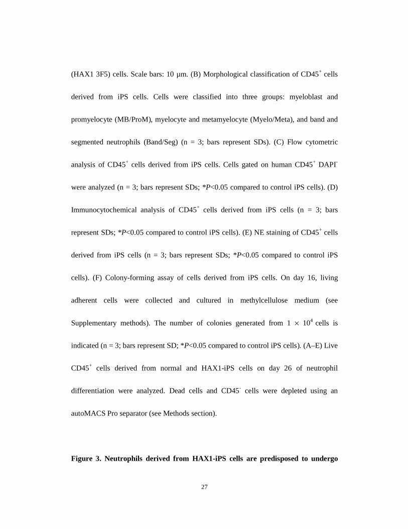
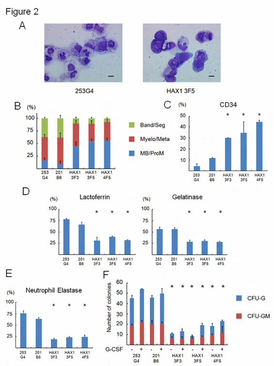
Figure 2. Maturation arrest at the progenitor level in neutrophil differentiation
from HAX1-iPS cells.
(A) May-Giemsa staining of CD45+ cells derived from normal (253G4) and HAX1-iPS
27
(HAX1 3F5) cells. Scale bars: 10 µm. (B) Morphological classification of CD45+ cells
derived from iPS cells. Cells were classified into three groups: myeloblast and
promyelocyte (MB/ProM), myelocyte and metamyelocyte (Myelo/Meta), and band and
segmented neutrophils (Band/Seg) (n = 3; bars represent SDs). (C) Flow cytometric
analysis of CD45+ cells derived from iPS cells. Cells gated on human CD45+ DAPI-
were analyzed (n = 3; bars represent SDs; *P<0.05 compared to control iPS cells). (D)
Immunocytochemical analysis of CD45+ cells derived from iPS cells (n = 3; bars
represent SDs; *P<0.05 compared to control iPS cells). (E) NE staining of CD45+ cells
derived from iPS cells (n = 3; bars represent SDs; *P<0.05 compared to control iPS
cells). (F) Colony-forming assay of cells derived from iPS cells. On day 16, living
adherent cells were collected and cultured in methylcellulose medium (see
Supplementary methods). The number of colonies generated from 1 × 10⁴ cells is
indicated (n = 3; bars represent SD; *P<0.05 compared to control iPS cells). (A–E) Live
CD45+ cells derived from normal and HAX1-iPS cells on day 26 of neutrophil
differentiation were analyzed. Dead cells and CD45- cells were depleted using an
autoMACS Pro separator (see Methods section).
Figure 3. Neutrophils derived from HAX1-iPS cells are predisposed to undergo

28
apoptosis due to their reduced Δψm.
Annexin V assay (A) and mitochondrial membrane potential assay (B) of iPS
cell-derived cells on day 26 of neutrophil differentiation using flow cytometry. Cells
gated on human CD45+ were analyzed (n = 3; bars represent SDs; *P<0.05 to control
iPS cells).
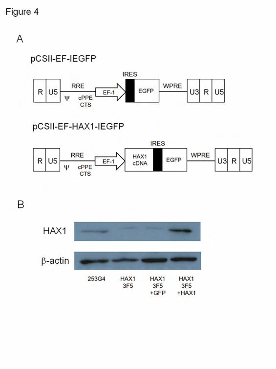
Figure 4. Lentiviral transduction of HAX1-iPS cells.
(A) Lentiviral vector constructs with only EGFP (pCSII-EF-IEGFP), and HAX1 cDNA
and EGFP (pCSII-EF-HAX1-IEGFP). (B) Western blot analysis for HAX1 protein in
lentivirally-transduced HAX1-iPS cells. β-actin was used as a loading control.
Figure 5. Lentiviral transduction of HAX1 cDNA improves maturation arrest of
HAX1-iPS cells.
(A) May-Giemsa staining of CD45+ cells derived from HAX1 3F5+GFP and HAX1
3F5+HAX1 cells. Scale bars: 10 µm. (B) Morphological classification of CD45+ cells
derived from lentivirally-transduced iPS cells. (n = 3; bars represent SDs). (C) Flow
cytometric analysis of CD45+ cells derived from lentivirally-transduced iPS cells. Cells
gated on GFP+ human CD45+ DAPI- were analyzed (n = 3; bars represent SDs;

29
*P<0.05). (D) Immunocytochemical analysis of CD45+ cells derived from
lentivirally-transduced iPS cells (n = 3; bars represent SDs; *P<0.05). (E) NE staining
of CD45+ cells derived from lentivirally-transduced iPS cells (n = 3; bars represent SDs;
*P<0.05). (F) Colony-forming assay of lentivirally-transduced cells derived from iPS
cells. The number of colonies derived from 1 × 10⁴ cells is indicated (n = 3; bars
represent SD; *P<0.05 compared to HAX1 3F5 and HAX1 3F5+GFP). (A-E) Live
CD45+ cells derived from lentivirally-transduced iPS cells on day 26 of neutrophil
differentiation were analyzed. Dead cells and CD45- cells were depleted using an
autoMACS Pro separator (see Methods section).
Figure 6. Lentiviral transduction of HAX1 cDNA prevents HAX1-iPS cells being
predisposed to undergo apoptosis.
Annexin V assay (A) and mitochondrial membrane potential assay (B) of
lentivirally-transduced iPS cell-derived cells on day 26 of neutrophil differentiation.
Cells gated on GFP+ human CD45+ were analyzed (n = 3; bars represent SDs; *P<0.05).


1
Supplemental methods
RNA isolation and qPCR
RNA was isolated using the RNeasy mini kit (Qiagen, Valencia, CA) and
subjected to reverse transcription (RT) with the Omniscript-RT kit (Qiagen). All
procedures were performed by following the manufacturer’s instructions. Quantitative
polymerase chain reaction (qPCR) was performed with a 7900HT Fast Real-Time PCR
system (Applied Biosystems, Carlsbad, CA) and SYBR Premix Ex Taq II (Takara,
Shiga, Japan). Primer sequences were described previously1.
Antibodies
Fluorescein isothiocyanate (FITC)-conjugated anti-human TRA 1-85 (R&D
Systems), anti-human CD45 (Becton-Dickinson, Franklin Lakes, NJ), phycoerythrin
(PE)-conjugated anti-human SSEA4 (R&D Systems), anti-human CD34 (Beckman
Coulter, Fullerton, CA), anti-human CD45 (Becton-Dickinson), allophycocyanin
(APC)-conjugated Annexin V (Becton-Dickinson), and anti-human CD45
(Becton-Dickinson) antibodies were used for flow cytometric analysis. Goat anti-human
lactoferrin (Santa Cruz Biotechnology, Santa Cruz, CA) and rabbit anti-human MMP9

2
(gelatinase) (Abcam, Cambridge, UK) antibodies were used for immunocytochemical
analysis. Biotinylated horse anti-goat or anti-rabbit IgG (Vector Laboratories,
Burlingame, CA) was used as the secondary antibody. Mouse anti-human -actin (Santa
Cruz Biotechnology) and mouse anti-human HAX1 (Becton-Dickinson) antibodies
were used for Western blotting. Horseradish peroxidase (HRP)-conjugated anti-mouse
IgG (Santa Cruz Biotechnology) was used as the secondary antibody.
DNA sequencing analysis
Genomic DNA was isolated from iPS cells using the QIAamp DNA blood mini
kit (Qiagen). PCR was performed with primers that spanned all exons of HAX1. The
PCR product was sequenced directly using the BigDye Terminator v3.1 Cycle
Sequencing kit and an ABI 3130xl genetic analyser (Applied Biosystems). Primer
sequences were described previously2.
Teratoma formation
Approximately 2 106 iPS cells were injected subcutaneously into the dorsal
flank of immunocompromised NOD/SCID/cnull (NOG) mice. Tissue masses were
excised 8–10 weeks after injection and fixed with phosphate-buffered saline (PBS)
3
containing 4% paraformaldehyde (PFA, wt/vol). Paraffin-embedded tissues were
sectioned and stained with hematoxylin and eosin.
Karyotyping and short tandem repeat analysis
Chromosomal G-banding analyses were performed at the Nihon Gene Research
Laboratories (Miyagi, Japan). For short tandem repeat analysis, genomic DNA was used
for PCR with the Powerplex 16 system (Promega, Fitchburg, WI). Analysis was
performed using an ABI 3100 sequencer and Gene Mapper v3.5 software (Applied
Biosystems).
Cytostaining
Cytospin and cytostaining were performed as described previously3. In brief,
collected cells were centrifuged onto glass slides using a Shandon Cytospin® 4
cytocentrifuge (Thermo, Pittsburgh, PA) and analyzed by microscopy after
May-Giemsa staining. For immunocytochemical analysis, cells centrifuged onto glass
slides were fixed immediately with PBS containing 4% PFA (wt/vol). Immunostaining
was performed as described previously3. For neutrophil elastase staining, cells were
transferred to glass slides by centrifugation and fixed. Cells were stained using the
4
elastase staining kit and the elastase AS-D staining kit (Muto Pure Chemicals, Tokyo,
Japan) according to the manufacturer’s instructions.
Colony-forming assay
The colony-forming assay was performed as described previously4 with some
modifications. On day 16 of culture, adherent cells were treated with Stempro®
Accutase® (Gibco, Carlsbad, CA), harvested, and incubated in a new tissue culture dish
(Becton–Dickinson) for 10 min to eliminate adherent non-haematopoietic cells5.
Floating cells were collected and dispersed using 40-mm strainers. Dead cells were
eliminated using the Dead Cell Removal kit (Miltenyi Biotec). Live hematopoietic cells
were plated at a density of 1 104 cells/ml in 35-mm petri dishes (Becton-Dickinson)
using 1 ml/dish of MethoCult H4230 semisolid medium (STEMCELL Technologies,
Vancouver, BC, Canada) containing stem cell factor (50 ng/mL, R&D Systems), IL-3
(50 ng/mL, R&D Systems), and thrombopoietin (5 ng/mL, kindly provided by Kyowa
Hakko Kirin), in the absence or presence of granulocyte colony-stimulating factor (50
ng/mL, also kindly provided by Kyowa Hakko Kirin). Colonies were counted after 14
days of incubation.
5
Mitochondrial membrane potential (m) assay
m was analysed using the MitoProbeTM DiIC1(5) Assay Kit (Molecular
Probes, Carlsbad, CA) according to the manufacturer’s instructions.
Lentiviral transduction of HAX1-iPS cells
Total RNA was extracted from peripheral blood mononuclear cells obtained
from a healthy volunteer, and RNA was reverse transcribed using the Omniscript-RT kit
(Qiagen). The human HAX1 gene was amplified by PCR using KOD-Plus v2
(TOYOBO, Osaka, Japan) and the following primers:
5’-CCGCGGCCGCCCACCATGAGCCTCTTTGATCTCTTCCGGGGCTTT-3’(sense)
and 5’-CGCGGATCCCTACCGGGACCGGAACCAACGTCCCAGGAA-3’
(antisense). The PCR product was gel-purified, digested with NotI and BamHI, and
cloned into the NotI/BamHI-digested pBlueScript II plasmid. The isolated clone
(pBlueScriptII-HAX1) was verified by DNA sequencing. To construct
pCSII-EF-IEGFP, the pGCDNsamIRESEGFP vector6 was digested with HindIII. After
creating blunt ends, the vector was further digested with NotI, and the resultant
fragment containing the internal ribosome entry site (IRES) and enhanced green
fluorescent protein (EGFP) cassette was inserted into NotI/HpaI-sites of the
6
pCSII-EF-MCS plasmid (kindly provided by Dr. Hiroyuki Miyoshi, The RIKEN
BioResource Center, Ibaraki, Japan). To generate pCSII-EF-HAX1-IEGFP, the
NotI/BamHI fragment of pBlueScriptII-HAX1 containing the HAX1 sequence was
cloned into NotI/BamHI-sites of the pCSII-EF-IEGFP plasmid. Viral supernatants were
collected as previously described (see
http://www.brc.riken.jp/lab/cfm/Subteam_for_Manipulation_of_Cell_Fate/Protocols.ht
ml). For lentiviral transduction, the lentiviral supernatant was added to human iPS cell
cultures in the presence of 4 µg/mL polybrene, followed by incubation at 37°C in an
atmosphere of 5% CO2 (vol/vol) for 24 hours. These cells were expanded, GFP-positive
cells were sorted with a FACSAriaII flow cytometer (Becton-Dickinson), and cultured
on mitomycin-C-treated SNL feeder cells in the presence of Y-276327 (10 mg/mL) for
24 hours. Single cell-derived GFP-positive colonies were then expanded, and these cell
lines were used for subsequent experiments.
Western blot analysis
iPS cell extracts were subjected to SDS-PAGE, and proteins were transferred
onto Immobilon-P membranes (Millipore, Billerica, MA). Membranes were blocked,
incubated with primary antibodies, washed, and incubated with corresponding

7
secondary antibodies. Immunoreactions were visualized using an ECL Western Blotting
Detection system (GE Healthcare, Waukesha, WI).
References
1. Tanaka T, Takahashi K, Yamane M, Tomida S, Nakamura S, Oshima K, et al.
Induced pluripotent stem cells from CINCA syndrome patients as a model for dissecting
somatic mosaicism and drug discovery. Blood. 2012 Aug 9;120(6):1299-308.
2. Ishikawa N, Okada S, Miki M, Shirao K, Kihara H, Tsumura M, et al.
Neurodevelopmental abnormalities associated with severe congenital neutropenia due to
the R86X mutation in the HAX1 gene. J Med Genet. 2008 Dec;45(12):802-7.
3. Morishima T, Watanabe K, Niwa A, Fujino H, Matsubara H, Adachi S, et al.
Neutrophil differentiation from human-induced pluripotent stem cells. J Cell Physiol. 2011
May;226(5):1283-91.
4. Niwa A, Heike T, Umeda K, Oshima K, Kato I, Sakai H, et al. A novel serum-free
monolayer culture for orderly hematopoietic differentiation of human pluripotent cells via
mesodermal progenitors. PLoS One. 2011;6(7):e22261.
5. Suwabe N, Takahashi S, Nakano T, Yamamoto M. GATA-1 regulates growth and
differentiation of definitive erythroid lineage cells during in vitro ES cell differentiation.
Blood. 1998 Dec 1;92(11):4108-18.
6. Iwama A, Osawa M, Hirasawa R, Uchiyama N, Kaneko S, Onodera M, et al.
Reciprocal roles for CCAAT/enhancer binding protein (C/EBP) and PU.1 transcription
factors in Langerhans cell commitment. J Exp Med. 2002 Mar 4;195(5):547-58.
7. Watanabe K, Ueno M, Kamiya D, Nishiyama A, Matsumura M, Wataya T, et al. A
ROCK inhibitor permits survival of dissociated human embryonic stem cells. Nat Biotechnol.
2007 Jun;25(6):681-6.
8
Supplemental Figure Legends
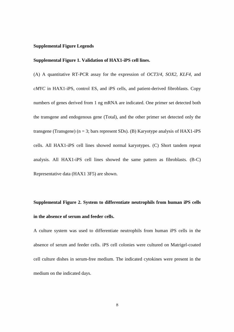
Supplemental Figure 1. Validation of HAX1-iPS cell lines.
(A) A quantitative RT-PCR assay for the expression of OCT3/4, SOX2, KLF4, and
cMYC in HAX1-iPS, control ES, and iPS cells, and patient-derived fibroblasts. Copy
numbers of genes derived from 1 ng mRNA are indicated. One primer set detected both
the transgene and endogenous gene (Total), and the other primer set detected only the
transgene (Transgene) (n = 3; bars represent SDs). (B) Karyotype analysis of HAX1-iPS
cells. All HAX1-iPS cell lines showed normal karyotypes. (C) Short tandem repeat
analysis. All HAX1-iPS cell lines showed the same pattern as fibroblasts. (B-C)
Representative data (HAX1 3F5) are shown.
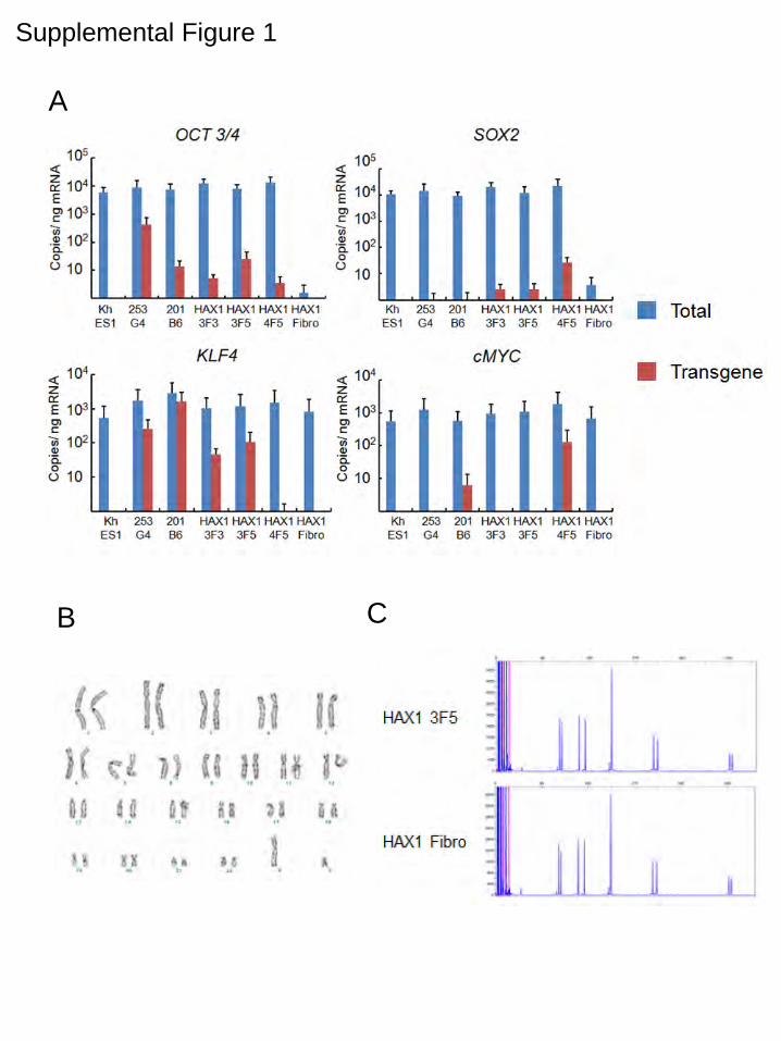
Supplemental Figure 2. System to differentiate neutrophils from human iPS cells
in the absence of serum and feeder cells.
A culture system was used to differentiate neutrophils from human iPS cells in the
absence of serum and feeder cells. iPS cell colonies were cultured on Matrigel-coated
cell culture dishes in serum-free medium. The indicated cytokines were present in the
medium on the indicated days.

9
Supplemental Figure 3. Mitochondrial membrane potential assay of
undifferentiated iPS cells.
m was analysed in undifferentiated iPS cells (n = 3; bars represent SD).
Supplemental Figure 1
A
B C
Supplemental Figure 2

Supplemental Figure 3