issues of beta cell dysfunction
TRANSCRIPT
Issues of Beta Cell Dysfunction Gordon C. Weir, M.D.
Joslin Diabetes Center
Beta Cell Meeting
Endocrine Society
January 10, 2009

A Diabetes Puzzle
How does diabetes start?
What is going on when someoneʼs fasting glucose rises from 83 to 95 mg/dl or when postprandial glucose level start to rise?
Hypothesis: Not enough β-cells

A Simple Hypothesis: Primacy of Reduced β-Cell Mass in Diabetes
Relative/Absolute β-Cell Mass
ß-Cell Function
Glucotoxicity Loss of β-Cell Phenotype
Reduced islet mass in T2DM
Autopsy Studies Maclean, Ogilvie - 1955 Westermark, Wilander - 1978 Saito, et al, 1978,1979 Kloppel, et al - 1985 Butler, et al - 2003 Yoon, et al - 2003
All these studies show islet mass 40-60% of normal
The debate is over!

β cell volume in human type 2 diabetics is about 50% of non diabetic
Lean Obese IGT Diabetic
Rela
tive β
cell
volu
me
(%)
0
1
2
3
Lean Obese Non-diabetic
Obese 16 30 19 16 41
Butler et al, Diabetes Jan 2003
Individual type 2 diabetic resected
0 5 10 15 20
Rela
tive β
cell
volu
me
(%)
3
2
1
0
Normal organ donor n=9 Non-diabetic resected n=10
Yoon et al, JCEM May 2003
Years after diagnosis

Beta cell mass versus glucose - humans
0 2 4 6 60
80
100
120
140
160
180
OD IFG OND
200 250 300
8 12
r = 0.50
beta-cell area [%]
FPG
[mg/
dl]
Ritzel, Butlers, et al, Diabtes Care, 2006
Ways to have too few β-cells en route to T2DM
β-cell death problem: Apoptosis
β-cell birth problem:
1. Not enough at the beginning: Intra-uterine growth retardation
2. Inadequate β-cell replication
3. Inadequate neogenesis
Hard to study: No good test for β-cell mass Rates of β-cell birth and death are very slow in humans.
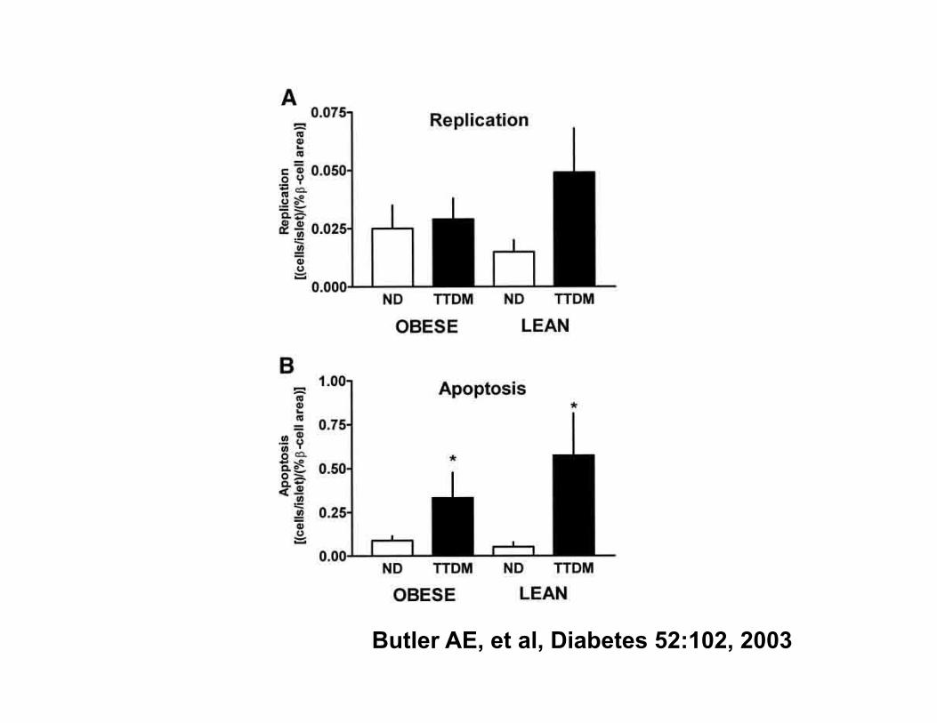
Butler AE, et al, Diabetes 52:102, 2003

10 20 30 40 50 60 70
150
100
50
β-ce
ll m
ass Successful
Compensation
No T2DM Obesity Phase of Life
Years

10 20 30 40 50 60 70
150
100
50
β-ce
ll m
ass
Successful Compensation
Obesity phase of life
β-cell Death Problem
IGT
T2DM
Years

10 20 30 40 50 60 70
150
100
50
β-ce
ll m
ass
Successful Compensation
β-Cell Death Problem
β-Cell Birth Problem
(Intra-uterine Growth Retardation)
Years

Progressive Loss of Glycemic Control in Obese Patients UKPDS
Open – Conventional
Cross – Metformin
Diamond - Intensive
UKPDS – Diabetes 44: 1249, 1995

Progressive Loss of Beta Cell Function in UKPDS – HOMA Studies
Black diamonds- Sulfonylureas
Open circles – Diet
Crosses - Metformin
UKPDS – Diabetes 44: 1249, 1995

What Might Cause Accelerated β-cell Apoptosis in T2D?
• Areas of focus – Glucotoxicity – Lipotoxicity – Oxidative injury – Amyloid toxicity – ER stress

Five Stages
Stage 1: Compensation - Glucose “normal”
Stage 2: Stable Adaptation 5.0-7.3 mM (89-130 mg/dl)
Stage 3: Unstable Early Decompensation 7.3-16 mM (130-285 mg/dl)
Stage 4: Stable Decompensation 16-20 mM (285-350 mg/dl)
Stage 5: Severe Decompensation - DKA

The Relationship Between Insulin Secretion and Insulin Resistance
Kahn SE, J Clin Endoc Metab. 86: 4047, 2001

With compensation, can a given β-cell mass put out much more insulin?
Absolutely yes!
Obesity has only about a 50% increase in β-cell mass (Kloppel, Butlers), but insulin secretory output increases 100%. (24 hr output of insulin 468 versus 235 nmol)
Camastra S, et al. Diabetes 54:2382, 2005

Insulin secretion rates over 24 hours in obese versus lean subjects
Camastra S, et al. Diabetes 54:2382, 2005
Stage 2: Stable Adaptation
Approximate glucose levels 90-130 mg/dl - Includes IGT and IFG
Not compensation - glucose levels not “normal”.
Beta cell phenotype altered - GSIS reduced.
Stable - Diabetes Prevention Program (DPP) IGT progresses to diabetes at 11% per year, and with diet and exercise only 5% per year.
Occurs in pre-T1DM and remissions, but not as durable due to autoimmune destruction.

Effect of Fasting Plasma Glucose (FPG)on the Acute Insulin Response
Rela
tive
acut
e in
sulin
resp
onse
(% in
crea
se)
Time (min)


With glucotoxicity in T2DM, does a given β-cell mass put out much less insulin?
Absolutely yes!
In T2DM β-cell mass is reduced to about 50% of normal, but insulin output to maximum stimulus of glucose and arginine is only about 15% of normal.


Glucotoxicity Hypothesis
β-cells exposed to even mild chronic hyperglycemia develop changes in phenotype characterized by dysfunctional insulin secretion associated with altered gene and protein expression.

Competing hypotheses Glucotoxicity: Excellent correlation between rising glucose levels and β-cell dysfunction. Molecular basis not yet established.
Lipotoxicity: Little evidence to support. High FFA in obesity associated with terrific insulin secretion. Fat may be good for β-cells.
Gluco-lipotoxicity: Fallback position. Could be true, but most evidence is from in vitro cell studies, which may not be applicable to in vivo situation. Molecular basis not yet established. Lipid accumulation in β-cells may be modestly increased but may not be harmful.

Stage 4: Stable Decompensation
Frank diabetes
Stable because in T2DM DKA is rare and considerable amounts of insulin are produced for decades.
There is attrition of beta cells, which often leads to oral agent failure, but beta cell mass remains at 30-50% of normal.
T1DM progresses to Stage 5.

Stage 4
Natural Forces
Treatment
Stage 4
Stage 2

Diabetes Prevention Program (DPP) Research Group
Diabetes. 2005; 54:1150.

The DREAM (Diabetes Reduction Assessment with ramipril and rosiglitazone Medication)
Lancet . 2006;368:1096.

Are We Delaying the Onset of Diabetes
or
Preventing Diabetes?
Debate about DPP

Because the problem with both type 1 and 2 diabetes is not enough β cells.
(Of course, autoimmune destruction
must be prevented too.)
Why β-cell Replenishment?


The Dream of β-cell Regeneration

β-cell Regeneration in Diabetes: The Task
• Type 1 Diabetes: Shut off autoimmunity, stimulate β-cell replication and neogenesis, inhibit apoptosis
• Type 2 Diabetes: Stimulate β-cell replication and neogenesis, inhibit apoptosis, and reduce insulin resistance

β-cell Regeneration