kp 9 autoimmune diseases_htr
TRANSCRIPT

Autoimmune Diseases
HTR_UMM

Pendahuluan
Penyakit Autoimmune terjadi dari kerusakandalam tubuh dengan adanya autoantibodiesatau autoreactive cells
Sekitar 2% populasi terkena penyakit ini
Terjadinya self tolerance pada individu ini
Self tolerance merupakan mekanisme clonal deletion dari sel efektor, pengaturan aktif oleh selT dan pengaturan melalui jalur idiopatik
Umumnya sel T yang diproses memalui Thymus tidak bertahan dan self reactive T cells dihancurkan
Kehilangan sel T autoAbs
Introduction

Pendahuluan
Terjadi situasi dimana antibody diproduksi
untuk melawan antibody itu sendiri
Ketika antibody diproduksi pertama kalinya,
area tersebut dikenali sebagai benda asing
oleh host
Kemudian host akan memproduksi antibody
untuk melawannya
anti-idiotypic Ab mungkin memiliki reaksi
melawan self Ags

Pendahuluan
Anti-idiotypic Abs ditemukan pada penyakitseperti myasthenia gravis, diabetes mellitus, and Graves’ disease
Kerusakan pada NK sel, dalam sekresi interleukin, dalam phagocytosis dan complements dapatterjadi
Hormon khususnya estrogen ditemukan untukmeningkatkan aktivasi sel B dan menekanpengaturan aktivitas sel T
Kondisi lingkungan seperti agen-agen infeksiseperti virus, bakteri dan obat dapatmenyebabkan autoimunitas

Systemic Lupus Erythematosus
Peradangan sistemik kronis yang ditandai olehexaserbasi dan remisi
1/2000 orang terinfeksi
Usia biasanya 20-40 tahun
Wanita lebih tinggi dari pria (13/1)
Penyebab tidak diketahui tetapi kemugkinankarena genetic dan faktor2 lingkungan
Pada orang berkulit putih kemungkinan besardihubungkan dengan HLA-DR3 or DR2 danDQB1

Systemic Lupus
ErythematosusClinical Signs:
The first signs to appear are usually nonspecific such as fatigue, weight loss, malaise, fever, and anorexia
Joint involvement of small joints of the hands, wrist and knees is reported in over 90% of cases
A skin manifestation of an erythematous rash may appear in the area of body exposed to UV light
A classic butterfly rash across the nose and cheeks which is responsible for the name lupus may appear
Some patients may also exhibit renal involvement
Systemic Lupus Erythematosus
Renal involvement is usually in the form of lesions the most dangerous of which is glomerulonephritis
Immune complexes may deposit in the subendothelial tissue and thickening of the basement membrane results
All of these can result in renal failure and death
There might also be cardiac involvement with pericarditis, tachycardia, or ventricular enlargement.
CNS manifestation such as seizures, psychoses, or depression
Hematologic abnormalities such as anemia, leukopenia or lymphopenia are exhibited
Systemic Lupus Erythematosus
Immunological Findings:
The LE cell is a neutrophil that has engulfed the
antibody-coated nucleus of another
neutrophil
SLE is associated with more than 28
autoantibodies
Lupus patients appear to have overactive B
cells especially the subset with the CD5 marker
which increase in number

Systemic Lupus Erythematosus
The increased prevalence of the disease in women may be traced to the fact that estrogen enhance B cell activity
Estrogen also suppresses regulator activities of T cells as a result, there is a decrease in the absolute number of T cells
Complement is activated which results in decrease in the serum level of complement components
At the same time there is an increase in the level of breakdown products of complements such as C3d and C3a
IgG is most pathogenic and it forms complexes that are deposited in the glomerular basement membrane

Systemic Lupus Erythematosus
Accumulation of immune complex with complement activation is responsible for the damage to the kidney
Drug induced lupus differs from the chronic form of the disease: when the drug is withdrawn symptoms disappear
It is a milder form of the disease and manifest as arthritis or rashes
Drugs that have been implicated include procainamide hydrochloride, hydralazine hydrochloride, methyldopa etc.
Systemic Lupus Erythematosus
Laboratory Diagnosis of SLE:
A screening test for antinuclear antibody (ANA) is
usually the first test done
Fluorescent antinuclear antibody (FANA) testing is
most widely used
The test is sensitive but the specificity is low because
many of the Abs are associated with other diseases
The test is an indirect IFA test which employs
antihuman globulin tagged with a fluorescent
marker.
If FANA is positive, then a profile testing should be
done for individual Abs
More than 99% of patients with SLE will have a
positive result for one or more tests

Rheumatoid Arthritis
Rheumatoid arthritis is a systemic autoimmune disorder
It involves the synovial membrane of multiple joints
Women are more likely to be affected than men and usually strikes between the ages of 20 and 40
Spontaneous remission may be experienced by some patients otherwise the disease may progress and result in deformity and disability
RA has been associated with certain of the HLA class II molecules.
HLA-DR1 and DR4 occur in 70% of patients with RA
Rheumatoid Arthritis
Clinical Signs:
Symptoms include morning stiffness around the joints lasting at least 1 hour, swelling of the soft tissue around three or more joints
Others include swelling of the proximal interphalangeal, metacarpophalangeal, or wrist joints, symetric arthritis, subcutaneous nodules, a positive RF test
Also included is a radiographic evidence of erosion of the joints of the hands, the wrist, or both

Rheumatoid Arthritis
Usually begins with nonspecific symptoms such as fever, malaise, weight loss, and transient joint pain
Stiffness and joint pain that gradually improves during the day are characteristics exhibited by most patients
Joint are involved progressively to larger joints in a symmetric manner from the knees, hips, elbows, shoulders and cervical spine
About 25% of patients have nodules over the bones
Nodules may also be found in the myocardium, pericardium, heart valve, pleural, lungs, spleen, and larynx

Rheumatoid Arthritis
Immunologic Findings:
The main immunologic finding is the presence
of RF
It has been suggested that RF may be anti-
idiotypic antibodies involved in the regulation
of immune response
In RA, polyclonal activation of B cells may
occur resulting in overwhelming amount of
antibody to IgG

Rheumatoid Arthritis
These Abs may cause immune complex
formation with the activation of complement
which contribute to pathogenesis
Joint damage is due to invasion of inflammatory
cells such as neutrophils, and macrophages
Proliferation of fibroblast, macrophages, mast
cells, and stellar cells result in the formation of a
pannus, an organized mass of cells that grow
into the joint space

Laboratory Diagnosis of Rheumatoid
Arthritis:
Diagnosis is based on a combination of clinical
manifestations, radiographic findings and lab tests
Laboratory screening test for RF
Quantitative test are also available which involve
nephelometry and ELISA techniques
RF is found in other diseases such as syphilis, viral
infections, leprosy, chronic liver disease, neoplasm
and other inflammatory processes
C-reactive protein and Erythrosit sedimentation
rate (ESR) are usually elevated and complements
are normal or elevated

Rheumatoid Arthritis
Treatment:
Treatment include palliatives with rest and
nonsteroidal anti-inflammatory drugs like salicylates
and ibuprofen
Slow-acting antirheumatic drugs (SAARDS) may
also be used to treat the condition
New therapy include the use of monoclonal Abs
that target T cells
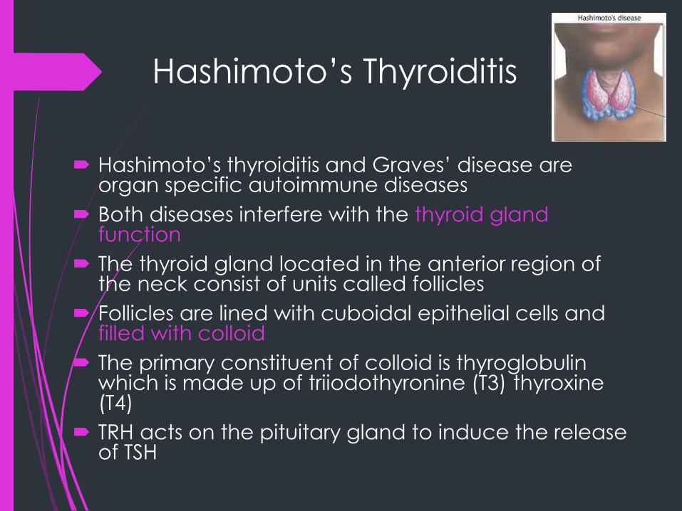
Hashimoto’s Thyroiditis
Hashimoto’s thyroiditis and Graves’ disease are organ specific autoimmune diseases
Both diseases interfere with the thyroid gland function
The thyroid gland located in the anterior region of the neck consist of units called follicles
Follicles are lined with cuboidal epithelial cells and filled with colloid
The primary constituent of colloid is thyroglobulin which is made up of triiodothyronine (T3) thyroxine(T4)
TRH acts on the pituitary gland to induce the release of TSH

Hashimoto’s Thyroiditis
TSH binds to receptors on the cell membrane of the thyroid gland causing break down of thyroglobulin into T3 and T4
AutoAbs may interfere with this process and cause under or overactivity of the thyroid
Hashimoto’s thyroditis is most often seen in womenbetween the ages of 30 and 40 years
Patients develop a combination of goiter or enlarged thyroid, hypothyroidism and thyroid autoantibodies
An association with HLA antigens DR4 and DR5 has been noted. DQA1 and DQB1 genes seem to confer resistance

Hashimoto’s Thyroiditis
Immunologic Findings:
Lymphocytic infiltration is seen with development of germinal centers that almost replace the normal glandular architecture of the thyroid
Cell infiltrates include T and B cells, macrophages and plasma cells
Both CD4 and CD8 cells are found thus the disease is characterized by a cellular and a humoral response
Autoantibodies are found in up to 80% of cases

Hashimoto’s Thyroiditis
Laboratory Testing:
The autoantibodies present include Abs to
thyroglobulin and to thyroid microsomal antigen
now known as thyroid peroxidase
Some peroxidase Abs inhibit enzyme activity while
others may mediate the cytotoxicity due to natural
killer cells
Abs to thyroglobulin help to produce hypothyroid
conditions
Other Abs include colloid Ab (CA2) thyrotropin-
binding inhibitory immunoglobulin (TBII)
Hashimoto’s Thyroiditis
Others are thyroid stimulating immunoglobulin (TSI) and thyroid growth-stimulating immunoglobulin (TGSI)
Peroxidase Abs can be measured by particle agglutination assays, complement fixation, RIA and Indirect IFA
Abs to thyroglobulin can be measured by precipitaion in agar, indirect IFA, passive agglutination, RIA, and EIA
Indirect IFA use human or monkey thyroid tissue fixed to a slide
Antithyroglobulin Abs are found in about 80% of patients with the disease

Graves’ Disease
Graves’ disease is another autoimmune disease that affects the thyroid gland
Graves’ disease produces hyperthyroidism
It the most common cause of hyperthyroidism and affects about 0.5% of the population
Women are more susceptible than men by a margin of 7:1 and usually present between the ages of 30 and 40.
In whites, the disease is associated with the HLA antigen DR3 while in Asians HLA Bw35 and Bw36 occur more frequently

Graves’ Disease
HLA DQB1 appears to confer resistance to the disease
Clinical Signs:
Disease is presented as thyrotoxicosis with a diffusely enlarged goiter that is soft instead of rubbery
Signs include nervousness, insomnia, depression, weight loss, heat intolerance, sweating, rapid heart beat
Other signs include fatigue, cardiac dysrhythmias, restlessness, and exopthalmus

Graves’ Disease
Immunologic Findings:
The thyroid presents with Hyperplasia with a patchy infiltration of lymphocytes
Both CD4 and CD8 cells are present and the T cells appear to play a central role in the pathogenesis of the disease
The most significant Ab present is thyroid stimulating hormone receptor antibody (TRab)
Ag-Ab combination result in the stimulation of the receptor resulting in the release of the thyroid hormones
Another group of Abs called thyroid stimulating antibodies or immunoglobulins (TSab or TSI) may have different specificity

Graves’ Disease
Laboratory Diagnosis:
A key finding in Graves’ disease is elevated levels of total and free T3 and T4
In addition, TSH levels are low due to Ab stimulation of the thyroid
Measurement of the thyroid Abs may be undertaken if the above assays are unclear
Treatment:
Antithyroid medication may be employed. Radioiodine which emits beta particles may be used. Surgery is also an option.

Insulin-Dependent Diabetes
This disease is characterized by insufficient
production of insulin due to an autoimmune
destruction of the beta cells of the pancreas
Peak onset is b/w 10 and 14 years of age
Disease may be attributed to genetic and
environmental conditions
About 95% of white diabetics carry the HLA-DR3
or DR4 genes
It appears that true susceptibility genes for
IDDM may occur in the HLA-DQ region

Insulin-Dependent Diabetes
Viral infections have been linked with diabetes
Mumps virus, rubella virus, CMV, and Coxsakie B4 virus have all been inconclusively linked to diabetes
Congenital rubella infection is the only one for which a link has been definitively identified
There appears to be similarity between coxsakieviral protein P2-C and the enzyme glutamic acid decarboxylase.
Antibodies are formed against glutamic acid decarboxylase in IDDM
Molecular mimicry could initiate Ab production against self Ag

Insulin-Dependent Diabetes
Immunopathology:
Inflammation of the islets of Langerhans in the pancreas leads to fibrosis and destruction of most of the beta cells
CD4 and CD8 B cells and macrophages are all involved in the destructive process of the islet cells
Cellular and humoral immunity are involved in this process
The subset of T cells that is activated determines whether the response is cellular or humoral

Insulin-Dependent Diabetes
Laboratory Testing:
IDDM is usually diagnosed by the presence of
hyperglycemia
Abs to islet cells may be screened for by
indirect IFA with human or rat islet cells
Islet cell may be detected in the sera of newly
diagnosed diabetic cases
Abs to insulin may be detected by ELISA or RIA
methods

Insulin-Dependent Diabetes
Treatment:
Insulin has been the standard form of
treatment
New treatment methods center around the use
of immunosuppressive agents
Agents that have been tried include
azathioprine, cyclosporine A and prednisone
All these agents have potentially toxic effects

Multiple Sclerosis
Destruction of the myelin sheath results in the formation of lesions known as plaques in the white matter of the brain and spinal cord
Genetic and environmental factors predispose one to this disease
MS is associated with the inheritance of HLA antigen DRw15 and DRw6
An inflammatory response to bacteria or virus may trigger the autoimmune process
Disease is most often seen in those b/w the ages of 20 & 50 and is more common in women

Multiple Sclerosis
90% of patients alternate between remissions and
relapses for many years
Within the plaques, CD4 cells, plasma cells and
macrophages are found along with immunoglobulin
Immunoglobulin is increased in the spinal fluid in 60
to 80% of patients
They usually produce oligoclonal bands on protein
electrophoresis
RIA (radioimuno assay) is used to detect the Abs
Therapy include use of corticosteroids

Myasthenia Gravis
Symptoms of disease include facial weakness, difficulty in chewing, and swallowing, difficulty breathing
Others include inability to maintain support of the trunk, the neck or the head
Antibody mediated damage to the acetylcholine receptors in the skeletal muscle leads to muscle weakness
May be associated with the presence of other autoimmune diseases such as SLE
Appears to be linked with either HLA-B8 or DRw3 antigens

Myasthenia Gravis
80 to 90% of patients have Abs to acetylcholine
receptors which may be the main contributor
to pathogenesis
Combination of the Abs to the receptors blocks
the binding of acetylcholine which destroy the
receptors
Abs can be detected with RIA methods
Anticholinesterase agents are employed in
therapy

Goodpasture’s Syndrome
Goodpasture’s syndrome is a
glomerulonephritis due to Abs reacting
specifically with Ags in the kidney
Necrosis of the glomerulus is triggered by an
Ab that reacts with glycoprotein present in
the basement membrane of the glomerulus
Results in immune complex deposit and
complement fixation which causes the
damage to the kidney
This may eventually produce renal failure