mellitus: a case control study diastolic dysfunction in
TRANSCRIPT

Page 1/14
Association Between Cortisol and Left VentricularDiastolic Dysfunction in Patients With DiabetesMellitus: A Case Control StudyRIKAKO SAGARA
Kyushu University: Kyushu Daigaku https://orcid.org/0000-0003-2908-5522Tomoaki Inoue ( [email protected] )
Kyushu University https://orcid.org/0000-0002-3829-3908Noriyuki Sonoda
Kyushu University: Kyushu DaigakuChieko Yano
Kyushu University: Kyushu DaigakuMisato Motoya
Kyushu University: Kyushu DaigakuHironobu Umakoshi
Kyushu University: Kyushu DaigakuRyuichi Sakamoto
Kyushu University: Kyushu DaigakuYoshihiro Ogawa
Kyushu University: Kyushu Daigaku
Original investigation
Keywords: cortisol, cardiac dysfunction, diabetes
Posted Date: April 20th, 2021
DOI: https://doi.org/10.21203/rs.3.rs-415041/v1
License: This work is licensed under a Creative Commons Attribution 4.0 International License. Read Full License

Page 2/14
Abstract
Introduction:Diabetes mellitus (DM) is a major risk factor for the development of cardiovascular diseases. Heartfailure with preserved ejection fraction is characterized by left ventricular diastolic dysfunction (LVDD). Ithas been reported that excess cortisol found in patients with Cushing’s syndrome was associated withthe development of LVDD. However, the relationship between cortisol concentration and LVDD in patientswith DM has not been addressed.
Research Design and Methods:We enrolled 109 patients with DM and 104 patients without DM who had undergone echocardiographicexamination at Kyushu University Hospital, Japan, between November 2016 and March 2019. Leftventricular function was evaluated and the ratio of early diastolic velocity from transmitral in�ow to earlydiastolic velocity (E/e ) was used as an index of diastolic function. Plasma cortisol concentrations,glycemic control, lipid pro�les, treatment with anti-diabetic drugs, and other clinical characteristics wereevaluated, and their associations with E/e were determined using univariate and multivariate analyses.
ResultsMultivariate linear regression analysis showed that log E/e was positively correlated with age (p = 0.017), log systolic blood pressure (p = 0.004), and cortisol (p = 0.037) and negatively correlated witheGFR (p = 0.016) and the usage of SGLT2 inhibitors (p = 0.042) in patients with DM. Multivariate analysisshowed that cortisol was positively correlated with age (p = 0.016) and HbA1c (p = 0.011). There was noassociation between E/e and cortisol in patients without DM.
ConclusionsIncreased cortisol levels may increase the risk of developing LVDD in DM patients.
BackgroundDiabetes mellitus (DM) is a major risk factor for the development of cardiovascular diseases. Heartfailure with preserved ejection fraction (HFpEF), which is characterized by left ventricular diastolicdysfunction (LVDD), is clinically important in patients with DM. Indeed, the prevalence of DM isapproximately 45% in patients with HFpEF(1).
Patients with normal left ventricular wall contraction may have symptoms of heart failure. Therefore, it isimportant to evaluate left ventricular diastolic function separately from left ventricular systolic

Page 3/14
function(2). Doppler echocardiography is widely used for the noninvasive assessment of diastolic �llingof the left ventricle(3). Tissue Doppler imaging (TDI) of mitral annular motion has been proposed tocorrect for the in�uence of myocardial relaxation on transmitral �ows and shown to be an excellentpredictor of LVDD(3).
Cushing’s syndrome, including Cushing’s disease and adrenal Cushing’s syndrome, is characterized byexcess blood levels of cortisol and confers an approximately 4-fold increase in mortality compared withthe general population(4). The increased mortality is due mainly to cardiovascular complications(5). Ithas been recognized that patients with Cushing’s syndrome have a high incidence of left ventricularhypertrophy and dysfunction(6). Additionally, the incidence of cardiovascular outcomes in patients withsubclinical Cushing’s syndrome was more than three times greater than in patients with nonfunctioningadrenal adenoma(7)(8). It is likely that cortisol affects cardiac structure and function, although itsrelationship with LVDD in patients with DM has not been addressed. Therefore, we designed a cross-sectional study to determine the relationship between cortisol and LVDD in patients with DM who did nothave overt cardiovascular diseases.
MethodsSome of the methods used in this study were described previously(9).
Subjects
Between November 2016 and March 2019, we consecutively recruited 109 patients with DM and 104patients without DM who had undergone echocardiographic examination at the metabolic ward ofKyushu University Hospital, Fukuoka, Japan. Patients were excluded if they 1) were taking steroids, 2)were undergoing hemodialysis treatment, or 3) had overt heart failure. Patients without DM consisted ofthose who were hospitalized for hypertension, gastrointestinal polyps, and/or adrenal mass whosediagnosis �nally became nonfunctional. All of the patients underwent clinical evaluation, laboratoryassessment, and echocardiographic examination. Blood concentrations of fasting plasma glucose,HbA1c, total cholesterol, high-density lipoprotein cholesterol, triglycerides, uric acid, creatinine, estimatedglomerular �ltration rate (eGFR), ACTH, and cortisol (at 8:00 am, fasting) were measured. Serum cortisolconcentrations were determined by electrochemiluminescence immunoassay (ECLusys Cortisol Kits,Roche in Vitro Diagnostics, Tokyo, Japan). Clinical data and information regarding treatment of thepatients with anti-diabetic drugs were obtained from medical records. HbA1c levels were determinedusing the criteria of the National Glycohemoglobin Standardization Program. The study protocol wasapproved by the Clinical Ethics Committee of Kyushu University Hospital (protocol #29-645). This studywas performed in accordance with the Declaration of Helsinki.
Echocardiography
All echocardiographic examinations were performed with an Aplio i900 TUS-AI900 imager (CanonMedical Systems, Inc., Tokyo, Japan). Chamber dimensions and left ventricular ejection fractions (LVEF)

Page 4/14
were measured in accordance with the recommendations of the American Society ofEchocardiography(10). The left ventricular mass index (LVMI) was calculated according to the Devereuxformula and expressed as a ratio of the left ventricular mass to body surface area(10). The relative wallthickness (RWT) was based on the end-diastolic posterior wall thickness (PWTd) and the end-diastolicleft ventricular dimension (LVDd). RWT was calculated as (2 × PWTd)/LVDd. The following mitral pulsewave Doppler and tissue Doppler parameters were measured to assess diastolic function. Peak velocitiesof E and A waves of mitral in�ow, the E/A ratio, and deceleration time of the E wave were measured fromthe mitral �ow velocity pattern using pulse wave Doppler imaging. The peak early diastolic myocardialvelocity (e velocity) was measured using tissue Doppler imaging, and the ratio of E velocity to e velocity(E/e ) was calculated.
Data Analysis
All statistical analyses were performed using JMP® statistical software, version 14 (SAS Institute Inc.,Cary, NC, USA). For univariate analysis of the relationships between each parameter and E/e , continuousand categorical variables were analyzed using Spearman’s rank-order correlation and the Mann-Whitney Utest, respectively. Variables that were signi�cant in the univariate model were entered into a multivariatelinear regression analysis. Gender and glucose-lowering therapy were coded as dummy variables.Continuous variables were logarithmically transformed if they were not normally distributed according tothe Kolmogorov-Smirnov test. Categorical variables are presented as number (%) or median (lowerquartile–upper quartile). A p-value < 0.05 was considered statistically signi�cant.
ResultsThe clinical, anthropometric, and metabolic characteristics of the study participants are shown in Table 1.The DM group was older than the non-DM group, and there was no signi�cant difference in the sex ratiobetween the 2 groups. Fasting plasma glucose, HbA1c, systolic blood pressure (SBP), and cortisolconcentrations were signi�cantly higher, and eGFR and high-density lipoprotein (HDL) cholesterolconcentrations were signi�cantly lower in the DM group than in the non-DM group.

Page 5/14
Table 1Demographic and clinical characteristics of the 2 patient cohorts (N = 213)
Patient characteristics Control (N = 104) DM (N = 109) p value
Age, years 54 (43–69) 66 (56–72) < 0.001
Sex, male/female, % 53 (51.0)/51(49.0) 54 (49.5) /55 (50.5) 0.836
Body mass index, kg/m2 23.7 (21.3–26.1) 25.5 (21.3–29.9) 0.147
Duration of diabetes, years 10 (5–18)
SBP, mmHg 119 (112–129) 126 (111–141) 0.038
DBP, mmHg 70 (65–79) 75 (68–84) 0.107
Fasting plasma glucose, mg/dl 91 (84–98) 143 (119–183) < 0.001
HbA1c, % 5.6 (5.4–5.7) 8.9 (7.7–10.2) < 0.001
Total cholesterol, mg/dl 187 (161–211) 176 (151–202) 0.099
HDL-C, mg/dl 51 (43–63) 45 (37–54) 0.003
TG, mg/dl 109 (80–146) 123 (85–181) 0.062
UA, mg/dl 5.5 (4.2–6.4) 5.7 (4.6–6.7) 0.222
Cre, mg/dl 0.64 (0.58–0.77) 0.69 (0.60–0.96) 0.009
eGFR, mL/min/1.73m2 83.5 (70-98.5) 72.5 (57.0–87.0) 0.001
ACTH, pg/ml 31.1 (20.9–44.9) 36.0 (21.6–52.6) 0.186
Cortisol, µg/dl 10.35 (7.4–14.0) 12.1 (10.0-15.2) 0.003
Glucose-lowering therapies
Biguanide 46 (42)
Sulfonylureas 27 (25)
Dipeptidyl peptidase-4 inhibitors 49 (45)
Thiazolidinediones 4 (4)
α-Glucosidase inhibitor 13 (12)
Glinide 8 (7)
Categorical variables are presented as number (%) or median (lower quartile–upper quartile).
Abbreviations: DM, diabetes mellitus; SBP, systolic blood pressure; DBP, diastolic blood pressure;HbA1c, hemoglobin A1c; HDL-C, high-density lipoprotein cholesterol; TG, triglycerides; UA, uric acid;Cre, creatinine; eGFR, estimate glomerular �ltration rate; ACTH, adrenocorticotropic hormone

Page 6/14
Patient characteristics Control (N = 104) DM (N = 109) p value
Glucagon-like peptide-1 agonists 20 (18)
Sodium glucose cotransporter 2 inhibitor 21 (20)
Insulin 36 (33)
Categorical variables are presented as number (%) or median (lower quartile–upper quartile).
Abbreviations: DM, diabetes mellitus; SBP, systolic blood pressure; DBP, diastolic blood pressure;HbA1c, hemoglobin A1c; HDL-C, high-density lipoprotein cholesterol; TG, triglycerides; UA, uric acid;Cre, creatinine; eGFR, estimate glomerular �ltration rate; ACTH, adrenocorticotropic hormone
The echocardiographic data is shown in Table 2. LVEF was preserved in both the DM and non-DM groups[69% (IQR 64–73%) vs. 70% (IQR 66–73%), p = 0.141]. However, the DM group had a signi�cantly higherE/e ratio compared with the non-DM group [10.4 (IQR 8.2–13.3) vs. 8.3 (IQR 6.3–10.1), p < 0.001],suggesting diastolic dysfunction in the patients with DM. The LVMI and RWT were signi�cantly higher inthe DM group than in the non-DM group [83g/m2 (IQR 74–104) vs 79 g/m2 (IQR 64–95), p < 0.003].

Page 7/14
Table 2Echocardiographic data from each patient cohort (N = 213)
Echocardiograph �ndings Control (N = 104) DM (N = 109) p value
LAD, mm 33 (30–37) 35 (30–40) 0.023
LVDd, mm 46 (43–49) 45 (42–49) 0.636
LVDs, mm 28 (25–30) 28 (25–31) 0.690
IVSd, mm 8 (7–10) 9 (8–11) < 0.001
PWd, mm 9 (8–10) 9 (9–10) < 0.001
LVMI, g/m2 79 (64–95) 83 (74–104) 0.003
RWT 0.38 (0.34–0.44) 0.40 (0.37–0.46) < 0.001
LVEF, % 70 (66–73) 69 (64–73) 0.141
E wave, cm/sec 67 (57–77) 65 (54–79) 0.657
A wave, cm/sec 63 (54–80) 78 (64–92) < 0.001
E/A 1.0 (0.8–1.3) 0.8 (0.6-1.0) < 0.001
DcT (ms) 193 (169–222) 203 (169–245) 0.202
e’ 8.0 (6.1–10.6) 6.1(4.9–7.8) < 0.001
E/e’ 8.3 (6.3–10.1) 10.4 (8.2–13.3) < 0.001
Categorical variables are presented as number (%) or median (lower quartile–upper quartile).
Abbreviations: DM, diabetes mellitus; LAD, left atrial dimension; LVDd, left ventricular end-diastolicdimension; LVDs, left ventricular end-systolic dimension; IVSd, interventricular septal wall dimension;PWd, posterior wall thickness dimension; LVMI, left ventricular mass index; RWT, relative wallthickness; LVEF, left ventricular ejection fraction; Dct, deceleration time of mitral E wave.
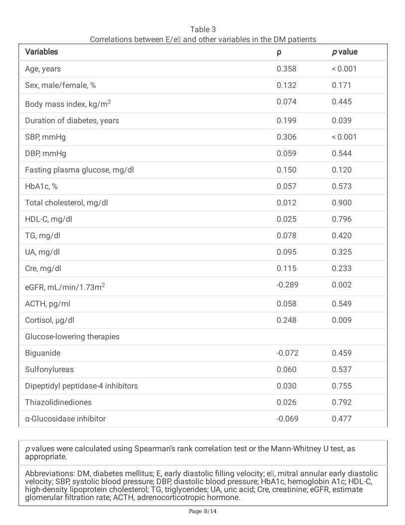
Because the E/e ratio and plasma cortisol concentrations were higher in the DM group than in non-DMgroup, we evaluated the relationships between the E/e ratio and other variables, including cortisol(Table 3). In this analysis, age (p < 0.001), duration of diabetes (p = 0.039), SBP (p < 0.001), and cortisol (p = 0.009) were positively associated with the E/e ratio, and eGFR (p = 0.002) and the use of sodiumglucose cotransporter 2 (SGLT2) inhibitors (p < 0.001) were inversely associated with the E/e ratio. Therewas no association between HbA1c and the E/e ratio. The factors that were associated with the E/eratio in the univariate analysis were included in a multivariate linear regression model. The parametersthat were independently associated with the E/e ratio in the DM group are shown in Table 4. The log ofthe E/e ratio was positively correlated with age (p = 0.017), the log of SBP (p = 0.004), and cortisol (p = 0.037) and negatively correlated with eGFR and the use of SGLT2 inhibitors (p = 0.042).
Page 8/14
Table 3Correlations between E/e and other variables in the DM patients
Variables ρ p value
Age, years 0.358 < 0.001
Sex, male/female, % 0.132 0.171
Body mass index, kg/m2 0.074 0.445
Duration of diabetes, years 0.199 0.039
SBP, mmHg 0.306 < 0.001
DBP, mmHg 0.059 0.544
Fasting plasma glucose, mg/dl 0.150 0.120
HbA1c, % 0.057 0.573
Total cholesterol, mg/dl 0.012 0.900
HDL-C, mg/dl 0.025 0.796
TG, mg/dl 0.078 0.420
UA, mg/dl 0.095 0.325
Cre, mg/dl 0.115 0.233
eGFR, mL/min/1.73m2 -0.289 0.002
ACTH, pg/ml 0.058 0.549
Cortisol, µg/dl 0.248 0.009
Glucose-lowering therapies
Biguanide -0.072 0.459
Sulfonylureas 0.060 0.537
Dipeptidyl peptidase-4 inhibitors 0.030 0.755
Thiazolidinediones 0.026 0.792
α-Glucosidase inhibitor -0.069 0.477
p values were calculated using Spearman’s rank correlation test or the Mann-Whitney U test, asappropriate.
Abbreviations: DM, diabetes mellitus; E, early diastolic �lling velocity; e , mitral annular early diastolicvelocity; SBP, systolic blood pressure; DBP, diastolic blood pressure; HbA1c, hemoglobin A1c; HDL-C,high-density lipoprotein cholesterol; TG, triglycerides; UA, uric acid; Cre, creatinine; eGFR, estimateglomerular �ltration rate; ACTH, adrenocorticotropic hormone.

Page 9/14
Variables ρ p value
Glinide -0.058 0.552
Glucagon-like peptide-1 agonists 0.061 0.526
Sodium glucose cotransporter 2 inhibitor -0.312 < 0.001
Insulin -0.004 0.967
p values were calculated using Spearman’s rank correlation test or the Mann-Whitney U test, asappropriate.
Abbreviations: DM, diabetes mellitus; E, early diastolic �lling velocity; e , mitral annular early diastolicvelocity; SBP, systolic blood pressure; DBP, diastolic blood pressure; HbA1c, hemoglobin A1c; HDL-C,high-density lipoprotein cholesterol; TG, triglycerides; UA, uric acid; Cre, creatinine; eGFR, estimateglomerular �ltration rate; ACTH, adrenocorticotropic hormone.
Table 4Multivariate linear regression analysis of factors associated with log E/e in the DM patients
Variables β p value
Age 0.22 0.017
Duration of diabetes 0.06 0.515
Log SBP 0.25 0.004
eGFR -0.21 0.016
Cortisol 0.18 0.037
Use of SGLT2 inhibitor -0.18 0.042
Abbreviation: DM, diabetes mellitus; E, early diastolic �lling velocity; e , mitral annular early diastolicvelocity; SBP, systolic blood pressure; eGFR, estimate glomerular �ltration rate; SGLT2, sodiumglucose cotransporter 2.
We examined the factors that were associated with plasma cortisol concentrations in the diabetic state.Univariate analysis showed that age (p = 0.021), fasting plasma glucose (p = 0.003), HbA1c (p = 0.008),and HDL cholesterol (p = 0.010) were positively associated with cortisol, and uric acid (p = 0.039) wasnegatively associated with cortisol (Table S1). Multivariate linear regression analysis showed that cortisolwas positively and independently correlated with the log of HbA1c (p = 0.011) and age (p = 0.016) (TableS2). These �ndings suggest that an elevated cortisol concentration in the blood is associated withdiastolic dysfunction in patients with DM. In contrast, there was no correlation between E/e and cortisolin the non-DM group (Table S3).
Discussion

Page 10/14
This study demonstrated a signi�cant correlation between the E/e ratio and cortisol, age, SBP, eGFR, andusage of SGLT2 inhibitors in patients with DM. It has been reported that the E/e ratio is linearlyassociated with invasively measured LV �lling pressures(11)(3). Therefore, the E/e ratio may serve as asurrogate measure of diastolic function.
The key �ndings of this study were that cortisol concentrations were signi�cantly higher in the DM groupthan in the non-DM group, and cortisol levels were independently and positively associated with the E/eratio in patients with DM. There was no signi�cant correlation between E/e and cortisol concentrationsin patients without DM. A previous study reported that DM patients with microangiopathy andmacroangiopathy had higher blood cortisol concentrations than DM patients without diabeticcomplications and non-DM patients.(12). It is conceivable that pathological excess of cortisol, such asthat found in Cushing’s syndrome, and mild cortisol excess found in DM plays a critical role in thedevelopment of cardiomyopathy.
Glucocorticoid receptors are abundantly expressed in the heart(13)(14). Therefore, cortisol may havedirect effects on myocardial tissue. Indeed, in an animal study using several rodent models,glucocorticoids played an important role in the development of cardiac hypertrophy and progression toheart failure(15). Additionally, the mineralocorticoid receptor (MR) is present in cardiac tissue and hashigh a�nity for both mineralocorticoids and glucocorticoids(16). Since glucocorticoids typically circulateat levels 100-fold higher than mineralocorticoids, the MR is likely to be constitutively occupied byglucocorticoids(16). In mineralocorticoid target tissues, the enzyme 11β-hydroxysteroid dehydrogenasetype 2 (11 β-HSD2) inactivates cortisol, which protects the MR from binding to glucocorticoids. Unlikeother MR target tissues, there is no appreciable dehydrogenase activity in the heart, and glucocorticoidsare free to activate the MR(16). Animal studies have shown that activation of the MR induces ventricularremodeling, hypertrophy, and �brotic changes in the heart(17)(18) Furthermore, high concentrations ofglucose stimulate protein kinase C β signaling, which leads to MR stabilization and induction of itstranscriptional activities(19). Taken together, cortisol may be involved in the development of diastolicdysfunction via activation of the MR in patients with DM. In this study, multivariate linear regressionanalysis showed that cortisol was positively correlated with HbA1c and age in the DM group. Patientswith poor glycemic control and older patients with diabetes have higher cortisol concentrations, whichmay lead to the development of LVDD.
The data from this study are consistent with previous reports that the prevalence of LVDD is associatedwith age, blood pressure, eGFR(20)(21)(22)(23). Recent studies have shown that the SGLT2 inhibitorsempagli�ozin and canagli�ozin signi�cantly reduced cardiovascular-mediated death, overall mortality,and hospitalization for heart failure in patients with type 2 DM (T2DM)(23)(24)(25). Additionally, anotherstudy reported that canagli�ozin improved LVDD(26), which is consistent with the association betweenLVDD and SGLT2 inhibitors found in this study. Taken together, these observations suggest that SGLT2inhibitors may protect against the development of HFpEF, which is characterized by LVDD, in patientswith T2DM.

Page 11/14
This study had several limitations. First, our study design was cross-sectional, and, therefore, cause-and-effect relationships could not be determined. Second, we did not collect 24-hour urine samples tomeasure cortisol, and cortisol measurements in blood may have been affected by diurnal �uctuations.Third, the number of study participants was relatively small. Future studies with larger cohorts will berequired to validate the correlations that were observed in our study.
ConclusionsThis study is the �rst demonstration of a positive correlation between cortisol and diastolic dysfunctionin patients with DM. This study facilitates a greater understanding of the pathogenesis of LVDD and mayprovide a mechanism for predicting the development of LVDD in patients with DM.
AbbreviationsDMDiabetes mellitus; LVDD:left ventricular diastolic dysfunction; HFpEF:Heart failure with preserved ejectionfraction; eGFR:estimated glomerular �ltration rate; LVEF:left ventricular ejection fractions; LVMI:leftventricular mass index; RWT:relative wall thickness; PWTd:end-diastolic posterior wall thickness;LVDd:end-diastolic left ventricular dimension; e velocity:peak early diastolic myocardial velocity;E/e :ratio of E velocity to e velocity; SBP:systolic blood pressure; HDL:high-density lipoprotein;SGLT2:sodium glucose cotransporter 2; MR:mineralocorticoid receptor; 11 β-HSD2:11β-hydroxysteroiddehydrogenase type 2; T2DM:type 2 DM.
DeclarationsAcknowledgments
The authors would also like to thank Ms. Chitose Matsuzaki for her assistance with clinicalexaminations. We thank Susan Zunino, PhD, from Edanz Group (https://en-author-services.edanz.com/ac) for editing a draft of this manuscript.
Authors’ Contributions
R.S. wrote the manuscript and researched the data. T.I. contributed to the discussion and reviewed/editedthe manuscript. C.Y., M.M., N.S., and Y.O. provided advice on the interpretation of the results. R.S. and T.I.are the guarantors of this work and, as such, had full access to all of the data in the study. R.S. and T.I.also take responsibility for the integrity of the data and the accuracy of the data analysis. All namedauthors meet the International Committee of Medical Journal Editors (ICMJE) criteria for authorship forthis manuscript, take responsibility for the integrity of the work as a whole, and have given �nal approvalto the version to be published.
Funding

Page 12/14
No funding or sponsorship was received for this study or publication of this article.
Availability of data and materials
The datasets generated and/or analyzed during this study are available from the corresponding authoron reasonable request.
Ethics approval and consent to participate
This article is compliant with all ethical guidelines and permission was obtained to conduct this studyfrom the Clinical Ethics Committee of Kyushu University Hospital (No. 29-33). All procedures conducted inthis study were in accordance with the ethical standards of the responsible committee on humanexperimentation (institutional and national) and with the Helsinki Declaration of 1964, as revised in 2013.Informed consent was obtained from all patients who were included in the study.
Consent for publication
Not applicable.
Competing interests
The authors declare that they have no competing interests.
References1. McHugh K, DeVore AD, Wu J, Matsouaka RA, Fonarow GC, Heidenreich PA, Yancy CW, Green JB,
Altman N, Hernandez AF. Heart Failure With Preserved Ejection Fraction and Diabetes: JACC State-of-the-Art Review. Journal of the American College of Cardiology 2019. doi:10.1016/j.jacc.2018.11.033.
2. Tanaka S, Hayashi T, Kihara Y, Takenaka K, Akaishi M, Ito H, Ishizuka N, Ohte N, Otsuji Y, Fukuda N,Mikami T, Mizushige K. Standard measurement of cardiac function indexes. Journal of MedicalUltrasonics 2006. doi:10.1007/s10396-006-0100-4.
3. Ommen SR, Nishimura RA, Appleton CP, Miller FA, Oh JK, Red�eld MM, Tajik AJ. Clinical utility ofDoppler echocardiography and tissue Doppler imaging in the estimation of left ventricular �llingpressures: A comparative simultaneous Doppler-catheterization study. Circulation 2000.doi:10.1161/01.CIR.102.15.1788.
4. Graversen D, Vestergaard P, Stochholm K, Gravholt CH, Jørgensen JOL. Mortality in Cushing’ssyndrome: A systematic review and meta-analysis. European Journal of Internal Medicine 2012.doi:10.1016/j.ejim.2011.10.013.
5. Lambert JK, Goldberg L, Fayngold S, Kostadinov J, Post KD, Geer EB. Predictors of mortality andlong-term outcomes in treated cushing’s disease: A study of 346 patients. Journal of ClinicalEndocrinology and Metabolism 2013. doi:10.1210/jc.2012-2893.

Page 13/14
�. Muiesan ML, Lupia M, Salvetti M, Grigoletto C, Sonino N, Boscaro M, Agabiti Rosei E, Mantero F, FalloF. Left ventricular structural andnfunctional characteristics in Cushing’s syndrome. Journal of theAmerican College of Cardiology 2003. doi:10.1016/S0735-1097(03)00493-5.
7. Park J, de Luca A, Dutton H, Malcolm JC, Doyle MA. Cardiovascular outcomes in autonomouscortisol secretion and nonfunctioning adrenal adenoma: A systematic review. Journal of theEndocrine Society 2019. doi:10.1210/js.2019-00090.
�. Debono M, Bradburn M, Bull M, Harrison B, Ross RJ, Newell-Price J. Cortisol as a marker forincreased mortality in patients with incidental adrenocortical adenomas. Journal of ClinicalEndocrinology and Metabolism 2014. doi:10.1210/jc.2014-3007.
9. Inoue T, Maeda Y, Sonoda N, Sasaki S, Kabemura T, Kobayashi K, Inoguchi T. Hyperinsulinemia andsulfonylurea use are independently associated with left ventricular diastolic dysfunction in patientswith type 2 diabetes mellitus with suboptimal blood glucose control. BMJ Open Diabetes Researchand Care 2016. doi:10.1136/bmjdrc-2016-000223.
10. Lang RM, Bierig M, Devereux RB, Flachskampf FA, Foster E, Pellikka PA, Picard MH, Roman MJ,Seward J, Shanewise JS, Solomon SD, Spencer KT, St John Sutton M, Stewart WJ.Recommendations for chamber quanti�cation: A report from the American Society ofEchocardiography’s guidelines and standards committee and the Chamber Quanti�cation WritingGroup, developed in conjunction with the European Association of Echocardiograph. Journal of theAmerican Society of Echocardiography 2005. doi:10.1016/j.echo.2005.10.005.
11. Nagueh SF, Appleton CP, Gillebert TC, Marino PN, Oh JK, Smiseth OA, Waggoner AD, Flachskampf FA,Pellikka PA, Evangelisa A. Recommendations for the evaluation of left ventricular diastolic functionby echocardiography. European Journal of Echocardiography 2009. doi:10.1093/ejechocard/jep007.
12. Chiodini I, Adda G, Scillitani A, Coletti F, Morelli V, di Lembo S, Epaminonda P, Masserini B, Beck-Peccoz P, Orsi E, Ambrosi B, Arosio M. Cortisol secretion in patients with type 2 diabetes: Relationshipwith chronic complications. Diabetes Care 2007. doi:10.2337/dc06-1267.
13. Funder JW, Duval D, Meyer P. Cardiac glucocorticoid receptors: The binding of tritiateddexamethasone in rat and dog heart. Endocrinology 1973. doi:10.1210/endo-93-6-1300.
14. Sylvén C, Jansson E, Sotonyi P, Waagstein F, Barkhem T, Brönnegård M. Cardiac nuclear hormonereceptor mrNA in heart failure in man. Life Sciences 1996. doi:10.1016/S0024-3205(96)00539-5.
15. Ohtani T, Mano T, Hikoso S, Sakata Y, Nishio M, Takeda Y, Otsu K, Miwa T, Masuyama T, Hori M,Yamamoto K. Cardiac steroidogenesis and glucocorticoid in the development of cardiac hypertrophyduring the progression to heart failure. Journal of Hypertension 2009.doi:10.1097/HJH.0b013e328326cb04.
1�. Gray GA, White CI, Castellan RFP, McSweeney SJ, Chapman KE. Getting to the heart of intracellularglucocorticoid regeneration: 11β-HSD1 in the myocardium. Journal of Molecular Endocrinology2017. doi:10.1530/JME-16-0128.
17. Bauersachs J, Jaisser F, Toto R. Mineralocorticoid receptor activation and mineralocorticoid receptorantagonist treatment in cardiac and renal diseases. Hypertension 2015.

Page 14/14
doi:10.1161/HYPERTENSIONAHA.114.04488.
1�. Funder JW. Is aldosterone bad for the heart? Trends in Endocrinology and Metabolism 2004.doi:10.1016/j.tem.2004.03.006.
19. Hayashi T, Shibata H, Kurihara I, Yokota K, Mitsuishi Y, Ohashi K, Murai-Takeda A, Jo R, Ohyama T,Sakamoto M, Tojo K, Tajima N, Utsunomiya K, Itoh H. High glucose stimulates mineralocorticoidreceptor transcriptional activity through the protein kinase C β signaling. International Heart Journal2017. doi:10.1536/ihj.16-649.
20. Miyatake K, Okamoto M, Kinoshita N, Owa M, Nakasone I, Sakakibara H, Nimura Y. Augmentation ofatrial contribution to left ventricular in�ow with aging as assessed by intracardiac doppler �owmetry.The American Journal of Cardiology 1984. doi:10.1016/0002-9149(84)90035-3.
21. Owan TE, Hodge DO, Herges RM, Jacobsen SJ, Roger VL, Red�eld MM. Trends in Prevalence andOutcome of Heart Failure with Preserved Ejection Fraction. New England Journal of Medicine 2006.doi:10.1056/nejmoa052256.
22. Otsuka T, Suzuki M, Yoshikawa H, Sugi K. Left ventricular diastolic dysfunction in the early stage ofchronic kidney disease. Journal of Cardiology 2009. doi:10.1016/j.jjcc.2009.05.002.
23. Steiner S. Empagli�ozin, cardiovascular outcomes, and mortality in type 2 diabetes. Zeitschrift furGefassmedizin 2016. doi:10.1056/nejmoa1504720.
24. S G, A A, J B, T P, H W, J W. Heart failure hospitalization risk associated with use of two classes oforal antidiabetic medications: an observational, real-world analysis. Cardiovascular diabetology2017.
25. Neal B, Perkovic V, Mahaffey KW, de Zeeuw D, Fulcher G, Erondu N, Shaw W, Law G, Desai M,Matthews DR. Canagli�ozin and Cardiovascular and Renal Events in Type 2 Diabetes. New EnglandJournal of Medicine 2017. doi:10.1056/nejmoa1611925.
2�. Matsutani D, Sakamoto M, Kayama Y, Takeda N, Horiuchi R, Utsunomiya K. Effect of canagli�ozin onleft ventricular diastolic function in patients with type 2 diabetes. Cardiovascular Diabetology 2018.doi:10.1186/s12933-018-0717-9.
Supplementary Files
This is a list of supplementary �les associated with this preprint. Click to download.
renamed4da68.docx