mixed tumor of the soft tissue (arm) - core.ac.uk · cavity. no skin tumor was found. the patient...
TRANSCRIPT
www.humanpathologycasereports.com
Human Pathology: Case Reports (2014) 1, 52–57
Mixed tumor of the soft tissue (arm)
Tadashi Terada MD, PhD⁎Department of Pathology, Shizuoka City Shimizu Hospital, Miyakami 1231 Shimizu-Ku, Shizuoka 424–8636, Japan
Received 28 June 2014; revised 4 September 2014; accepted 18 September 2014
h2
Keywords:Soft tissue;Mixed tumor;Histopathology;Immunohistochemistry
Abstract Mixed tumor of soft tissue is extremely rare; only 15 cases have been reported in Englishliterature. A 56-year-old man presented with a tumor in right arm. Physical examination showed atumor measuring 3 × 3 × 3 cm of deep portion of right arm. MRI showed a well-defined deeptumor measuring 3 × 3 × 4 cm of arm next to fascia. The tumor was resected, and it was foundthat the tumor is located in deep subcutaneous tissue outside the fascia. Histologically, the tumorwas well-defined and contained a fibrous capsule. The tumor cells showed no significant nuclearatypia. The tumor was composed of epithelial, myoepithelial, and stromal elements. The epithelialelement formed tubules, and myoepithelial element showed solid nests and periepithelial linings.The epithelial element showed frequent apocrine features including apocrine snouts. The stromalelement was composed of cartilage and myxoid tissues. Immunohistochemically, the epithelialelement was positive for cytokeratin (CK) AE1/3, CK CAM5.2, CK34BE12, CK7, CK8, CK18,CK19, EMA, CEA, CD56, p53, and Ki-67 (labeling = 80%), but negative for CK5/6, CK14,CK20, vimentin, p63, S100 protein, CD10, CD34, ASMA, CD68, myoglobin, bcl-2, BMP-2, TGF-β1, CDK-4, and MDM2. The myoepithelial element was positive for CK AE1/3, CK CAM5.2,CK34BE12, CK5/6, CK7, CK8, CK14, CK18, p63, vimentin, CD10, S100 protein, bcl-2, p53, andKi-67 (labeling = 20%), but negative for CK19, CK20, EMA, CEA, CD34, ASMA, CD68, CD56,myoglobin, BMP-2, TGF-β1, CDK-4 and MDM2. The stromal element was positive for vimentin,S100 protein (cartilage), and Ki-67 (labeling = 6%), but negative for other antigens examinedincluding bcl-2, BMP-2, TGF-β1, CDK-4 and MDM2. No findings of metastasis, invasion, andrecurrence were recognized clinically. The author speculates that the current mixed tumor wasderived from misplaced apocrine glands in the deep soft tissue.
© 2014 The Author. Published by Elsevier Inc. Open access under CC BY-NC-ND license.
1. Introduction usually designated as pleomorphic adenoma or mixed tumor
Mixed tumor of the soft tissue is extremely rare; only 15cases have been reported [1–4], to the best of author'sknowledge. Mixed tumor is most common in major andminor salivary glands, and mixed tumor of these locations is
⁎ Tel.: +81 54 336 1111; fax: +81 54 336 1315.E-mail address: [email protected].
ttp://dx.doi.org/10.1016/j.ehpc.2014.09.004214-3300/© 2014 TheAuthor. Published by Elsevier Inc. Open access under CC B
[5]. Mixed tumor is also seen in the skin, and such cutaneousmixed tumor is usually called chondroid syringoma [6].Mixed tumor is extremely rare in the soft tissue [1–4,7].According to WHO blue book [7], mixed tumor of softtissue is included in a group of mixed tumor/myoepithe-lioma/parachordoma. Mixed tumor is composed of epithe-lial, myoepithelial, and mesenchymal cells which oftenshow cartilage, and shows pleomorphic diverse features of
Y-NC-ND license.

53Mixed tumor of soft tissue
histology [1–7]. According to WHO blue book [7], in thesection of mixed tumor/myoepithelioma/parachordomagroup of soft tissue, mixed tumor is used for tumor havingepithelial tubular element, and myoepithelioma for tumorlacking epithelial tubular element [7]. Parachordoma is avague entity, and should be included in mixed tumor ormyoepithelioma [7]. Thus, mixed tumor and myoepithe-lioma are different histological entities [7], although thehistology is mutually similar. There has been only one studyof soft tissue mixed tumor with immunohistochemicalstudy, which was performed by Kilpatrick et al. [1]. Hereinreported is a case of mixed tumor of the soft tissue of arm.An extensive immunohistochemical study was performed inthis case report.
2. Case report
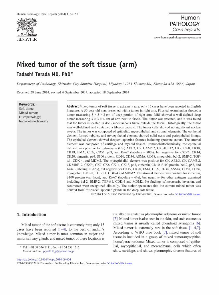
A 56-year-old man noticed a tumor in right arm, andconsulted to our hospital. Physical examination showed adeep tumor measuring 3 × 3 × 3 cm in the right arm. Noother skin tumor was found. MRI showed a well-defineddeep tumor measuring 3 × 3 × 4 cm of the arm next tofascia (Fig. 1). The resonance density of the tumor isheterogenous (Fig. 1). The tumor was excised. During theoperation, it was found that the tumor was located in deepsubcutaneous tissue next to fascia. It was not within thestriated muscles. No tumor was found in the overlying skinand upper subcutaneous tissue.
Fig. 1 MRI findings of the right arm tumor. MRI showed awell-defined deep tumor measuring 3 × 3 × 4 cm of the armnext to fascia. The density of the tumor is heterogenous.
Histologically, the tumor was well-defined and con-tained a fibrous capsule (Fig. 2A). The resected sectionincluded the epidermis and dermis, and the distancebetween superficial part of the tumor and superficialepidermis was 12 mm. The tumor cells showed nosignificant nuclear atypia. The tumor showed pleomorphicdiverse histologies, but basically the tumor was composedof epithelial, myoepithelial, and stromal elements (Fig. 2Aand B). The epithelial element formed tubules (Fig. 2C),and myoepithelial element showed solid nests (Fig. 2D)and periepithelial linings (Fig. 2C). The epithelial elementshowed frequent apocrine features including apocrinesnouts and ample acidophilic cytoplasm (Fig. 2E). Someareas of the myoepithelial element were composed ofmyoepithelial cells with clear cytoplasm. The stromalelement was composed of cartilage (Fig. 2F) and myxoidtissue. There were gradual transitions between themyoepithelial element and stromal element. No apparentcarcinoma areas were seen.
An immunohistochemical analysis was performed bythe Envision method (Dako Corp, Glostrup, Denmark), aspreviously reported [8–13]. The results are shown inTable 1. Immunohistochemically, the epithelial elementwas positive for cytokeratin (CK) AE1/3, CK CAM5.2(Fig. 3A), CK34BE12, CK7 (Fig. 3B), CK8, CK18(Fig. 3C), CK19, epithelial membrane antigen (EMA)(Fig. 3D), carcinoembryonic antigen (CEA) (Fig. 3E),CD56 (Fig. 3F), p53, and Ki-67 (labeling = 80%)(Fig. 4A), but negative for CK5/6 (Fig. 4B), CK14(Fig. 4C), CK20, vimentin (Fig. 4D), p63 (Fig. 4E), S100protein (Fig. 4F), CD10, CD34, α-smooth muscle antigen(ASMA), CD68, myoglobin, bcl-2, bone morphogenicprotein-2 (BMP-2), transforming growth factor-β1(TGF-β1), cyclin dependent kinase (CDK-4), andMDM2. The myoepithelial element was positive for CKAE1/3, CK CAM5.2 (Fig. 3A), CK34BE12, CK5/6(Fig. 4B), CK7 (Fig. 3B), CK8, CK14 (Fig. 4C), CK18(Fig. 3C), p63 (Fig. 4E), vimentin (Fig. 4D), S100 protein(Fig. 4F), CD10, bcl-2, p53, and Ki-67 (labeling = 20%),but negative for CK19, CK20, EMA (Fig. 3D), CEA(Fig. 3E), CD34,ASMA,CD68,CD56,myoglobin, BMP-2,TGF-β1, CDK-4 and MDM2. The stromal element waspositive for vimentin, S100 protein (cartilage), and Ki-67(labeling = 6%), but negative for any types of CK, EMA,p63, CD10, CD34, CD68, p53, ASMA, CD56, myoglo-bin, bcl-2, BMP-2, TGF-β1, CDK-4 and MDM2. Thestromal element was positive for vimentin, S100 protein(cartilage), and Ki-67 (labeling = 6%), but negative forany types of CK, EMA, p63, CD10, CD34, CD68, p53,ASMA, CD56, myoglobin, bcl-2, BMP-2, TGF-β1,CDK-4 and MDM2.
No findings of metastasis, invasion or recurrence werenoted clinically. Clinical examination identified no tumors inhead and neck regions including the salivary glands and oral
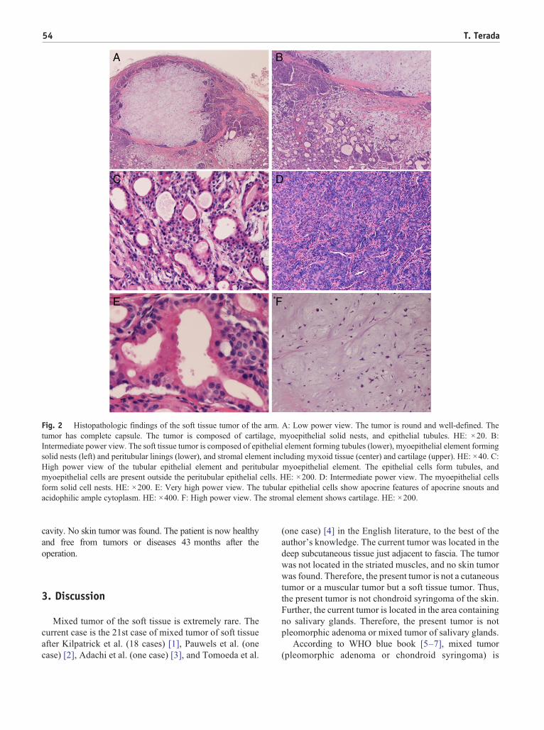
Fig. 2 Histopathologic findings of the soft tissue tumor of the arm. A: Low power view. The tumor is round and well-defined. Thetumor has complete capsule. The tumor is composed of cartilage, myoepithelial solid nests, and epithelial tubules. HE: ×20. B:Intermediate power view. The soft tissue tumor is composed of epithelial element forming tubules (lower), myoepithelial element formingsolid nests (left) and peritubular linings (lower), and stromal element including myxoid tissue (center) and cartilage (upper). HE: ×40. C:High power view of the tubular epithelial element and peritubular myoepithelial element. The epithelial cells form tubules, andmyoepithelial cells are present outside the peritubular epithelial cells. HE: ×200. D: Intermediate power view. The myoepithelial cellsform solid cell nests. HE: ×200. E: Very high power view. The tubular epithelial cells show apocrine features of apocrine snouts andacidophilic ample cytoplasm. HE: ×400. F: High power view. The stromal element shows cartilage. HE: ×200.
54 T. Terada
cavity. No skin tumor was found. The patient is now healthyand free from tumors or diseases 43 months after theoperation.
3. Discussion
Mixed tumor of the soft tissue is extremely rare. Thecurrent case is the 21st case of mixed tumor of soft tissueafter Kilpatrick et al. (18 cases) [1], Pauwels et al. (onecase) [2], Adachi et al. (one case) [3], and Tomoeda et al.
(one case) [4] in the English literature, to the best of theauthor's knowledge. The current tumor was located in thedeep subcutaneous tissue just adjacent to fascia. The tumorwas not located in the striated muscles, and no skin tumorwas found. Therefore, the present tumor is not a cutaneoustumor or a muscular tumor but a soft tissue tumor. Thus,the present tumor is not chondroid syringoma of the skin.Further, the current tumor is located in the area containingno salivary glands. Therefore, the present tumor is notpleomorphic adenoma or mixed tumor of salivary glands.
According to WHO blue book [5–7], mixed tumor(pleomorphic adenoma or chondroid syringoma) is
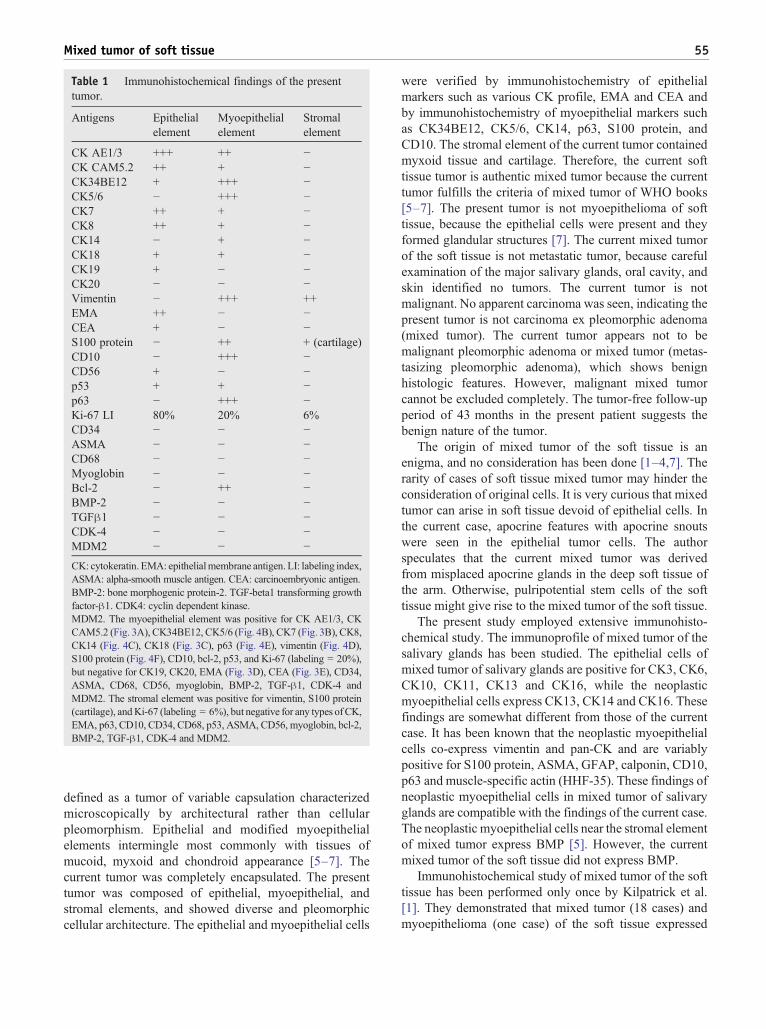
Table 1 Immunohistochemical findings of the presenttumor.
Antigens Epithelialelement
Myoepithelialelement
Stromalelement
CK AE1/3 +++ ++ −CK CAM5.2 ++ + −CK34BE12 + +++ −CK5/6 − +++ −CK7 ++ + −CK8 ++ + −CK14 − + −CK18 + + −CK19 + − −CK20 − − −Vimentin − +++ ++EMA ++ − −CEA + − −S100 protein − ++ + (cartilage)CD10 − +++ −CD56 + − −p53 + + −p63 − +++ −Ki-67 LI 80% 20% 6%CD34 − − −ASMA − − −CD68 − − −Myoglobin − − −Bcl-2 − ++ −BMP-2 − − −TGFβ1 − − −CDK-4 − − −MDM2 − − −
CK: cytokeratin. EMA: epithelialmembrane antigen. LI: labeling index,ASMA: alpha-smooth muscle antigen. CEA: carcinoembryonic antigen.BMP-2: bone morphogenic protein-2. TGF-beta1 transforming growthfactor-β1. CDK4: cyclin dependent kinase.MDM2. The myoepithelial element was positive for CK AE1/3, CKCAM5.2 (Fig. 3A), CK34BE12, CK5/6 (Fig. 4B), CK7 (Fig. 3B), CK8,CK14 (Fig. 4C), CK18 (Fig. 3C), p63 (Fig. 4E), vimentin (Fig. 4D),S100 protein (Fig. 4F), CD10, bcl-2, p53, and Ki-67 (labeling = 20%),but negative for CK19, CK20, EMA (Fig. 3D), CEA (Fig. 3E), CD34,ASMA, CD68, CD56, myoglobin, BMP-2, TGF-β1, CDK-4 andMDM2. The stromal element was positive for vimentin, S100 protein(cartilage), andKi-67 (labeling = 6%), but negative for any types of CK,EMA, p63, CD10, CD34, CD68, p53, ASMA, CD56, myoglobin, bcl-2,BMP-2, TGF-β1, CDK-4 and MDM2.
55Mixed tumor of soft tissue
defined as a tumor of variable capsulation characterizedmicroscopically by architectural rather than cellularpleomorphism. Epithelial and modified myoepithelialelements intermingle most commonly with tissues ofmucoid, myxoid and chondroid appearance [5–7]. Thecurrent tumor was completely encapsulated. The presenttumor was composed of epithelial, myoepithelial, andstromal elements, and showed diverse and pleomorphiccellular architecture. The epithelial and myoepithelial cells
were verified by immunohistochemistry of epithelialmarkers such as various CK profile, EMA and CEA andby immunohistochemistry of myoepithelial markers suchas CK34BE12, CK5/6, CK14, p63, S100 protein, andCD10. The stromal element of the current tumor containedmyxoid tissue and cartilage. Therefore, the current softtissue tumor is authentic mixed tumor because the currenttumor fulfills the criteria of mixed tumor of WHO books[5–7]. The present tumor is not myoepithelioma of softtissue, because the epithelial cells were present and theyformed glandular structures [7]. The current mixed tumorof the soft tissue is not metastatic tumor, because carefulexamination of the major salivary glands, oral cavity, andskin identified no tumors. The current tumor is notmalignant. No apparent carcinoma was seen, indicating thepresent tumor is not carcinoma ex pleomorphic adenoma(mixed tumor). The current tumor appears not to bemalignant pleomorphic adenoma or mixed tumor (metas-tasizing pleomorphic adenoma), which shows benignhistologic features. However, malignant mixed tumorcannot be excluded completely. The tumor-free follow-upperiod of 43 months in the present patient suggests thebenign nature of the tumor.
The origin of mixed tumor of the soft tissue is anenigma, and no consideration has been done [1–4,7]. Therarity of cases of soft tissue mixed tumor may hinder theconsideration of original cells. It is very curious that mixedtumor can arise in soft tissue devoid of epithelial cells. Inthe current case, apocrine features with apocrine snoutswere seen in the epithelial tumor cells. The authorspeculates that the current mixed tumor was derivedfrom misplaced apocrine glands in the deep soft tissue ofthe arm. Otherwise, pulripotential stem cells of the softtissue might give rise to the mixed tumor of the soft tissue.
The present study employed extensive immunohisto-chemical study. The immunoprofile of mixed tumor of thesalivary glands has been studied. The epithelial cells ofmixed tumor of salivary glands are positive for CK3, CK6,CK10, CK11, CK13 and CK16, while the neoplasticmyoepithelial cells express CK13, CK14 and CK16. Thesefindings are somewhat different from those of the currentcase. It has been known that the neoplastic myoepithelialcells co-express vimentin and pan-CK and are variablypositive for S100 protein, ASMA, GFAP, calponin, CD10,p63 and muscle-specific actin (HHF-35). These findings ofneoplastic myoepithelial cells in mixed tumor of salivaryglands are compatible with the findings of the current case.The neoplastic myoepithelial cells near the stromal elementof mixed tumor express BMP [5]. However, the currentmixed tumor of the soft tissue did not express BMP.
Immunohistochemical study of mixed tumor of the softtissue has been performed only once by Kilpatrick et al.[1]. They demonstrated that mixed tumor (18 cases) andmyoepithelioma (one case) of the soft tissue expressed

Fig. 3 Immunohistochemical findings. Cytokeratin (CK) CAM5.2 (A), CK7 (B), and CK18 (C) are expressed in both epithelial cellsand myoepithelial cells. EMA (D), CEA (E), and CD56 (F) are expressed in epithelial cells but not in myoepithelial cells. ×200.
56 T. Terada
pan-CK in 16/16, S100 protein in 16/17, ASMA in 6/14,muscle specific actin (HHF-35) in 2/10, desmin in 2/10,GFAP in 3/11, and EMA in 3/16. Immunohistochemicalresult of the present case (Table 1) is basically similar tothe data of Kilpatrick et al. [1]. The present case of mixedtumor of the soft tissue revealed the CK immunoprofile inthe epithelial and myoepithelial cells. Of interest is thatCD5/6, CK34BE12, and CK14 were expressed in themyoepithelial cells but not in the epithelial cells. Thesetypes of CK are known to be expressed in myoepithelialcells. CD10 expression showed a similar pattern; CD10 iswell known to be expressed in myoepithelial cells [1,10].
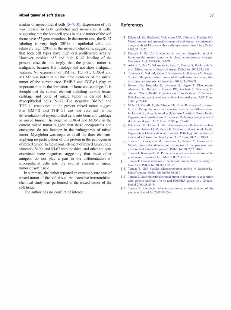
Fig. 4 Immunohistochemical expressions. Ki-67 labeling is high in epitCK20, vimentin (D), p63 (E) and S100 protein (F) are expressed in myoe
Vimentin, S100 protein and p63 labeled myoepithelialcells but not epithelial cells, being compatible with otherstudy [1,5–7,10]. EMA and CEA were expressed inepithelial cells but not in myoepithelial cells. These arenew findings. Expression of bcl-2 was seen in myoepithelialcells but not in epithelial cells, suggesting that bcl-2may be amarker of myoepithelial cells. This is a novel finding. CD56(NCAM) expression was seen in epithelial cells but not inmyoepithelial cells, suggesting that epithelial cells of mixedtumor of the soft tissue show neuroendocrine features. In thepresent case, no ASMA expression is seen in both epithelialcells and myoepithelial cells, although ASMA is a good
helial cells (labeling = 80%) (A). In contrast, CK5/6 (B), CK14 (C),pithelial cells, but not in epithelial cells. ×200.
57Mixed tumor of soft tissue
marker of myoepithelial cells [5–7,10]. Expression of p53was present in both epithelial and myoepithelial cells,suggesting that the both cell types in mixed tumor of the softtissue have p53 gene mutations. In the current case, the Ki-67labeling is very high (80%) in epithelial cells andrelatively high (20%) in the myoepithelial cells, suggestingthat both cell types have high cell proliferative activity.However, positive p53 and high Ki-67 labeling of thepresent case do not imply that the present tumor ismalignant, because HE histology did not show malignantfeatures. No expression of BMP-2, TGF-β1, CDK-4 andMDM2 was noted in all the three elements of the mixedtumor of the current case. BMP-2 and TGF-β1 play animportant role in the formation of bone and cartilage. It isthought that the stromal element including myxoid tissue,cartilage and bone of mixed tumor is derived frommyoepithelial cells [5–7]. The negative BMP-2 andTGF-β1 reactivities in the present mixed tumor suggestthat BMP-2 and TGF-β1 are not essential in thedifferentiation of myoepithelial cells into bone and cartilagein mixed tumor. The negative CDK-4 and MDM2 in thecurrent mixed tumor suggest that these oncoproteins andoncogenes do not function in the pathogenesis of mixedtumor. Myoglobin was negative in all the three elements,implying no participation of this protein in the pathogenesisof mixed tumor. In the stromal element of mixed tumor, onlyvimentin, S100, and Ki-67 were positive, and other antigensexamined were negative, suggesting that these otherantigens do not play a part in the differentiation ofmyoepithelial cells into the stromal element in mixedtumor of soft tissue.
In summary, the author reported an extremely rare case ofmixed tumor of the soft tissue. An extensive immunohisto-chemical study was performed in the mixed tumor of thesoft tissue.
The author has no conflict of interest.
References
[1] Kilpatrick SE, Hitchcock MG, Kraus MD, Calonje E, Fletcher CD.Mixed tumors and myoepitheliomas of soft tissue: a clinicopath-ologic study of 19 cases with a unifying concept. Am J Surg Pathol1997;21:13-22.
[2] Pauwels P, Dal Cin P, Roumen R, van den Berghe H, Sciot R.Intramuscular mixed tumor with clonal chromosomal changes.Virchows Arch 1999;434:167-71.
[3] Adachi T, Oda Y, Sakamoto A, Saito T, Tamiya S, Hachitanda Y,et al. Mixed tumor of deep soft tissue. Pathol Int 2003;53:35-9.
[4] Tomoeda M, Yuki M, Kubo C, Yoshizawa H, Kitamura M, NagatoS, et al. Malignant mixed tumor of the soft tissue occurring aftertotal knee arthroplasty. Orthopedics 2011;34:e768-71.
[5] Eveson JW, Kusafuka K, Stenman G, Nagao T. Pleomorphicadenoma. In: Barnes L, Eveson JW, Reichart P, Sidransky D,editors. World Health Organization Classification of Tumours.Pathology and genetics of head and neck tumorsLyon: IARC Press;2005. p. 254-8.
[6] McNiff J, Vassallo C,McCalmont TH, Rosso R, Requena L, BorroniG, et al. Benign tumours with apocrine and eccrine differentiation.In: LeBoit PE, Burg G, Weedon D, Sarasin A, editors. World HealthOrganization Classification of Tumours. Pathology and genetics ofskin tumorsLyon: IARC Press; 2006. p. 139-48.
[7] Kilpatrick SE, Limon J. Mixed tumour/myoepithelioma/parachor-doma. In: Fletcher CDM, Unni KK, Mertens F, editors. World HealthOrganization Classification of Tumours. Pathology and genetics oftumors of soft tissue and boneLyon: IARC Press; 2002. p. 198-9.
[8] Terada T, Kawaguchi M, Furukawa K, Sekido Y, Osamura Y.Minute mixed ductal-endocrine carcinoma of the pancreas withpredominant intraductal growth. Pathol Int 2002;52:740-6.
[9] Terada T, Kawaguchi M. Primary clear cell adenocarcinoma of theperitoneum. Tohoku J Exp Med 2005;271:271-5.
[10] Terada T. Ductal adenoma of the breast: immunohistochemistry oftwo cases. Pathol Int 2008;58:801-5.
[11] Terada T. Gall bladder adenocarcinoma arising in Rokitansky-Schoff sinuses. Pathol Int 2008;58:806-9.
[12] Terada T. Gastrointestinal stromal tumor of the uterus: A case reportwith genetic analyses of c-kit and PDGFRA genes. Int J GynecolPathol 2009;28:29-34.
[13] Terada T. Intraductal tubular carcinoma, intestinal type, of thepancreas. Pathol Int 2009;59:53-8.