molecular signaling of the hmgb1/rage axis contributes to cholesteatoma ... · original article...
TRANSCRIPT
ORIGINAL ARTICLE
Molecular signaling of the HMGB1/RAGE axis contributesto cholesteatoma pathogenesis
Miroslaw J. Szczepanski & Michal Luczak & Ewa Olszewska & Marta Molinska-Glura &
Mariola Zagor & Antoni Krzeski & Henryk Skarzynski & Jan Misiak & Karolina Dzaman &
Mikolaj Bilusiak & Tomasz Kopec & Malgorzata Leszczynska & Henryk Witmanowski &Theresa L. Whiteside
Received: 21 January 2014 /Revised: 25 September 2014 /Accepted: 1 October 2014 /Published online: 12 November 2014# The Author(s) 2014. This article is published with open access at Springerlink.com
AbstractCholesteatoma represents progressive expansion of thekeratinizing squamous epithelium in the middle ear withsubsequent chronic inflammation in subepithelial connectivetissues. The hypothesis was tested that receptor for advancedglycation endproduct (RAGE) and its ligand, high-mobilitybox 1 (HMGB1), are overexpressed in cholesteatoma, and theRAGE/HMGB1 axis might contribute to its pathogenesis.Cholesteatoma samples (n=36) and 27 normal skin specimenswere studied by immunohistochemistry (IHC) for HMGB1and RAGE expression. Effects of HMGB1 signaling on pro-liferation, migration, cytokine production, and apoptosis ofhuman immortalized keratinocytes (HaCaTs) and normal
keratinocytes were studied by quantitative reverse transcrip-tion (qRT)-PCR, IHC,Western blots, and flow cytometry aftercell co-incubation with HMGB1. While all studied tissuesexpressed HMGB1, its expression was higher incholesteatoma than in normal skin (p<0.0001). All cases ofcholesteatoma also showed elevated RAGE expression levels,and only 7/27 (26 %) of normal skin specimens were weaklypositive for RAGE. Proliferation and migration of HaCaTcells incubated with HMGB1 were up-regulated (p<0.05).HMGB1 also prevented HaCaT cell apoptosis and inducedactivation of several molecular signaling pathways inkeratinocytes. The data suggest that in cholesteatoma,HMGB1 released from stressed or necrotic epithelial cells
Electronic supplementary material The online version of this article(doi:10.1007/s00109-014-1217-3) contains supplementary material,which is available to authorized users.
M. J. Szczepanski :M. Zagor :A. Krzeski :K. DzamanDepartment of Otorhinolaryngology, Faculty of Medicine andDentistry, Medical University of Warsaw, Warsaw, Poland
M. J. Szczepanski : J. MisiakDepartment of Clinical Immunology, University of MedicalSciences, Poznan, Poland
M. Molinska-GluraDepartment of Computer Science and Statistics, University ofMedical Sciences, Poznan, Poland
M. Bilusiak : T. Kopec :M. LeszczynskaDepartment of Otolaryngology, University of Medical Sciences,Poznan, Poland
E. OlszewskaDepartment of Otolaryngology, Medical University of Bialystok,Bialystok, Poland
M. LuczakDepartment of Pathology and Laboratory Medicine, BrownUniversity, Providence, RI 02912, USA
H. SkarzynskiInstitute of Physiology and Pathology of Hearing, Warsaw, Poland
H. WitmanowskiDepartment of Physiology, University of Medical Sciences, Poznan,Poland
T. L. Whiteside (*)Department of Pathology, University of Pittsburgh School ofMedicine, University of Pittsburgh Cancer Institute, 5117 CentreAvenue, Pittsburgh, PA 15213, USAe-mail: [email protected]
J Mol Med (2015) 93:305–314DOI 10.1007/s00109-014-1217-3
and binding to RAGE overexpressed in keratinocytes initiatesmolecular signaling that culminates in pro-inflammatory cy-tokine release and chronic inflammation.
Key message& HMGB1 signaling engages multiple activation pathways
in RAGE-positive keratinocytes.& HMGB1 protects RAGE-positive keratinocytes from
drug-induced apoptosis.& Keratinocyte proliferation is controlled via RAGE and
HMGB1 molecular signaling.& Molecular signaling of the HMGB1/RAGE axis contrib-
utes to cholesteatoma pathogenesis.
Keywords RAGE/HMGB1 axis . Keratinocytes .
Cholesteatoma pathogenesis . Inflammation
Introduction
The immune system has evolved to respond not only topathogens, but also to signals released from dying cells.Sensing the presence of pathogens provides an initial signalfor the immune system to respond to the invader [1, 2]. Thisprocess requires the presence of pattern recognition receptors(PRRs) that detect pathogen-associated molecular patterns(PAMPs). The best studied of these receptors are Toll-likereceptors (TLRs). The immune system is also able to recog-nize endogenous danger signals or damage-associated molec-ular patterns (DAMPs), which involve molecules released bynecrotic or dying cells [3]. When cells die through necrosis,they induce several molecular pathways. In contrast, apoptoticcell death induces tolerance [4]. The DAMP superfamilyincludes various breakdown products of the extracellular ma-trix, e.g., the DNA-binding high-mobility box 1 (HMGB1)protein which has been widely studied [5]. HMGB1 plays acrucial role in molecular responses to cell damage/necrosis viaactivation of the nuclear factor kappa B (NF-κB) transcriptionfactor and subsequent induction of pro-inflammatory cyto-kines, interleukin (IL)-1 and tumor necrosis factor alpha(TNF-α) [6]. The receptor for advanced glycationendproducts (RAGEs) is the major receptor for DAMPs [4].RAGE activation via its multiple ligands, including HMGB1,plays a key role in various diseases, including acute or chronicinflammatory conditions such as sepsis and diseases such asrheumatoid arthritis, diabetic nephropathy, and cancer [6–9].
Cholesteatoma is a destructive disease characterized by theprogressive expansion of keratinizing squamous epithelium inthe middle ear and mastoid and chronic inflammatory reactionof the subepithelial connective tissue which is accompaniedby bone destruction with intracranial extensions. Many theo-ries of its pathogenesis have been proposed but failed toprovide insights into the molecular background of disease,
prevention, or cure. Cholesteatoma tissue is composed ofproliferating, migrating, and highly keratinizing epitheliumknown as the matrix and of subepithelial connective tissueinfiltrated by inflammatory cells, antigen-presenting cells,fibroblasts, and endothelial cells and known as the perimatrix[10, 11]. Cholesteatoma tissue is also the source of pro-inflammatory cytokines such as IL-1, transforming growthfactor alpha (TGF-α), pro-angiogenic factors, and variousenzymes [12–15]. It is suspected but not proven that thismix of soluble factors activates osteoclasts, which are engagedin bone destruction [10].
Here, we report for the first time the co-expression ofHMGB1 and RAGE in cholesteatoma tissues and hypothesizethat their molecular interaction resulting in activation of sev-eral major intracellular signaling pathwaysmay be responsiblefor cellular hyperplasia, cell migration, and resistance to apo-ptosis characteristic of cholesteatoma. Although a Mongoliangerbil animal model of cholesteatoma has been described [16],concerns exist about its relevance to human disease [17].Since we have not been able to establish an in vivocholesteatoma model, we have resorted to a series of molec-ular in vitro studies with human keratinocytes to investigatethe potential role of HMGB1 in the cholesteatoma pathogen-esis. The results of our ex vivo studies were correlatedwith theexpression in human cholesteatoma tissues of the proteinsshown to be involved in keratinocyte activation and prolifer-ation. Based on our ex vivo and in situ studies, we proposethat high RAGE expression levels in cholesteatoma tissuesmight be the key factor contributing to the diseaseprogression.
Materials and methods
Patients Thirty-six tissue samples were obtained from 15females and 21 males diagnosed with acquired middle earcholesteatoma who underwent surgery for the first time. Thepatients’ ages ranged from 17 to 78 years (median=48). Thepresence of pathologically documented middle earcholesteatoma in surgically removed tissues was required forentry into the study. Tissues were fixed in 10% formaldehyde,and sections stained with hematoxylin–eosin (H + E) wereprepared and evaluated by light microscopy. Control tissuesincluded noninflammatory skin specimens from the externalauditory canal (n=9) or from the retroauricular area (n=18).Seven fresh middle ear cholesteatoma tissue samples andseven control tissue samples were used for protein extraction.The study was approved by the Local Ethics Committees atthe Poznan University of Medical Sciences (#290/04 to MJS)and at the Medical University of Bialystok (#R-I-002/89/2010and #R-I-002/125/2011 to EO) and was conducted as recom-mended by the Helsinki Declaration.
306 J Mol Med (2015) 93:305–314
Cell cultures Normal human keratinocyte cell line humanimmortalized keratinocyte (HaCaT) and Normal Adult Hu-man Primary Epidermal Kerat inocytes (ATCC®PCS200011™) were purchased from CLS Cell Lines ServiceGmbH (Eppelheim, Germany) and from ATCC, respectively.HaCaTcells were maintained in RPMI-1640medium (Sigma-Aldrich Chemie GmbH, Steinheim, Germany) containing10 % (v/v) heat-inactivated fetal calf serum (FCS), 2 mMglutamine, 100 mg/mL streptomycin, and 100 U/mL penicil-lin. Normal Adult Human Primary Epidermal Keratinocyteswere maintained in serum-free Dermal Cell Basal Medium(ATCC® PCS200030) supplemented with components of thekeratinocyte growth kit (ATCC® PCS200040) that containsthe following growth supplements: bovine pituitary extract(BPE), rhTGF-α, L-glutamine, hydrocortisone hemisuccinate,insulin, epinephrine, and apotransferrin. Both cell lines weregrown at 37 °C in a 5 % CO2 humidified atmosphere. Cells inconfluence were harvested, washed twice with phosphate-buffered saline (PBS), and detached from culture flasks by abrief treatment with 0.02 % EDTA solution (Sigma-AldrichChemie GmbH).
Reverse transcription and real-time quantitative PCR analysisof receptor for advanced glycation endproduct and Toll-likereceptor 4 transcripts The primers used for RAGE amplifica-tion were F=5′-GAAGGGAGTATCTGTGAAGGAA-3′ andR=5′-AGCTACAGGAGAAGGTGGGA-3′; for TLR4 am-plification, F=5′-ACAACCTCCCCTTCTCAACCA-3′ andR=5′-GTGGCTTAGGCTCTGATATGC-3′; and for PBGD(a housekeeping gene) amplification, F=5′-GCCAAGGACCAGGACATC-3′ and R=5′-TCAGGTACAGTTGCCCATC-3′. The quantitative real-time reverse transcription (RT)-PCRwas performed as previously described [18].
Flow cytometryABecton Dickinson flow cytometer equippedwith FACSDiva v6.1.2 software was used to evaluate surfaceRAGE expression in cells. To evaluate intracellular HMGB1expression, cells were permeabilized with saponin (0.1 % inPBS) for 20 min, washed, and incubated with anti-HMGB1antibody (Ab). At least 2×10⁴ HaCaT cells were acquired foranalysis. The following Abs were used for staining: polyclon-al rabbit anti-human HMGB1 (Abcam, Cambridge, USA),polyclonal rabbit anti-human RAGE (LifeSpan BioscienceInc., Seattle, WA, USA), and secondary Ab FITC-labeleddonkey anti-rabbit IgG (Jackson ImmunoResearch Laborato-ries, Inc., West Grove, USA). Before staining, all Abs, includ-ing isotype control Ab, were pretitrated to establish optimalstaining dilutions, and the staining was performed as previ-ously described [18].
Immunohistochemistry The following primary Abs were usedfor immunostaining of tissue sections: rabbit polyclonal anti-human: HMGB1 (Abcam), RAGE (LifeSpan Bioscience
Inc.), phosphorylated Akt (pAkt), pc-Jun, pMAPKp44/p42,pMEK1/2, pSTAT3, pNF-κβ p65 (all from Cell SignalingTechnology, Danvers, MA, USA), and isotype control IgG(Dako, Gdynia, Poland). Paraffin sections of normal skin,cholesteatoma, and head and neck squamous cell cancer(HNSCC) tissues were handled as previously described [19,20]. As a positive control, HNSCC tissues were stained forAkt (pAkt), pc-Jun, pMAPKp44/p42, pMEK1/2, pSTAT3,and pNF-κβ p65. After standard de-paraffinization, the En-Vision+ System (Dako) was used for staining according to themanufacturer’s instructions. Slides were evaluated in a lightmicroscope (at ×200 or ×400 magnification) or in an invertedOlympus FluoView 10 laser scanning confocal microscopeunder an oil immersion objective. For digital image analysis,the AnalySIS^B software was used. All stained sections wereanalyzed and scored by two independent investigators (M.J.S.andM.L.) to avoid bias, and the two scores were averaged andrecorded. The sections were scored according to the % ofcholesteatoma tissue staining (positivity <25 %=0, 25–75 %=50, and >75 %=100). The level of staining intensitywas recorded as none=0, weak=1, moderate=2, or strong=3.Using these values, the H score was calculated for eachsample by multiplying positivity by intensity.
Silencing of Toll-like receptor 4 expression with lentivirusparticles HMGB1 is a ligand for RAGE; however, it alsoactivates TLR4, and therefore, to avoid co-signaling, TLR4was silenced in HaCaT cells using five small hairpin RNA(shRNA) Lentiviral Clones (Sigma-Aldrich Chemie GmbH)targeting the NM_003266 sequence. The TLR4 gene KO waspreformed according to the manufacturer’s protocol as previ-ously described by Pi et al. [21]. The TRC1.5 pLKO.1-puroEmpty Vector Control Transduction Particles (Sigma-AldrichChemie GmbH) and TRC1.5 pLKO.1-puro-CMV-TurboGFPPositive Control Transduction Particles (Sigma-AldrichChemie GmbH) were used as a missense control and positivecontrol for measuring transduction efficiency and optimizingshRNA delivery, respectively.
Treatment of cell lines with high-mobility box 1 Cells wereseeded in wells of 6- or 24-well plates (5×10⁵/mL). HMGB1(Sigma-Aldrich) was added at the concentrations of 10–200 ng/mL, and cells were incubated for various time periods.Cells were also cultured in medium alone or without FCS(controls). In all experiments, HaCaT cells permanently si-lenced for TLR4 with lentivirus particles (Sigma-Aldrich)were used. For functional assays, plates were incubated at37 °C for 6 to 96 h. Supernatants were collected and storedfrozen at −20 °C for cytokine analyses. Each sample wasevaluated in triplicate.
Blocking of high-mobility box 1 effects In some experiments,anti-RAGE blocking Abs or isotype control Abs (both from
J Mol Med (2015) 93:305–314 307
R&D Systems Inc., Minneapolis, MN, USA) were used todetermine whether interference with HMGB1 signaling in-hibits cell proliferation and migration. In preliminary titrationexperiments, the Ab concentration of 10 μg/mL was found tobe able to almost completely block HMGB1-mediated effects.
SDS-PAGE and Western blots for signaling pathway compo-nents in human immortalized keratinocyte cells The Abs usedfor immunostaining of phosphoproteins were also used forWestern blots. Rabbit polyclonal anti-GAPDH (FL-335) andgoat anti-rabbit IgG HRP-conjugated Abs were purchased(Santa Cruz Biotechnology, Santa Cruz, CA, USA). Westernblots were performed as previously described [18] using RIPAbuffer (Sigma-Aldrich Chemie GmbH) supplemented withHalt Protease and Phosphatase Inhibitors (Thermo Scientific,Rockford, IL, USA) and PMSF (Sigma-Aldrich ChemieGmbH). The Western blots were quantified using ImageJ1.46r software (National Institutes of Health, USA).
Cell proliferation and migration assays Cell proliferation andmigration assays were performed using the xCELLigenceSystem (Roche Diagnostics GmbH) with RTCA DP Instru-ment (Roche Diagnostics GmbH) according to the manufac-turer’s protocol. When the cells entered the logarithmicgrowth phase, culture medium was aspirated and replacedwith fresh culture medium ± HMGB1 at the concentration of10–200 ng/ml. Cell proliferation and migration were continu-ously monitored every 5 min using the RTCA DP Instrumentvia calculation of the “cell index” (to reflect the surface areacovered by the cells) for each plate. Each sample was evalu-ated in triplicate.
Measurements of cytokines in cell culture supernatants Thequantitative determinations of IL-1β and TNF-α levels insupernatants were performed using a Cytometric Bead ArrayFlex Set (BD Biosciences) and analyzed by a FACSCantoflow cytometer (BD Biosciences) equipped with Fax DivaSoftware, according to the manufacturer’s instructions. IL-8was detected in supernatants from HaCaT cultures using ahuman IL-8 Quantikine ELISA kit (R&D Systems) used asper the manufacturers’ instructions.
Annexin V binding assaysApoptosis after treatment of HaCaTcells with HMGB1 followed by incubation with cisplatin wasanalyzed by flow cytometry, and Annexin V binding to cellswas measured using an Annexin V kit purchased fromBeckman Coulter as previously described [20].
NF-κB and STAT3 translocation and signaling moleculephosphorylation To evaluate NF-κB or STAT3 expressionand Akt, c-Jun, MAPK, and MEK1/2 activation, the trans-location to nucleus of the NF-κB p65 subunit and of STAT3and pAkt, c-Jun, MAPK, and MEK1/2 expression were
measured in an Olympus FluoView 10 confocal microscopeusing HaCaT cells stained with rabbit: anti-human NF-κBp65 (Santa Cruz Biotechnology) and anti-human STAT3(Cell Signaling Technology) Abs and Abs specific forpAkt, c-Jun, MAPKs, and MEK1/2 as previously described[20].
Statistical analysis Data were summarized by descriptivestatistics. Fisher’s exact tests were used to determine if therewas a difference in RAGE and HMGB1 expression among thetissue types. Adjustments to p values were made using theBonferroni step-down procedure. The paired Student’s t testwas used to evaluate differences between treated vs. untreatedpairs of cell lines. The P value of <0.05 was considered to besignificant.
Results
RAGE and HMGB1 expression in cells and tissues The pres-ence of epithelium in paraffin sections of normal skin andcholesteatoma tissues was the requirement for evaluatingRAGE and HMGB1 expression. Using immunohistochemis-try and Western blot techniques, we found significant differ-ences in RAGE expression levels between cholesteatomaepithelium and normal skin epithelium (p<0.0001) (Fig. 1a).In cholesteatoma tissues, RAGE was detectable in the cyto-plasm and in cell nuclei (Fig. 1a (6)). In normal skin, RAGEexpression was absent in 74 % of specimens or weak in 26 %(Fig. 1a (5)). RAGE expression in the normal skin epitheliumwas confined to the basal layers (23/27=85 %) or to the basal/granular layers (4/27=15 %). We also found that RAGE wasexpressed in sebaceous glands of normal skin, where itsstaining intensity ranged from moderate to strong (data notshown). RAGE was expressed in all cases of cholesteatoma,and RAGE staining intensity ranged from moderate to strong(Fig. 1a (6)). In 31 patients (86 %), the staining intensity wasstrong, and in 5 (14 %), it was moderate. In cholesteatoma, allepithelial layers were RAGE positive. In the dermis, RAGEexpression was confined to single fibroblasts or immune cells(Fig. 1a (7)). In the cholesteatoma perimatrix, a large numberof inflammatory cells were evident, and these cells (Fig. 1a(8)) as well as endothelial cells (not shown) were stronglyRAGE positive.
We also observed significant differences in HMGB1 ex-pression between normal skin and cholesteatoma tissues(p<0.0001). In normal skin, HMGB1 was expressed in allviable nucleated cells (Fig. 1a (9)) and was not expressedextracellularly. In cholesteatoma tissues, HMGB1 was alsoexpressed in all viable and nucleated cells in the matrix andperimatrix. However, abundant extracellular accumulations ofHMGB1 were also found in the debris released from necrotic
308 J Mol Med (2015) 93:305–314
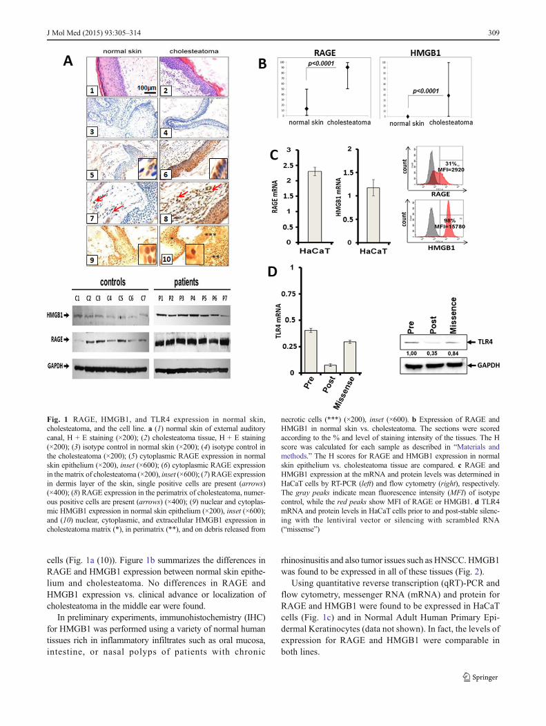
cells (Fig. 1a (10)). Figure 1b summarizes the differences inRAGE and HMGB1 expression between normal skin epithe-lium and cholesteatoma. No differences in RAGE andHMGB1 expression vs. clinical advance or localization ofcholesteatoma in the middle ear were found.
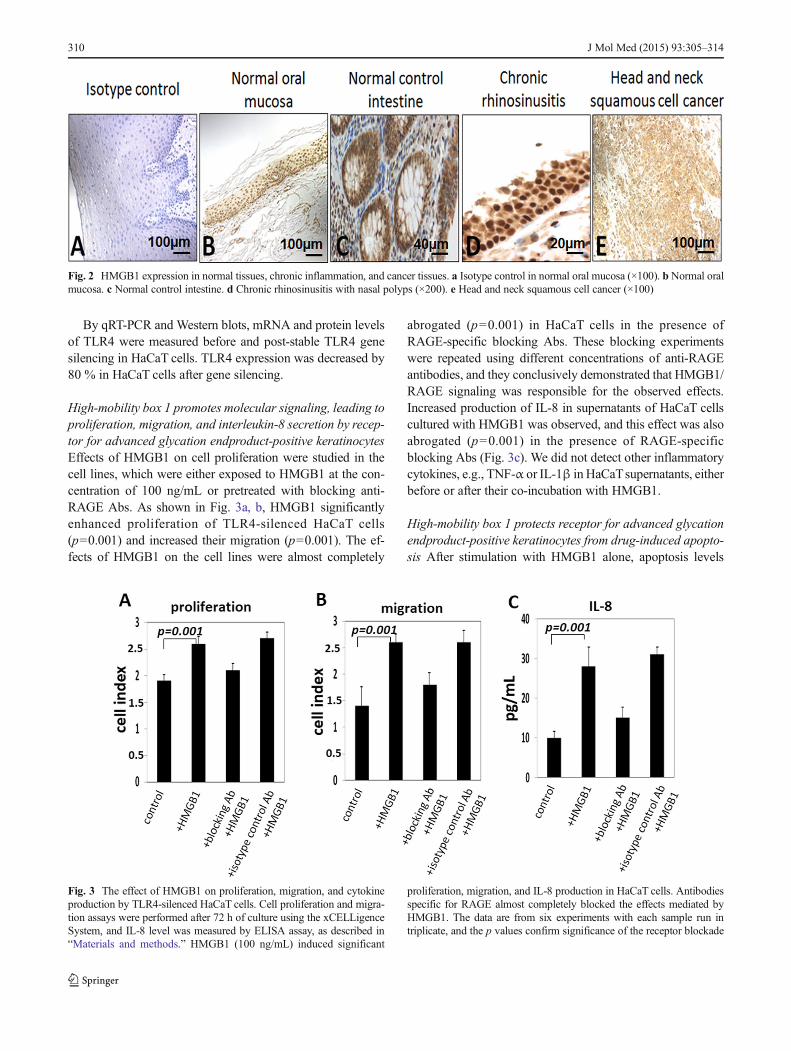
In preliminary experiments, immunohistochemistry (IHC)for HMGB1 was performed using a variety of normal humantissues rich in inflammatory infiltrates such as oral mucosa,intestine, or nasal polyps of patients with chronic
rhinosinusitis and also tumor issues such as HNSCC. HMGB1was found to be expressed in all of these tissues (Fig. 2).
Using quantitative reverse transcription (qRT)-PCR andflow cytometry, messenger RNA (mRNA) and protein forRAGE and HMGB1 were found to be expressed in HaCaTcells (Fig. 1c) and in Normal Adult Human Primary Epi-dermal Keratinocytes (data not shown). In fact, the levels ofexpression for RAGE and HMGB1 were comparable inboth lines.
Fig. 1 RAGE, HMGB1, and TLR4 expression in normal skin,cholesteatoma, and the cell line. a (1) normal skin of external auditorycanal, H + E staining (×200); (2) cholesteatoma tissue, H + E staining(×200); (3) isotype control in normal skin (×200); (4) isotype control inthe cholesteatoma (×200); (5) cytoplasmic RAGE expression in normalskin epithelium (×200), inset (×600); (6) cytoplasmic RAGE expressionin thematrix of cholesteatoma (×200), inset (×600); (7) RAGE expressionin dermis layer of the skin, single positive cells are present (arrows)(×400); (8) RAGE expression in the perimatrix of cholesteatoma, numer-ous positive cells are present (arrows) (×400); (9) nuclear and cytoplas-mic HMGB1 expression in normal skin epithelium (×200), inset (×600);and (10) nuclear, cytoplasmic, and extracellular HMGB1 expression incholesteatoma matrix (*), in perimatrix (**), and on debris released from
necrotic cells (***) (×200), inset (×600). b Expression of RAGE andHMGB1 in normal skin vs. cholesteatoma. The sections were scoredaccording to the % and level of staining intensity of the tissues. The Hscore was calculated for each sample as described in “Materials andmethods.” The H scores for RAGE and HMGB1 expression in normalskin epithelium vs. cholesteatoma tissue are compared. c RAGE andHMGB1 expression at the mRNA and protein levels was determined inHaCaT cells by RT-PCR (left) and flow cytometry (right), respectively.The gray peaks indicate mean fluorescence intensity (MFI) of isotypecontrol, while the red peaks show MFI of RAGE or HMGB1. d TLR4mRNA and protein levels in HaCaT cells prior to and post-stable silenc-ing with the lentiviral vector or silencing with scrambled RNA(“missense”)
J Mol Med (2015) 93:305–314 309
By qRT-PCR and Western blots, mRNA and protein levelsof TLR4 were measured before and post-stable TLR4 genesilencing in HaCaT cells. TLR4 expression was decreased by80 % in HaCaT cells after gene silencing.
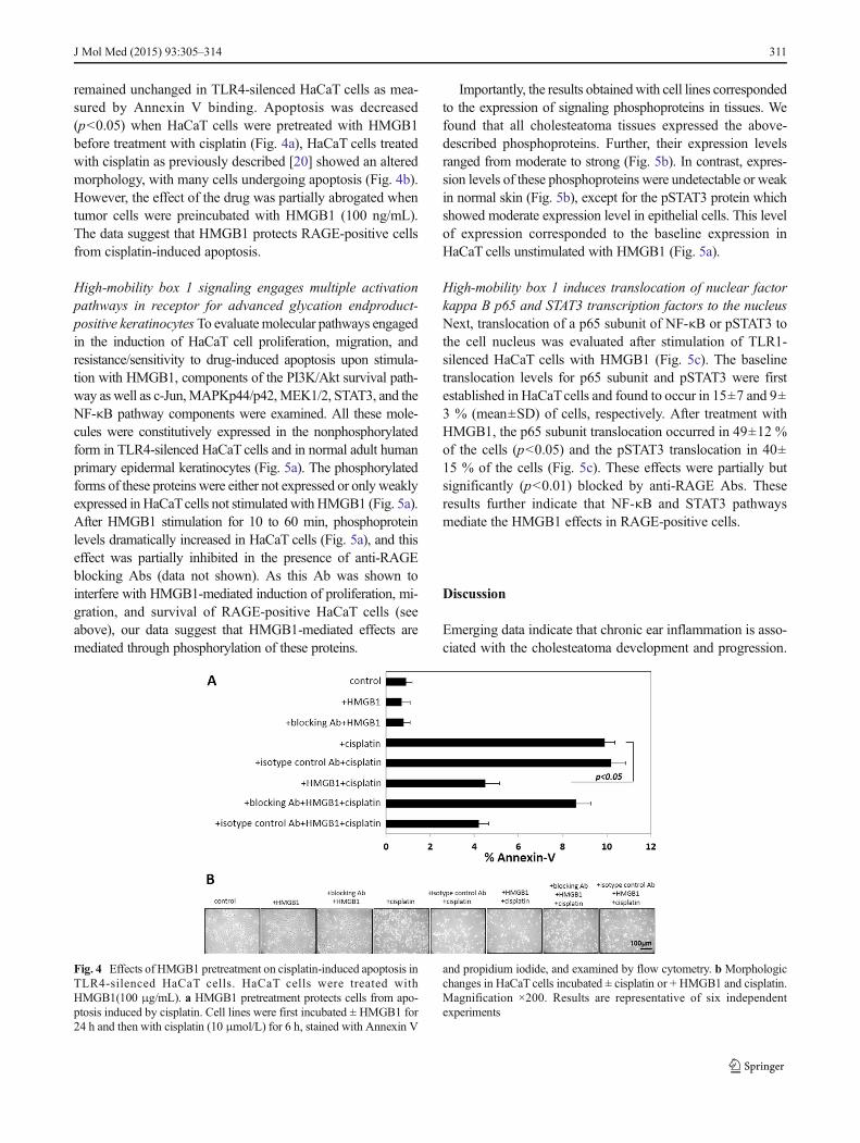
High-mobility box 1 promotes molecular signaling, leading toproliferation, migration, and interleukin-8 secretion by recep-tor for advanced glycation endproduct-positive keratinocytesEffects of HMGB1 on cell proliferation were studied in thecell lines, which were either exposed to HMGB1 at the con-centration of 100 ng/mL or pretreated with blocking anti-RAGE Abs. As shown in Fig. 3a, b, HMGB1 significantlyenhanced proliferation of TLR4-silenced HaCaT cells(p=0.001) and increased their migration (p=0.001). The ef-fects of HMGB1 on the cell lines were almost completely
abrogated (p=0.001) in HaCaT cells in the presence ofRAGE-specific blocking Abs. These blocking experimentswere repeated using different concentrations of anti-RAGEantibodies, and they conclusively demonstrated that HMGB1/RAGE signaling was responsible for the observed effects.Increased production of IL-8 in supernatants of HaCaT cellscultured with HMGB1 was observed, and this effect was alsoabrogated (p=0.001) in the presence of RAGE-specificblocking Abs (Fig. 3c). We did not detect other inflammatorycytokines, e.g., TNF-α or IL-1β in HaCaTsupernatants, eitherbefore or after their co-incubation with HMGB1.
High-mobility box 1 protects receptor for advanced glycationendproduct-positive keratinocytes from drug-induced apopto-sis After stimulation with HMGB1 alone, apoptosis levels
Fig. 2 HMGB1 expression in normal tissues, chronic inflammation, and cancer tissues. a Isotype control in normal oral mucosa (×100). b Normal oralmucosa. c Normal control intestine. d Chronic rhinosinusitis with nasal polyps (×200). e Head and neck squamous cell cancer (×100)
Fig. 3 The effect of HMGB1 on proliferation, migration, and cytokineproduction by TLR4-silenced HaCaT cells. Cell proliferation and migra-tion assays were performed after 72 h of culture using the xCELLigenceSystem, and IL-8 level was measured by ELISA assay, as described in“Materials and methods.” HMGB1 (100 ng/mL) induced significant
proliferation, migration, and IL-8 production in HaCaT cells. Antibodiesspecific for RAGE almost completely blocked the effects mediated byHMGB1. The data are from six experiments with each sample run intriplicate, and the p values confirm significance of the receptor blockade
310 J Mol Med (2015) 93:305–314
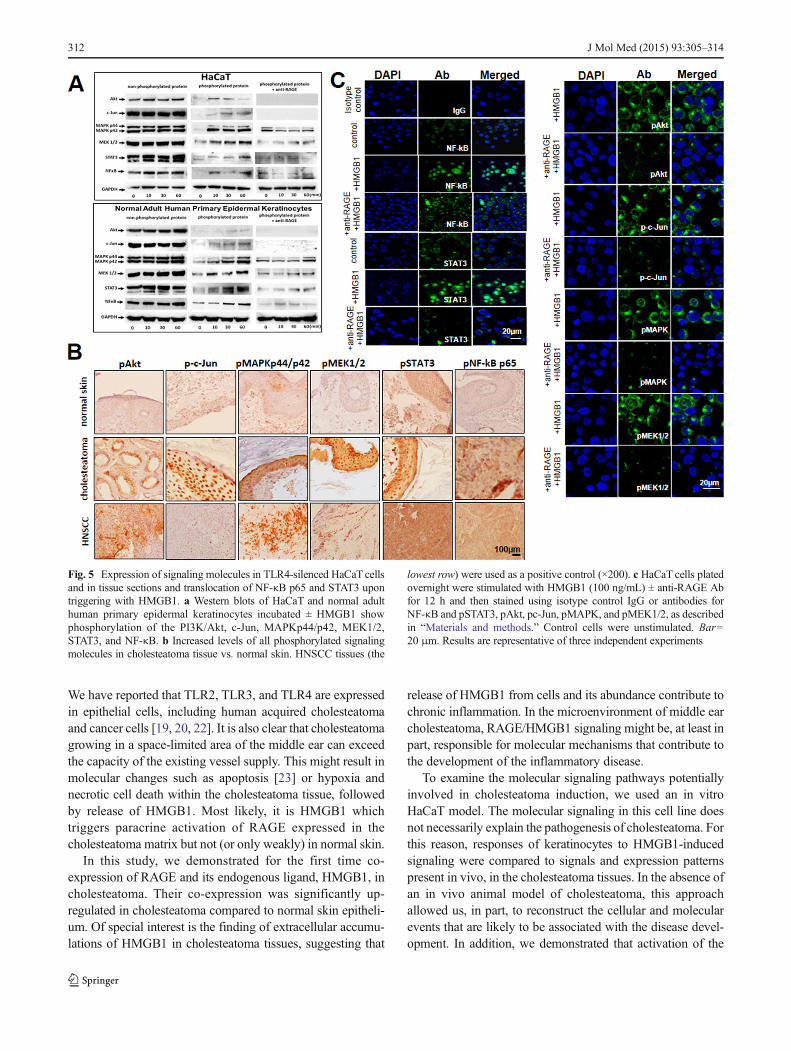
remained unchanged in TLR4-silenced HaCaT cells as mea-sured by Annexin V binding. Apoptosis was decreased(p<0.05) when HaCaT cells were pretreated with HMGB1before treatment with cisplatin (Fig. 4a), HaCaT cells treatedwith cisplatin as previously described [20] showed an alteredmorphology, with many cells undergoing apoptosis (Fig. 4b).However, the effect of the drug was partially abrogated whentumor cells were preincubated with HMGB1 (100 ng/mL).The data suggest that HMGB1 protects RAGE-positive cellsfrom cisplatin-induced apoptosis.
High-mobility box 1 signaling engages multiple activationpathways in receptor for advanced glycation endproduct-positive keratinocytes To evaluate molecular pathways engagedin the induction of HaCaT cell proliferation, migration, andresistance/sensitivity to drug-induced apoptosis upon stimula-tion with HMGB1, components of the PI3K/Akt survival path-way as well as c-Jun,MAPKp44/p42,MEK1/2, STAT3, and theNF-κB pathway components were examined. All these mole-cules were constitutively expressed in the nonphosphorylatedform in TLR4-silenced HaCaT cells and in normal adult humanprimary epidermal keratinocytes (Fig. 5a). The phosphorylatedforms of these proteins were either not expressed or only weaklyexpressed in HaCaTcells not stimulated with HMGB1 (Fig. 5a).After HMGB1 stimulation for 10 to 60 min, phosphoproteinlevels dramatically increased in HaCaT cells (Fig. 5a), and thiseffect was partially inhibited in the presence of anti-RAGEblocking Abs (data not shown). As this Ab was shown tointerfere with HMGB1-mediated induction of proliferation, mi-gration, and survival of RAGE-positive HaCaT cells (seeabove), our data suggest that HMGB1-mediated effects aremediated through phosphorylation of these proteins.
Importantly, the results obtainedwith cell lines correspondedto the expression of signaling phosphoproteins in tissues. Wefound that all cholesteatoma tissues expressed the above-described phosphoproteins. Further, their expression levelsranged from moderate to strong (Fig. 5b). In contrast, expres-sion levels of these phosphoproteins were undetectable or weakin normal skin (Fig. 5b), except for the pSTAT3 protein whichshowed moderate expression level in epithelial cells. This levelof expression corresponded to the baseline expression inHaCaT cells unstimulated with HMGB1 (Fig. 5a).
High-mobility box 1 induces translocation of nuclear factorkappa B p65 and STAT3 transcription factors to the nucleusNext, translocation of a p65 subunit of NF-κB or pSTAT3 tothe cell nucleus was evaluated after stimulation of TLR1-silenced HaCaT cells with HMGB1 (Fig. 5c). The baselinetranslocation levels for p65 subunit and pSTAT3 were firstestablished in HaCaTcells and found to occur in 15±7 and 9±3 % (mean±SD) of cells, respectively. After treatment withHMGB1, the p65 subunit translocation occurred in 49±12 %of the cells (p<0.05) and the pSTAT3 translocation in 40±15 % of the cells (Fig. 5c). These effects were partially butsignificantly (p<0.01) blocked by anti-RAGE Abs. Theseresults further indicate that NF-κB and STAT3 pathwaysmediate the HMGB1 effects in RAGE-positive cells.
Discussion
Emerging data indicate that chronic ear inflammation is asso-ciated with the cholesteatoma development and progression.
Fig. 4 Effects of HMGB1 pretreatment on cisplatin-induced apoptosis inTLR4-silenced HaCaT cells. HaCaT cells were treated withHMGB1(100 μg/mL). a HMGB1 pretreatment protects cells from apo-ptosis induced by cisplatin. Cell lines were first incubated ± HMGB1 for24 h and then with cisplatin (10 μmol/L) for 6 h, stained with Annexin V
and propidium iodide, and examined by flow cytometry. b Morphologicchanges in HaCaT cells incubated ± cisplatin or + HMGB1 and cisplatin.Magnification ×200. Results are representative of six independentexperiments
J Mol Med (2015) 93:305–314 311
We have reported that TLR2, TLR3, and TLR4 are expressedin epithelial cells, including human acquired cholesteatomaand cancer cells [19, 20, 22]. It is also clear that cholesteatomagrowing in a space-limited area of the middle ear can exceedthe capacity of the existing vessel supply. This might result inmolecular changes such as apoptosis [23] or hypoxia andnecrotic cell death within the cholesteatoma tissue, followedby release of HMGB1. Most likely, it is HMGB1 whichtriggers paracrine activation of RAGE expressed in thecholesteatoma matrix but not (or only weakly) in normal skin.
In this study, we demonstrated for the first time co-expression of RAGE and its endogenous ligand, HMGB1, incholesteatoma. Their co-expression was significantly up-regulated in cholesteatoma compared to normal skin epitheli-um. Of special interest is the finding of extracellular accumu-lations of HMGB1 in cholesteatoma tissues, suggesting that
release of HMGB1 from cells and its abundance contribute tochronic inflammation. In the microenvironment of middle earcholesteatoma, RAGE/HMGB1 signaling might be, at least inpart, responsible for molecular mechanisms that contribute tothe development of the inflammatory disease.
To examine the molecular signaling pathways potentiallyinvolved in cholesteatoma induction, we used an in vitroHaCaT model. The molecular signaling in this cell line doesnot necessarily explain the pathogenesis of cholesteatoma. Forthis reason, responses of keratinocytes to HMGB1-inducedsignaling were compared to signals and expression patternspresent in vivo, in the cholesteatoma tissues. In the absence ofan in vivo animal model of cholesteatoma, this approachallowed us, in part, to reconstruct the cellular and molecularevents that are likely to be associated with the disease devel-opment. In addition, we demonstrated that activation of the
Fig. 5 Expression of signaling molecules in TLR4-silenced HaCaT cellsand in tissue sections and translocation of NF-κB p65 and STAT3 upontriggering with HMGB1. a Western blots of HaCaT and normal adulthuman primary epidermal keratinocytes incubated ± HMGB1 showphosphorylation of the PI3K/Akt, c-Jun, MAPKp44/p42, MEK1/2,STAT3, and NF-κB. b Increased levels of all phosphorylated signalingmolecules in cholesteatoma tissue vs. normal skin. HNSCC tissues (the
lowest row) were used as a positive control (×200). c HaCaT cells platedovernight were stimulated with HMGB1 (100 ng/mL) ± anti-RAGE Abfor 12 h and then stained using isotype control IgG or antibodies forNF-κB and pSTAT3, pAkt, pc-Jun, pMAPK, and pMEK1/2, as describedin “Materials and methods.” Control cells were unstimulated. Bar=20 μm. Results are representative of three independent experiments
312 J Mol Med (2015) 93:305–314
RAGE/HMGB1 signaling axis in normal keratinocytes in-duced their proliferation and migration, decreased the sensi-tivity to drug-induced apoptosis, activated pro-survival mole-cules, and stimulated intracellular kinase signaling pathwaysfollowed by activation of NF-κB and STAT3 transcriptionfactors. Our results are consistent with previous studies whichshowed that topical application of HMGB1 to skin wounds inmouse models of diabetes enhanced vessel density, accelerat-ed wound healing, and consistently had chemotactic effects onskin fibroblasts and keratinocytes in vitro [24]. It is necessaryto stress, however, that RAGE/HMGB1 signaling is not re-stricted to keratinocytes. HMGB1 expression has been ob-served by us in normal intestinal mucosa, in chronicrhinosinusitis, and in tumors rich in inflammatory infiltratessuch as HNSCC (data not shown). HMGB1 is a chaperone,which has been reported to be present in all nucleated cells andto be expressed in normal and pathological conditions, includ-ing inflammation, cancer, and autoimmune disorders [25–27].Thus, its overexpression and potential involvement in molec-ular signaling are not specific to cholesteatoma but are man-ifestations of the inflammatory cascade that HMGB1 sustainsvia the HMGB1/RAGE axis.
The PI3K/Akt/NF-κB pathway is now recognized as one ofthe critical pathways in regulating cell survival/apoptosis,migration, and proliferation in pathological conditions suchas chronic inflammation or cancer [8, 9, 20].We demonstratedthat triggering of RAGE by HMGB1 activated Akt, inducedNF-κB p65 translocation, and resulted in increased prolifera-tion and decreased sensitivity to drug-induced apoptosis inkeratinocytes. In agreement with other reports [1, 14, 15], wefound that levels of pAkt and pNF-κB molecules were signif-icantly increased and the phosphorylated forms of c-Jun,MEK1, MEK2, MAPKp44/p42 (Erk1/2), and STAT3 wereoverexpressed in cholesteatoma tissue vs. normal skin epithe-lium. Furthermore, phosphorylation of all the above-listedmolecules was triggered by HMGB1/RAGE interactions incell lines and correspondingly also in cholesteatoma tissues.
The abnormal behavior of cholesteatoma epitheliumseems to be related to the presence of infiltrating immunecells releasing high levels of various cytokines and growthfactors. Although IL-1α and TNF-α were not secreted byHaCaT cells, HMGB1 triggered IL-8 production in thesecells, confirming previous reports that HMGB1/RAGE in-teractions can induce IL-8, a cytokine best known for itspro-inflammatory effects on immune cells [27–29]. Wesuggest, therefore, that RAGE and HMGB1 regulate boneremodeling in chronic inflammation via up-regulation ofthe IL-8 production, a mediator of bone destruction, intissue samples of cholesteatoma. The role of RAGE/HMGB1 interactions in bone remodeling has beenreviewed [30], and a higher expression of RAGE withincholesteatoma perimatrix, which is adjacent to ossicle sur-face or to other parts of the temporal bone, might be
interpreted as indicative of the RAGE/HMGB1 involve-ment in this process.
The immune response is critical for the primary defenseagainst middle ear infections. HMGB1 is released extracellu-larly in response to stress or necrosis and behaves as a cyto-kine, promoting inflammation [31]. It is possible thatkeratinocyte proliferation is promoted and sustained byHMGB1 via its interaction with RAGE on keratinocytes. Itis also plausible that RAGE/HMGB1 interactions may triggerthe signaling pathways involved in the regulation of chronicinflammation as well as bone resorption in cholesteatoma. Thecholesteatoma molecular pathogenesis, growth, and inflam-mation might be related to the co-expression and functionalinteractions between HMGB1 and RAGE. Their molecularinteractions may not be specific to cholesteatoma but, as aprominent part of the inflammatory milieu, are involved in thepathogenesis of this middle ear disease. This suggests thatblocking of these interactions might represent a new poten-tially effective therapy, especially early in disease develop-ment. A development of an in vivo animal model ofcholesteatoma is crucial for in vivo testing the validity of theproposed mechanisms.
Acknowledgments This work was supported in part by the grant of thePolish Ministry of Science and Higher Education NN403590638 to EOand the NIH grants PO-1 CA109688 and R0-1 CA168628 to TLW. Wethank Andrzej Kluk for technical assistance.
Conflict of interest The authors declare no conflict of interest.
Open Access This article is distributed under the terms of the CreativeCommons Attribution License which permits any use, distribution, andreproduction in any medium, provided the original author(s) and thesource are credited.
References
1. Kumar H, Kawai T, Akira S (2011) Pathogen recognition by theinnate immune system. Int Rev Immunol 30:16–34
2. Palm NW, Medzhitov R (2009) Pattern recognition receptors andcontrol of adaptive immunity. Immunol Rev 227:221–233
3. Lotze MT, Zeh HJ, Rubartelli A, Sparvero LJ, Amoscato AA,Washburn NR, Devera ME, Liang X, Tor M, Billiar T (2007) Thegrateful dead: damage-associated molecular pattern molecules andreduction/oxidation regulate immunity. Immunol Rev 220:60–81
4. Seong SY, Matzinger P (2004) Hydrophobicity: an ancient damage-associated molecular pattern that initiates innate immune responses.Nat Rev Immunol 4:469–478
5. Li G, LiangX, LotzeMT (2013) HMGB1: the central cytokine for alllymphoid cells. Front Immunol 4:68
6. Rauvala H, Rouhiainen A (2007) RAGE as a receptor of HMGB1(Amphoterin): roles in health and disease. Curr Mol Med 7:725–734
7. Steenvoorden MM, Toes RE, Ronday HK, Huizinga TW, Degroot J(2007) RAGE activation induces invasiveness of RA fibroblast-likesynoviocytes in vitro. Clin Exp Rheumatol 25:740–742
J Mol Med (2015) 93:305–314 313
8. Kim J, Sohn E, Kim CS, Jo K, Kim JS (2011) The role of high-mobility group box-1 protein in the development of diabetic nephrop-athy. Am J Nephrol 33:524–529
9. Yamamoto Y, Harashima A, Saito H, Tsuneyama K, Munesue S,Motoyoshi S, Han D,Watanabe T, AsanoM, Takasawa S et al (2011)Septic shock is associated with receptor for advanced glycation endproducts ligation of LPS. J Immunol 186:3248–3257
10. Olszewska E,WagnerM, Bernal-SprekelsenM, Ebmeyer J, Dazert S,Hildmann H, Sudhoff H (2004) Etiopathogenesis of cholesteatoma.Eur Arch Otorhinolaryngol 261:6–24
11. Albino AP, Kimmelman CP, Parisier SC (1998) Cholesteatoma: amolecular and cellular puzzle. Am J Otol 19:7–19
12. Olszewska E, Olszewski S, Borzym-Kluczyk M, Zwierz K (2007)Role of N-acetyl-beta-d-hexosaminidase in cholesteatoma tissue.Acta Biochim Pol 54:365–370
13. Bujia J, Kim C, Ostos-Aumente P, Lopez-Villarejo J, Kastenbauer E(1997) Enhanced epithelial proliferation due to elevated levels ofinterleukin-1 receptors in middle ear cholesteatomas. Eur ArchOtorhinolaryngol 254:6–8
14. Sudhoff H, Borkowski G, Bujia J, Hildmann H, Fisseler-Eckhoff A(1997) Immunohistochemical studies with middle ear mucosal rem-nants in cholesteatoma. HNO 45:630–635
15. Sudhoff H, Dazert S, Gonzales AM, Borkowski G, Park SY,Baird A, Hildmann H, Ryan AF (2000) Angiogenesis and angio-genic growth factors in middle ear cholesteatoma. Am J Otol 21:793–798
16. McGinn MD, Chole RA, Henry KR (1982) Cholesteatoma.Experimental induction in the Mongolian Gerbil, MerionesUnguiculaus. Acta Otolaryngol 93:61–67
17. Choufani G, Roper N, Delbrouck C, Hassid S, Gabius HJ (2007)Animal model for cholesteatoma induced in the gerbil: will theprofiles of differentiation/growth-regulatory markers be similar tothe clinical situation? Laryngoscope 117:706–711
18. Szczepanski MJ et al (2013) PRAME expression in head and neckcancer correlates with markers of poor prognosis and might help inselecting candidates for retinoid chemoprevention in pre-malignantlesions. Oral Oncol 49:144–151
19. Szczepanski M, Stelmachowska M, Stryczynski L, Golusinski W,Samara H, Mozer-Lisewska I, Zeromski J (2007) Assessment ofexpression of toll-like receptors 2, 3 and 4 in laryngeal carcinoma.Eur Arch Otorhinolaryngol 264:525–530
20. Szczepanski MJ, Czystowska M, Szajnik M, Harasymczuk M,Boyiadzis M, Kruk-Zagajewska A, Szyfter W, Zeromski J, WhitesideTL (2009) Triggering of Toll-like receptor 4 expressed on human headand neck squamous cell carcinoma promotes tumor development andprotects the tumor from immune attack. Cancer Res 69:3105–3113
21. Pi J, Leung L, Xue P, WangW, Hou Y, Liu D, Yehuda-Shnaidman E,Lee C, Lau J, Kurtz TWet al (2010) Deficiency in the nuclear factorE2-related factor-2 transcription factor results in impaired adipogen-esis and protects against diet-induced obesity. J Biol Chem 285:9292–9300
22. Szczepanski M, Szyfter W, Jenek R, Wrobel M, Lisewska IM,Zeromski J (2006) Toll-like receptors 2, 3 and 4 (TLR-2, TLR-3and TLR-4) are expressed in the microenvironment of human ac-quired cholesteatoma. Eur Arch Otorhinolaryngol 263:603–607
23. Olszewska E, Chodynicki S, Chyczewski L (2006) Apoptosis in thepathogenesis of cholesteatoma in adults. Eur Arch Otorhinolaryngol263:409–413
24. Straino S et al (2008) High-mobility group box 1 protein in humanand murine skin: involvement in wound healing. J Invest Dermatol128:1545–1553
25. Sims GP, Rowe DC, Rietdijk ST, Herbst R, Coyle AJ (2010)HMGB1 and RAGE in inflammation and cancer. Annu RevImmunol 28:367–388
26. Xie J, Mendez JD, Mendez-Valenzuela V, Aguilar-Hernandez MM(2013) Cellular signaling of the receptor for advanced glycation andproducts (RAGE). Cell Signal 25:2185–2197
27. Bucciarelli LG et al (2002) RAGE is a multiligand receptor of theimmunoglobulin superfamily: implications for homeostasis andchronic disease. Cell Mol Life Sci 59:1117–1128
28. Mukaida N (2003) Pathophysiological roles of interleukin-8/CXCL8in pulmonary diseases. Am J Physiol Lung Cell Mol Physiol 284:L566–L577
29. Dejean E, Foisseau M, Lagarrigue F, Lamant L, Prade N, Marfak A,Delsol G, Giuriato S, Gaits-Iacovoni F, Meggetto F (2012) ALK+ALCLs induce cutaneous, HMGB-1-dependent IL-8/CXCL8 pro-duction by keratinocytes through NF-kappaB activation. Blood119:4698–4707
30. Zhou Z, Xiong WC (2011) RAGE and its ligands in bone metabo-lism. Front Biosci (Schol Ed) 3:768–776
31. Ito Y et al (2012) Involvement of HMGB1 and RAGE in IL-1beta-induced gingival inflammation. Arch Oral Biol 57:73–80
314 J Mol Med (2015) 93:305–314