nerve injury enhances rat neuronal glutamate transporter ... · nerve injury enhances rat neuronal...
TRANSCRIPT

The Journal of Neuroscience, December 1995, 15(12): 7872-7878
Nerve Injury Enhances Rat Neuronal Glutamate Transporter Expression: Identification by Differential Display PCR
Sumiko Kiryu, Gui Lan Yao, Naonori Morita, Hidemasa Kato, and Hiroshi Kiyama
Department of Neuroanatomy, Biomedical Research Center, Osaka University Medical School, Osaka 565, Japan
An increase in neuronal glutamate transporter expression after nerve injury was demonstrated by means of differ- ential display PCR (DD-PCR) coupled with in situ hybrid- ization. DD-PCR was carried out to compare differences in expression of mFlNAs between axotomized and normal hy- poglossal motoneurons in the rat. The expression of sev- eral gene fragments were found to be increased following nerve injury; the full length cDNA corresponing to one frag- ment was cloned by subsequent rat cDNA library screen- ing. The close homology of glutamate transporters with our rat cDNA led us to conclude that this clone corresponds to the rat neuronal glutamate transporter (rat EAACl). We speculate that the upregulation of this glutamate uptake system may increase the resistance of these cells against neurotoxic glutamate accumulation during the process of nerve regeneration.
[Key words: nerve regeneration, hypoglossal nerve, neu- ronal high affinity glutamate transporter, glutamate foxici- fy, cell death]
Nerve regeneration is a complex process involving many met- abolic and catabolic pathway. Previous reports have demonstrat- ed that molecules such as growth factors (Heumann et al., 1987a,b; Eckenstein et al., 1991; Kobayashi et al., 1993), some peptides (Saika et al., 1991) and cytoskeltonal proteins (Tetzlaff et al., 1991; Tsui et al., 1991) are upregulated during peripheral nerve regeneration. However, the molecular events associated with axotomy are still not well understood.
Recently, we have focused our attention on several molecules that are involved in the signal transduction pathways during hy- poglossal nerve regeneration (Ohno et al., 1994; Saika et al., 1994; Kiryu et al., 199.5; Morita et al., 1995). These studies implicate some signal transduction pathways such as that in- volving Ras pathway in regeneration associated signaling path- way. By examining individual intracellular signaling molecules associated with regeneration, we hope to gain insight into the phenomena underlying of successful nerve regeneration. How- ever, it is likely that a number of other so far unknown molecules and genes will play an important role in nerve regeneration.
Received June 9, 1995; revised Aug. 18, 1995; accepted Aug. 24, 1995. We are grateful to Dr. M. Tohyama for encouragement, Drs. P C. Emson
and S.J. Augood for critical reading and English correction and Drs. Y. Kanai, and K. Tanaka for useful advice. This work was supported by Ciba-Geigy science foundation and Uehara memorial foundation, and Grant-in-Aid for Sci- entific Research from the Ministry of Education, Science and Culture Japan.
Correspondence should be addressed to Dr. Hiroshi Kiyama, Department of Neuroanatomy, Osaka University Medical School, 2-2 Yamadaoka, Suita, Osa- ka 565, Japan. Copyright 0 1995 Society for Neuroscience 0270-6474/95/157872-07$05.00/O
Therefore, an alternative method to quickly isolate those.genes which may be involved in the nerve regeneration event, has been sought. The development of a new technique termed differential display PCR (DD-PCR) has considerably simplified the identi- fication of genes upregulated after axotomy or lesion, replacing earlier subtractive strategies (Liang et al., 1992; Liang and Par- dee, 1992). The method is based on directly comparing the mRNAs expressed in two or more cell populations, separating their reverse transcription-PCR products and comparing band patterns. We have used this method to identify a gene not pre- viously implicated in peripheral nerve regeneration. Using this approach, we identified the rat homolog of the neuronal high affinity glutamate transporter (rabbit EAAC 1 or human EAAT3) (Kanai and Hediger, 1992; Meister et al., 1993; Arriza et al., 1994) and showed that this glutamate transporter is upregulated in injured motoneurons. High affinity glutamate transporter have recently been defined by molecular cloning (Kanai and Hediger et al., 1992; Pines et al., 1992; Storck et al., 1992; Arriza et al., 1994) and represent a new family of Na+ and K+ coupled elec- trogenic transporters, which have no significant primary struc- tural homology to the superfamily of Na+ and Cll coupled trans- porters (Amara, 1993). These transporters mediate the cellular uptake of acidic and neutral amino acids from the synaptic cleft into glial cells (GLAST, GLTl) or nerve endings (EAACl). Since glutamate is neurotoxic (Benveniste et al., 1984; McBean et al., 1985; Choi et al., 1988) as well as being the principal excitatory amino acid neurotransmitter (Fonnum et al., 1984), transporter-mediated uptake systems may serve to maintain ex- tracellular glutamate concentrations below toxic levels (Nicholls et al., 1990; Eliasof et al., 1993). It has also been suggested that several neurodegenerative disorders are due to dysfunction of glutamate transporters (Rothstein et al., 1992). In this study, we show that rat EAACl, cloned by DD-PCR, is associated with axotomy-induced nerve regeneration. In addition, the distribu- tion of rat EAACl mRNA was demonstrated in the adult rat brain.
Materials and Methods Animals. Seventy male Wistar rats weighing about 100 gm were anes- thetized with pentobarbital (45 mg/kg), positioned supine and their right hypoglossal nerve cut with scissors, the hypoglossal nuclei were then dissected from the operated- and normal-sides (Fig. 1A). Seventy hy- poglossal nuclei (operated and normal) were collected and frozen in liquid nitrogen. For in situ hybridization, rats were sacrificed 1, 3, 5, 7, 14, 21, 28, and 35 d afer the operation (three rats each point).
Differential display PCR (DD-PCR). DD-PCR was carried out (Fig. 1A) as previously described (Liang et al., 1992; Liang and Pardee, 1992; Bauer et al., 1993). Total RNA (approximately 70 kg) was obtained from either the operated or the normal hypoglossal nuclei 7 d after surgery. Total RNA (each 0.2 kg) was converted to cDNA with super- script reverse transcriptase (GIBCO/BRL) and nucleotide oligo dT,,.

A
mi / A
RNA(control) RNA(injured)
\ /
DD-PCR
The Journal of Neuroscience, December 1995, 15(12) 7873
B cant ope
Figure 1. A, Preparation of tissue for DD-PCR. Differential display comparing mRNAs from both the normal (left) and injured (right) hypoglossal nuclei; tissue was carefully dissected and total RNA was prepared from each hypoglossal nucleus. B shows autoradiography of amplified ?S-labeled PCR products (after electrophoresis in a 5% polyacrylamide gel) using two arbitrary primers. The left lane corresponds to PCR products derived from the normal hypoglossal nucleus and the right lane is from the operated side. Arrowhead indicates a differentially expressed band which is located at about 200 bp in size.
Subsequently, l/10 volume of the each pool of cDNA was amplified by PCR in the presence of a-‘5S dATP (New England Nuclear-Du Pont, Natick, MA) using two arbitrary primers, 5’-AGGGGAACTGCTG- GGGTCGTCCCGGTGGTC-3’ and 5’.GGCCTTCATGTTAATGA- TGCAATTAAGGTC-3’, which were selected among 87 arbitary prim- ers by chance. The cycling parameters are as follows: denaturation at 94°C for 5 min, 40 cycles with denaturation at 94°C for 30 set, an- nealing at 42°C for 1 min and extension at 72°C for 1.5 min and an additional extention period at 72°C for 5 min. Radiolabeled PCR prod- ucts were analyzed by electrophoresis on a 5% sequencing gel, and visualized by autoradiography. Differentially upregulated bands were
recovered from dried denaturing polyacrylamide gels and reamplified in a 40 cycles PCR using the same primers, Reamplified cDNA products were cloned into PCRYI vectors using the TA cloning kit (lnvitrogen, San Diego, CA).
In situ hybridizarion. Animals were decapitated 1, 2, 3, 5, 7, 14, and 21 d after surgery. Their brains were removed quickly and frozen in powdered dry ice; 20 urn thick sections were cut on a cryostat, thaw- mounted onto 3-aminopropyltriethoxysilane coated slides, and stored at -80°C until used. Just before use, the sections were fixed in 4% para- formaldehyde in 0.1 M phosphate buffer (PB) for 20 min, washed in PB, treated with 10 pg/ml proteinase-K in 50 mM Trrs-HCL and 5 mM
Figure 2. Histological display by in situ hybridization using the cRNA probe derived from cDNA fragment shown in Figure 1B (arrowhead). The section was obtained from an animal whose uni- lateral hypoglossal nerve was axotomi- zed 7 d before (right side is the operated side). The counterstained emulsion au- toradiography is observed under semi- darkfield observation. Note the accu- mulation of silver grain is observed only on the injured motoneurons. Scale bar, 40 pm.
7874 Kiryu et al. * Nerve injury Upregulates Glutamate Transporter Expression
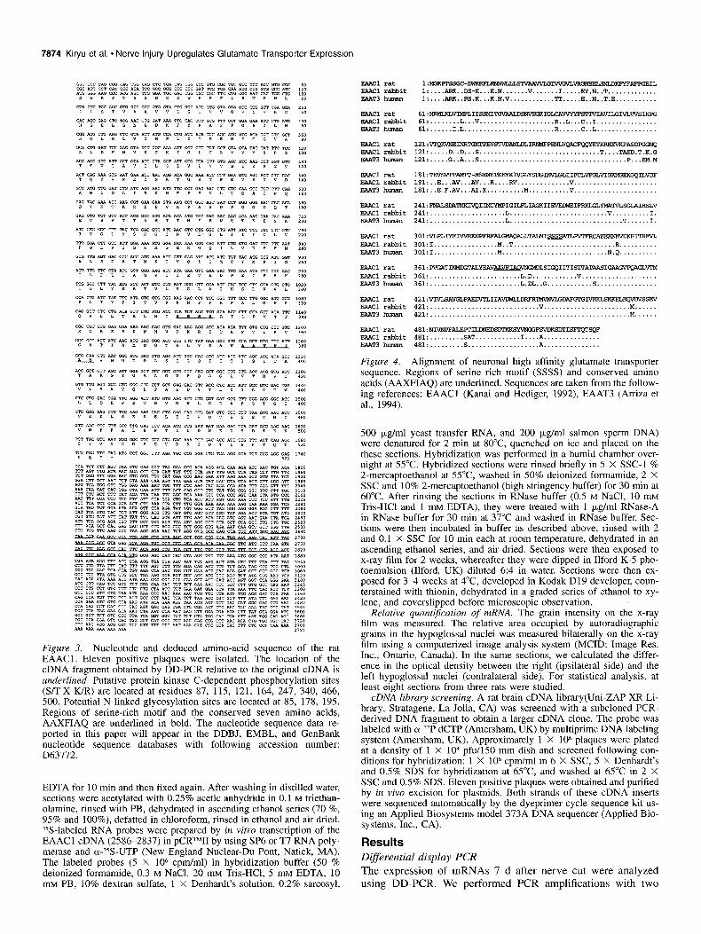
Figure 3. Nucleotide and deduced amino-acid sequence of the rat EAACl. Eleven positive plaques were isolated. The location of the cDNA fragment obtained by DD-PCR relative to the original cDNA is underlined. Putative protein kinase C-dependent phosphorylation sites (S/l-X-K/R) are located at residues 87, 115, 121, 164, 247, 340, 466, 500. Potential N-linked glycosylation sites are located at 85, 178, 195. Regions of serine-rich motif and the conserved seven amino acids, AAXFIAQ are underlined in bold. The nucleotide sequence data re- ported in this paper will appear in the DDBJ, EMBL, and GenBank nucleotide sequence databases with following accession number: D63772.
EDTA for 10 min and then fixed again. After washing in distilled water, sections were acetylated with 0.25% acetic anhydride in 0.1 M triethan- olamine, rinsed with PB, dehydrated in ascending ethanol series (70 %, 95% and lOO%), defatted in chloroform, rinsed in ethanol and air dried. ?S-labeled RNA probes were prepared by in vitro transcription of the EAACl cDNA (25862837) in pCRTMII by using SP6 or T7 RNA poly- merase and a-?S-UTP (New England Nuclear-Du Pont, Natick, MA). The labeled probes (5 X lo6 cpm/ml) in hybridization buffer (50 % deionized formamide, 0.3 M NaCl, 20 mM Tris-HCI, 5 m.~ EDTA, 10 mM PB, 10% dextran sulfate, 1 X Denhardt’s solution, 0.2% sarcosyl,
Eracl rat EAAClrabbit aAT3 human
E?ACl rat EARcl rabbit D.&T3 h""mn
E?Acl rat !?AACl rabbit EAAT3human
241:-m~IvQIPIG~~~II svEu4E-m~sGL?.IHsL" 241:....................L.........................."..........I. 241:....................L..............................T......1.
EA?cl rat -Cl rabbit EAATShuman
301:vLPLm- s.IGNAQmT~~TLm-Icu~ 301:1.................M..T...........................R.......... 301:I.................M............................N.Q..........
EAACl rat FAX1 rabbit EAAT3h-
EAAcl rat EAACl rabbit EAAT3h-
421vrJLsAvGLPAEnvTLIIA--~m~sm
421:.............................".......................M...... 421:.....................................................M......
!aACl rat -Cl rabbit F.&&T3 h-
4*1:NIvNPFALEPPIL~ =mcmc?~ 481:.........sAT............I....A............... 481:.........S...................A...............
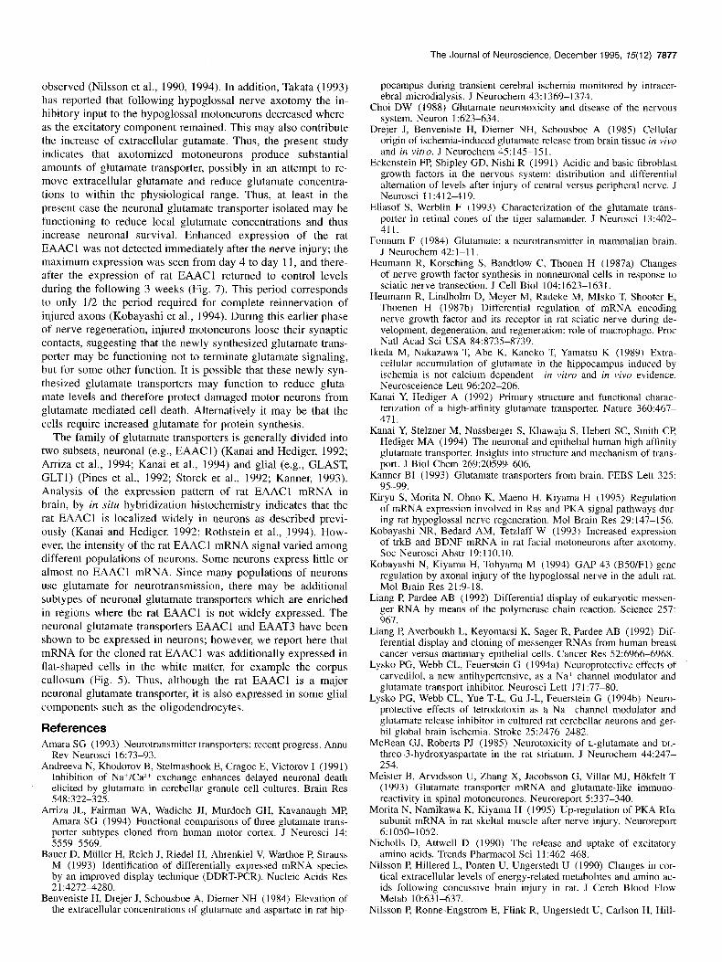
Figure 4. Glignment of neuronal high affinity glutamate transporter sequence. Regions of serine rich motif (SSSS) and conserved amino acids (AAXFIAQ) are underlined. Sequences are taken from the follow- ing references: EAACl (Kanai and Hediger, 1992), EAAT3 (Arriza et al., 1994).
500 yg/ml yeast transfer RNA, and 200 pg/ml salmon sperm DNA) were denatured for 2 min at 80°C quenched on ice and placed on the these sections. Hybridization was performed in a humid chamber over- night at 55°C. Hybridized sections were rinsed briefly in 5 X SSC-1 % 2-mercaptoethanol at 55°C washed in 50% deionized formamide, 2 X SSC and 10% 2-mercaptoethanol (high stringency buffer) for 30 min at 60°C. After rinsing the sections in RNase buffer (0.5 M NaCl, 10 mM Tris-HCl and 1 mu EDTA), they were treated with 1 pg/ml RNase-A in RNase buffer for 30 min at 37°C and washed in RNase buffer. Sec- tions were then incubated in buffer as described above, rinsed with 2 and 0.1 X SSC for 10 min each at room temperature, dehydrated in an ascending ethanol series, and air dried. Sections were then exposed to x-ray film for 2 weeks, whereafter they were dipped in Ilford K-5 pho- toemulsion (Ilford, UK) diluted 6:4 in water. Sections were then ex- posed for 34 weeks at 4°C developed in Kodak D19 developer, coun- terstained with thionin, dehydrated in a graded series of ethanol to xy- lene, and coverslipped before microscopic observation.
Relative quantijcation of mRNA. The grain intensity on the x-ray film was measured. The relative area occupied by autoradiographic grains in the hypoglossal nuclei was measured bilaterally on the x-ray film using a computerized image analysis system (MCID: Image Res. Inc., Ontario, Canada). In the same sections, we calculated the differ- ence in the optical density between the right (ipsilateral side) and the left hypoglossal nuclei (contralateral side). For statistical analysis, at least eight sections from three rats were studied.
cDNA library screening. A rat brain cDNA library(Uni-ZAP XR Li- brary, Stratagene, La Jolla, CA) was screened with a subcloned PCR- derived DNA fragment to obtain a larger cDNA clone. The probe was labeled with &*P dCTP (Amersham, UK) by multiprime DNA labeling system (Amersham, UK). Approximately 1 X lo6 plaques were plated at a density of 1 X lo4 pfu/l50 mm dish and screened following con- ditions for hybridization: 1 X lo6 cpm/ml in 6 X SSC, 5 X Denhardt’s and 0.5% SDS for hybridization at 65°C and washed at 65°C in 2 X SSC and 0.5% SDS. Eleven positive plaques were obtained and purified by in vivo excision for plasmids. Both strands of these cDNA inserts were sequenced automatically by the dyeprimer cycle sequence kit us- ing an Applied Biosystems model 373A DNA sequencer (Applied Bio- systems. Inc., CA).
Results Differential display PCR The expression of mRNAs 7 d after nerve cut were analyzed using DD-PCR. We performed PCR amplifications with two
The Journal of Neuroscience, December 1995, 75(12) 7875
primer combinations. The pattern of amplified cDNA fragments is indicated in Fignre 1B. A cDNA fragment located at about 200 bp (arrowhead) was amplified to a greater degree on the operated side. This 200 bp band was excised and used for further analysis. In order to eliminate possible false positive signal, the observed ncrease in mRNA expression was confirmed on tissue sections by in situ hybridization. The cDNA fragment recovered from the differential display analysis was reamplified and used as a probe for in situ hybridization. This histological survey revealed a significant increase in mRNA expression in cells in the injured hypoglossal nucleus (Fig. 2).
Molecular cloning
We screened a rat brain cDNA library to isolate a full length cDNA clone using radio-labeled cDNA fragments recovered by DD-PCR. Among the 11 positive plaques identified, 4 plaques contained a full length clone. Searching of the nucleotide data- base, DDBJ, revealed that this clone had high homology with a neuronal high affinity glutamate transporter isolated from rabbit small intestine EAACl (Kanai et al., 1992) and human brain EAAT3 (Arriza et al., 1994). The nucleotide sequence of this clone is shown in Figure 3. cDNA sequencing revealed an open reading frame from nucleotides 121-1689 which encoded a 523 amino acid protein. Protein homology revealed a 89% sequence identity with rabbit EAACl and a 90% identity with the human EAAT3. Alignment of the amino acid sequences illustrates the
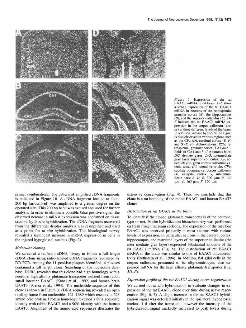
Figure 5. Expression of the rat EAAC 1 mRNA in rat brain. A-C show a strong expression of the rat EAACl mRNA in neurons of the retrosplenial granular cortex (A), the hippocampus (B), and the superior colliculus (C). D- F indicate the rat EAACI mRNA ex- pression in the corpus callosum (gee, cc) at three different levels of the brain. In addition, intense hybridization signal is also observed in various regions such as the CPU (D), cerebral cortex (E, F) and S (E, F). Abbreviations: RSG, re- trosplenial granular cortex; CA1 and 3, fields of CA1 and 3 of Ammon’s horn; DG, dentate gyms; ZnG, intermediate gray layer superior colliculus; Aq, aq- ueduct; gee, genu corpus callosum; IT, tenia tecta; LV, lateral ventricle; CPU, caudate putamen; cc, corpus callosum; Oc, occipital cortex; S, subiculum. Scale bars: A, D, E, 300 brn; B, 140 pm; C, 165 urn; F, 120 pm.
extensive conservation (Fig. 4). Thus, we conclude that this clone is a rat homolog of the rabbit EAACl and human EAAT3 clones.
Distribution of rat EAACl in the brain
To identify if the cloned glutamate transporter is of the neuronal type or not, in situ hybridization histochemistry was performed on fresh frozen rat brain sections. The expression of the rat clone EAACl was observed primarily in most neurons with various levels of expression. In particular, neurons in the cerebral cortex, hippocampus, and restricted layers of the superior colliculus (the inter mediate gray layer) expressed substantial amounts of the rat EAACl mRNA (Fig. 5). The distribution of rat EAACl mRNA in the brain was similar to that of EAACl imunoreac- tivity (Rothstein et al., 1994). In addition, flat glial cells in the corpus callosum, presumed to be oligodendrocyte, also ex- pressed mRNA for the high affinity glutamate transporter (Fig. SD-F).
Expression projle of the rat EAACI during nerve regeneration
We carried out in situ hybridization to evaluate changes in ex- pression of the rat EAACl clone over time during nerve regen- eration (Fig. 6, 7). A slight increase in the rat EAACI hybrid- ization signal was detected initially in the ipsilateral hypoglossal nucleus 1 d after the nerve cut, however the intensity of the hybridization signal markedly increased to peak levels during

7876 Kiryu et al. * Nerve Injury Upregulates Glutamate Transporter Expression
Figure 6. Expression of the rat EAACl mRNA in the hypoglossal nucleus 1 d (A), 7 d (B), and 21 d (C) after unilateral hypoglossal nerve transection (right side). 4V, Fourth ventricle; 12, hypoglossal nucleus. Scale bar, 75 km.
the following 3 d (Fig. 7) and persisted at this level for about 7 d. Thereafter, the hybridization signal gradually decreased to control level over the following 3 weeks.
Discussion In the present study, the technique of DD-PCR was used to identify genes associated with hypoglossal nerve regeneration. In particular, one candidate cDNA fragment (200 bp) was iso- lated and sequenced (Fig. 1). Although this method can be used successfully to isolate candidate cDNAs, it has some technical disadvantages over traditional differential expression cloning methods; one disadvantage is the high incidence of false posi- tives (Nishio et al., 1994). To eliminate these false positives, in
Figure 7. Expression profile of rat EAACl mRNA in both resection side (solid circle) and control side (open circle). Each point shows the average intensity of the positive signals and its SD. Asterisks denote statistically significant differences (Student’s t test) from control: *, p < 0.05; **, p < 0.01.
situ hybridization histochemistry was used as a secondary screening method. This histological approach, in situ differential display, allows false positive clones to be effectively eliminated. About 60-70% of bands initially displayed on the gel were false positives when assessed using in situ hybridization.
Subsequent screening of a cDNA library showed that the clone isolated here corresponded to the rat homolog of a neu- ronal high affinity glutamate transporter. Comparing the amino acid sequences of the rat clone with the rabbit and human se- quences revealed that the rat homolog also conserves an AAX- FIAQ structural motif and serine-rich sequences (SSSS) (Figs. 3, 4). In situ hybridization further suggested that the present clone was expressed primarily in neurons although some glial expression was observed. Taking all this data into consideration, we suggest that the clone derived by DD-PCR is the rat homolog of the rabbit EAACllhuman EAAT3 sequences. Further, ex- pression of this neuronal type of glutamate transporter is upre- gulated following peripheral nerve injury.
Glutamate is the major excitatory neurotransmitter in the ner- vous system as well as a neurotoxin. Under normal conditions, glutamate signal transmission is terminated by the rapid uptake of glutamate into presynaptic nerve terminals and into surround- ing glia (Nicholls and Attwell, 1990). It is conceivable that ab- normal glutamate uptake may be closely related to an increase in extracellular glutamate concentrations as observed in certain neurodegenerative diseases (Drejer et al., 1985; Silverstein et al., 1986; Ikeda et al., 1989). However, the kinetics of the glutamate transport system in such conditions is still controversial. Several studies have demonstrated that an excess of glutamate in the synaptic cleft results from a reduction in the density of glutamate transporter in spinal cord and brain tissues from patients with amyotrophic lateral sclerosis (ALS) (Rothstein et al., 199\2). Fur- ther, in ischemia or anoxia, rundown of Na+, K+ and voltage gradients is expected to initiate reversal of the Na+-dependent and electrogenic glutamate uptake carrier, releasing excess glu- tamate into the extracellular space (Szatkowski et al., 1990; Lys- ko et al., 1994a,b). This abnormal Ca2+-independent nonvesi- cular release of glutamate results in a reduction of glutamate clearance at the synaptic cleft, activation of NMDA receptors and subsequent Ca2+ influx leading to neural death (Andreeva et al., 1991; Waxman et al., 1991). After traumatic brain injury, an increase in extracellular concentrations of glutamate is also
The Journal of Neuroscience, December 1995, 15(12) 7877
observed (Nilsson et al., 1990, 1994). In addition, Takata (1993) has reported that following hypoglossal nerve axotomy the in- hibitory input to the hypoglossal motoneurons decreased where- as the excitatory component remained. This may also contribute the increase of extracellular gutamate. Thus, the present study indicates that axotomized motoneurons produce substantial amounts of glutamate transporter, possibly in an attempt to re- move extracellular glutamate and reduce glutamate concentra- tions to within the physiological range. Thus, at least in the present case the neuronal glutamate transporter isolated may be functioning to reduce local glutamate concentrations and thus increase neuronal survival. Enhanced expression of the rat EAACl was not detected immediately after the nerve injury; the maximum expression was seen from day 4 to day 11, and there- after the expression of rat EAACl returned to control levels during the following 3 weeks (Fig. 7). This period corresponds to only l/2 the period required for complete reinnervation of injured axons (Kobayashi et al., 1994). During this earlier phase of nerve regeneration, injured motoneurons loose their synaptic contacts, suggesting that the newly synthesized glutamate trans- porter may be functioning not to terminate glutamate signaling, but for some other function. It is possible that these newly syn- thesized glutamate transporters may function to reduce gluta- mate levels and therefore protect damaged motor neurons from glutamate mediated cell death. Alternatively it may be that the cells require increased glutamate for protein synthesis.
The family of glutamate transporters is generally divided into two subsets, neuronal (e.g., EAACl) (Kanai and Hediger, 1992; Arriza et al., 1994; Kanai et al., 1994) and glial (e.g., GLAST, GLTl) (Pines et al., 1992; Storck et al., 1992; Kanner, 1993). Analysis of the expression pattern of rat EAACl mRNA in brain, by in situ hybridization histochemistry indicates that the rat EAACl is localized widely in neurons as described previ- ously (Kanai and Hediger, 1992; Rothstein et al., 1994). How- ever, the intensity of the rat EAACl mRNA signal varied among different populations of neurons. Some neurons express little or almost no EAACl mRNA. Since many populations of neurons use glutamate for neurotransmission, there may be additional subtypes of neuronal glutamate transporters which are enriched in regions where the rat EAACl is not widely expressed. The neuronal glutamate transporters EAACl and EAAT3 have been shown to be expressed in neurons; however, we report here that mRNA for the cloned rat EAACI was additionally expressed in flat-shaped cells in the white matter, for example the corpus cullosum (Fig. 5). Thus, although the rat EAACl is a major neuronal glutamate transporter, it is also expressed in some glial components such as the oligodendrocytes.
References Amara SG (1993) Neurotransmitter transporters: recent progress. Annu
Rev Neurosci 16:73-93. Andreeva N, Khodorov B, Stelmashook E, Cragoe E, Victorov I (1991)
Inhibition of Na+/Ca*+ exchange enhances delayed neuronal death elicited by glutamate in cerebellar granule cell cultures. Brain Res 548:322-325.
Arriza JL, Fairman WA, Wadiche JI, Murdoch GH, Kavanaugh MP, Amara SC (1994) Functional comparisons of three glutamate trans- porter subtypes cloned from human motor cortex. J Neurosci 14: 555995569.
Bauer D, Mtiller H, Reich J, Riedel H, Ahrenkiel V, Warthoe P, Strauss M (1993) Identification of differentially expressed mRNA species by an improved display technique (DDRT-PCR). Nucleic Acids Res 21:4272-4280.
Benveniste H, Drejer J, Schousboe A, Diemer NH (1984) Elevation of the extracellular concentrations of glutamate and aspartate in rat hip-
pocampus during transient cerebral ischemia monitored by intracer- ebral microdialysis. J Neurochem 43:1369-1374.
Choi DW (1988) Glutamate neurotoxicity and disease of the nervous system. Neuron 1:623-634.
Drejer J, Benveniste H, Diemer NH, Schousboe A (1985) Cellular origin of ischemia-induced glutamate release from brain tissue in viva and in vitro. J Neurochem 45:145-151.
Eckenstein FP, Shipley GD, Nishi R (1991) Acidic and basic fibroblast growth factors in the nervous system: distribution and differential alternation of levels after injury of central versus peripheral nerve. J Neurosci 11:412+19.
Eliasof S, Werblin F (1993) Characterization of the glutamate trans- porter in retinal cones of the tiger salamander. J Neurosci 13:402- 411.
Fonnum F (1984) Glutamate: a neurotransmitter in mammalian brain. J Neurochem 42: l-l 1.
Heumann R, Korsching S, Bandtlow C, Thonen H (1987a) Changes of nerve growth factor synthesis in nonneuronal cells in response to sciatic nerve transection. J Cell Biol 104: 1623-163 1.
Heumann R, Lindholm D, Meyer M, Radeke M, Mlsko T, Shooter E, Thoenen H (1987b) Differential regulation of mRNA encoding nerve growth‘factor’and its receptor in rat sciatic nerve during de- velopment, degeneration, and regeneration: role of macrophage. Proc Nat1 Acad Sci USA 84:8735-8739.
Ikeda M, Nakazawa T, Abe K, Kaneko T, Yamatsu K (1989) Extra- cellular accumulation of glutamate in the hippocampus induced by ischemia is not calcium dependent--in vitro and in vivo evidence. Neurosceience Lett 96:202-206.
Kanai Y, Hediger A (1992) Primary structure and functional charac- terization of a high-affinity glutamate transporter. Nature 360:467- 471.
Kanai Y, Stelzner M, Nussberger S, Khawaja S, Hebert SC, Smith CP, Hediger MA (1994) The neuronal and epithelial human high affinity glutamate transporter. Insights into structure and mechanism of trans- port. J Biol Chem 269:20599-606.
Kanner BI (1993) Glutamate transporters from brain. FEBS Lett 325: 95-99.
Kiryu S, Morita N, Ohno K, Maeno H, Kiyama H (1995) Regulation of mRNA expression involved in Ras and PKA signal pathways dur- ing rat hypoglossal nerve regeneration. Mot Brain Res 29:147-156.
Kobayashi NR, Bedard AM, Tetzlaff W (1993) Increased expression of trkB and BDNF mRNA in rat facial motoneurons after axotomy. Sot Neurosci Abstr 19:110.10.
Kobayashi N, Kiyama H, Tohyama M (1994) GAP-43 (B50/Fl) gene regulation by axonal injury of the hypoglossal nerve in the adult rat. Mol Brain Res 21:9-18.
Liang P, Pardee AB (1992) Differential display of eukaryotic messen- ger RNA by means of the polymerase chain reaction. Science 257: 967.
Liang P, Averboukh L, Keyomarsi K, Sager R, Pardee AB (1992) Dif- ferential display and cloning of messenger RNAs from human breast cancer versus mammary epithelial cells. Cancer Res 52:6966-6968.
Lysko PG, Webb CL, Feuerstein G (1994a) Neuroprotective effects of carvedilol, a new antihypertensive, as a Na+ channel modulator and glutamate transport inhibitor. Neurosci Lett 17 1:77-80.
Lysko PG, Webb CL, Yue T-L, Gu J-L, Feuerstein G (1994b) Neuro- protective effects of tetrodotoxin as a Na+ channel modulator and glutamate release inhibitor in cultured rat cerebellar neurons and ger- bil global brain ischemia. Stroke 25:2476-2482.
McBean GJ, Roberts PJ (1985) Neurotoxicity of L-glutamate and DL- threo-3-hydroxyaspartate in the rat striatum. J Neurochem 44:2477 254.
Meister B, Arvidsson U, Zhang X, Jacobsson G, Villar MJ, Hbkfelt T (1993) Glutamate transporter mRNA and glutamate-like immuno- reactivity in spinal motoneurones. Neuroreport 5:337-340.
Morita N, Namikawa K, Kiyama H (1995) Up-regulation of PKA RIol subunit mRNA in rat skeltal muscle after nerve injury. Neuroreport 6:1050-1052.
Nicholls D, Attwell D (1990) The release and uptake of excitatory amino acids. Trends Pharmacol Sci 11:462-468.
Nilsson P, Hillered L, Ponten U, Ungerstedt U (1990) Changes in cor- tical extracellular levels of energy-related metabolites and amino ac- ids following concussive brain injury in rat. J Cereb Blood Flow Metab 10:631-637.
Nilsson P, Ronne-Engstrom E, Flink R, Ungerstedt U, Carlson H, Hill-

7878 Kiryu et al. * Nerve Injury Upregulates Glutamate Transporter Expression
ered L (1994) Epileptic seizure activity in the acute phase following cortical impact trauma in rat. Brain Res 637:227-232.
Nishio Y, Aiello LP King GL (1994) Glucose induced genes in bovine aortic smooth muscle cells identified by mRNA differential display. FASEB J 8:103-106.
Ohno K, Kitahara T, Takeda N, Kubo T, Kiyama H (1994) Gene reg- ulation of CAMP-dependent protein kinase subunits (Co, B: Rio, B and RIIo, B) in the rat facial motoneurons after nerve transection. Neuroscience 63: 1 loll1 109.
Pines G, Danbolt N, Bjoras M, Zhang Y, Bendahan A, Eide L, Koepsell H, Stormmathisen J, Seeberg E, Kanner BI (1992) Cloning and ex- pression of a rat brain L-glutamate transporter. Nature 360:464467.
Rothstein JD, Martin LJ, Kuncl RW (1992) Decreased glutamate trans- port by the brain and spinal cord in amyotrophic lateral sclerosis. N Engl J Med 326:1464-1468.
Rothstein JD, Martin L, Levey AI, Dykes-Hoberg M, Jin L, Wu D, Nash N, Kuncl RW (1994) Localization of neuronal and glial glu- tamate transporters. Neuron 13:713-725
Saika T, Senba E, Noguchi K, Sato M, Kubo T, Matsunaga T, Tohyama M (1991) Changes in expression of peptides in rat facial motoneu- rons after facial nerve crushing and resection. Mol Brain Res 11: 187-196.
Saika T, Kiyama H, Matsunaga T, Tohyama M (1994) Differential reg-
ulation of phospholipase C isozymes in the rat facial nucleus follow- ing axotomy. Neuroscience 59:121-129.
Silverstein FS, Buchanan K, Johnston MV (1986) Perinatal hypoxia- ischemia disrupts striatal high-affinity [‘HIglutamate uptake into syn- aptosomes. J Neurochem 47:1614-1619.
Storck T, Schulte S, Hofmann K, Stoffel W (1992) Structure, expres- sion, and functional analysis of a Na+-dependent glutamate/asparatate transporter from rat brain. Proc Nat1 Acad Sci USA 89: 10955-10959.
Szatkowski M. Barbour B. Attwell D (1990) Non-vesicular release of glutamate from glial cells by reversed electrogenic glutamate uptake. Nature 348:443446.
Takata M (1993) Two types of imhibitory postsynaptic potentials in the hypoglossal motoneurons. Prog Neurobiol 40:385411.
Tetzlaff W, Alexander SW, Miller FD, Bisby MA (1991) Response of facial and rubrospinal neurons to axotomy: changes in mRNA ex- pression for cytoskeltal protein and GAP-43. J Neurosci 11:2528- 2544.
Tsui BJ, Cassar SL, Tetzlaff W (1991) Changing in mRNA levels for GAP-43, tubulin and neurofilament-M in rat spinal motoneurons after proximal versus distal axotomy. Sot Neurosci Abstr 17:24.6.
Waxman S, Ransom BR, Stys PK (1991) Non-synaptic mechanisms of Caz+-mediated injury in CNS white matter. Trends Neurosci 14:461- 468.