pediatric continuous eeg monitoring (ceeg)€“-daniel-arndt-md.pdf · seizure diagnosis: clinical...
TRANSCRIPT

Michigan Society of Electroneurodiagnostic Technologists
9.27.13
Daniel Arndt, MD
Director, Pediatric Epilepsy
Beaumont Children‟s Hospital & Health System
PEDIATRIC CONTINUOUS EEG MONITORING
(CEEG)

LEARNING OBJECTIVES
1. History of cEEG
2. What‟s what of common cEEG terminology
3. Update current practice of cEEG
NICU – Neonatal Intensive Care Unit
PICU – Pediatric Intensive Care Unit
4. Epidemiology – Subclinical Seizures / S.E.
5. Impact – Electrographic Seizures / S.E.
6. ACNS Guidelines & Critical Care EEG Terminology
Pediatric / Adult
Neonatal

CONTINUOUS EEG VIDEO MONITORING (CEEG)
Monitoring brain’s electrical activity Video correlation w/ clinical signs (reported sx’s)
Indications: Cerebral function monitoring (i.e. CEA, Anesthesia)
Event identification
Detect subclinical Sz AND CLINICAL Sz
EMU/Presurgical, ICU
Where: PICU, NICU, Floors

CEEG DEVELOPMENT OVER THE YEARS……..
0
10
20
30
40
50
60
70
80
90
19
67
19
69
19
70
19
73
19
74
19
75
19
76
19
77
19
78
19
79
19
80
19
81
19
82
19
83
19
84
19
85
19
86
19
87
19
88
19
89
19
90
19
91
19
92
19
93
19
94
19
95
19
96
19
97
19
98
19
99
20
00
20
01
20
02
20
03
20
04
20
05
20
06
20
07
20
08
20
09
20
10
20
11
20
12
20
13
# of cEEG monitoring studies by year
PubMed Search 9.25.13
QEEG for CEA, Anesthesia, Brain Trauma DeGeorgio ‟92, Jordan „93, 94-04, 01-05
1978: 1st video EEG

WHAT’S WHAT – COMMON CEEG TERMINOLOGY
Electroclinical Seizure = Clinical + EEG
Clinical Seizure = Clinical +/- EEG
Subclinical Seizure = Subtle Clinical or EEG-only
Nonconvulsive Seizure = Subclinical Seizure
*Or = Subtle Clinical Seizure
Electrographic Seizure = EEG +/- Clinical Signs
*Or = Subtle Clinical or Nonconvulsive Seizure
Electrographic-Only Seizure = EEG only
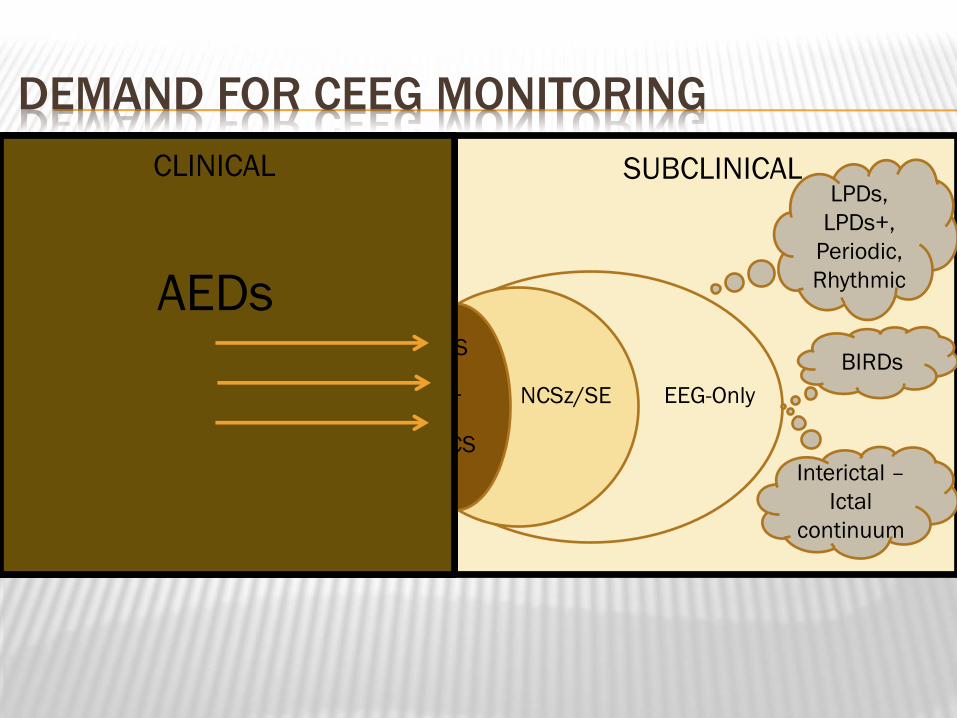
SUBCLINICAL CLINICAL
DEMAND FOR CEEG MONITORING
Clinical
Sz/S.E.
CS
+ NCSz/SE
NCS
Psychogenic
Nonepileptic
Sz
EEG-Only
Posturing,
Twitching, etc.
LPDs,
LPDs+,
Periodic,
Rhythmic
BIRDs
Interictal –
Ictal
continuum
CLINICAL
AEDs

DEMAND FOR CEEG MONITORING (CONT.)
Nonconvulsive seizures/NCSE occur commonly
NCSz/NCSE potentially:
Worsen Acute Brain Injury
Increased Risk for Future Neurocognitive Morbidity
Increased Risk of Development of Epilepsy
NCSE → ↑ Morbidity/Mortality

SUPPLY/RESOURCES FOR CEEG
↑ ↑ Utilization of cEEG in PICU/NICU over past 5yrs
Resource Development & EEG reimbursement ∆s
Institutional Guidelines – Standardize Monitoring
ACNS Guidelines: Neonatal, Pediatric, & Adult
Research Consortia:
Pediatric Critical Care EEG Group (PCCEG)
Critical Care EEG Monitoring Research Consortium
(CCEMRC)
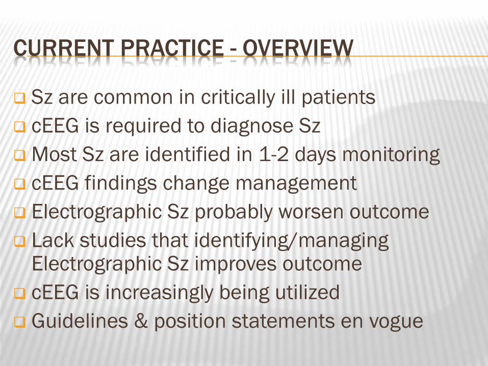
CURRENT PRACTICE - OVERVIEW
Sz are common in critically ill patients
cEEG is required to diagnose Sz
Most Sz are identified in 1-2 days monitoring
cEEG findings change management
Electrographic Sz probably worsen outcome
Lack studies that identifying/managing Electrographic Sz improves outcome
cEEG is increasingly being utilized
Guidelines & position statements en vogue
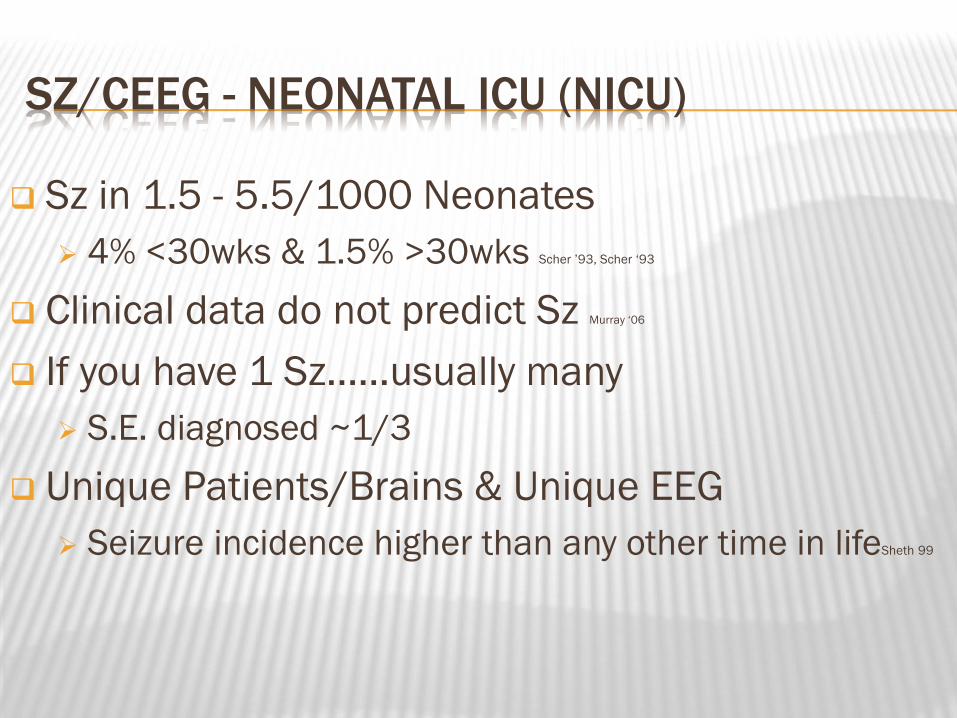
SZ/CEEG - NEONATAL ICU (NICU)
Sz in 1.5 - 5.5/1000 Neonates
4% <30wks & 1.5% >30wks Scher ‟93, Scher „93
Clinical data do not predict Sz Murray „06
If you have 1 Sz……usually many
S.E. diagnosed ~1/3
Unique Patients/Brains & Unique EEG
Seizure incidence higher than any other time in lifeSheth 99

SZ/CEEG – NEONATAL ICU (NICU) (CONT.)
Unique Sz Semiology
Accurate recognition of Clinical Sz is challengingScher 02
Experience clinicians frequently fail to recog Clin Sz
Staff ID 9% of 526 Clinical SzMurray 08
78% of 177 PBE (-EEG) Murray 08
Video Review: 50% accuracy (Poor interater agreement) Malone
„09
Sz frequently SubclinicalConnell 89, Hellstrom-Westas 85, Nash 11, Scher 03, Clancy 06

SZ/CEEG - NEONATAL ICU (NICU) (CONT.)
Connell article – data
4 or 8 channel EEG – 275 Consecutive NICU adm 25% critically ill
¾ preterm infants
20% Sz 42% Sz infants = Electrographic-only
40% of E-only Sz infants were medically paralyzed
Additional 36% Sz infants = Electrographic onset preceded Clin Sz Thus, 78% E-only portions of Sz (Would be similar to Clancy „06 #)
55% of Sz infants expired No diff +/- Clinical Sz (Few had Clinical only Sz)

SZ/CEEG - NEONATAL ICU (NICU) (CONT.)
Nonparalyzed infants: Only 20% ESz provoke Clin SignsClancy 06
>42% seen in Connell ‟89 – Better EEG
Electroclinical uncoupling – Phenobarbital – 50%
Slide adapted Abend ‟13, PHB Data: Scher ‟03, Connell „89

CURRENT NICU CEEG INDICATIONS
HIE & THERAPEUTIC HYPOTHERMIA
Nash ‟11
N=41, 34% Sz → 10% S.E., 43% Subclinical
Wusthoff ‟11
N=26, 65% Sz → 23% S.E., 47% Subclinical
Cardiac Surgery
Peri-operative Subclinical Sz 6-20%Chock 06, Clancy 05, Helmers 97, Gaynor 05, Schmitt 05
ECMO
Subclinical Sz 11-30% Campbell 91, Hahn 93, Horan 07

CURRENT NICU CEEG UTILIZATION
Glass „12: International Survey
Monitor “at risk” newborns: EEG 24%, aEEG 24%, Both 19% None 34%
Seizure Diagnosis: Clinical 8%, EEG 58%, aEEG or EEG 38%
EEG Duration: <60min 31%, 24hrs 17%, Sz-free 24hrs 49%
Boylan „10: EEG Monitoring Access = 90% (EEG 27%, aEEG 22%, Both 51%)
Confident or Very Confident interpreting = 28%
aEEG (Amplitude integrated EEG): Sensitivity (single channel w/out raw EEG single channel for
confirmation): <50%Rennie 04, Shellhaas 07
Addition of 2nd aEEG channel w/ ability to review raw EEG improves sensitivity to 76%, specificity 78%Shah 08
But Sz detection remains difficult with this tool
It has been shown to reduce total seizure duration in neonatesVan Rooij 10

CEEG NICU – ACNS GUIDELINE
Shellhaas „11: Idealized Goals – NOT Mandated Standard of Care
Indications: Differential Diagnosis – Abnormal Paroxysmal Events
Detection of Electrographic Sz in High Risk Populations
Monitoring Burst Suppression
Judge severity of encephalopathy
Procedures for monitoring
Duration of monitoring: 24hrs – Routine EEG little value
Training of caretakers
EEG interpretation & reporting 1st hour reported asap & >2x in 24hrs
Data Retention & Storage
Digital trending & analyses

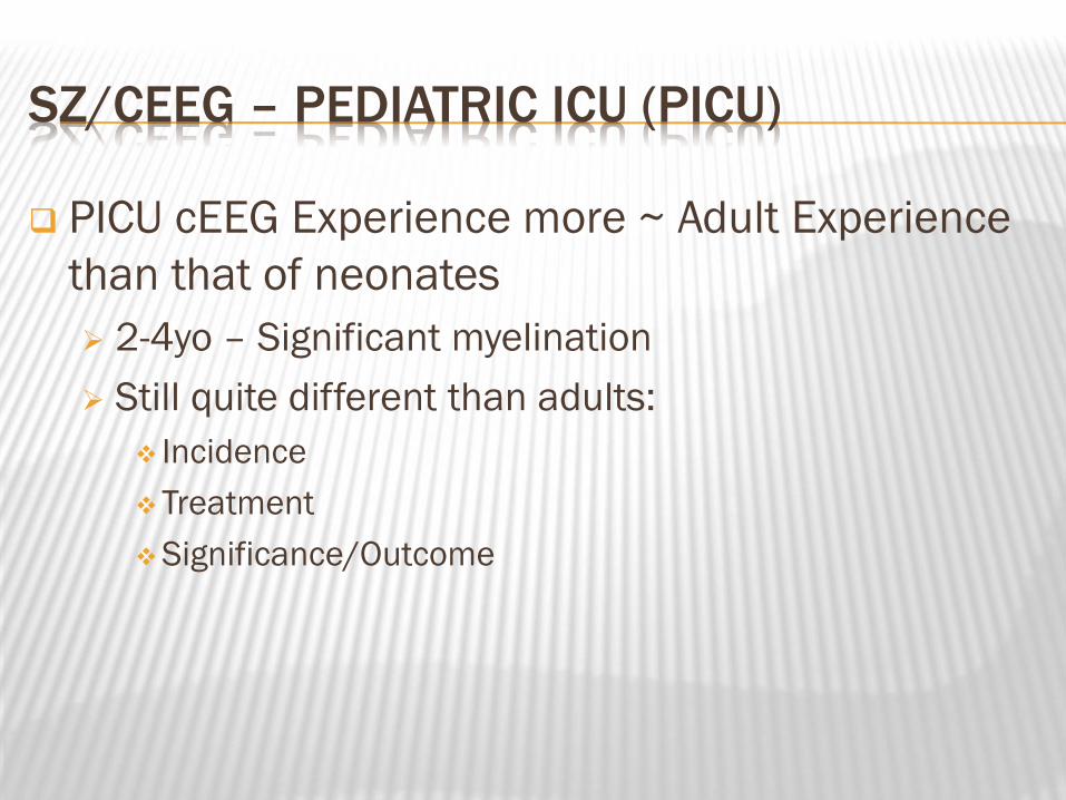
SZ/CEEG – PEDIATRIC ICU (PICU)
PICU cEEG Experience more ~ Adult Experience
than that of neonates
2-4yo – Significant myelination
Still quite different than adults:
Incidence
Treatment
Significance/Outcome

1ST PAPER – CEEG PICU
44% Sz (75% EEG-only)
23% S.E. (89% NCSE)

SUMMARY – ELECTROGRAPHIC SZ INCIDENCE
PICU
Prior single-studies – varying incidences of ESz:
7 - 48%Abend 11, Hosain 05, Jette 06, Abend 07, Alehan 01, Tay 06, Saengpattrachai 06, Shahwan 10, Abend 09, Williams 11, Greiner 12, Kirkham 12
Variability:
Small sample size
Case mix variability across institutions
Interinstitution variabilities in cEEG indications
Range of studies performed over a decade – cEEG/Crit Care evolved
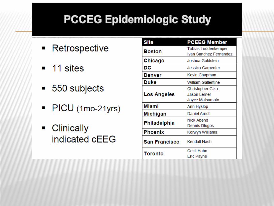


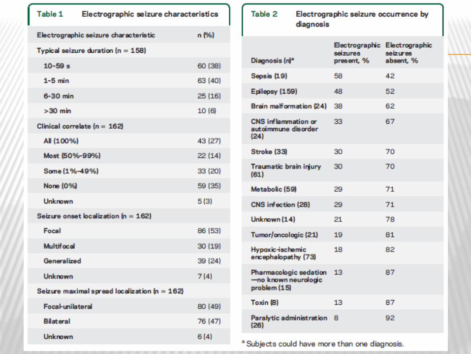
PCCEG EPIDEMIOLOGIC DATAABEND ‘13
Electrographic Sz – 30% (162/550)
Electrographic S.E. – 38% Sz >30min – 46%
Recurrent Sz >50% of 1hr Epoch – 56%
Sz w/ Clinical correlate?: All – Only 27%
Some – 34%
None – 36%
Sz risk factors: (multivariate analysis) Younger Age
Clinical Sz prior to cEEG
Abnormal initial EEG background
IEDs
Epilepsy Diagnosis
NCSz risk factors: (reported elsewhere) Younger AgeAbend 11, Williams 11, Schreiber 12
Convulsive SEWilliams 11
Acute SeizuresMcCoy 11, Greiner 12, Schreiber 12
Structural Brain Injury & TBIMcCoy 11, Greiner 12, Williams 11
EEG: Lack of ReactivityJette 06, Epileptiform D/cWilliams 11, J 06, McCoy 11, Abend 09, Background DiscontAbend 09

PCCEG EPIDEMIOLOGIC DATAABEND ’13 (CONT.)
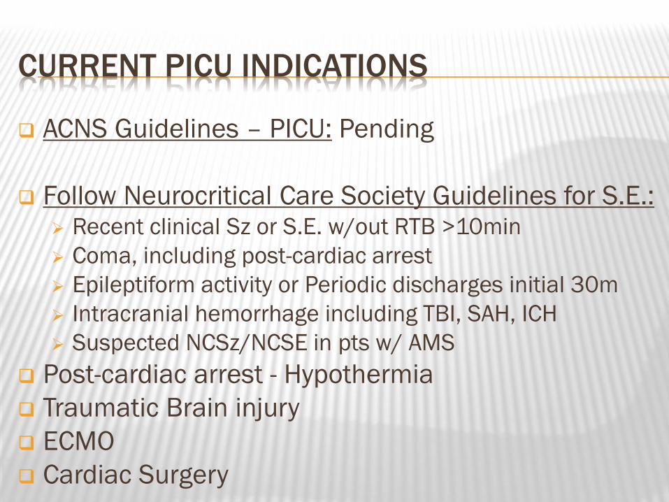
CURRENT PICU INDICATIONS
ACNS Guidelines – PICU: Pending
Follow Neurocritical Care Society Guidelines for S.E.: Recent clinical Sz or S.E. w/out RTB >10min
Coma, including post-cardiac arrest
Epileptiform activity or Periodic discharges initial 30m
Intracranial hemorrhage including TBI, SAH, ICH
Suspected NCSz/NCSE in pts w/ AMS
Post-cardiac arrest - Hypothermia
Traumatic Brain injury
ECMO
Cardiac Surgery
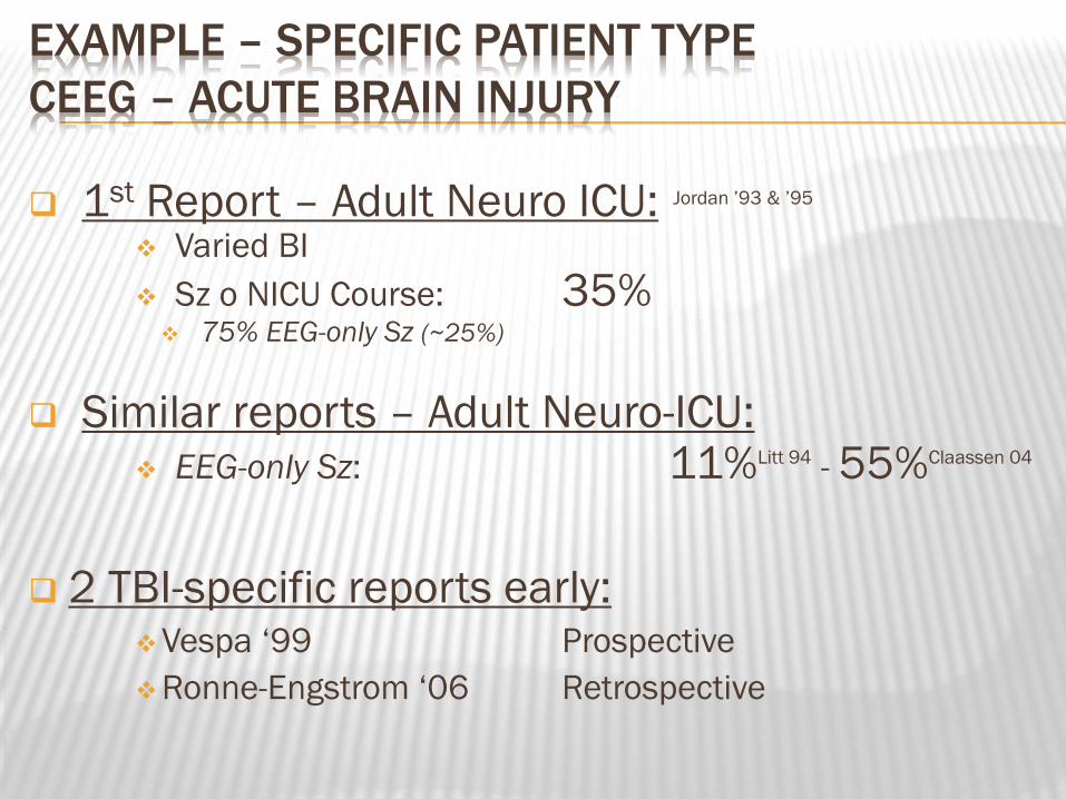
EXAMPLE – SPECIFIC PATIENT TYPE
CEEG – ACUTE BRAIN INJURY
1st Report – Adult Neuro ICU: Jordan ‟93 & ‟95
Varied BI
Sz o NICU Course: 35% 75% EEG-only Sz (~25%)
Similar reports – Adult Neuro-ICU: EEG-only Sz: 11%Litt 94 - 55%Claassen 04
2 TBI-specific reports early: Vespa „99 Prospective
Ronne-Engstrom „06 Retrospective
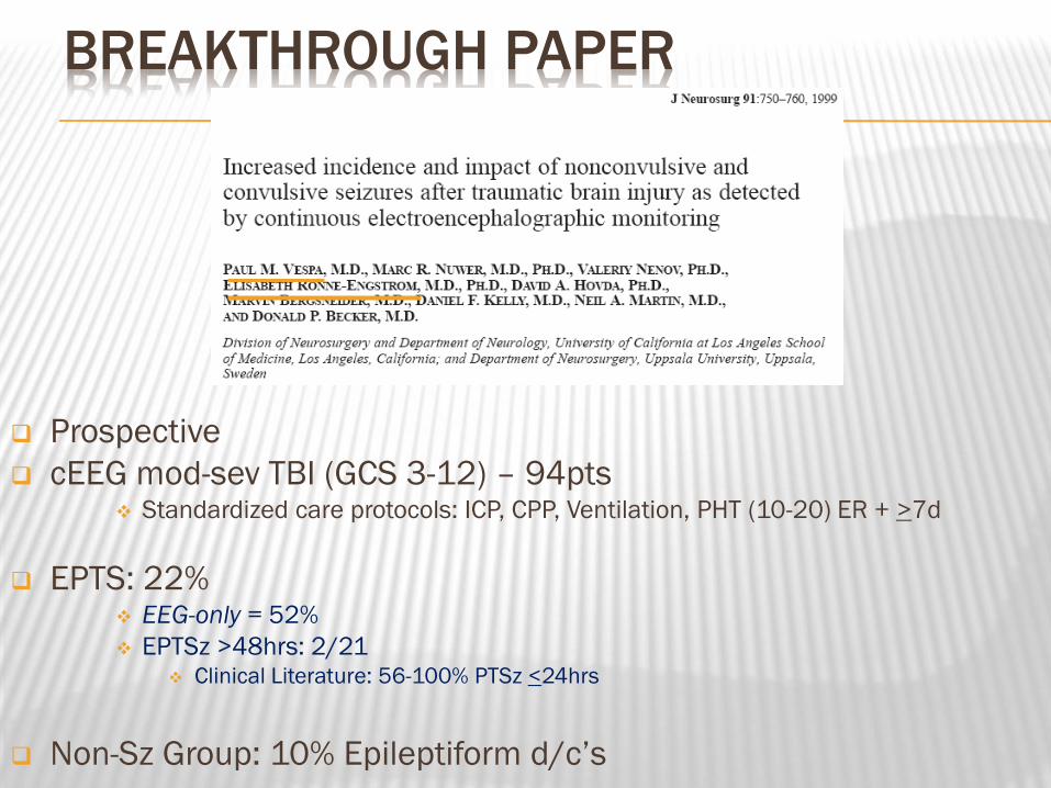
BREAKTHROUGH PAPER
Prospective
cEEG mod-sev TBI (GCS 3-12) – 94pts Standardized care protocols: ICP, CPP, Ventilation, PHT (10-20) ER + >7d
EPTS: 22% EEG-only = 52%
EPTSz >48hrs: 2/21 Clinical Literature: 56-100% PTSz <24hrs
Non-Sz Group: 10% Epileptiform d/c‟s

Retrospective cEEG >24hrs, Standardized care protocols: ICP, CPP, Ventilation
EPTS: 33%
“Significant %” EEG-only
Non-Sz Group: 16% epileptiform d/c‟s
FOLLOW-UP PAPER

Animals: Prince et al. ‟09: (Lit Review)
Electrographic only (no clinical signs) focal Sz in TBI simulated Rats
Depth electrodes & video EEG
Pitkanen et al.
PRECLINICAL PTSz Detection
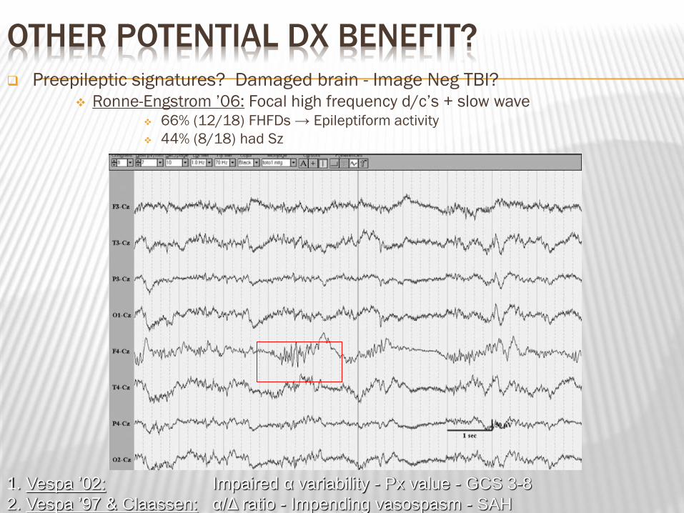
OTHER POTENTIAL DX BENEFIT?
Preepileptic signatures? Damaged brain - Image Neg TBI? Ronne-Engstrom ‟06: Focal high frequency d/c‟s + slow wave
66% (12/18) FHFDs → Epileptiform activity
44% (8/18) had Sz
1. Vespa ’02: Impaired α variability - Px value - GCS 3-8
2. Vespa ’97 & Claassen: α/Δ ratio - Impending vasospasm - SAH

ELECTROGRAPHIC (SUBCLIN/NONCONV) PTSZ
↑ ICP & METABOLIC STRESS
Results: +EPTSz group: Overall ↑ ICP & Lactate
↑ >100hrs
Conclusions: 1. PTSz NOT just benign epiphenomina!
Direct evidence: ↑ ICP, ∆ IC Hemodynamics, & potentiate metab stress
2. PTSz Therapeutic target for TBI patients
3. Consider cEEG in TBI pts w/ ICP refractory to conventional measures ↑ICP >96hrs post-injury
Detecting & Rx Sz (E/C) may improve ICP control

EARLY SEIZURES CAUSE HIPPOCAMPAL
ATROPHY Mod-Sev TBI w/ cEEG
N=140
+ acute & chronic MRI
N=29
6/29 had Sz & were
compared w/
10 controls w/o Sz
Slide courtesy of Chris
Giza, MD

43% Sz rate RF: Younger age, AHT
16% Subclinical Sz (6.9% only Subclinical Sz) RF: Younger age, AHT, & Intraaxial bleed
18.4% S.E. RF: Younger age, AHT, & Intraaxial bleed
13.8% Subclinical S.E. RF: Younger age, AHT, & Intraxial bleed
Subclinical Sz:
Lower Hospital D/c KOSCHI score
S.E. & Subclinical S.E.
Increased Hospital LOS
Lower Hospital D/c KOSCHI score

Acute Symptomatic Sz after brain injury ARE NOT BENIGN Vulnerable State
HOWEVER, No clinical class I/II trials:
Sz provoked injury affects outcome
Absolutely Chg Mgmt for these Sz
HOWEVER, Substantial evidence mounting
Acutely injured brains
EEG-only Sz
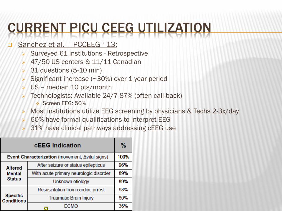
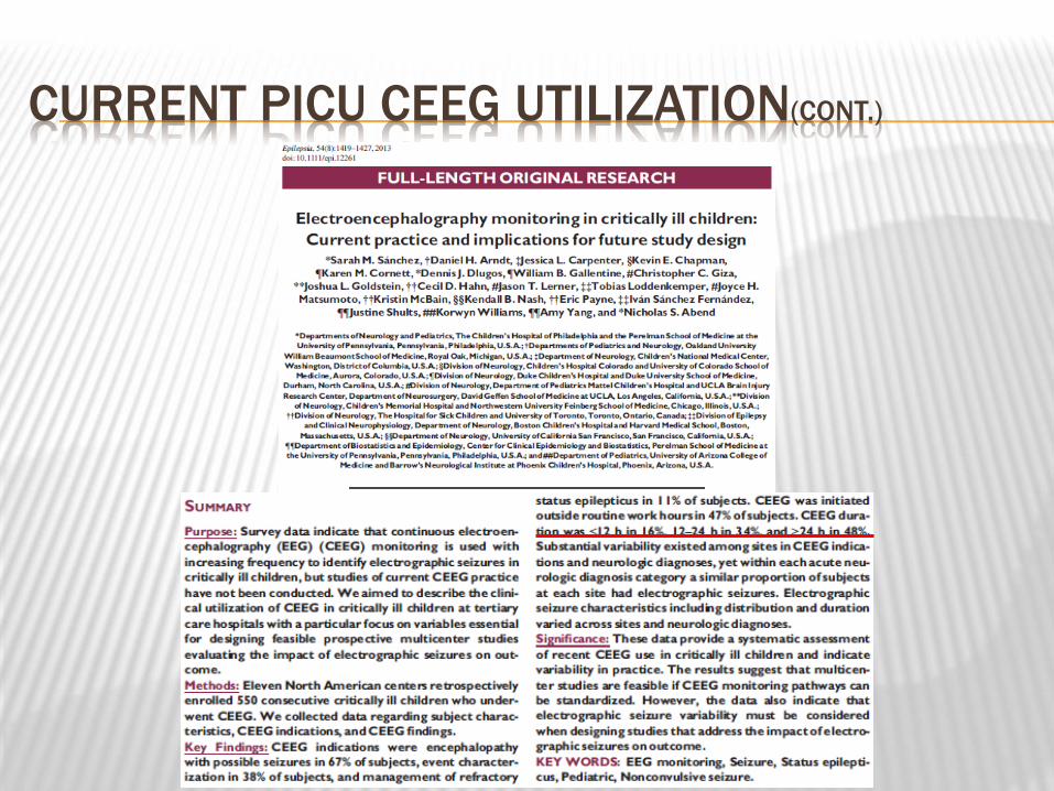
CURRENT PICU CEEG UTILIZATION Sanchez et al. – PCCEEG „ 13:
Surveyed 61 institutions - Retrospective
47/50 US centers & 11/11 Canadian
31 questions (5-10 min)
Significant increase (~30%) over 1 year period
US – median 10 pts/month
Technologists: Available 24/7 87% (often call-back) Screen EEG: 50%
Most institutions utilize EEG screening by physicians & Techs 2-3x/day
60% have formal qualifications to interpret EEG
31% have clinical pathways addressing cEEG use
CURRENT PICU CEEG UTILIZATION(CONT.)

HOW LONG DO WE MONITOR PATIENTS??
Most patients will have Sz 1-2 days monitoring
ACNS Neonate Guidelines:
High Risk Neonates – Conventional EEG x 24hrs
If Sz detected – EEG monitoring >24hrs Sz-free
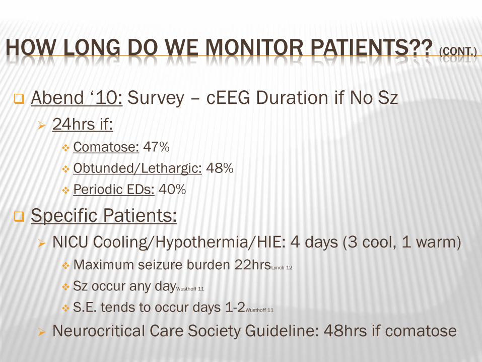
HOW LONG DO WE MONITOR PATIENTS?? (CONT.)
Abend „10: Survey – cEEG Duration if No Sz
24hrs if:
Comatose: 47%
Obtunded/Lethargic: 48%
Periodic EDs: 40%
Specific Patients:
NICU Cooling/Hypothermia/HIE: 4 days (3 cool, 1 warm)
Maximum seizure burden 22hrsLynch 12
Sz occur any dayWusthoff 11
S.E. tends to occur days 1-2Wusthoff 11
Neurocritical Care Society Guideline: 48hrs if comatose

CEEG IMPACT ON MANAGEMENT/OUTCOME
Little data
Mostly Sz impacts/does not impact outcome
Few electrographic Sz occurrence papers show it
impacts outcome

CEEG IMPACT ON MANAGEMENT/OUTCOME
NEONATAL ICU (NICU)
Van Rooj „10:
33 HIE Neonates
aEEG = ↓ Sz Burden
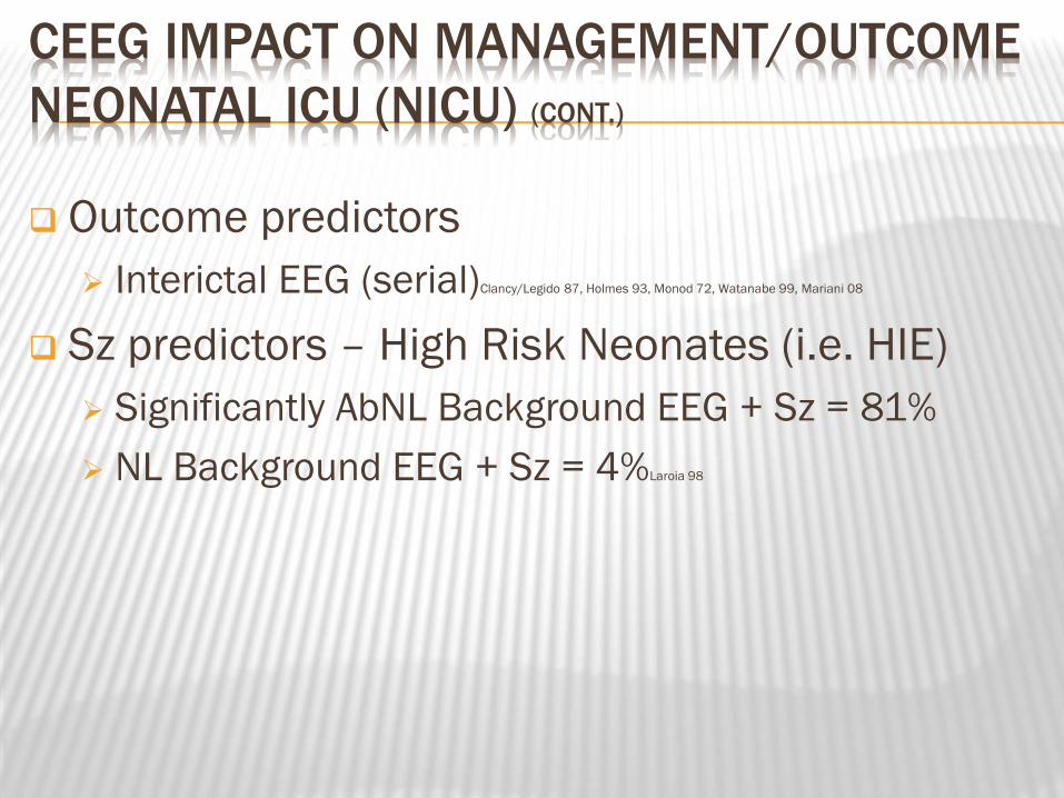
CEEG IMPACT ON MANAGEMENT/OUTCOME
NEONATAL ICU (NICU) (CONT.)
Outcome predictors
Interictal EEG (serial)Clancy/Legido 87, Holmes 93, Monod 72, Watanabe 99, Mariani 08
Sz predictors – High Risk Neonates (i.e. HIE)
Significantly AbNL Background EEG + Sz = 81%
NL Background EEG + Sz = 4%Laroia 98
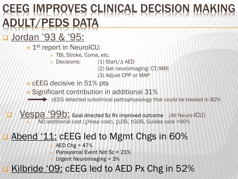
CEEG IMPROVES CLINICAL DECISION MAKING
ADULT/PEDS DATA
Jordan ‟93 & ‟95: 1st report in NeuroICU:
TBI, Stroke, Coma, etc.
Decisions: (1) Start/∆ AED
(2) Get neuroimaging: CT/MRI
(3) Adjust CPP or MAP
cEEG decisive in 51% pts
Significant contribution in additional 31% cEEG detected subclinical pathophysiology that could be treated in 82%
Vespa „99b: Goal directed Sz Rx improved outcome (All Neuro-ICU) NO additional cost (↓Hosp cost), ↓LOS, ↑GOS, Guides care >90%
Abend „11: cEEG led to Mgmt Chgs in 60% AED Chg = 47%
Paroxysmal Event Not Sz = 21%
Urgent Neuroimaging = 3%
Kilbride „09: cEEG led to AED Px Chg in 52%
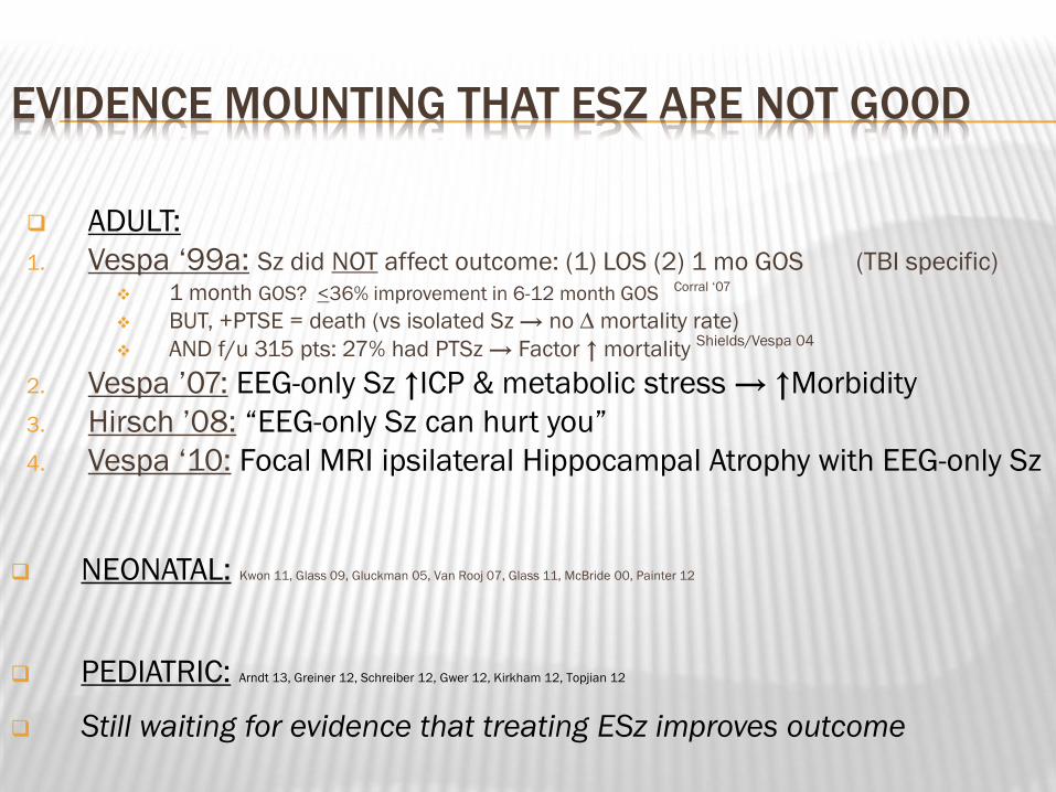
EVIDENCE MOUNTING THAT ESZ ARE NOT GOOD
ADULT:
1. Vespa „99a: Sz did NOT affect outcome: (1) LOS (2) 1 mo GOS (TBI specific)
1 month GOS? <36% improvement in 6-12 month GOS Corral „07
BUT, +PTSE = death (vs isolated Sz → no ∆ mortality rate)
AND f/u 315 pts: 27% had PTSz → Factor ↑ mortality Shields/Vespa 04
2. Vespa ‟07: EEG-only Sz ↑ICP & metabolic stress → ↑Morbidity
3. Hirsch ‟08: “EEG-only Sz can hurt you”
4. Vespa „10: Focal MRI ipsilateral Hippocampal Atrophy with EEG-only Sz
NEONATAL: Kwon 11, Glass 09, Gluckman 05, Van Rooj 07, Glass 11, McBride 00, Painter 12
PEDIATRIC: Arndt 13, Greiner 12, Schreiber 12, Gwer 12, Kirkham 12, Topjian 12
Still waiting for evidence that treating ESz improves outcome

Cited prior evidence NCSz are harmful: NCSz or Periodic d/c‟s → Independ predictors worse outcome in multiple populations
Epilepsy (w/out TBI) + Prolonged NCSz → Permanent neurologic injury, albeit rarely
NSE (neuronal injury) ↑ p NCSE (even w/out brain injury)
Pericontusional elect d/c‟s → 2º brain injury
Preclinical rat MCA occlusion stroke → NCSz → ↑ infarct & mortality
Preclinical rat pilocarpine-induced NCSE → Long-term motor & behav deficits
Hemorrhagic stroke + NCSz → ↑ ML shift (28% incidence)
Mitchell ‟02: Pediatric SE paper cited similar reasons to argue for treating NCSE
NCSE: Delayed Dx & Duration - Independent predictors of worse outcome Shneker 03 Duration: <10hrs (10% death) >20hrs (85%)
Delay in Dx: <30min (36% death) >24hrs (75%)
Etiology: Epilepsy related (3%) & Cryptogenic (18%) Acute Symp (27% death)
In contrast, Aggressive Rx often required in critically ill to stop NCSz Potentially harmful → Ongoing controversy → “Rx or No Rx?”

NEUROCRITICAL CARE SOCIETY
GUIDELINE FOR STATUS EPILEPTICUSBROPHY 12
S.E.: >5min (1) continuous clinical and/or electrographic Sz activity (2) recurrent Sz activity w/out recovery (baseline) between Sz
S.E. Treatment: Should occur rapidly & continue sequentially until electrographic Sz are halted
cEEG is usually required for treatment of S.E.
cEEG should be initiated <1hr S.E. onset:
If ongoing Sz suspected
Duration of cEEG monitoring: 48hrs in comatose
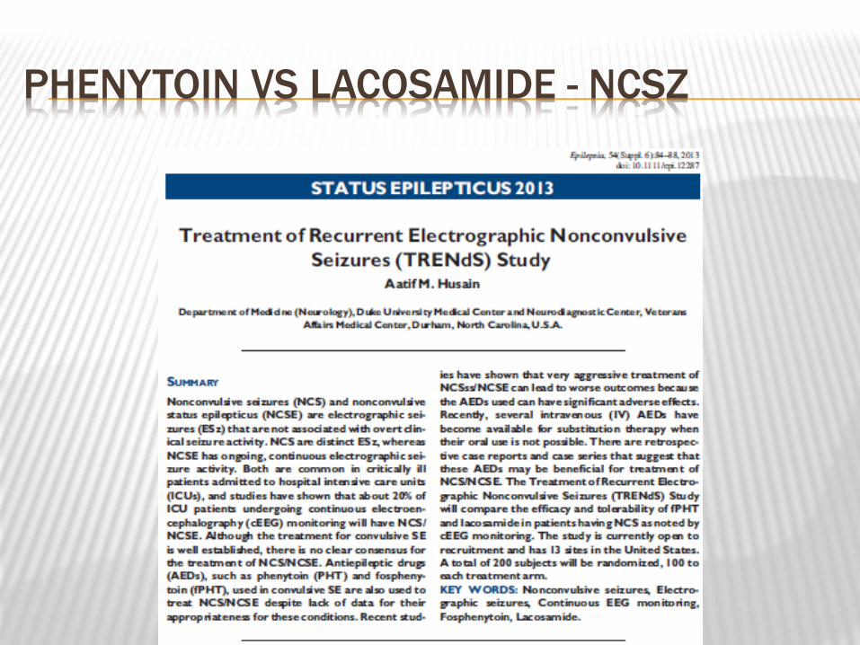
PHENYTOIN VS LACOSAMIDE - NCSZ

ACNS GUIDELINES
Critical Care EEG Terminology
Adult: J Clin Neurophys Volume 30, Number 1, 2013
Neonate: Volume 30, Number 2, 2013
cEEG Monitoring Guidelines
Neonate
Pending: Children & Adult
*Update cEEG monitoring PICU / NICU
J Clin Neurophys Volume 30, Number 2, 2013

ADDITIONAL ICU EEG ISSUES
Ictal-Interictal Continuum
Nomenclature, Significance
EEG background / prognosis
Guide for real-time Mgmt
Quantitative EEG / Persyst / Trending
Efficient Sz identification
Identification of interval interictal background chgs
THANK YOU!
