positioning in anaesthesia mgmc
TRANSCRIPT
Positioning in anaesthesia
Dr. S. Parthasarathy MD., DA., DNB, MD (Acu),
Dip. Diab. DCA, Dip. Software statistics PhD (physio)
Goals
• Avoid pressure on the chest cavity• To maintain circulation• To prevent nerve damage• To maintain patient’s airway • To provide adequate exposure of the
operative site• To provide comfort and safety to the patient
Positions – common
• Patient is not aware of the damage and he cant tell that my eye is getting compressed
• supine,• lithotomy, • sitting,• head-down,• prone, • lateral decubitus
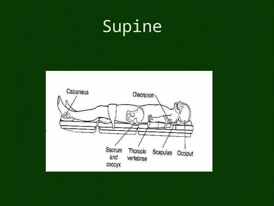
Supine
Supine
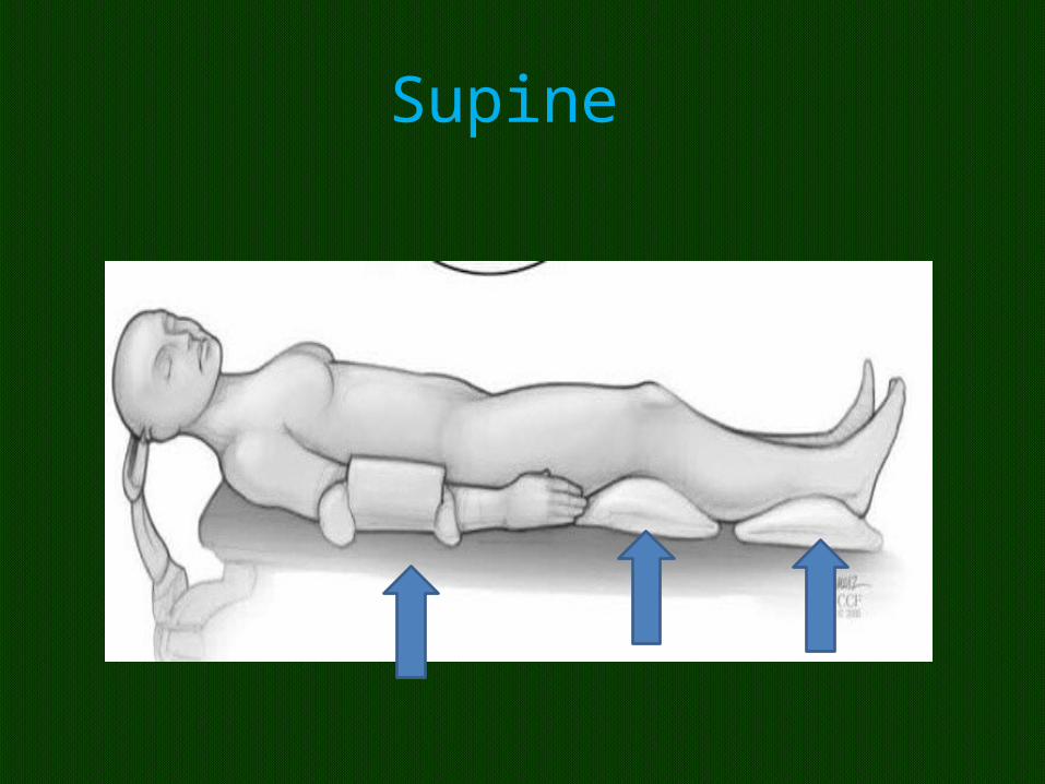
• We spent most of our life like this • Be careful • patients with morbid obesity, • mediastinal masses, • poor cardiac function and • term parturients prone to aortocaval
compression• .
Supine
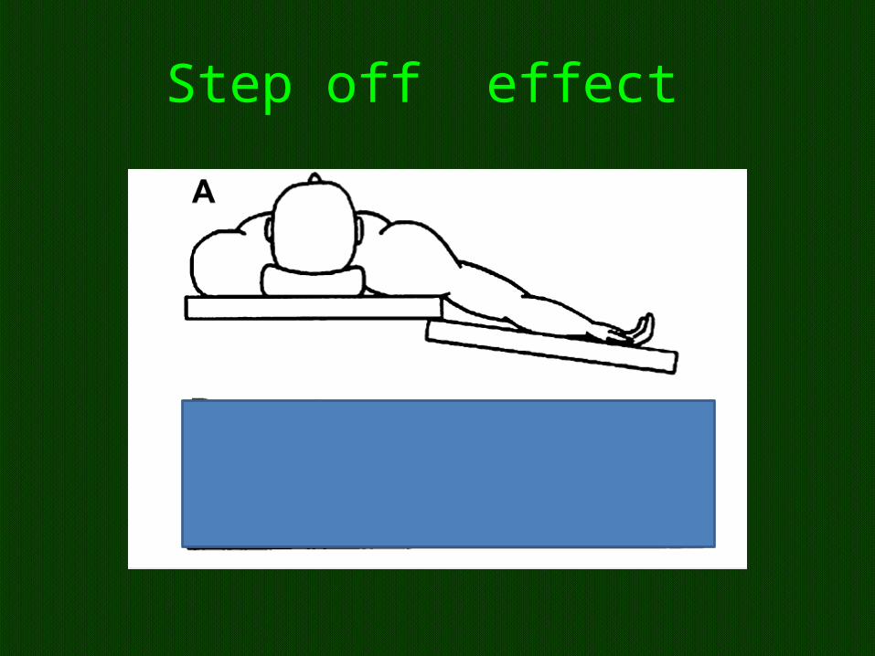
Step off effect
Supine
• prolonged contact of the back of the head may result in alopecia
• ulnar neuropathy is the most common- males • 0.25 % may be delayed upto 3 days • Brachial plexus, femoral cutaneous nerves are
next common. • Brachial or ulnar ??
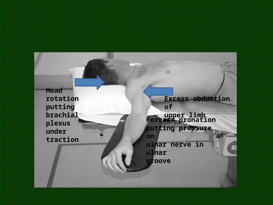
Head rotationputting brachialplexus undertraction
Excess abduction ofupper limb
Forearm pronationputting pressure onulnar nerve in ulnargroove
CVS in supine
• MAP, heart rate (HR),venous return rises peripheral vascular resistance decrease
• cardiac output and stroke volume increase.
• Offset anaesthetic action
RS
• cephalad movement of the abdominal contents.• The main complications are airway obstruction
and decreased tidal volumes• The resulting reduction in functional residual
capacity (FRC) is detrimental to gas exchange• increase in ventilation–perfusion mismatching
and decrease in pulmonary compliance.
• loss of the natural lumbar lordosis • associated with postoperative low back pain.
• The occiput, sacrum and heel are at risk of developing pressure sores
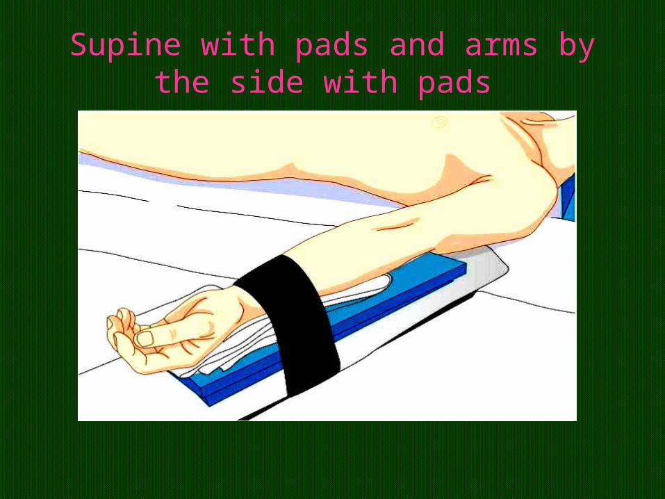
Supine with pads and arms by the side with pads
Lawn Chair Position
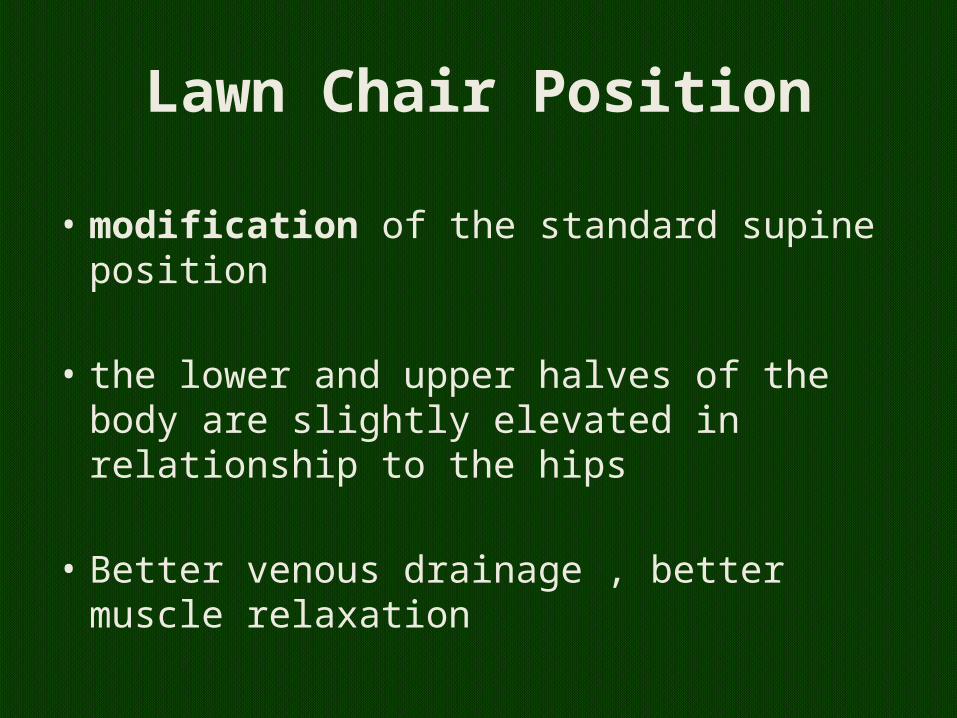
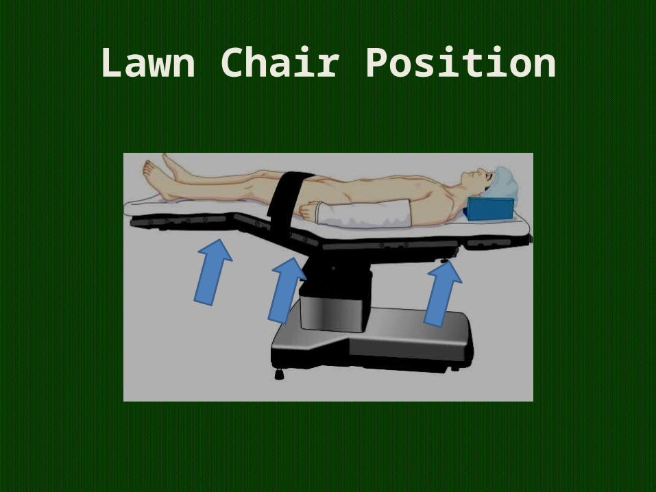
Lawn Chair Position
• modification of the standard supine position
• the lower and upper halves of the body are slightly elevated in relationship to the hips
• Better venous drainage , better muscle relaxation
beach chair position

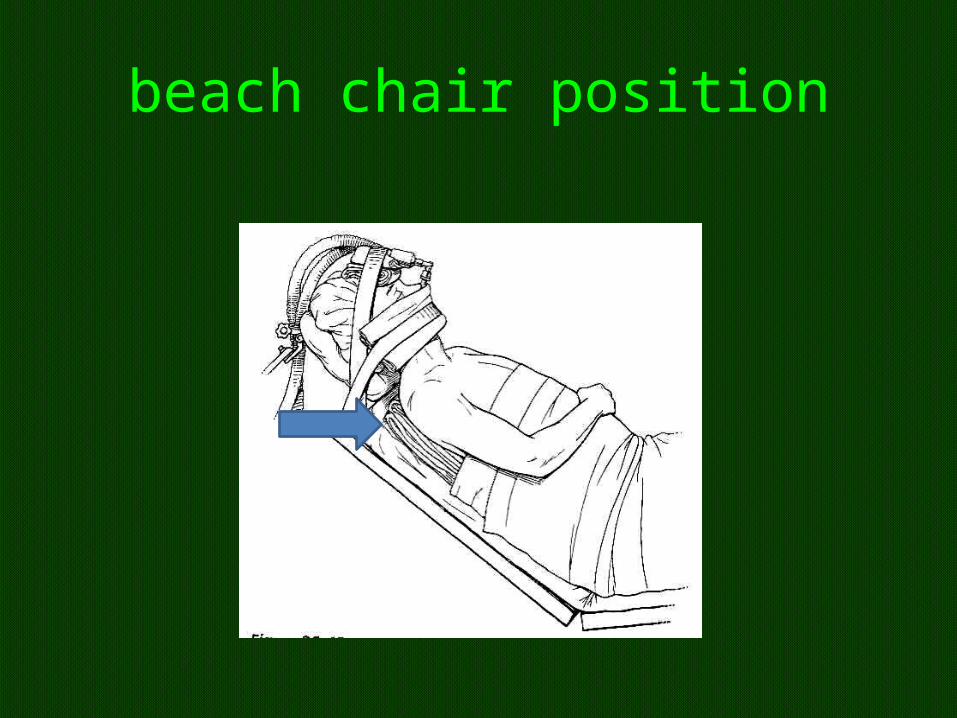
beach chair position
• beach chair position is associated with the risk for cerebral underperfusion.
• Blood pressure must be maintained at a level that guarantees a perfusion pressure of 60 to 70 mm Hg measured at the level of the foramen magnum
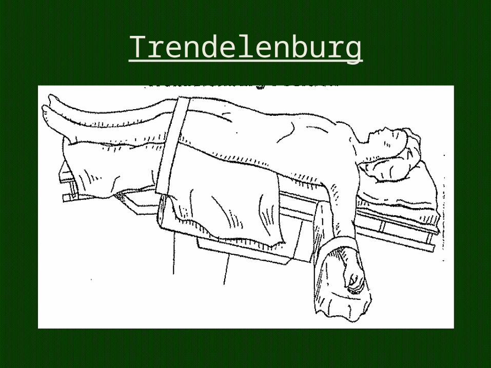
Trendelenburg
Trendelenburg• Central blood volume increase by 1 litre.
swelling of the face, conjunctiva, larynx, and tongue ?? postoperative upper airway obstruction.
• The cephalic movement of abdominal viscera against the diaphragm also decreases functional residual capacity and pulmonary compliance.
May 3, 2023 20
Effects of Trendelenberg’ s position
• ↑ CVP• ↑ ICP• ↑ IOP• ↑ myocardial work• ↑ pulmonary venous pressure• ↓ pulmonary compliance• ↓ FRC• Swelling of face, eyelids, conjunctiva & tongue observed in long surgeries
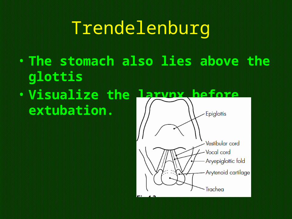
Trendelenburg
• The stomach also lies above the glottis• Visualize the larynx before extubation.
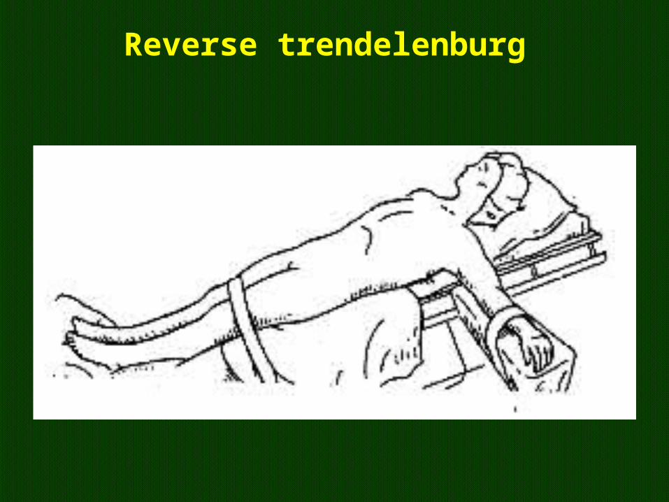
Reverse trendelenburg
Reverse Trendelenburg position(head-up tilt)
• to facilitate upper abdominal surgery by shifting the abdominal contents caudad.
• This position is popular because of the growing number of laparoscopic surgeries.
• slipping on the table, • monitoring of arterial blood pressure.
Reverse Trendelenburg position
• hypotension and increased risk of venous air embolism (VAE).
• the position of the head above the heart reduces perfusion pressure to the brain
Lithotomy
• This position is most often used for• genitourinary, gynecologic, and colorectal• Procedures.• Hips flexed 100 deg 30-40 deg. abduction at
the hips • . Knees 90 approx 30 deg
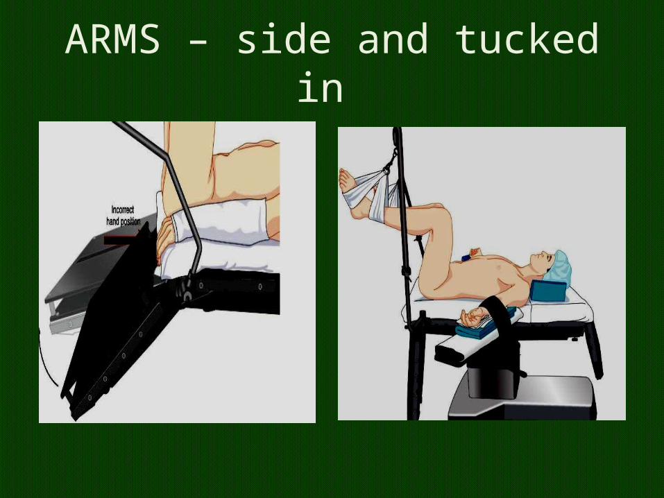
ARMS – side and tucked in
Martin and Warner have proposed a standardized classification
• low, standard, • high,• hemi, • exaggerated, • tilted• Martin JT, Warner MA (Eds): Positioning in
Anesthesia and Surgery, 3rd edition. Philadelphia, WB Saunders, 1997
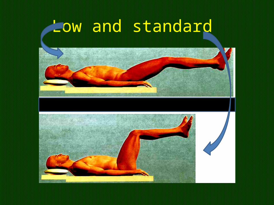
Low and standard
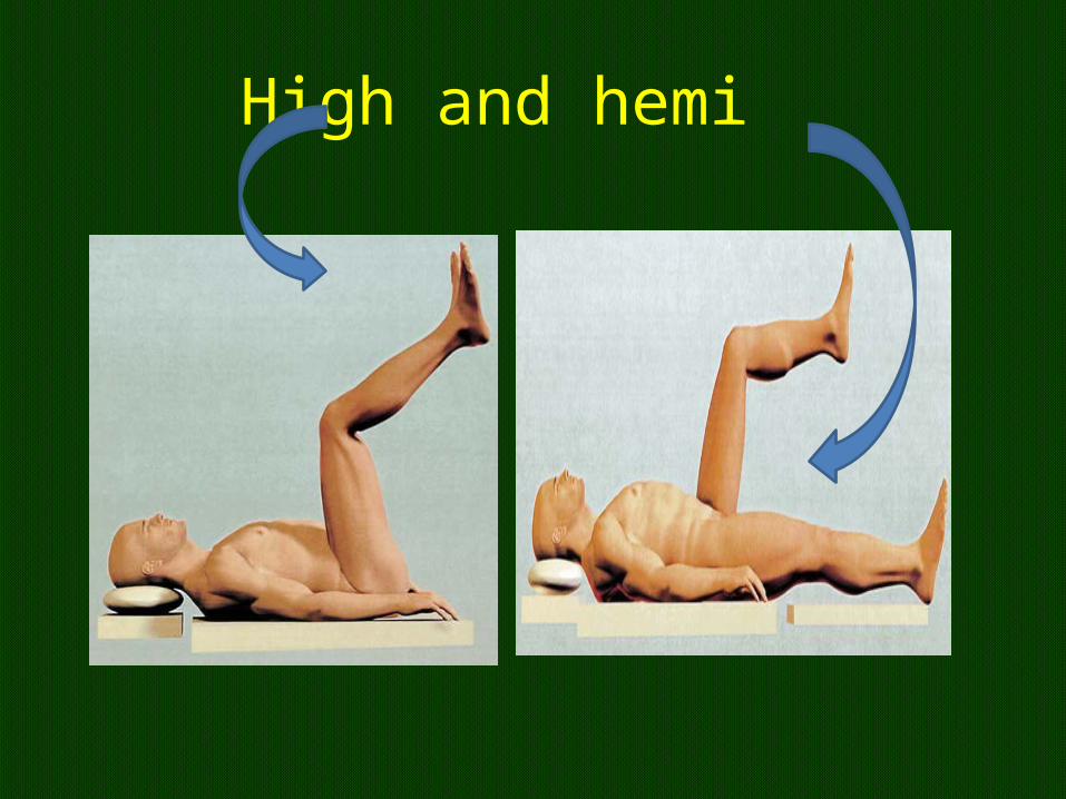
High and hemi
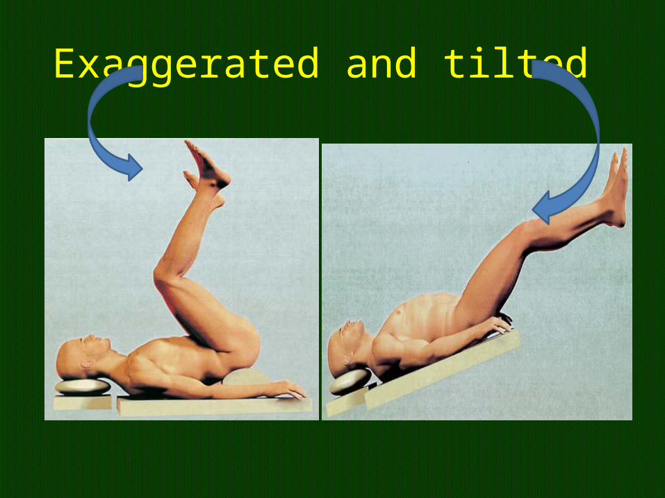
Exaggerated and tilted
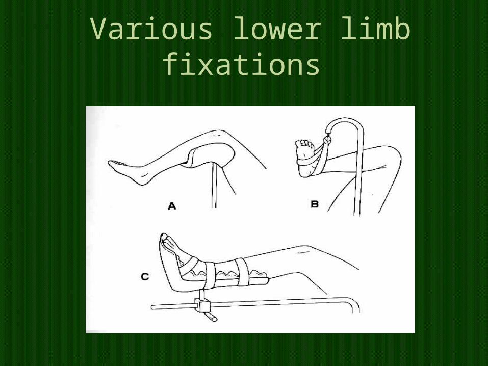
Various lower limb fixations
Lithotomy
• coordinated positioning of the lower extremities by two assistants to avoid torsion of the lumbar spine.
• Both legs should be raised together, flexing the hips and knees simultaneously.
• Slow removal • Hands beware
lithotomy from the supine position
• Unanticipated stimulation of the carina with bronchospasm or endobronchial intubation may result.
• In the lithotomy position, calf compression is almost inevitable and this predisposes to venous thrombo embolism and compartment syndrome ( surgery > 5 hours)
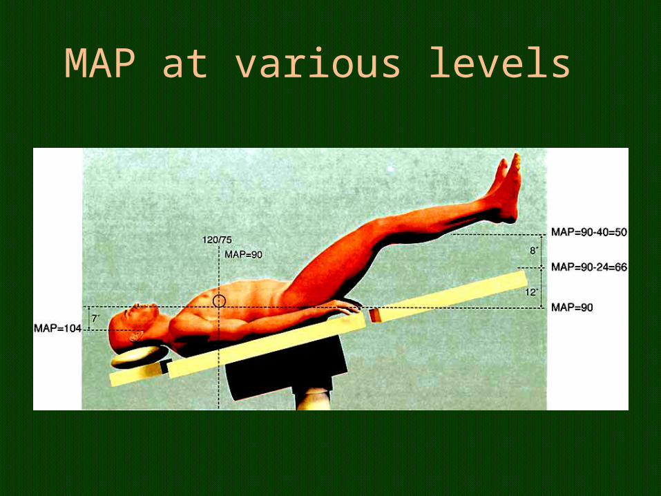
MAP at various levels
Lithotomy
• Lower extremity compartment syndrome is a rare complication associated with the lithotomy position.
• perfusion to an extremity is inadequate, resulting in ischemia, edema
extensive rhabdomyolysis from increased tissue pressure within a fascial compartment
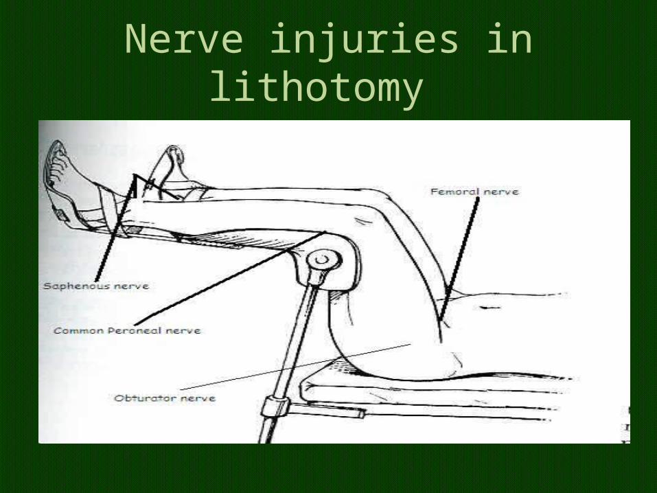
Nerve injuries
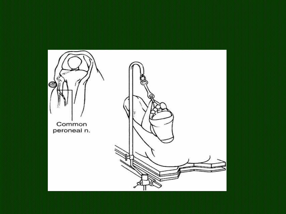
• injury to the common peroneal nerve was the most common lower extremity motor neuropathy, representing 78% of nerve injuries.
• A potential cause of the injury was the compression of the nerve between the lateral head of the fibula and the bar holding the legs.
Nerve injuries in lithotomy
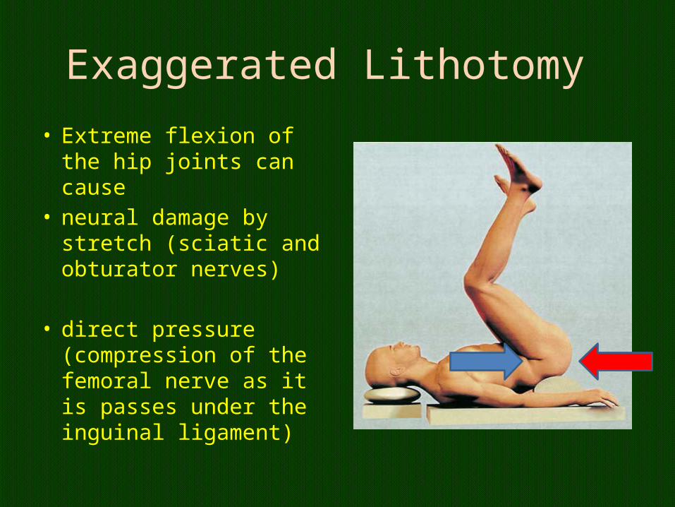
Exaggerated Lithotomy • Extreme flexion of the hip
joints can cause• neural damage by stretch
(sciatic and obturator nerves)
• direct pressure (compression of the femoral nerve as it is passes under the inguinal ligament)
Hemodynamics and RS
• preload increases, transient increase in cardiac output
• Cerebral venous and intracranial pressure in otherwise healthy patients.
• causes the abdominal viscera to displace the diaphragm cephalad, reducing lung compliance and potentially resulting in a decreased tidal volume
The frog-leg position
• hips and knees are flexed• hips are externally rotated with the soles of the
feet facing each other, • allows access to the perineum, medial thighs,
genitalia, and rectum.• Care must be taken to minimize stress and
postoperative pain in the hips and prevent dislocation by supporting the knees appropriately
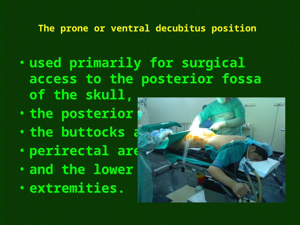
The prone or ventral decubitus position
• used primarily for surgical access to the posterior fossa of the skull,
• the posterior spine, • the buttocks and • perirectal area, • and the lower • extremities.
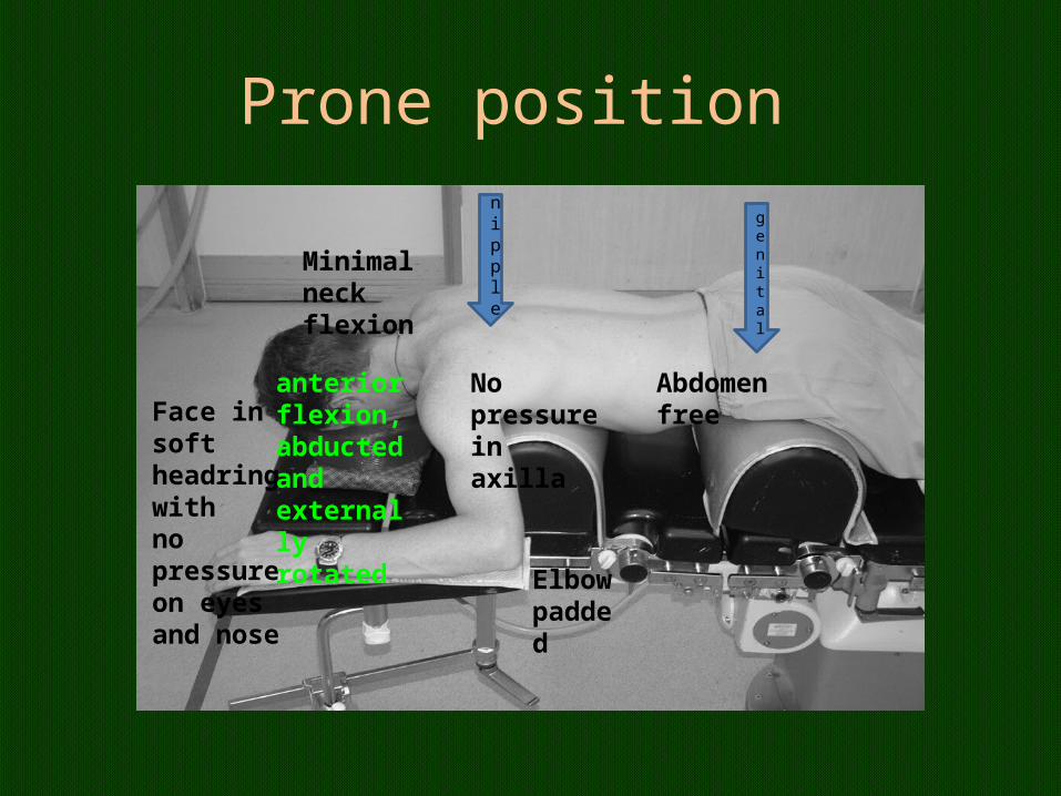
Prone position
Minimal neckflexion
Face insoft headringwithnopressureon eyesand nose
Elbowpadded
No pressure inaxilla
Abdomenfree
anterior flexion, abducted andexternally rotated
genital
nipple
Abdomen pressure in prone
• inferior vena caval compression,• reduced venous return and subsequent poor
cardiac output.• Associated pulmonary problems are caused by
an increase in transdiaphragmatic pressure leading to reduced thoracic compliance.
RS – better
• An increase in FRC, changes in diaphragmatic excursions and improved ventilation–perfusion matching can significantly improve oxygenation in the prone position.
• for treatment of refractory hypoxaemia and in early ARDS
• 70–80% of patients turned prone initially benefit from improved oxygenation
Prone position
• Complete obstruction of the contralateral• vertebral blood flow with rotation of the head
>80• Beware in old CVAs • ‘Concorde’ position with the neck flexed and
the chin approximately one finger-breadth from the sternum
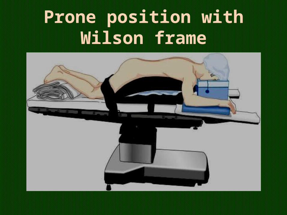
Prone position with Wilson frame
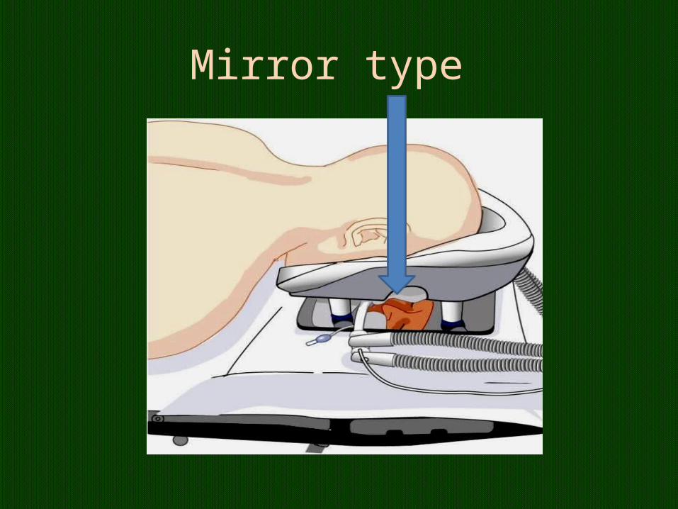
Mirror type
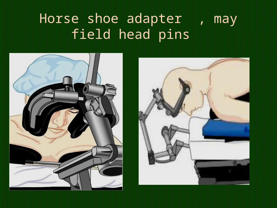
Horse shoe adapter , may field head pins
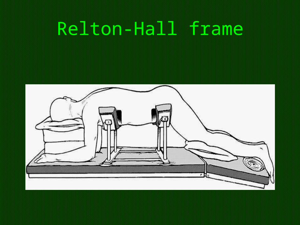
Relton-Hall frame
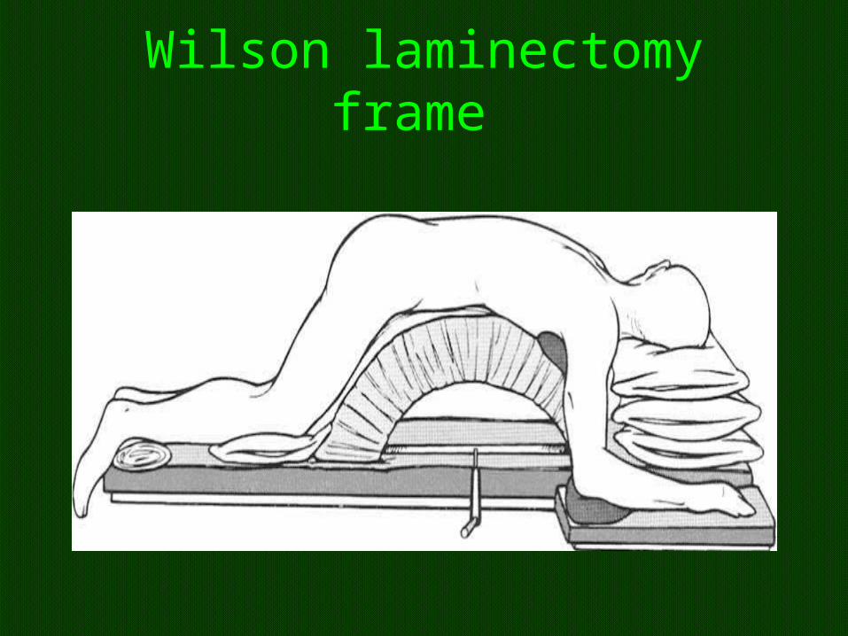
Wilson laminectomy frame
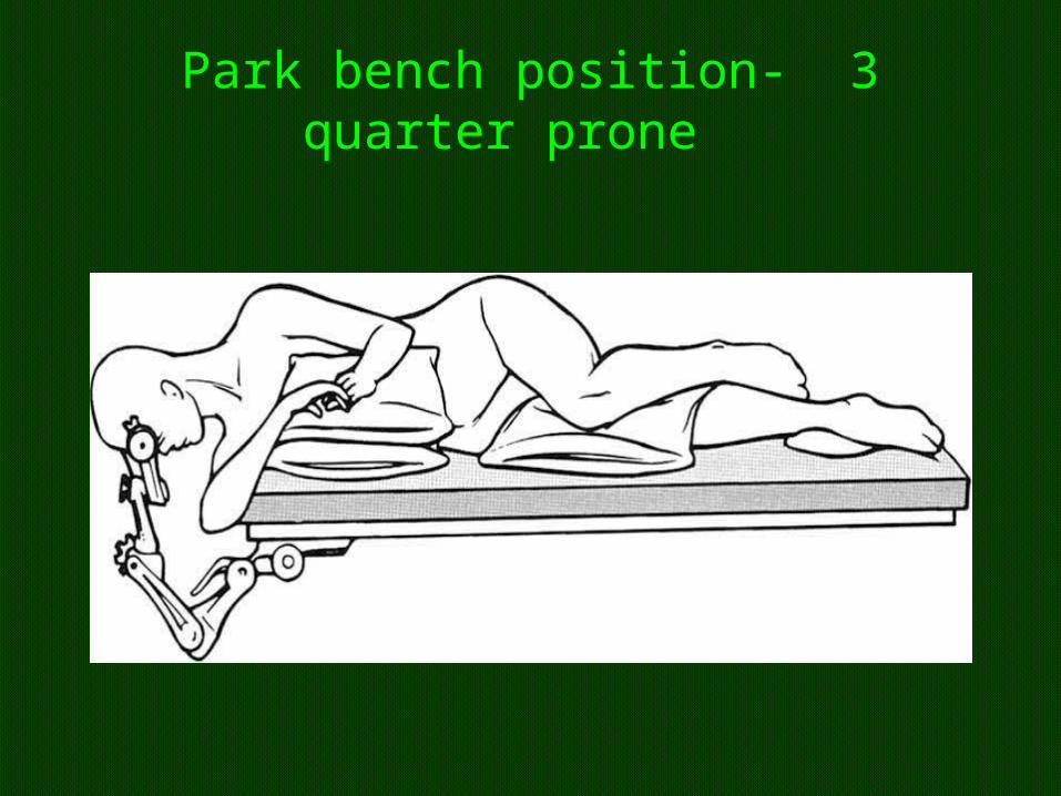
Park bench position- 3 quarter prone
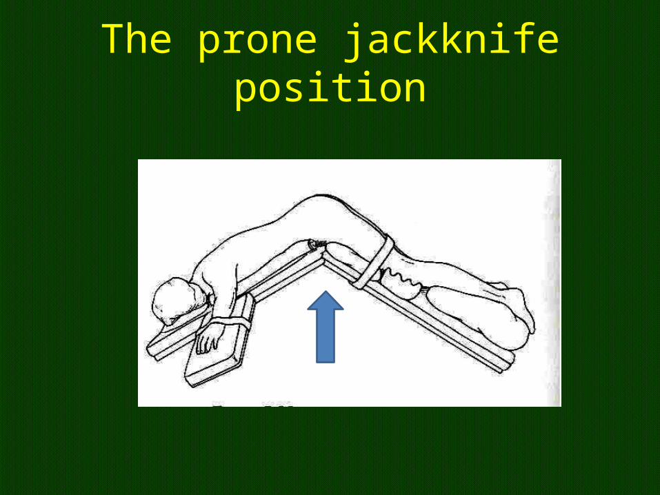
The prone jackknife position
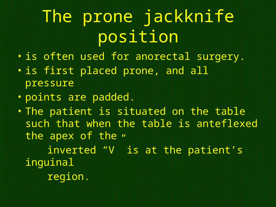
The prone jackknife position
• is often used for anorectal surgery. • is first placed prone, and all pressure• points are padded. • The patient is situated on the table such that
when the table is anteflexed the apex of the inverted “V” is at the patient’s inguinal region.
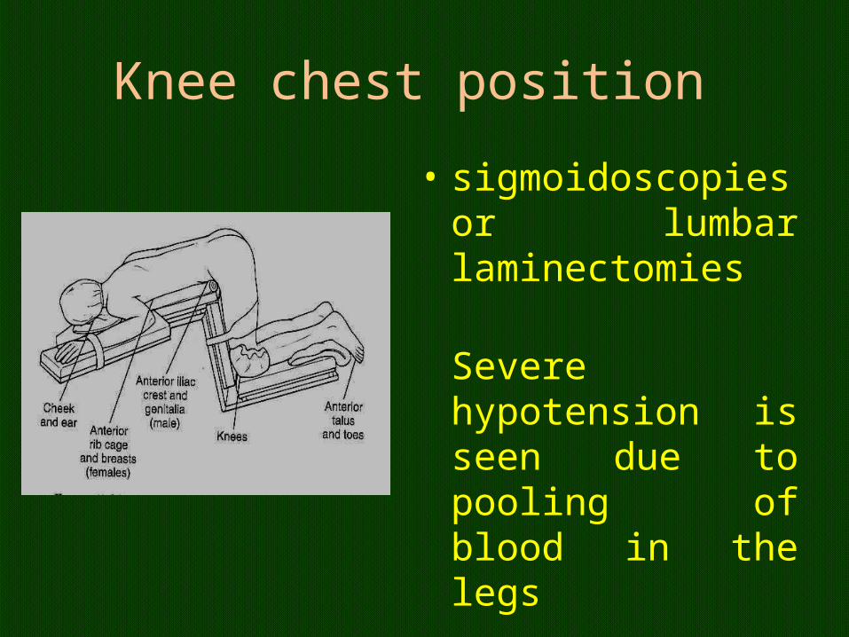
Knee chest position
• sigmoidoscopies or lumbar laminectomies
Severe hypotension is seen due to pooling of blood in the legs
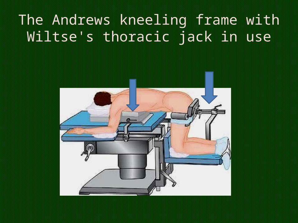
The Andrews kneeling frame with Wiltse's thoracic jack in use
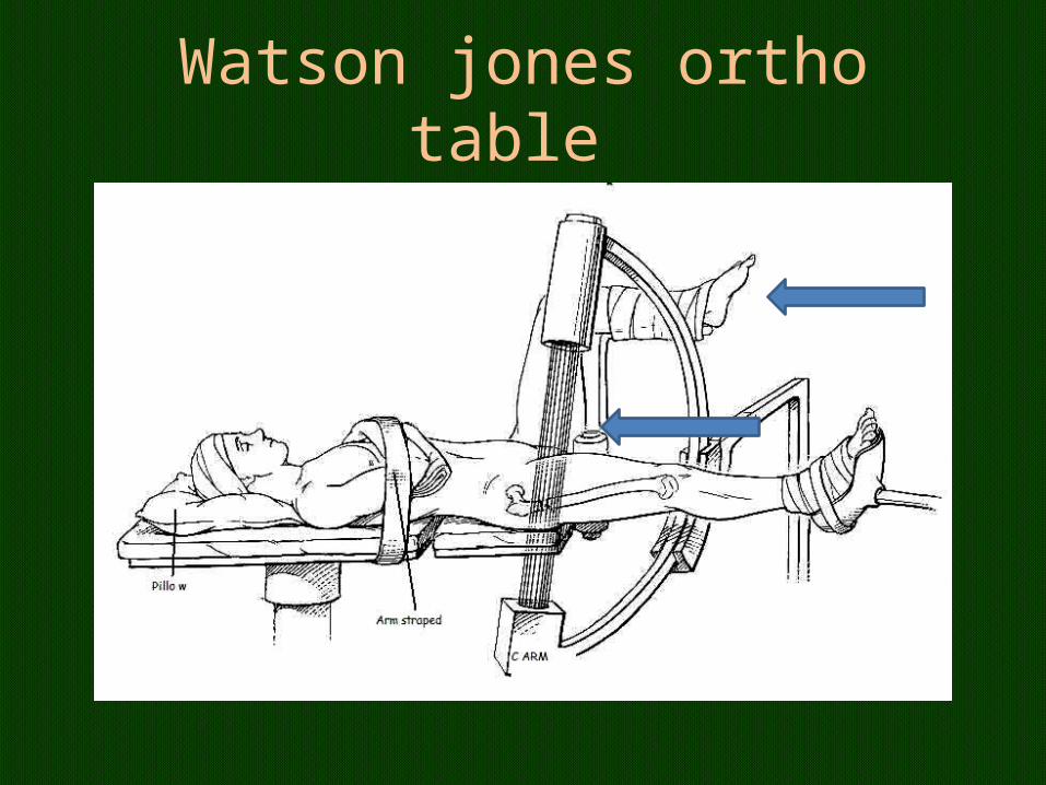
Watson jones ortho table
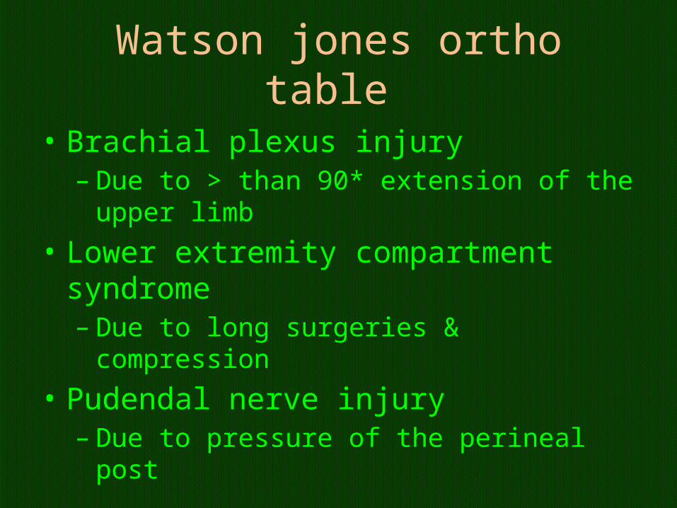
Watson jones ortho table
• Brachial plexus injury– Due to > than 90* extension of the upper limb
• Lower extremity compartment syndrome– Due to long surgeries & compression
• Pudendal nerve injury– Due to pressure of the perineal post
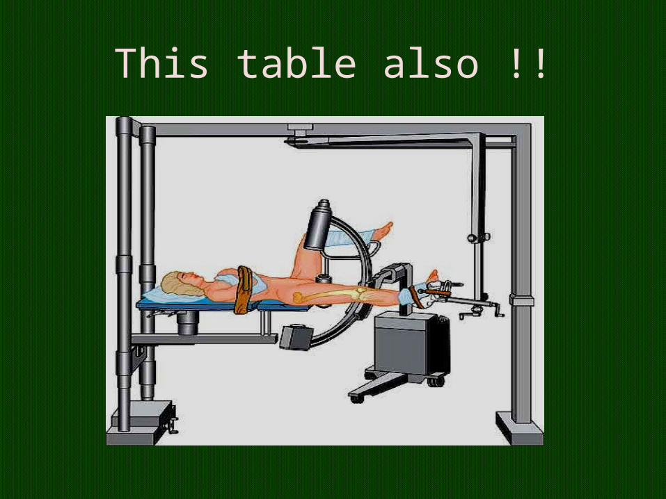
This table also !!
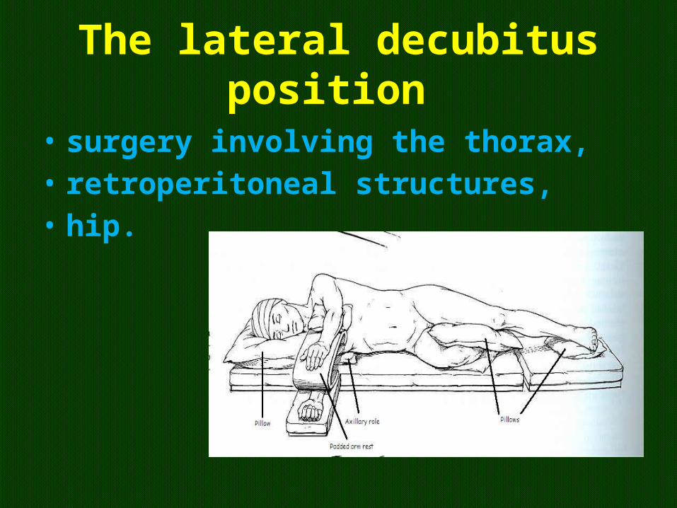
The lateral decubitus position
• surgery involving the thorax,• retroperitoneal structures, • hip.
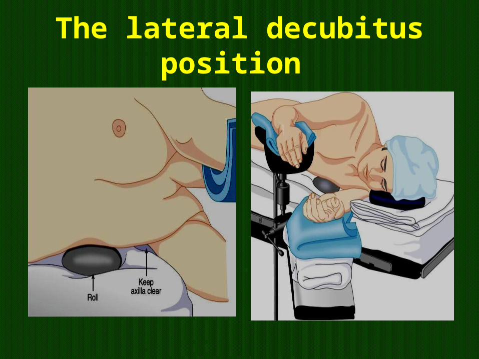
The lateral decubitus position
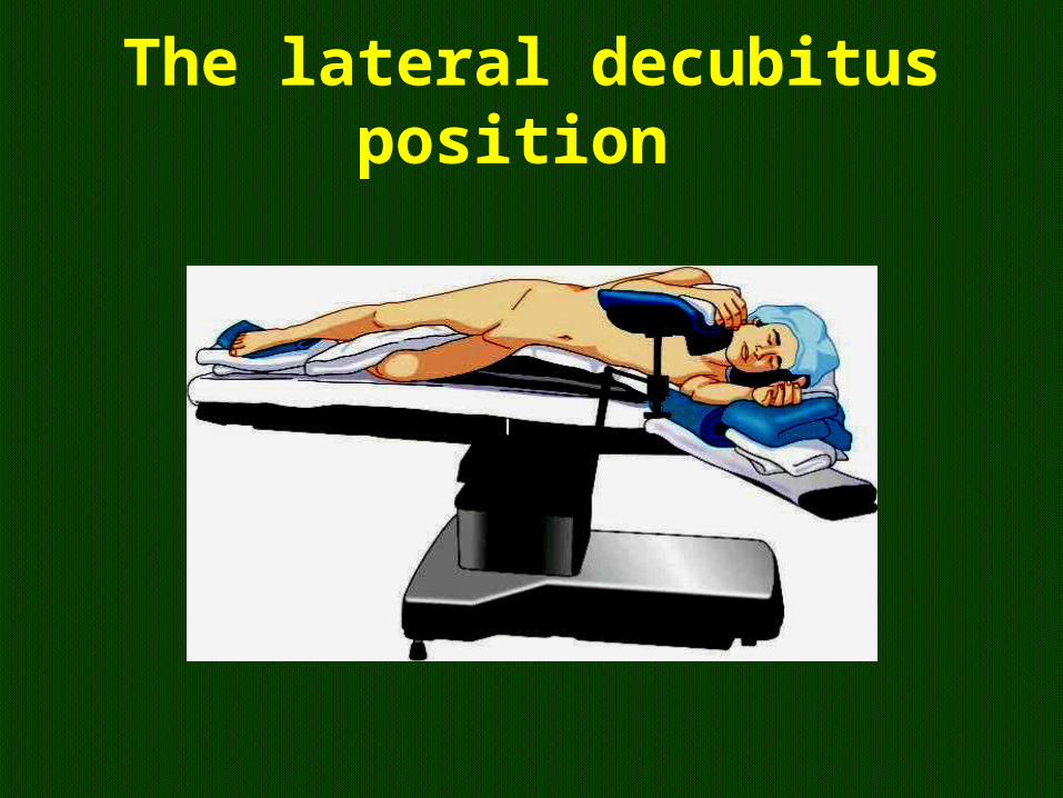
The lateral decubitus position
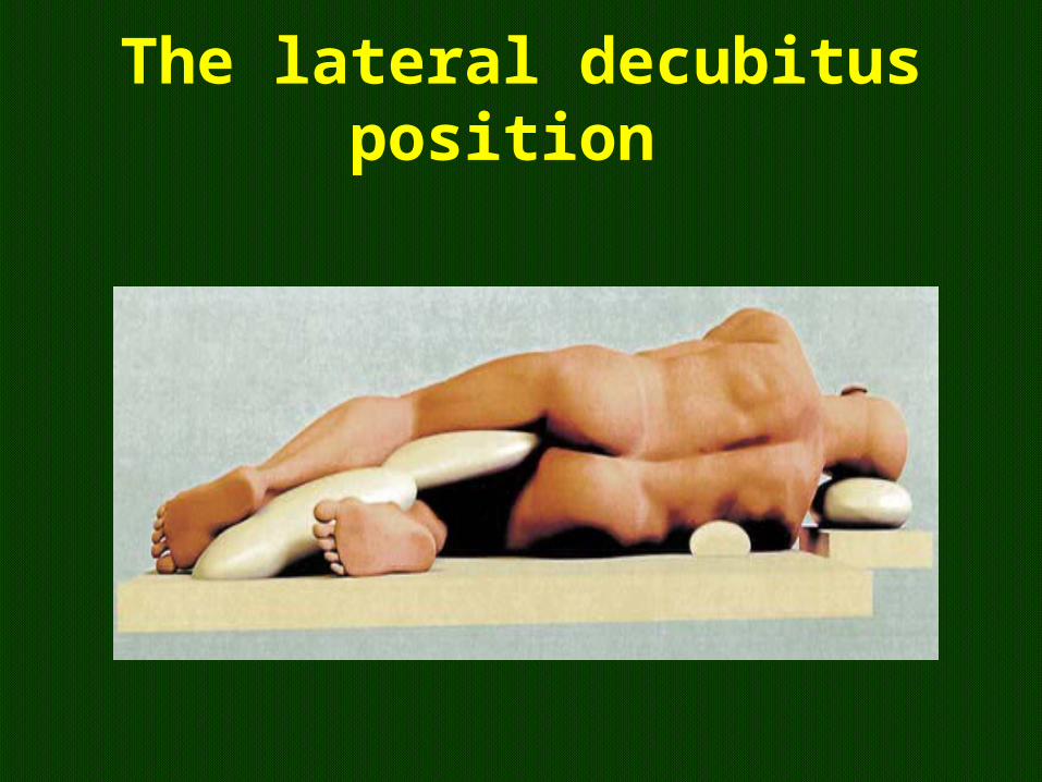
The lateral decubitus position
The lateral decubitus position
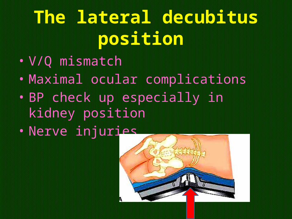
• V/Q mismatch • Maximal ocular complications • BP check up especially in kidney position • Nerve injuries

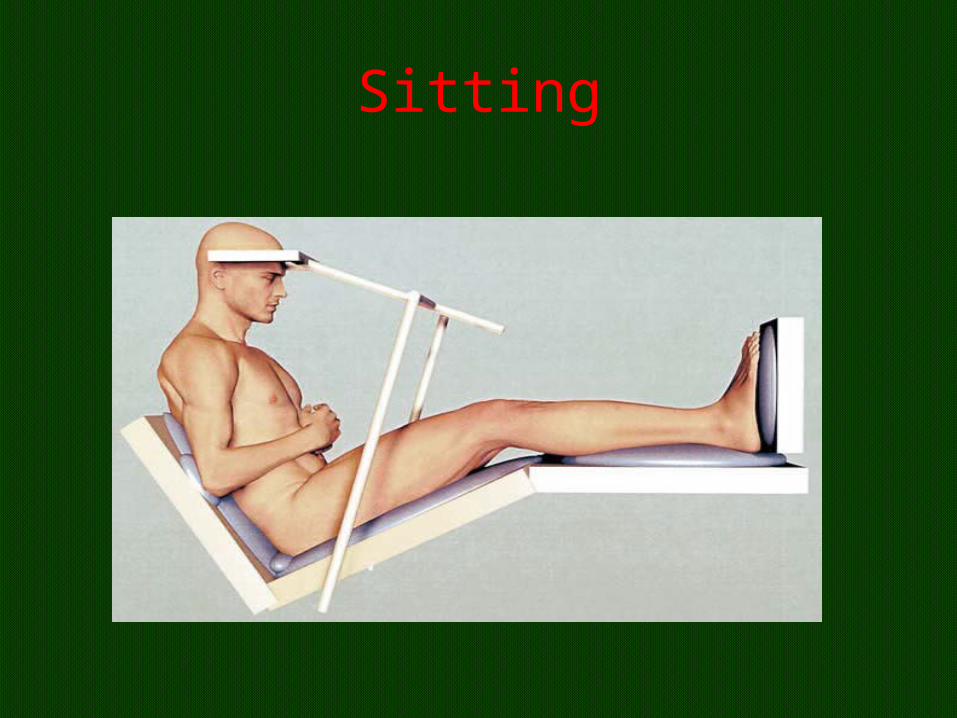
Sitting
• Not frequently used • Craniotomy • Venous return decrease and cardiac output
decrease • HR no change
• Venous air embolism
Sitting
Sitting
• overall increase in ventilation with increased VC and FRC.
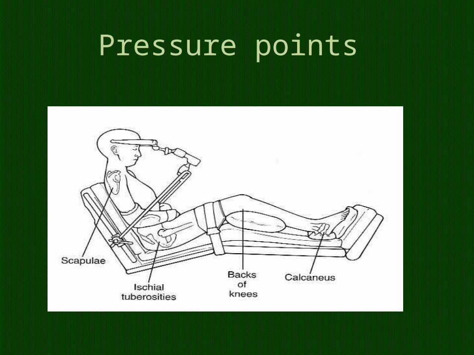
Pressure points
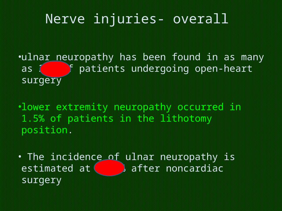
Nerve injuries- overall
• ulnar neuropathy has been found in as many as 26% of patients undergoing open-heart surgery
• lower extremity neuropathy occurred in 1.5% of patients in the lithotomy position.
• The incidence of ulnar neuropathy is estimated at 0.46% after noncardiac surgery
Overall mechanism of nerve injuries
• (i) stretch,• (ii) compression,• (iii) generalized ischaemia, • (iv) metabolic derangement.
all predispose to perioperative nerve injury
• Peripheral vascular disease, • diabetes, • hereditary neuropathy, and • anatomic variation (eg, cervical rib),
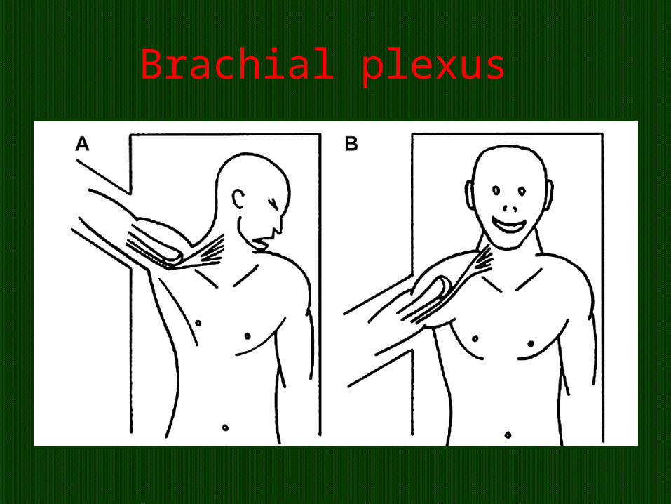
Brachial plexus
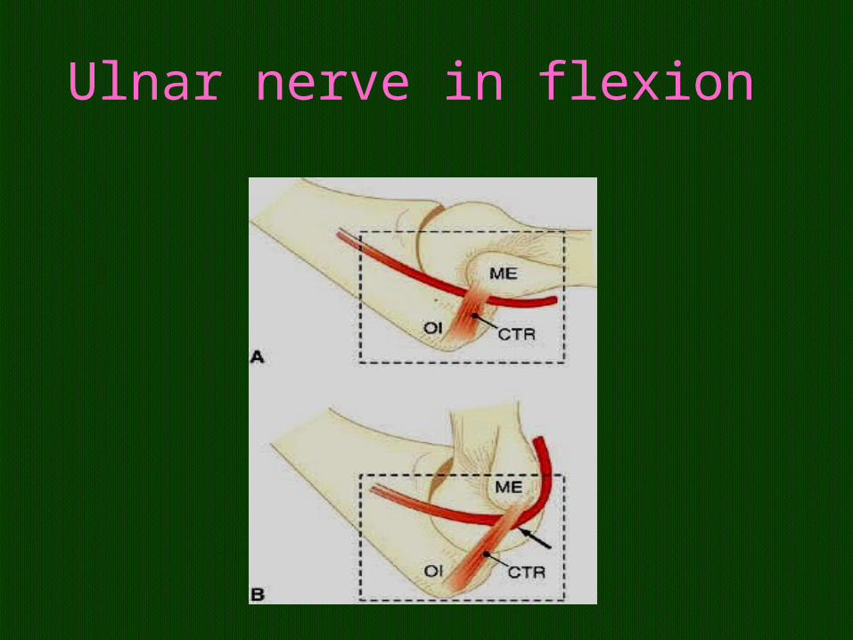
Ulnar nerve in flexion
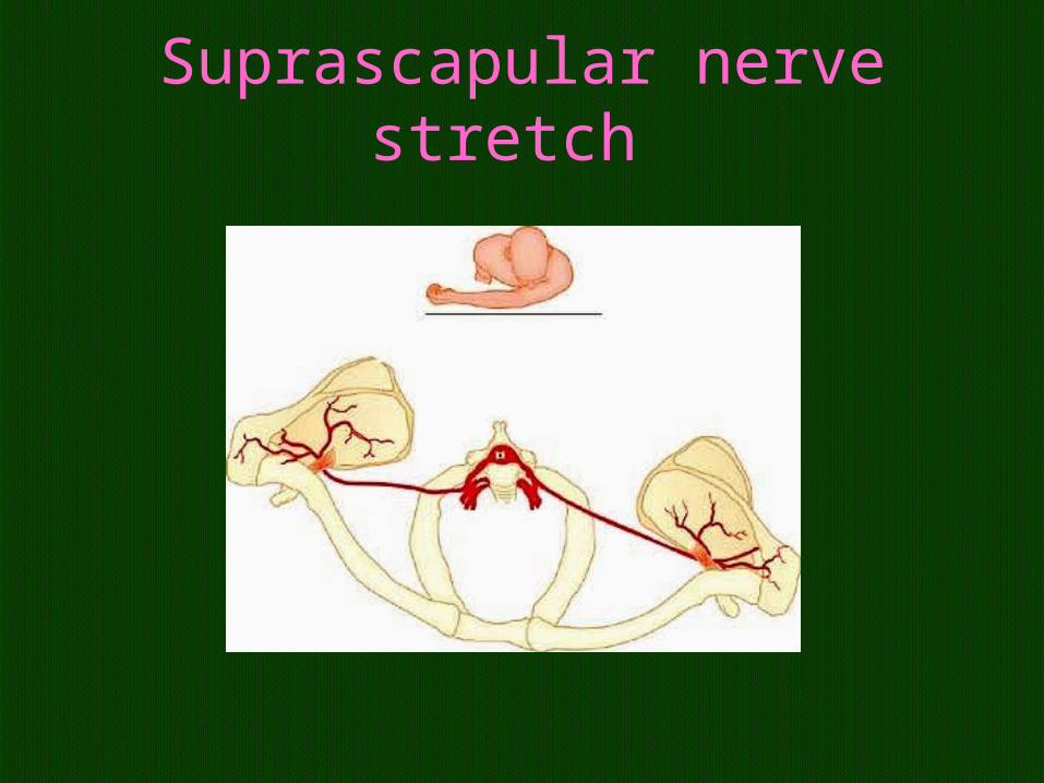
Suprascapular nerve stretch
Wedge in pregnant
• A rare complication of this positioning is sciatic neuropathy, suggesting that time in this position should be minimized
• Early intervention within 48 hours with EMG studies
• no significant difference in the incidence of ulnar neuropathy in patients undergoing general anaesthesia, regional anaesthesia or sedation.
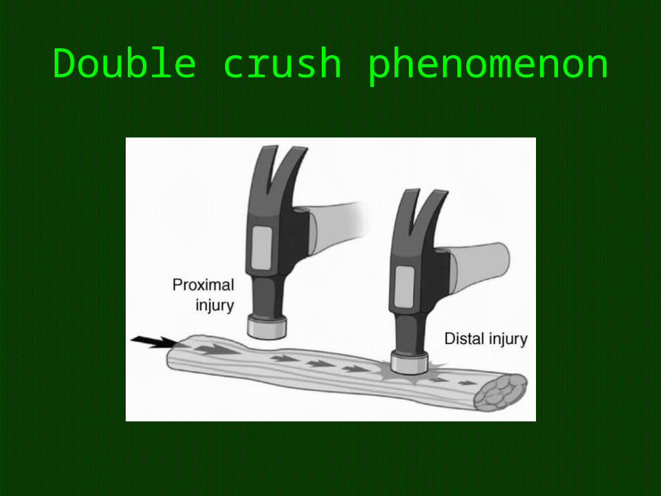
Double crush phenomenon
Effects of Positioning - Obese Patients
• Lateral:–Well tolerated– Correct sizing and placement of axillary roll is
important– Ensure that pendulous abdomen does not hang
over side of OR bed• Head-Up: (Reverse Trendelenburg/Semi-recumbent)–Most safe–Weight of abdominal contents unloaded from
diaphragm– Use of well-padded footboard to prevent sliding
Ocular injuries
• The frequency of eye injury during anaesthesia and surgery is very low (<0.1% of anaesthetics),
• As little as 10 min• Corneal abrasions, periorbital,and conjunctival
edema, ocular hemorrhage,• vitreous loss, retinal detachment,• central retinal artery occlusion,• ischemic optic neuropathy
Causes
• Patient movement,• chemical irritation from prep solutions,• direct trauma from face mask,• pressure from the laryngoscopic blade,• pressure effects on the globe from lateral• and prone positioning, (duration ) • intraoperative hypotension, and anemia
Contributing patient comorbidconditions
• hypertension, diabetes,• obesity, smoking history,
hypercholesterolemia,• alcohol abuse, atherosclerosis,• anemia, Graves disease,• and renal transplantation• Tape ok !!! Ointment ??
Don’t Forget:• Good positioning starts with an assessment• Prevent surgical team members from leaning• Arm board pads should be level with table pads• Cushioning of all pressure points is a priority -• Procedures longer than 2 ½ to 3 ??• During a longer procedure, shifting the patient,
adjusting the table, or adding/removing a positioning device
• assess extremities at regular intervals for signs of circulatory compromise
• Documentation of the positioning process- accurate and complete
Summary
• Change – check all • Cardiac • Respiratory• Nerve injuries • Pressure sores • Visual loss • Follow up for some days
Comfortable position
Uncomfortable position
Uncomfortable position but happy
Thank you all