respiratory system 2nd
TRANSCRIPT

Respiratory
System
2nd
Practical Pulmonary
Pathology
A Diagnostc Approach 2005

COVID-192021-03-01

A koronavírusok lipidburokkal rendelkező, egyszálú
RNS vírusok, amelyek légzőszervi és bélrendszeri
infekciókat okoznak állatokban és emberekben. A
humán koronavírusok közül négy folyamatosan jelen
van környezetünkben, légzőszervi megbetegedéseket
okoz, endemikus megjelenésük a téli hónapokra
tehető.
Coronaviruses are enveloped, nonsegmented, single-stranded, positive-sense RNA viruses that have a characteristic appearance on electron microscopy negative staining

Guarner J. Three Emerging Coronaviruses in Two Decades. Am J Clin Pathol. 2020 Mar 9;153(4):420-421. doi: 10.1093/ajcp/aqaa029. PMID: 32053148; PMCID: PMC7109697.
SARS-CoV and MERS-CoVhave had different behaviors, SARS-CoV-2 will likely have unique features of its own


Cui J, Li F, Shi ZL. Origin and evolution of pathogenic coronaviruses. Nat Rev Microbiol. 2019 Mar;17(3):181-192.
Animal origins of human coronaviruses.

Receptor recognition by SARS-CoV and MERS-CoV.
Cui J, Li F, Shi ZL. Origin and evolution of pathogenic coronaviruses. Nat Rev Microbiol. 2019 Mar;17(3):181-192.

https://www.cas.org/blog/ace2-covid-19-target

Hoffmann M, Kleine-Weber H, Schroeder S, Krüger N, Herrler T, Erichsen S, Schiergens TS, Herrler G, Wu NH, Nitsche A, Müller MA, Drosten C, Pöhlmann S. SARS-CoV-2 Cell Entry Depends on ACE2 and TMPRSS2 and Is Blocked by a Clinically Proven Protease Inhibitor. Cell. 2020 Apr 16;181(2):271-280.e8. doi: 10.1016/j.cell.2020.02.052. Epub 2020 Mar 5. PMID: 32142651; PMCID: PMC7102627.

Vírus Átmeneti
gazda
Intermediate
host
Rezervoár Receptor Első
felbukkanás
SARS-COV-2
2019……..
? Denevér
Bat
ACE2 Vuhan, Kína
SARS-COV
2002-2003
Cibetmacska
Cat
Denevér
Bat
ACE2 Guangzhou, Kína
MERS-COV
2012----
Teve
Camel
Denevér
Bat
CD26/DPP4 Szaúd-Arábia
Wang H, Wei R, Rao G, Zhu J, Song B. Characteristic CT findings distinguishing2019 novel coronavirus disease (COVID-19) from influenza pneumonia. Eur Radiol.2020 Apr 22.

Published 11 months ago on April 10, 2020By Nick Routley


Decreased perfusion in the posterior lungs (B) corresponds to a small amount of consolidation seen in Panel A.
August 27, 2020N Engl J Med 2020; 383:886-889DOI: 10.1056/NEJMc2022068

Suess C, Hausmann R. Gross and histopathological pulmonary findings in a COVID-19 associated death during self-isolation. Int J Legal Med. 2020 Jul;134(4):1285-1290. doi: 10.1007/s00414-020-02319-8. Epub 2020 Jun 5. PMID: 32504146; PMCID: PMC7273129.
a Representative image of post-mortem chest CT scan revealing bilaterally diffuse ground-glass opacities and consolidations. b Lung, gross (inset: hemorrhage on the pleural surface). c Gross cross section of the right lung. d Gross cross section of the right lower lobe with fluid-filled bronchi

Pulmonary pathology of COVID-19: a
review of
autopsy studiesAlain C. Borczuk

KEY POINTSThe histology of COVID-19 lung injury is diffuse
alveolar damage, often temporally heterogeneous.
Thrombosis, especially microthrombi, are common in
lung and extra-pulmonary sites.
Lung injury can lead to lung fibrosis.
A less common neutrophil-rich COVID-19 pneumonia
may provide insight into disease pathogenesis, perhapsin a subset of severe cases.


a Low power demonstrating the predominance of acute diffuse alveolar damage. b Intermediate powerdemonstrating edema, hemorrhage, and fibrin deposition. c High power demonstrates atypical enlargedintra-alveolar cells characterised by large nuclei with increased mitotic figures (arrow). d Immunhistochemical staining with TTF-1 confirmed the atypical enlarged cells with type II pneumocytes

a High power demonstrating collections of intra-aveolar foamy macrophages. b Immunhistochemical staining with CD68 highlighted the abundance of macrophages in lung tissue. c High power demonstrating reactive changes of the bronchial epithelium with enlarged nuclei and increased mitotic figures (arrow). d Low power demonstrating patchy nonspecific pericardial infiltration with aggregates of inflammatory cells (inset: high power demonstrating the predominance of lymphocytes mixed with plasma cells without neutrophils or granulomas)

Histological–ultrasonographical correlation of pulmonary involvement in severe COVID-19Renata Aparecida Almeida Monteiro, Ellen Pierre de Oliveira, Paulo Hilário Nascimento Saldiva, Marisa Dolhnikoff & Amaro Nunes Duarte-Neto & BIAS – Brazilian Image Autopsy Study Group
Original Source: Intensive Care Medicine (2020)
Three distinct histological patterns were identified in severe COVID-19 affected lungs:A. Acute pulmonary injury: defined as exudative inflammatory changes that include exudative diffuse alveolar damage (DAD), alveolar edema, neutrophilic pneumonia and hemorrhage;
B. Early fibroproliferative changes: defined as a mixed pattern of acute and fibroproliferative changes, with organization of the exudative process and deposition of loose extracellular matrix;
C. Predominant pattern of fibroproliferation(fibroproliferative DAD).
We tested the agreement between US image patterns and histological alterations in 10 COVID-19 fatal cases by blindly comparing the diagnosis made by ultrasound and those obtained by histopathological analysis.

The hyper-activation of the extrinsic blood coagulation cascade and the suppression of the plasminogen activation system in SARS-CoV-2 infected epithelial cells may drive diverse coagulopathies in the lung and distal organ systems.
The gene transcription pattern in SARS-CoV-2 infected epithelial cells is distinct from IAV infected epithelial cells with regards to the regulation of blood coagulation.
Unique transcriptionalchanges in coagulationcascade genes in SARS-CoV-2-infected lungepithelial cells: A potential factor in COVID-19 coagulopathiesEthan S. FitzGerald, View ORCID ProfileAmanda M. Jamiesondoi: https://doi.org/10.1101/2020.07.06.182972
Gross and Histopathology of COVID-19 WithFirst Histology Report of Olfactory BulbChangesGeorge S. Stoyanov, Lilyana Petkova, Deyan L. Dzhenkov, Nikolay R. Sapundzhiev, IliyanTodorov

Necrotizing olfactory bulbitis as observed in both cases.A and C: severe edema (white arrows) and diffuse inflammatory cell infiltration (black arrows), H&E stain, original magnification 100x; B and D: diffuse degenerative changes, H&E stain, original magnification 400x.

Gavriatopoulou M, Korompoki E, Fotiou D, Ntanasis-Stathopoulos I, Psaltopoulou T, Kastritis E, Terpos E, Dimopoulos MA. Organ-specificmanifestations of COVID-19 infection. Clin Exp Med. 2020 Nov;20(4):493-506. doi: 10.1007/s10238-020-00648-x. Epub 2020 Jul 27. PMID: 32720223; PMCID: PMC7383117.

Lung diseases
• Acute lung injury (ARDS)
• Inflammation: pneumonia
nota bene: pneumonitis – non-organic hypersensitivereaction
• COPD (chronic obstructive lung disease)
• Restrictive lung diseases
• Neoplasma- primary & secondary

Chronic Obstructive Pulmonary Disease (COPD)
Major symptom - dyspnea
• cigarette smoking!!!
Site of disease:
bronchi- chronic bronchitis,
bronchioles-bronchiolitis,
acini- emphysema
„Non-smoking” etiology –Asthma
Bronchiectasis
• Obstructive airway disease
• increase in resistance toairflow due to obstruction atany level of aiways;
• reduced maximal airflowrates (FEV1)
(forced expiratory volume in 1 second, FEV1%VC)

• Chronic bronchitis
• Emphysema
• Bronchial asthma
• Bronchiectasis
Chronic Obstructive Pulmonary Disease (COPD)

Chronic Bronchitis
• Diagnosis: persistent cough with sputum for 3 months in 2 consecutive years
• More infections, purulent sputum, hypercapnia, hypoxia than emphysema; clinically called “blue bloaters”
• Causes: 4-10x more common in smokers, alsochronic irritation, infections
A person with chronic bronchitis who
demonstrates evidence of cyanosis
and pedal edema.

• Simple bronchitis: mucoid sputum wo obstruction
• Intermittant bronchial spasmus
• Chronic obstructive bronchitis w emphysema (heavy smokers)
Chronic Bronchitis
Reid index:
ratio of thickness of mucus gland layer to thickness of wall between epitheliumand cartilage;
normal is 0.4, increased inchronic bronchitis

Emphysema
• Permanent enlargement of air spaces distal to terminalbronchiole with walldestruction but withoutfibrosis
• Acinar and airspaceenlargement is usually dueto tobacco related walldestruction
Patients breathless
from CHRONIC lungdisease but still able
to maintain sufficient oxygenation of the
blood to avoid CYANOSIS.

emphysema
Emphysema

Emphysema
Subtypes
• Centriacinar/lobular - smoking
• Panacinar/lobular - (α-AT def)
• Distal acinar
• Irregular

COPDPathogenesis
Oxidants and
Free Radicals
Proteases(elastase)
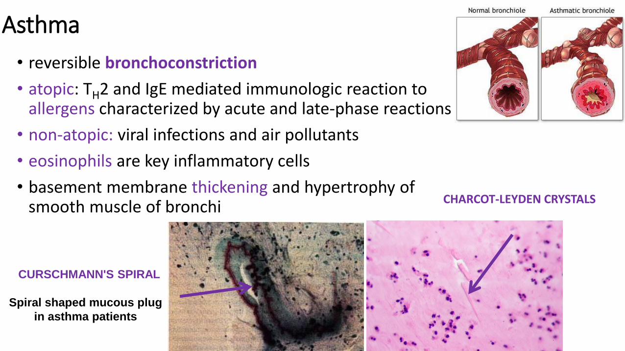
Asthma
• reversible bronchoconstriction
• atopic: TH2 and IgE mediated immunologic reaction toallergens characterized by acute and late-phase reactions
• non-atopic: viral infections and air pollutants
• eosinophils are key inflammatory cells
• basement membrane thickening and hypertrophy of smooth muscle of bronchi
CURSCHMANN'S SPIRAL
Spiral shaped mucous plug
in asthma patients
CHARCOT-LEYDEN CRYSTALS

Bronchiectasis
Etiology
• Bronchial obstruction
• Cystic fibrosis
• Chronic (necrotizing) infection of bronchi and bronchioles associatedwith permanent dilatation of theseairways
• Symptoms: cough, fever, purulentsputum
Sec amyloidosis!!!!
• Gross:
markedly distendedperipheral bronchi, usually inlower lobes, can trace topleural surface;

Kartagener Syndrome/Primary Ciliary Dyskinesia (PCD)
• Situs inversus, bronchiectasisand sinusitis, due to defective ciliary action
• PCD is a genetically heterogeneous disorder affecting motile of ciliawhich are made up of approximately 250 proteins.

NORMAL COPD RESTRICTIVE

Restrictive Lung Disease
reduced expansion (compliance) of lung parenchyma withdecrease in total lung capacity;
NORMAL FEV1
1.Fibrosing diseases
• Interstitial / infiltrative lungdiseases - ILD interstitialfibrosis
• Pneumoconioses
• Autoimmune disease
2.Granulomatous diseases(i.e. sarcoidosis)
3.Eosinophilic
4.Smoking-related

Fibrosing diseases
Idiopathic pulmonary fibrosis (IPF) (usual interstitial pneumonia - UIP)
• 60+ (male>female)
• recurrent alveolitis
• Diagnosis of exclusion
(no asbest, no vascular
disease etc)
TGF-beta 1!!
Non-specific interstitial pneumonia


Pneumoconioses
DiseasesSilicosisCoal-worker’s pneumoconiosis
Asbestosis(talcosis, siderosis,
aluminosis,berylliosis)
Definition
dust in the lung
diseases of the lung
related to the inhaled dust

a. Fibrotic nodules
b. Progressive massive fibrosis
c. Alveolar proteinosis
Silicotic lung
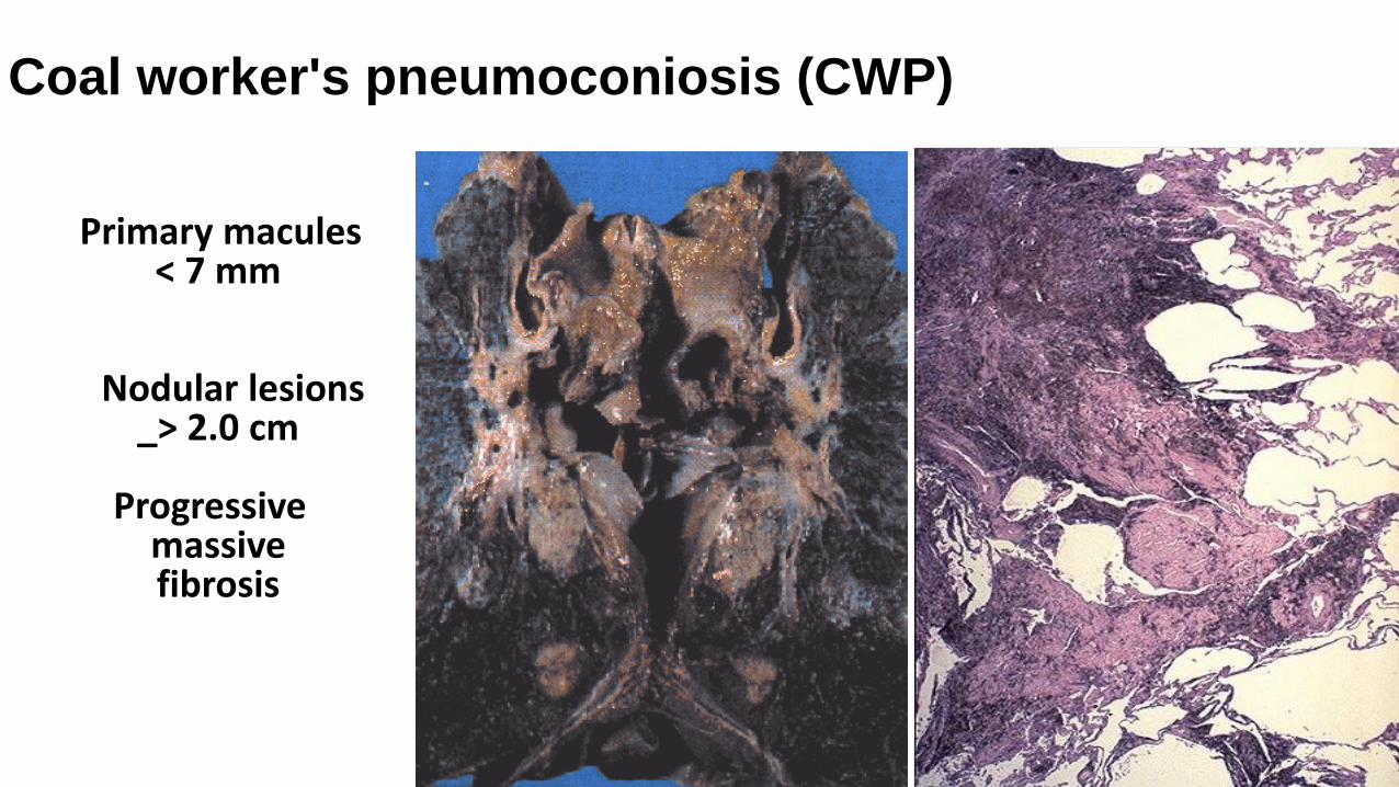
Primary macules< 7 mm
Nodular lesions_> 2.0 cm
Progressivemassivefibrosis
Coal worker's pneumoconiosis (CWP)
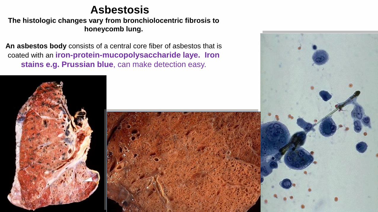
AsbestosisThe histologic changes vary from bronchiolocentric fibrosis to
honeycomb lung.
An asbestos body consists of a central core fiber of asbestos that is
coated with an iron-protein-mucopolysaccharide laye. Iron
stains e.g. Prussian blue, can make detection easy.

Granulomatous inflammation
(non-infectious)
Boeck’s sarcoidosis• Multisystemic disease of unknown
origin that involves lung in 90% of cases
• Presents as bilateral hilarlymphadenopathy (BHL)
• 65% recover without further problems; 20%have permanent pulmonary loss;
• Skin: erythema nodosum
• Diagnosis of exclusion, culture and specialstains – Ziehl-Neelsen
• Treatment: steroids for severe symptoms, advanced disease
Diff dg!!!!!!!
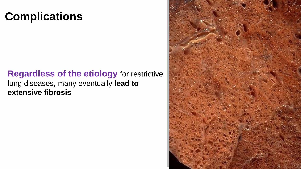
Regardless of the etiology for restrictive
lung diseases, many eventually lead to
extensive fibrosis
Complications

.
Both restrictive and obstructivelung diseases
can affect the pulmonary arterialcirculation.
The loss of normal lungparenchyma leads to pulmonary
hypertensionthat leads to thickening of the small
arteries
Complications

27% chronic obstructive pulmonary
disease (COPD), including emphysema;
16% idiopathic pulmonary fibrosis;
14% cystic fibrosis;
12% idiopathic (formerly known as "primary")
pulmonary hypertension;
5% alpha 1-antitrypsin deficiency;
2% replacing previously transplanted lungs
that have since failed;
24% other causes,
including bronchiectasis and sarcoidosis.
Lung transplantation