ryota higuchi, tatsuki fukami, miki nakajima, and tsuyoshi...
TRANSCRIPT

DMD #51714
1
Prilocaine- and Lidocaine-induced Methemoglobinemia is Caused by Human
Carboxylesterase-, CYP2E1- and CYP3A4-mediated Metabolic Activation
Ryota Higuchi, Tatsuki Fukami, Miki Nakajima, and Tsuyoshi Yokoi
Drug Metabolism and Toxicology, Faculty of Pharmaceutical Sciences, Kanazawa University,
Kakuma-machi, Kanazawa 920-1192, Japan (R. H., T. F., M. N., and T. Y.)
DMD Fast Forward. Published on March 25, 2013 as doi:10.1124/dmd.113.051714
Copyright 2013 by the American Society for Pharmacology and Experimental Therapeutics.
This article has not been copyedited and formatted. The final version may differ from this version.DMD Fast Forward. Published on March 25, 2013 as DOI: 10.1124/dmd.113.051714
at ASPE
T Journals on M
ay 6, 2018dm
d.aspetjournals.orgD
ownloaded from
DMD #51714
2
Running title: Prilocaine- and Lidocaine-induced Methemoglobinemia
To whom all correspondence should be sent:
Tatsuki Fukami, Ph.D.
Drug Metabolism and Toxicology
Faculty of Pharmaceutical Sciences
Kanazawa University
Kakuma-machi
Kanazawa 920-1192, Japan
Tel: +81-76-234-4438
Fax: +81-76-234-4407
E-mail: [email protected]
Number of text pages: 23
Number of tables: 2
Number of figures: 12
Number of references: 32
Number of words in abstract: 236 words
Number of words in introduction: 678 words
Number of words in discussion: 1,414 words
Abbreviations: AADAC, arylacetamide deacetylase; BNPP, bis-(4-nitrophenyl) phosphate;
CES, carboxylesterase; CYP, cytochrome P450; DFP, diisopropyl fluorophosphate; FMO,
flavin-containing monooxygenase; HLM, human liver microsomes; NADPH-GS,
NADPH-generating system; NPR, NADPH-P450 reductase; MEGX,
monoethylglycinexylidide; Met-Hb, methemoglobin.
This article has not been copyedited and formatted. The final version may differ from this version.DMD Fast Forward. Published on March 25, 2013 as DOI: 10.1124/dmd.113.051714
at ASPE
T Journals on M
ay 6, 2018dm
d.aspetjournals.orgD
ownloaded from
DMD #51714
3
Abstract
Prilocaine and lidocaine are classified as amide-type local anesthetics whose serious
adverse effects include methemoglobinemia. Although the hydrolyzed metabolites of
prilocaine (o-toluidine) and lidocaine (2,6-xylidine) have been suspected to induce
methemoglobinemia, the metabolic enzymes that are involved remain uncharacterized. In the
present study, we aimed to identify the human enzymes that are responsible for prilocaine-
and lidocaine-induced methemoglobinemia. Our experiments revealed that prilocaine was
hydrolyzed by recombinant human carboxylesterase (CES) 1A and CES2, whereas lidocaine
was hydrolyzed only by human CES1A. When the parent compounds (prilocaine and
lidocaine) were incubated with HLM, Met-Hb formation was lower than when the hydrolyzed
metabolites were incubated with HLM. In addition, Met-Hb formation when prilocaine and
o-toluidine were incubated with HLM was higher than that when lidocaine and 2,6-xylidine
were incubated with HLM. Incubation with diisopropyl fluorophosphate and
bis-(4-nitrophenyl) phosphate, which are general inhibitors of CES, significantly decreased
Met-Hb formation when prilocaine and lidocaine were incubated with HLM. An
anti-CYP3A4 antibody further decreased the residual formation of Met-Hb. Met-Hb
formation following the incubation of o-toluidine and 2,6-xylidine with HLM was only
markedly decreased by incubation with an anti-CYP2E1 antibody. o-Toluidine and
2,6-xylidine were further metabolized by CYP2E1 to 4- and 6-hydroxy-o-toluidine and
4-hydroxy-2,6-xylidine, respectively, and these metabolites were shown to more efficiently
induce Met-Hb formation than the parent compounds. Collectively, we found that the
metabolites produced by human CES-, CYP2E1- and CYP3A4-mediated metabolism were
involved in prilocaine- and lidocaine-induced methemoglobinemia.
This article has not been copyedited and formatted. The final version may differ from this version.DMD Fast Forward. Published on March 25, 2013 as DOI: 10.1124/dmd.113.051714
at ASPE
T Journals on M
ay 6, 2018dm
d.aspetjournals.orgD
ownloaded from
DMD #51714
4
Introduction
Prilocaine and lidocaine are classified as amide-type local anesthetics, which prevent and
relieve pain by interrupting nerve excitation and conduction via direct interaction with
voltage-gated Na+ channels to block the Na+ current (Lipkind and Fozzard, 2005). In general,
prilocaine and lidocaine are safely used in patients, although methemoglobinemia is
occasionally induced (Rehman, 2001; Maimo and Redick, 2004). Methemoglobinemia is
defined as a methemoglobin (Met-Hb) level > 1.0% in the blood. Met-Hb is an abnormal form
of hemoglobin in which iron is oxidized from the ferrous (Fe2+) to the ferric state (Fe3+).
Because Met-Hb cannot bind and transport oxygen, increased levels of Met-Hb are associated
with clinically severe symptoms (Moore et al., 2004). Met-Hb concentrations are normally
maintained at roughly 1% of total hemoglobin by the action of Met-Hb reductase (Guay,
2009). Cyanosis usually occurs when Met-Hb concentrations rise above 10%, and is followed
by anxiety, fatigue, and tachycardia when Met-Hb levels reach 20% to 50% of total
hemoglobin levels. When Met-Hb levels reach 50 – 70% of total hemoglobin levels, coma
and death may occur (Rodriguez et al., 1994).
When prilocaine was used for epidural analgesia and peripheral nerve block, some
patients were reported to develop methemoglobinemia (Climie et al., 1967; Vasters et al.,
2006). For example, when 288 mg of prilocaine was used in a 19-year-old Caucasian woman,
her Met-Hb levels were observed to be 37.8% of total hemoglobin levels approximately 4 hr
after injection (Kreutz and Kinni, 1983). Fetuses and infants under 6 months of age seem to
be more susceptible. Lidocaine also causes methemoglobinemia, although only rarely. In fact,
the number of articles that were published from 1949 to 2007 concerning lidocaine-related
methemoglobinemia (12 episodes) is lower than the number of articles concerning
prilocaine-related methemoglobinemia (68 episodes) (Guay, 2009). In humans, prilocaine and
lidocaine are hydrolytically metabolized to the aromatic amines o-toluidine and 2,6-xylidine,
This article has not been copyedited and formatted. The final version may differ from this version.DMD Fast Forward. Published on March 25, 2013 as DOI: 10.1124/dmd.113.051714
at ASPE
T Journals on M
ay 6, 2018dm
d.aspetjournals.orgD
ownloaded from
DMD #51714
5
respectively. These metabolites have been reported to cause increased levels of Met-Hb after
intravenous administration to cats or rats (Onji and Tyuma, 1965; Lindstrom et al., 1969).
Based on the results of these studies, prilocaine and lidocaine hydrolysis pathways have been
suggested to play an important role in methemoglobinemia, but the metabolic enzymes that
are involved in methemoglobinemia remain to be experimentally characterized. Moreover, it
is unclear whether differences in methemoglobinemia frequency after prilocaine and lidocaine
treatment are due to differences in enzymatic metabolism or differences in the potency of
Met-Hb formation by their metabolites.
Esterases, which are expressed in human liver, plasma, and other tissues, contribute to
the hydrolysis of approximately 10% of clinically therapeutic drugs, including ester, amide,
and thioester bonds (Fukami and Yokoi, 2012). In particular, human carboxylesterases (CES),
especially the CES1A and CES2 enzymes, are the major serine esterases that are responsible
for the hydrolysis of various drugs and xenobiotics (Imai et al., 2006). Recently, we
demonstrated that human arylacetamide deacetylase (AADAC) is involved in the metabolism
of drugs such as flutamide, phenacetin, and rifamycins (Watanabe et al., 2009; Watanabe et al.,
2010; Nakajima et al., 2011). We have more recently demonstrated that the hydrolysis of
phenacetin by AADAC, which produced p-phenetidine, an aromatic amine metabolite, is
predominantly involved in the phenacetin-induced formation of Met-Hb (Kobayashi et al.,
2012). Thus, it is conceivable that CES1A, CES2, and AADAC are involved in the hydrolysis
of prilocaine and lidocaine.
Ganesan et al. (2010) reported that dapsone-hydroxylamine, which is an N-hydroxylated
metabolite of dapsone, was suspected to be a cause of dapsone-induced methemoglobinemia.
The formation of dapsone-hydroxylamine is catalyzed by cytochrome P450 (CYP) 2C19,
CYP2E1, and CYP3A4. We recently reported that metabolic activation by CYP1A2 and
CYP2E1, as well as hydrolysis of AADAC, plays a predominant role in phenacetin-induced
methemoglobinemia (Kobayashi et al., 2012). Thus, it is conceivable that CYP(s) are also
This article has not been copyedited and formatted. The final version may differ from this version.DMD Fast Forward. Published on March 25, 2013 as DOI: 10.1124/dmd.113.051714
at ASPE
T Journals on M
ay 6, 2018dm
d.aspetjournals.orgD
ownloaded from

DMD #51714
6
involved in prilocaine- and lidocaine-induced methemoglobinemia.
Based on the background studies that were outlined above, in the present study, we
investigated the human enzymes responsible for the metabolism of prilocaine and lidocaine in
order to clarify the mechanisms of prilocaine- and lidocaine-induced methemoglobinemia. In
addition, the efficiencies of enzymatic metabolism and Met-Hb formation were compared
between prilocaine and lidocaine.
This article has not been copyedited and formatted. The final version may differ from this version.DMD Fast Forward. Published on March 25, 2013 as DOI: 10.1124/dmd.113.051714
at ASPE
T Journals on M
ay 6, 2018dm
d.aspetjournals.orgD
ownloaded from
DMD #51714
7
Materials and Methods
Chemicals and Reagents. Lidocaine hydrochloride, 2,6-xylidine, o-toluidine,
4-hydroxyl-o-toluidine, and diisopropyl fluorophosphate (DFP) were purchased from Wako
Pure Chemical Industries (Osaka, Japan). 6-Hydroxyl-o-toluidine and 4-hydroxyl-2,6-xylidine
were purchased from Tokyo Chemical Industry (Tokyo, Japan). Prilocaine hydrochloride and
bis-(4-nitrophenyl)-phosphate (BNPP) were obtained from Sigma (St. Louis, MO). Human
liver microsomes (HLM) (pooled, n = 50), recombinant human CYP1A2, CYP2A6, CYP2C8,
CYP2D6 (with NADPH-P450 reductase (NPR)), CYP2B6, CYP2C9, CYP2C19, CYP2E1
and CYP3A4 (with NPR and cytochrome b5) enzymes expressed in baculovirus-infected
insect cells, monoclonal mouse anti-human CYP1A2 antibody, anti-human CYP2E1 antibody,
and anti-human CYP3A4 antibody were purchased from BD Gentest (Woburn, MA). Other
chemicals were of the highest commercially available grade.
Mouse and Human Red Blood Cells. Animals were maintained in accordance with the
National Institutes of Health Guide for Animal Welfare of Japan, and the protocols were
approved by the Institutional Animal Care and Use Committee of Kanazawa University, Japan.
The use of human red blood cells was approved by the Ethics Committees of Kanazawa
University (Kanazawa, Japan). Mouse blood (pooled samples from 5 C57BL/6J mice: 6-week
old male, 20 - 25 g) that had been obtained from SLC Japan (Hamamatsu, Japan) and human
blood (5 healthy Japanese volunteers: 22 to 30-year old males) were obtained according to our
previous report (Kobayashi et al., 2012). All assays were performed immediately after the
separation of the red blood cells.
Prilocaine and Lidocaine Hydrolase Activities. Prilocaine and lidocaine hydrolase activities
This article has not been copyedited and formatted. The final version may differ from this version.DMD Fast Forward. Published on March 25, 2013 as DOI: 10.1124/dmd.113.051714
at ASPE
T Journals on M
ay 6, 2018dm
d.aspetjournals.orgD
ownloaded from

DMD #51714
8
were determined as follows: a typical incubation mixture (final volume of 0.2 ml) contained
100 mM potassium phosphate buffer (pH 7.4) and various enzyme sources (HLM or Sf21 cell
homogenates expressing esterases, 0.4 mg/ml). Sf21 cell homogenates expressing CES1A,
CES2, or AADAC were prepared as previously described (Fukami et al., 2010; Watanabe et
al., 2010). In a preliminary study, we confirmed that the formation rates of o-toluidine and
2,6-xylidine from prilocaine and lidocaine, respectively, were linear with respect to protein
concentration (< 2.0 mg/ml) and incubation time (< 120 min). Prilocaine and lidocaine were
dissolved in distilled water. The reactions were initiated by the addition of prilocaine and
lidocaine (0.2 - 10 mM for HLM or 0.1 - 4 mM for Sf21 cell homogenates expressing
esterases) after a 2-min preincubation at 37°C. After a 30-min incubation, the reactions were
terminated by the addition of 10 µl of ice-cold 60% perchloric acid. After removal of the
protein by centrifugation at 9,500 g for 5 min, a 60-µl portion of the supernatant was
subjected to high-performance liquid chromatography (HPLC). The HPLC analysis was
performed using an L-7100 pump (Hitachi, Tokyo, Japan), an L-7200 autosampler (Hitachi),
an L-7405 UV detector (Hitachi), and a D-2500 Chromato-Integrator (Hitachi) equipped with
a Wakopak® eco-ODS column (5-μm particle size, 4.6 mm i.d.×150 mm; Wako Pure
Chemical Industries). The eluent was monitored at 210 nm with a noise-base clean Uni-3
(Union, Gunma, Japan), which can reduce the noise by integrating the output and increase the
signal 3-fold by differentiating the output and 5-fold by further amplification with an internal
amplifier, resulting in a maximum 15-fold amplification of the signal. The mobile phase was
35% methanol containing 0.2% phosphoric acid and 2.2 mM sodium 1-octanesulfonate. The
flow rate was 1.0 ml/min. The column temperature was 35°C. The quantification of
o-toluidine and 2,6-xylidine was performed by comparing the HPLC peak height with that of
an authentic standard. The limit of quantification in the reaction mixture for o-toluidine and
2,6-xylidine was 100 nM with a coefficient of variation < 5.6%. The activity at each
concentration was determined as the mean value in triplicate. For kinetic analyses of the
This article has not been copyedited and formatted. The final version may differ from this version.DMD Fast Forward. Published on March 25, 2013 as DOI: 10.1124/dmd.113.051714
at ASPE
T Journals on M
ay 6, 2018dm
d.aspetjournals.orgD
ownloaded from
DMD #51714
9
prilocaine and lidocaine hydrolase activities, the parameters were estimated from the fitted
curves using a computer program (KaleidaGraph, Synergy Software, Reading, PA) that was
designed for use in nonlinear regression analyses.
Met-Hb Formation. A Met-Hb formation assay was conducted according to the methods
outlined in our previous study (Kobayashi et al., 2012), with a slight modification. A typical
incubation mixture (final volume of 0.2 ml) contained 5% of the mouse red blood cell fraction
except in (7) where human red blood cell fraction was used, 100 mM potassium phosphate
buffer (pH 7.4), an NADPH-generating system (NADPH-GS: 0.5 mM NADP+, 5 mM glucose
6-phosphate, 5 mM MgCl2, and 1 U/ml glucose-6-phosphate dehydrogenase), and various
enzyme sources.
(1) To investigate the time-dependence of Met-Hb formation, HLM (1.0 mg/ml) were
used as enzyme sources. The reactions were initiated by the addition of prilocaine, lidocaine,
o-toluidine, or 2,6-xylidine (1 mM) after a 2-min preincubation at 37°C. After the 0- to
120-min incubation at 37°C, the reaction was terminated by placing the samples on ice.
(2) To investigate the concentration-dependence of Met-Hb formation, HLM (1.0
mg/ml) were used as enzyme sources. The reactions were initiated by the addition of
prilocaine, lidocaine, o-toluidine, or 2,6-xylidine (0.01, 0.1, 1, or 10 mM), after a 2-min
preincubation at 37°C. After the 60-min incubation at 37°C, the reaction was terminated by
placing the samples on ice.
(3) To determine the CYP enzymes that were involved in prilocaine-, lidocaine-,
o-toluidine-, and 2,6-xylidine-induced Met-Hb formation, recombinant human CYP enzymes
(25 pmol /ml) were used as enzyme sources. The reactions were initiated by the addition of
prilocaine (10 mM), lidocaine (10 mM), o-toluidine (1 mM), or 2,6-xylidine (1 mM) after a
2-min preincubation at 37°C. After the 120-min incubations (prilocaine and lidocaine) and the
60-min incubations (o-toluidine and 2,6-xylidine) at 37°C, the reactions were terminated by
This article has not been copyedited and formatted. The final version may differ from this version.DMD Fast Forward. Published on March 25, 2013 as DOI: 10.1124/dmd.113.051714
at ASPE
T Journals on M
ay 6, 2018dm
d.aspetjournals.orgD
ownloaded from
DMD #51714
10
placing the samples on ice.
(4) To investigate the involvement of various esterase(s) in the HLM, inhibition analyses
of prilocaine- and lidocaine-induced Met-Hb formation were performed using the general
CES inhibitors DFP and BNPP (Watanabe et al., 2009). HLM (1.0 mg/ml) were used as
enzyme sources, and the concentrations of inhibitors were 100 µM. DFP and BNPP were
dissolved in distilled water. The reaction conditions were the same as those described under
(3) above.
(5) To investigate the involvement of CYP enzymes in the HLM in Met-Hb formation,
inhibition analyses of the formation of Met-Hb by prilocaine (10 mM), lidocaine (10 mM),
o-toluidine (1 mM), or 2,6-xylidine (1 mM) were performed using anti-CYP antibodies. HLM
(0.5 mg/ml) were used as enzyme sources. Ten microliters of antibody-mixtures [4 µl of
anti-CYP2E1 antibody mixed with 6 µL of 25 mM Tris-buffer (pH 7.5), or 10 µl of
anti-CYP1A2 or anti-CYP3A4 antibody] was incubated on ice for 30 min with enzyme
sources, after which point typical incubation mixtures (final volume of 0.2 ml) that included
the antibody mixtures were prepared. To investigate the involvement of CYP enzymes in
prilocaine- and lidocaine-induced Met-Hb formation in the absence of a hydrolysis reaction,
DFP (100 µM) was added into the incubation mixture. The reaction conditions were the same
as those described under (3) above.
(6) To investigate whether the hydroxylated metabolites of o-toluidine and 2,6-xylidine
induced Met-Hb formation, o-toluidine, 4-hydroxy-o-toluidine, 6-hydroxy-o-toluidine,
2,6-xylidine, and 4-hydroxy-2,6-xylidine (1 mM) were incubated with HLM (1.0 mg/mL) and
mouse red blood cells both in the presence and absence of an NADPH-GS. After a 60
min-incubation at 37°C, the reactions were terminated by placing the samples on ice.
(7) To investigate the sensitivity of human red blood cells to prilocaine- and
lidocaine-induced Met-Hb formation, a Met-Hb formation assay was conducted using human
red blood cells instead of mouse red blood cells. HLM (1.0 mg/ml) were used as enzyme
This article has not been copyedited and formatted. The final version may differ from this version.DMD Fast Forward. Published on March 25, 2013 as DOI: 10.1124/dmd.113.051714
at ASPE
T Journals on M
ay 6, 2018dm
d.aspetjournals.orgD
ownloaded from
DMD #51714
11
sources, and DFP (100 μM) was added into the incubation mixture. The reaction was initiated
by the addition of 10 mM prilocaine and lidocaine after a 2-min preincubation at 37°C. The
reaction conditions were the same as those described under (3) above.
Prilocaine and lidocaine were dissolved in distilled water. o-Toluidine and 2,6-xylidine
were dissolved in acetonitrile and the final concentration of acetonitrile in the incubation
mixture was 1%. It has been reported that 1% acetonitrile does not inhibit the activities of
CYP1A2, CYP2E1 and CYP3A4 (Chauret et al., 1998).
The Met-Hb levels in red blood cells were determined as the percent of total hemoglobin
according to the methods outlined in our previous study (Kobayashi et al., 2012)
Normalizing Met-Hb Formation by CYP Levels in HLM. Met-Hb formation was
normalized by the levels of each CYP enzyme in HLM and was calculated using the
following equation, where the CYPHLM activity and CYPexpression activity values are the marker
activities of each CYP in HLM and recombinant CYP expression systems, respectively:
Met-Hb formation normalized by CYP levels in HLM (%) = Met-Hb formation by
recombinant human CYP expression systems (%) × CYPHLM activity/ CYPexpression activity
The reaction conditions for Met-Hb formation by recombinant human CYP expression
systems were determined as follows: prilocaine, lidocaine (10 mM), and their hydrolyzed
metabolites (1 mM) were incubated with each CYP expression system (25 pmol CYP/ml), an
NADPH-GS, and mouse red blood cells. The incubation time was either 120 min (prilocaine
and lidocaine) or 60 min (o-toluidine and 2,6-xylidine).
The reaction conditions for Met-Hb formation in HLM were determined as described in
the above paragraph, except HLM (1.0 mg/mL) were used in place of the CYP expression
systems. Met-Hb formation was detected as described under section 2.4. (3) above.
This article has not been copyedited and formatted. The final version may differ from this version.DMD Fast Forward. Published on March 25, 2013 as DOI: 10.1124/dmd.113.051714
at ASPE
T Journals on M
ay 6, 2018dm
d.aspetjournals.orgD
ownloaded from

DMD #51714
12
Methoxyresorufin O-demethylase, chlorzoxazone 6-hydroxylase, and midazolam
1’-hydroxylase activities were measured as markers for the activities of CYP1A2, CYP2E1,
and CYP3A4, respectively, by HPLC, according to methods described in our previous reports
(Nakajima et al., 2002; Fukami et al., 2007; Fukami et al., 2008).
o-Toluidine and 2,6-Xylidine Hydroxylase Activities. The activities of o-toluidine and
2,6-xylidine hydroxylase were determined as follows: a typical incubation mixture (final
volume of 0.2 ml) contained o-toluidine or 2,6-xylidine (4 - 200 μM), 100 mM potassium
phosphate buffer (pH 7.4), an NADPH-GS and HLM (0.4 mg/ml). In a preliminary study, we
confirmed that the formation rates of 4-hydroxyl-o-toluidine and 6-hydroxyl-o-toluidine from
o-toluidine, and 4-hydroxyl-2,6-xylidine from 2,6-xylidine, in HLM were linear with respect
to protein concentration (< 1.0 mg/ml) and incubation time (< 60 min). o-Toluidine and
2,6-xylidine were dissolved in acetonitrile and the final concentration of acetonitrile in the
incubation mixture was 1%.
Inhibition analyses of o-toluidine or 2,6-xylidine hydroxylation were performed using
anti-CYP antibodies. HLM (0.4 mg/ml) were used as enzyme sources. Eight microliters of
antibody-mixtures [3.2 µl of anti-CYP2E1 antibody mixed with 4.8 µL of 25 mM Tris-buffer
(pH 7.5) or 8 µl of anti-CYP1A2 or anti-CYP3A4 antibody] was incubated with enzyme
sources on ice for 30 min. Then, typical incubation mixtures (final volume of 0.2 ml) were
prepared by the addition of o-toluidine (50 μM) or 2,6-xylidine (30 μM).
The reactions were initiated by the addition of an NADPH-GS after a 2-min
preincubation at 37°C. After a 30-min incubation, the reactions were terminated by the
addition of 10 µl of ice-cold 60% perchloric acid. After removal of the protein by
centrifugation at 9,500 g for 5 min, a 50-µl portion of the supernatant was subjected to HPLC.
The HPLC equipment was the same as that described under section 2.3. above. The mobile
phase for o-toluidine hydroxylation was 8.5% methanol/8% acetonitrile containing 0.2%
This article has not been copyedited and formatted. The final version may differ from this version.DMD Fast Forward. Published on March 25, 2013 as DOI: 10.1124/dmd.113.051714
at ASPE
T Journals on M
ay 6, 2018dm
d.aspetjournals.orgD
ownloaded from

DMD #51714
13
phosphoric acid and 2.2 mM sodium 1-octanesulfonate, and the mobile phase for 2,6-xylidine
hydroxylation was 10% acetonitrile containing 0.11% phosphoric acid and 2.2 mM sodium
1-octanesulfonate. The quantification of the hydroxylated metabolites of o-toluidine and
2,6-xylidine was performed by comparing the HPLC peak height with that of an authentic
standard. The activity at each concentration was determined as the mean value in triplicate.
For kinetic analyses of the o-toluidine and 2,6-xylidine hydroxylase activities, the parameters
were estimated as described under section 2.3. above.
Statistical Methods. Statistical analyses between two and multiple groups were performed
using an unpaired, two-tailed Student’s t test and ANOVA, followed by Tukey’s post-hoc test.
P < 0.05 was considered to be statistically significant.
This article has not been copyedited and formatted. The final version may differ from this version.DMD Fast Forward. Published on March 25, 2013 as DOI: 10.1124/dmd.113.051714
at ASPE
T Journals on M
ay 6, 2018dm
d.aspetjournals.orgD
ownloaded from
DMD #51714
14
Results
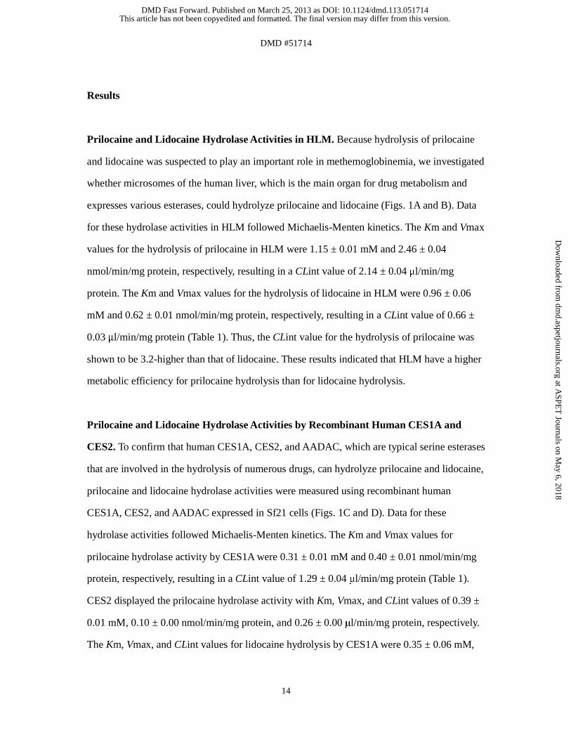
Prilocaine and Lidocaine Hydrolase Activities in HLM. Because hydrolysis of prilocaine
and lidocaine was suspected to play an important role in methemoglobinemia, we investigated
whether microsomes of the human liver, which is the main organ for drug metabolism and
expresses various esterases, could hydrolyze prilocaine and lidocaine (Figs. 1A and B). Data
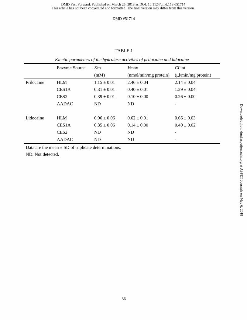
for these hydrolase activities in HLM followed Michaelis-Menten kinetics. The Km and Vmax
values for the hydrolysis of prilocaine in HLM were 1.15 ± 0.01 mM and 2.46 ± 0.04
nmol/min/mg protein, respectively, resulting in a CLint value of 2.14 ± 0.04 μl/min/mg
protein. The Km and Vmax values for the hydrolysis of lidocaine in HLM were 0.96 ± 0.06
mM and 0.62 ± 0.01 nmol/min/mg protein, respectively, resulting in a CLint value of 0.66 ±
0.03 μl/min/mg protein (Table 1). Thus, the CLint value for the hydrolysis of prilocaine was
shown to be 3.2-higher than that of lidocaine. These results indicated that HLM have a higher
metabolic efficiency for prilocaine hydrolysis than for lidocaine hydrolysis.
Prilocaine and Lidocaine Hydrolase Activities by Recombinant Human CES1A and
CES2. To confirm that human CES1A, CES2, and AADAC, which are typical serine esterases
that are involved in the hydrolysis of numerous drugs, can hydrolyze prilocaine and lidocaine,
prilocaine and lidocaine hydrolase activities were measured using recombinant human
CES1A, CES2, and AADAC expressed in Sf21 cells (Figs. 1C and D). Data for these
hydrolase activities followed Michaelis-Menten kinetics. The Km and Vmax values for
prilocaine hydrolase activity by CES1A were 0.31 ± 0.01 mM and 0.40 ± 0.01 nmol/min/mg
protein, respectively, resulting in a CLint value of 1.29 ± 0.04 μl/min/mg protein (Table 1).
CES2 displayed the prilocaine hydrolase activity with Km, Vmax, and CLint values of 0.39 ±
0.01 mM, 0.10 ± 0.00 nmol/min/mg protein, and 0.26 ± 0.00 μl/min/mg protein, respectively.
The Km, Vmax, and CLint values for lidocaine hydrolysis by CES1A were 0.35 ± 0.06 mM,
This article has not been copyedited and formatted. The final version may differ from this version.DMD Fast Forward. Published on March 25, 2013 as DOI: 10.1124/dmd.113.051714
at ASPE
T Journals on M
ay 6, 2018dm
d.aspetjournals.orgD
ownloaded from
DMD #51714
15
0.14 ± 0.00 nmol/min/mg protein, and 0.40 ± 0.02 μl/min/mg protein, respectively, whereas
CES2 did not display any lidocaine hydrolase activity (Table 1). AADAC did not display
hydrolase activities for either prilocaine or lidocaine. These results indicated that the
metabolic efficiency of prilocaine hydrolysis by CES1A was higher than that of lidocaine
hydrolysis. CES2 participated in prilocaine hydrolysis but not lidocaine hydrolysis.
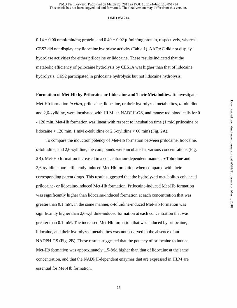
Formation of Met-Hb by Prilocaine or Lidocaine and Their Metabolites. To investigate
Met-Hb formation in vitro, prilocaine, lidocaine, or their hydrolyzed metabolites, o-toluidine
and 2,6-xylidine, were incubated with HLM, an NADPH-GS, and mouse red blood cells for 0
- 120 min. Met-Hb formation was linear with respect to incubation time (1 mM prilocaine or
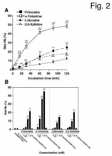
lidocaine < 120 min, 1 mM o-toluidine or 2,6-xylidine < 60 min) (Fig. 2A).
To compare the induction potency of Met-Hb formation between prilocaine, lidocaine,
o-toluidine, and 2,6-xylidine, the compounds were incubated at various concentrations (Fig.
2B). Met-Hb formation increased in a concentration-dependent manner. o-Toluidine and
2,6-xylidine more efficiently induced Met-Hb formation when compared with their
corresponding parent drugs. This result suggested that the hydrolyzed metabolites enhanced
prilocaine- or lidocaine-induced Met-Hb formation. Prilocaine-induced Met-Hb formation
was significantly higher than lidocaine-induced formation at each concentration that was
greater than 0.1 mM. In the same manner, o-toluidine-induced Met-Hb formation was
significantly higher than 2,6-xylidine-induced formation at each concentration that was
greater than 0.1 mM. The increased Met-Hb formation that was induced by prilocaine,
lidocaine, and their hydrolyzed metabolites was not observed in the absence of an
NADPH-GS (Fig. 2B). These results suggested that the potency of prilocaine to induce
Met-Hb formation was approximately 1.5-fold higher than that of lidocaine at the same
concentration, and that the NADPH-dependent enzymes that are expressed in HLM are
essential for Met-Hb formation.
This article has not been copyedited and formatted. The final version may differ from this version.DMD Fast Forward. Published on March 25, 2013 as DOI: 10.1124/dmd.113.051714
at ASPE
T Journals on M
ay 6, 2018dm
d.aspetjournals.orgD
ownloaded from
DMD #51714
16
Met-Hb Formation Following the Metabolic Activation of o-Toluidine and 2,6-Xylidine
by Human CYP(s). As outlined above, NADPH-dependent enzyme(s) are essential for
prilocaine- and lidocaine-induced Met-Hb formation. Therefore, the involvement of
representative NADPH-dependent enzymes (the CYP enzymes) in o-toluidine and
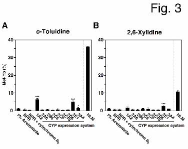
2,6-xylidine metabolism was investigated (Fig. 3). Either o-toluidine or 2,6-xylidine (1 mM)
was incubated with recombinant human CYPs, an NADPH-GS, and mouse red blood cells.
o-Toluidine-induced Met-Hb formation was significantly increased by CYP1A2 (6.2 ± 0.7%),
CYP2E1 (5.0 ± 0.4%) and CYP3A4 (1.3 ± 0.5%), and 2,6-xylidine-induced Met-Hb
formation was increased by CYP2E1 (2.3 ± 0.8%).
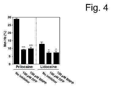
Prilocaine- and Lidocaine-induced Met-Hb Formation Inhibition Analyses in the
Presence of Esterase Inhibitors. To investigate the involvement of human CES in
prilocaine- and lidocaine-induced methemoglobinemia, inhibition analyses were performed
with DFP and BNPP, which are potent CES inhibitors (Watanabe et al., 2009) (Fig. 4). The
hydrolase activities of prilocaine and lidocaine (1 mM) in HLM (1.0 mg/ml) were
preliminarily confirmed by HPLC to be completely inhibited by 10 μM DFP and 10 μM
BNPP (data not shown). When prilocaine and lidocaine (10 mM) were incubated with HLM,
an NADPH-GS, and mouse red blood cells in the presence of DFP or BNPP (100 µM), both
prilocaine- and lidocaine-induced Met-Hb formation were decreased (prilocaine, % of control:
31.6% in the presence of DFP and 34.0% in the presence of BNPP; lidocaine, % of control:
56.6% in the presence of DFP and 56.6% in the presence of BNPP) (Fig. 4). DFP and BNPP
themselves did not alter Met-Hb formation (data not shown). Thus, CES were determined to
be involved in Met-Hb formation.
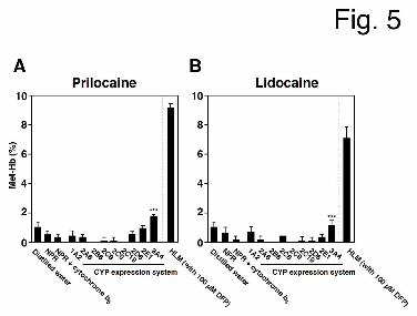
Met-Hb Formation Following the Metabolic Activation of Prilocaine and Lidocaine by
This article has not been copyedited and formatted. The final version may differ from this version.DMD Fast Forward. Published on March 25, 2013 as DOI: 10.1124/dmd.113.051714
at ASPE
T Journals on M
ay 6, 2018dm
d.aspetjournals.orgD
ownloaded from
DMD #51714
17
Human CYP(s). As shown in Fig. 4, DFP and BNPP could not completely inhibit prilocaine-
and lidocaine-induced Met-Hb formation, suggesting that the metabolic activation of
prilocaine and lidocaine by CYP enzyme(s) may mediate Met-Hb formation in the absence of
hydrolysis. When prilocaine and lidocaine (10 mM) were incubated with representative
recombinant human CYPs, an NADPH-GS, and mouse red blood cells, Met-Hb formation
increased in the presence of CYP3A4 (1.7 ± 0.2% for prilocaine and 1.1 ± 0.4% for lidocaine)
(Fig. 5). These results suggested that prilocaine- and lidocaine-induced Met-Hb formation in
the absence of hydrolysis is catalyzed by metabolic activation by human CYP3A4.
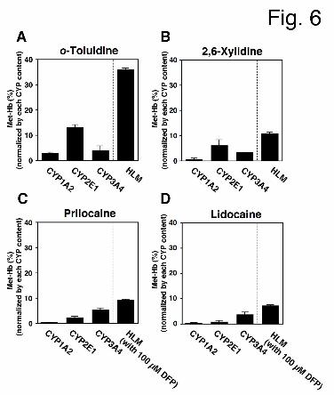
Normalizing Met-Hb Formation by CYP Levels in HLM. Because each CYP enzyme is
expressed at different levels in HLM, we were unable to simply compare the contribution of
each CYP enzyme to prilocaine-, lidocaine-, o-toluidine-, or 2,6-xylidine-induced Met-Hb
formation in HLM using human CYP expression systems. Therefore, to estimate the
contributions of CYP enzymes in HLM, Met-Hb formation in the presence of recombinant
CYP expression systems was normalized by the levels of each CYP enzyme in HLM (Fig. 6).
For o-toluidine- and 2,6-xylidine-induced Met-Hb formation, CYP2E1 had the highest
contribution in HLM, whereas CYP1A2 and CYP3A4 had relatively low contributions (Figs.
6A and 6B). For prilocaine- and lidocaine-induced Met-Hb formation, CYP3A4 had the
highest contribution in HLM (Figs. 6C and 6D). Although flavin-containing monooxygenase
(FMO) is also known to be an NADPH-dependent enzyme, Met-Hb formation that was
induced by prilocaine, lidocaine, and their-hydrolyzed metabolites in HLM was not inhibited
by 1 mM methimazole, which is a competitive FMO inhibitor (data not shown) (Rawden et al.,
2000). Thus, CYP2E1 was determined to contribute highly to o-toluidine- and
2,6-xylidine-induced Met-Hb formation, while CYP3A4 was determined to contribute highly
to prilocaine- and lidocaine-induced Met-Hb formation.
This article has not been copyedited and formatted. The final version may differ from this version.DMD Fast Forward. Published on March 25, 2013 as DOI: 10.1124/dmd.113.051714
at ASPE
T Journals on M
ay 6, 2018dm
d.aspetjournals.orgD
ownloaded from

DMD #51714
18
Met-Hb Formation Inhibition Analyses Following the Incubation of Prilocaine,
Lidocaine, and Their Hydrolyzed Metabolites with Anti-CYP Antibodies. To further
investigate the contributions of CYP1A2, CYP2E1 and CYP3A4 to the o-toluidine- and
2,6-xylidine-induced Met-Hb formation in HLM, inhibition analyses were performed using
anti-CYP antibodies (Fig. 7A). o-Toluidine- and 2,6-xylidine-induced Met-Hb formation were
markedly decreased by incubation with an anti-CYP2E1 antibody (from 19.5 ± 0.6% to 7.1 ±
0.4% for o-toluidine and from 6.2 ± 0.3% to 2.6 ± 0.2% for 2,6-xylidine) and slightly
decreased by incubation with an anti-CYP3A4 antibody (to 16.9 ± 0.5% for o-toluidine and to
4.3 ± 0.3% for 2,6-xylidine). Incubation with an anti-CYP1A2 antibody also slightly
decreased 2,6-xylidine-induced Met-Hb formation (to 5.0 ± 0.5%).
To investigate the contribution of CYP3A4 to prilocaine- and lidocaine-induced Met-Hb
formation in the absence of hydrolysis, inhibition analyses were performed using anti-CYP
antibodies (Fig. 7B). When 100 μM DFP was used to inhibit CES enzyme activity, incubation
with an anti-CYP3A4 antibody substantially decreased prilocaine- and lidocaine-induced
Met-Hb formation (from 5.7 ± 0.4% to 0.6 ± 0.3% for prilocaine and from 3.4 ± 0.4% to 0.7 ±
0.2% for lidocaine).
Met-Hb Formation in the Presence of the Hydroxylated Metabolites of o-Toluidine and
2,6-Xylidine. 4-Hydroxy-o-toluidine, 6-hydroxy-o-toluidine or 4-hydroxy-2,6-xylidine have
been detected in human urine after the administration of prilocaine or lidocaine (Hjelm et al.,
1972; Keenaghan and Boyes, 1972). To investigate whether these metabolites could induce
Met-Hb formation, in vitro analyses were performed. 4- and 6-Hydroxy-o-toluidine- and
4-hydroxy-2,6-xylidine-induced Met-Hb formation were markedly increased, both in the
presence and absence of an NADPH-GS (Fig. 8). A higher degree of Met-Hb formation was
detected in the presence of these metabolites than in the presence of o-toluidine and
2,6-xylidine. Thus, it was suggested that these hydroxylated metabolites of o-toluidine and
This article has not been copyedited and formatted. The final version may differ from this version.DMD Fast Forward. Published on March 25, 2013 as DOI: 10.1124/dmd.113.051714
at ASPE
T Journals on M
ay 6, 2018dm
d.aspetjournals.orgD
ownloaded from
DMD #51714
19
2,6-xylidine were one of the causative factors underlying prilocaine- and lidocaine-induced
methemoglobinemia.
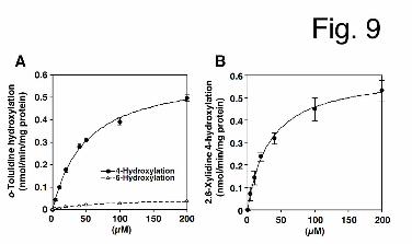
Formation of the Hydroxylated Metabolites of o-Toluidine and 2,6-Xylidine in HLM.
Because the hydroxylated metabolites of o-toluidine and 2,6-xylidine could induce Met-Hb
formation, we investigated whether HLM could catalyze the hydroxylation of o-toluidine and
2,6-xylidine (Figs. 9A and B). Data for these hydroxylase activities in HLM followed
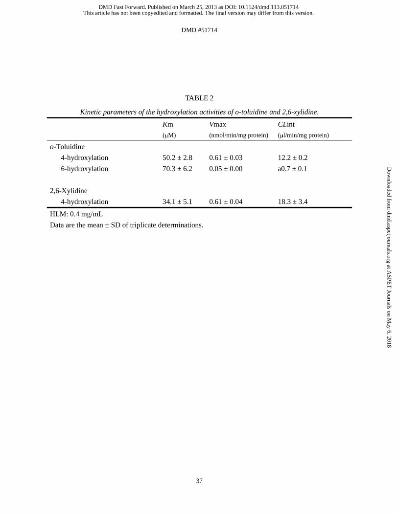
Michaelis-Menten kinetics. The Km and Vmax values for 4-hydroxylation of o-toluidine in
HLM were 50.2 ± 2.8 μM and 0.61 ± 0.03 nmol/min/mg protein, respectively, resulting in a
CLint value of 12.2 ± 0.2 μL/min/mg protein. The Km and Vmax values for 6-hydrolxylation
of o-toluidine in HLM were 70.3 ± 6.2 μM and 0.05 ± 0.00 nmol/min/mg protein, respectively,
resulting in a CLint value of 0.7 ± 0.1 μL/min/mg protein (Table 2). Thus, the CLint value for
4-hydroxylation of o-toluidine in HLM was shown to be 18-fold higher than that for
6-hydroxylation. The Km and Vmax values for 4-hydroxylation of 2,6-xylidine in HLM were
34.1 ± 5.1 μM and 0.61 ± 0.04 nmol/min/mg protein, respectively, resulting in a CLint value
of 18.3 ± 3.4 μL/min/mg protein. These results indicated that o-toluidine and 2,6-xylidine
were efficiently metabolized to their hydroxylated forms in HLM.
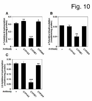
Inhibition Analyses of o-Toluidine and 2,6-Xylidine Hydroxylase Activities in the
Presence of Anti-CYP Antibodies. To further investigate the contributions of CYP1A2,
CYP2E1 and CYP3A4 to o-toluidine and 2,6-xylidine hydroxylation in HLM, inhibition
analyses were performed using anti-CYP antibodies (Fig. 10). o-Toluidine 4- and
6-hydroxylase activities were markedly decreased (% of control: 35.4 ± 1.6 for o-toluidine
4-hydroxylation and 47.1 ± 2.3 for o-toluidine 6-hydroxylation) by incubation with an
anti-CYP2E1 antibody. 2,6-Xylidine 4-hydroxylase activity was also markedly decreased by
incubation with an anti-CYP2E1 (% of control: 25.6 ± 0.9), and slightly decreased by
This article has not been copyedited and formatted. The final version may differ from this version.DMD Fast Forward. Published on March 25, 2013 as DOI: 10.1124/dmd.113.051714
at ASPE
T Journals on M
ay 6, 2018dm
d.aspetjournals.orgD
ownloaded from

DMD #51714
20
incubation with an anti-CYP3A4 antibody (% of control: 92.9 ± 0.3). Thus, it was suggested
that CYP2E1 could mainly catalyze o-toluidine and 2,6-xylidine hydroxylations, leading to
Met-Hb formation.
Prilocaine- and Lidocaine-induced Met-Hb Formation in Human Red Blood Cells. In the
Met-Hb formation analyses outlined above, mouse red blood cells were used. To assess
species specific differences in Met-Hb formation between mice and humans, an analysis using
human red blood cells that were obtained from 5 individuals was conducted as follows:
prilocaine or lidocaine (10 mM) was incubated with HLM, an NADPH-GS, and individual
human red blood cells that had been obtained from the 5 healthy donors. The levels of
Met-Hb formation in the absence of prilocaine or lidocaine were 0.6 – 1.5% (mean 1.1%).
High Met-Hb formation was observed after incubation with both prilocaine and lidocaine
(prilocaine: mean 34.8% (26.8 - 41.5%), lidocaine: mean 9.9% (9.0 - 11.3%)), but the
formation of Met-Hb was significantly decreased by incubation with 100 μM DFP [prilocaine:
mean 7.6% (6.8 - 8.6%), lidocaine: mean 6.4% (5.1 - 7.4%)] (Fig. 11). This result was similar
to that obtained using mouse red blood cells (Fig. 4). These results indicated the comparable
Met-Hb formation sensitivity between human and mouse red blood cells.
This article has not been copyedited and formatted. The final version may differ from this version.DMD Fast Forward. Published on March 25, 2013 as DOI: 10.1124/dmd.113.051714
at ASPE
T Journals on M
ay 6, 2018dm
d.aspetjournals.orgD
ownloaded from
DMD #51714
21
Discussion
Prilocaine and lidocaine are typical amide-type local anesthetics that carry the risk of a
serious adverse reaction known as methemoglobinemia (Maimo and Redick, 2004). Prilocaine
and lidocaine are hydrolytically metabolized to the aromatic amines o-toluidine and
2,6-xylidine, respectively. Although these metabolites were suspected to be causes of
prilocaine- and lidocaine-induced methemoglobinemia (Neuhaeuser et al., 2008), the enzymes
responsible for Met-Hb formation remained to be characterized. In the present study, we
found that metabolic activation by human CES, CYP2E1, and CYP3A4 was involved in
prilocaine- and lidocaine-induced methemoglobinemia.
We demonstrated that prilocaine was hydrolyzed by CES1A and CES2, whereas
lidocaine was specifically hydrolyzed by CES1A (Fig. 1), and that hydrolysis reactions that
were catalyzed by CES enzymes enhanced prilocaine- and lidocaine-induced Met-Hb
formation (Figs. 2 and 4). Because prilocaine and lidocaine hydrolase activities were detected
in HLM, but not in human plasma where cholinesterases and paraoxonases are expressed
(data not shown), these enzymes could be excluded from the candidate enzymes catalyzing
their hydrolysis. Recombinant human AADAC showed no activity (Fig. 1 and Table 1).
Moreover, prilocaine and lidocaine hydrolase activities at 1 mM were completely inhibited by
100 µM DFP and BNPP (data not shown), which are potent CES inhibitors (Watanabe et al.,
2009). Collectively, these results suggested that CES enzymes are major enzymes that are
responsible for the hydrolysis of prilocaine and lidocaine.
Met-Hb formation following the incubation of the parent compounds (prilocaine and
lidocaine) with HLM was lower than that following incubation of their hydrolyzed
metabolites (o-toluidine and 2,6-xylidine) (Fig. 2). Prilocaine- and lidocaine-induced Met-Hb
formation was significantly decreased by DFP and BNPP (Fig. 4), indicating that CES
enzymes may be involved in the prilocaine- and lidocaine-induced Met-Hb formation. When
This article has not been copyedited and formatted. The final version may differ from this version.DMD Fast Forward. Published on March 25, 2013 as DOI: 10.1124/dmd.113.051714
at ASPE
T Journals on M
ay 6, 2018dm
d.aspetjournals.orgD
ownloaded from
DMD #51714
22
metabolic efficiency was analyzed, prilocaine was shown to be more efficiently hydrolyzed in
HLM than lidocaine (Figs. 1A and 1B). Furthermore, prilocaine- and o-toluidine-induced
Met-Hb formation was higher that induced by incubating lidocaine and 2,6-xylidine (Figs. 2A
and 2B). These results supported the previous report (Guay, 2009) which indicated that the
number of prilocaine-related methemoglobinemia episodes is higher than that of
lidocaine-related methemoglobinemia.
We considered the possibility that hydrolysis may not be the sole cause of prilocaine-
and lidocaine-induced methemoglobinemia since Met-Hb formation was not completely
inhibited by DFP and BNPP. Supporting this assumption, we observed that metabolism by
CYP enzymes was also required for prilocaine- and lidocaine-induced Met-Hb formation in
the presence and absence of the hydrolysis reaction (Figs. 2, 3, and 5 - 7). Human CYP
enzymes, which are major NADPH-dependent enzymes, account for ~75% of the metabolism
of clinical drugs (Guengerich, 2008). In the absence of an NADPH-GS, Met-Hb formation
following incubation with prilocaine, lidocaine, and their hydrolyzed metabolites was not
increased (Fig. 2B). In fact, we demonstrated that Met-Hb formation was increased by
CYP3A4 (prilocaine and lidocaine) by using recombinant CYP enzymes in the absence of the
hydrolysis reaction (Fig. 5). The formation of Met-Hb following incubation with the
hydrolyzed metabolites was indeed increased by CYP1A2, CYP2E1, and CYP3A4
(o-toluidine), or CYP2E1 (2,6-xylidine) (Fig. 3). When we accounted for the levels of each
CYP enzyme in HLM, CYP2E1 appeared to contribute highly to the formations of Met-Hb
following incubation with the hydrolyzed metabolites (Fig. 6). These data were consistent
with those obtained in the analyses that were conducted in the presence of anti-CYP
antibodies (Fig. 7A). Met-Hb formation in the presence of HLM was similar to the sum of
Met-Hb formation by each of the individual CYP expression systems (Fig. 6), indicating that
these CYP enzymes may be responsible for Met-Hb formation in the presence of prilocaine,
lidocaine, o-toluidine, or 2,6-xylidine in the presence and absence of the hydrolysis reaction.
This article has not been copyedited and formatted. The final version may differ from this version.DMD Fast Forward. Published on March 25, 2013 as DOI: 10.1124/dmd.113.051714
at ASPE
T Journals on M
ay 6, 2018dm
d.aspetjournals.orgD
ownloaded from

DMD #51714
23
Collectively, our data indicate that two metabolic pathways, the hydrolysis pathway, which is
catalyzed by CES enzymes and CYP2E1, and the non-hydrolysis pathway, which is catalyzed
by CYP3A4, may be involved in prilocaine- and lidocaine-induced methemoglobinemia, as
outlined in Fig. 12.
By comparing the decreased levels of Met-Hb following an incubation with DFP to the
residual formation of Met-Hb as described in Fig. 4, the contributions of the hydrolytic and
non-hydrolytic metabolic pathways to prilocaine- and lidocaine-induced Met-Hb formation in
HLM can be roughly predicted. The decreases in prilocaine- and lidocaine-induced Met-Hb
formation were 2.16- and 0.82-fold greater, respectively, than the residual formation of
Met-Hb. Thus, the hydrolysis of prilocaine by CES enzymes highly contributed to
prilocaine-induced formation, whereas the hydrolytic and non-hydrolytic pathways of
lidocaine seemed to contribute equally to Met-Hb formation.
Methemoglobinemia is reported to be induced by certain types of clinically used drugs,
including the analgesic antipyretic phenacetin, the antileprosy drug dapsone, and the
antibiotic sulfamethoxazole (Reilly et al., 1999; Ganesan et al., 2010; Kobayashi et al., 2012),
which all possess an aromatic amine moiety. N-Hydroxylamines, which are N-hydroxylated
metabolites of aromatic amines, were suspected to be a cause of aromatic amine-induced
methemoglobinemia (Spooren and Evelo, 2000). For instance, in vitro experimental data
indicated that dapsone-hydroxylamine, which is an N-hydroxylated metabolite of dapsone that
is catalyzed by CYP2C19, CYP2E1, and CYP3A4, may cause dapsone-induced
methemoglobinemia (Reilly et al., 1999). Hence, N-hydroxyl-o-toluidine and
N-hydroxyl-2,6-xylidine, which are N-hydroxylated metabolites of o-toluidine and
2,6-xylidine, respectively, may cause Met-Hb formation following exposure to o-toluidine and
2,6-xylidine. Because N-hydroxylamines are generally unstable (Fuller, 1978),
N-hydroxyl-o-toluidine and N-hydroxyl-2,6-xylidine could not be obtained. 4- and
6-Hydroxy-o-toluidine or 4-hydroxy-2,6-xylidine (Fig. 8A) have been detected in human
This article has not been copyedited and formatted. The final version may differ from this version.DMD Fast Forward. Published on March 25, 2013 as DOI: 10.1124/dmd.113.051714
at ASPE
T Journals on M
ay 6, 2018dm
d.aspetjournals.orgD
ownloaded from
DMD #51714
24
urine after the administration of prilocaine or lidocaine, respectively (Hjelm et al., 1972;
Keenaghan and Boyes, 1972). We also confirmed that o-toluidine and 2,6-xylidine were
converted to the hydroxylated metabolites 4- and 6-Hydroxy-o-toluidine or
4-hydroxy-2,6-xylidine in HLM (Fig. 9), and that hydroxylation of o-toluidin and 2,6-xylidine
was mainly catalyzed by CYP2E1 (Fig. 10). These hydroxylated metabolites could efficiently
induce Met-Hb formation in the absence of an NADPH-GS (Fig. 8B), suggesting that 4- and
6-hydroxy-o-toluidine or 4-hydroxy-2,6-xylidine may also cause Met-Hb formation following
exposure to o-toluidine and 2,6-xylidine, respectively. Although o-toluidine more efficiently
induced Met-Hb formation than 2,6-xylidine did (Fig. 2), the metabolic efficiency of
o-toluidine 4-hydroxylation was shown to be lower than that of 2,6-xylidine 4-hydroxylation
(Table 2). This discrepancy may be accounted for by the N-hydroxylated metabolites. Because
we could not compare the induction potencies of Met-Hb formation between the
N-hydroxylated metabolites and the other hydroxylated metabolites, the metabolite that
contributed to Met-Hb formation to the largest degree could not be determined.
As shown in Figs. 6C, 6D, and 7B, CYP3A4 may be involved in prilocaine- and
lidocaine-induced Met-Hb formation in the absence of the hydrolysis pathway. Lidocaine is
known to be metabolized to monoethylglycinexylidide (MEGX) by CYP3A4, but we
confirmed that little Met-Hb was formed after incubation with MEGX (Supplemental Fig. 1).
In addition, lidocaine is metabolized to 3-hydroxylidocaine, but this metabolite was excluded
as a potential cause of Met-Hb formation because it is formed by CYP1A2 as well as
CYP3A4 (Wang et al., 2000). CYP enzymes have not been previously reported to participate
in the metabolism of prilocaine. It will be worthwhile to identify the metabolites of prilocaine
and lidocaine that cause methemoglobinemia and are catalyzed by CYP3A4 in the near future.
An in vitro assay to determined Met-Hb formation used human red blood cells that had
been obtained from 5 individuals, resulting in comparable sensitivity for Met-Hb formation
between human and mouse red blood cells (Figs. 4 and 11). Vasters et al. (2006) reported that
This article has not been copyedited and formatted. The final version may differ from this version.DMD Fast Forward. Published on March 25, 2013 as DOI: 10.1124/dmd.113.051714
at ASPE
T Journals on M
ay 6, 2018dm
d.aspetjournals.orgD
ownloaded from
DMD #51714
25
interindividual differences in the Met-Hb levels (17.1-fold) varied by more than the variation
observed after different dosages of prilocaine (1.4-fold). Neuhaeuser et al. (2008) reported
that there were large interindividual variations in Met-Hb levels (8.8-fold) in spite of the
similar dosages of lidocaine that had been administered to patients. In this study, no
interindividual variability in Met-Hb formation was observed in individual red blood cells that
were treated with prilocaine and lidocaine (Fig. 11). Therefore, we suggest that interindividual
variations in Met-Hb formation after treatment with prilocaine and lidocaine were not due to
hemoglobin in red blood cells per se, but rather to the metabolic potencies of enzymes, CES,
and CYPs, and Met-Hb reductases.
In conclusion, this study clarified that two metabolic pathways, the hydrolysis pathway,
which means the hydroxylation by CYP2E1 following the hydrolysis by CES(s), and the
non-hydrolysis pathway, which is catalyzed by CYP3A4, were involved in prilocaine- and
lidocaine-induced methemoglobinemia. Furthermore, the catalytic efficiencies of prilocaine
and lidocaine metabolism may be involved in the different incidences of methemoglobinemia
that were observed for prilocaine and lidocaine. The results obtained in this study will provide
valuable information regarding the importance of CES and CYP enzymes in drug toxicity.
This article has not been copyedited and formatted. The final version may differ from this version.DMD Fast Forward. Published on March 25, 2013 as DOI: 10.1124/dmd.113.051714
at ASPE
T Journals on M
ay 6, 2018dm
d.aspetjournals.orgD
ownloaded from

DMD #51714
26
AUTHORSHIP CONTRIBUTION
Participated in research design: Higuchi, Fukami, Nakajima and Yokoi.
Conducted experiment: Higuchi and Fukami.
Contributed new reagent or analytic tools: Higuchi and Fukami.
Performed data analysis: Higuchi.
Wrote or contributed to the writing of the manuscript: Higuchi, Fukami and Yokoi.
This article has not been copyedited and formatted. The final version may differ from this version.DMD Fast Forward. Published on March 25, 2013 as DOI: 10.1124/dmd.113.051714
at ASPE
T Journals on M
ay 6, 2018dm
d.aspetjournals.orgD
ownloaded from
DMD #51714
27
References
Chauret N, Gauthier A, and Nicoll-Griffith DA (1998) Effect of common organic solvents on
in vitro cytochrome P450-mediated metabolic activities in human liver microsomes.
26:1-4.
Climie CR, McLean S, Starmer GA, and Thomas J (1967) Methaemoglobinaemia in mother
and foetus following continuous epidural analgesia with prilocaine. Clinical and
experimental data. Br J Anaesth 39:155-160.
Fukami T, Katoh M, Yamazaki H, Yokoi T, and Nakajima M (2008) Human cytochrome P450
2A13 efficiently metabolizes chemicals in air pollutants: naphthalene, styrene, and
toluene. Chem Res Toxicol 21:720-725.
Fukami T, Nakajima M, Sakai H, Katoh M, and Yokoi T (2007) CYP2A13 metabolizes the
substrates of human CYP1A2, phenacetin, and theophylline. Drug Metab Dispos
35:335-339.
Fukami T, Takahashi S, Nakagawa N, Maruichi T, Nakajima M, and Yokoi T (2010) In vitro
evaluation of inhibitory effects of antidiabetic and antihyperlipidemic drugs on human
carboxylesterase activities. Drug Metab Dispos 38:2173-2178.
Fukami T and Yokoi T (2012) The emerging role of human esterases. Drug Metab
Pharmacokinet 27:466-477.
Fuller RW (1978) Structure-activity relationships among the halogenated amphetamines. Ann
N Y Acad Sci 305:147-159.
Ganesan S, Sahu R, Walker LA, and Tekwani BL (2010) Cytochrome P450-dependent
toxicity of dapsone in human erythrocytes. J Appl Toxicol 30:271-275.
Guay J (2009) Methemoglobinemia related to local anesthetics: a summary of 242 episodes.
Anesth Analg 108:837-845.
Guengerich FP (2008) Cytochrome P450 and chemical toxicology. Chem Res Toxicol
This article has not been copyedited and formatted. The final version may differ from this version.DMD Fast Forward. Published on March 25, 2013 as DOI: 10.1124/dmd.113.051714
at ASPE
T Journals on M
ay 6, 2018dm
d.aspetjournals.orgD
ownloaded from
DMD #51714
28
21:70-83.
Hjelm M, Ragnarsson B, and Wistrand P (1972) Biochemical effects of aromatic compounds.
3. Ferrihaemoglobinaemia and the presence of p-hydroxy-o-toluidine in human blood
after the administration of prilocaine. Biochem Pharmacol 21:2825-2834.
Imai T, Taketani M, Shii M, Hosokawa M, and Chiba K (2006) Substrate specificity of
carboxylesterase isozymes and their contribution to hydrolase activity in human liver
and small intestine. Drug Metab Dispos 34:1734-1741.
Keenaghan JB and Boyes RN (1972) The tissue distribution, metabolism and excretion of
lidocaine in rats, guinea pigs, dogs and man. J Pharmacol Exp Ther 180:454-463.
Kobayashi Y, Fukami T, Higuchi R, Nakajima M, and Yokoi T (2012) Metabolic activation by
human arylacetamide deacetylase, CYP2E1, and CYP1A2 causes phenacetin-induced
methemoglobinemia. Biochem Pharmacol 84:1196-1206.
Kreutz RW and Kinni ME (1983) Life-threatening toxic methemoglobinemia induced by
prilocaine. Oral Surg Oral Med Oral Pathol 56:480-482.
Lindstrom HV, Bowie WC, Wallace WC, Nelson AA, and Fitzhugh OG (1969) The toxicity
and metabolism of mesidine and pseudocumidine in rats. J Pharmacol Exp Ther
167:223-234.
Lipkind GM and Fozzard HA (2005) Molecular modeling of local anesthetic drug binding by
voltage-gated sodium channels. Mol Pharmacol 68:1611-1622.
Maimo G and Redick E (2004) Recognizing and treating methemoglobinemia: a rare but
dangerous complication of topical anesthetic or nitrate overdose. Dimens Crit Care
Nurs 23:116-118.
Moore TJ, Walsh CS, and Cohen MR (2004) Reported adverse event cases of
methemoglobinemia associated with benzocaine products. Arch Intern Med
64:1192-1196.
Nakajima A, Fukami T, Kobayashi Y, Watanabe A, Nakajima M, and Yokoi T (2011) Human
This article has not been copyedited and formatted. The final version may differ from this version.DMD Fast Forward. Published on March 25, 2013 as DOI: 10.1124/dmd.113.051714
at ASPE
T Journals on M
ay 6, 2018dm
d.aspetjournals.orgD
ownloaded from
DMD #51714
29
arylacetamide deacetylase is responsible for deacetylation of rifamycins: rifampicin,
rifabutin, and rifapentine. Biochem Pharmacol 82:1747-1756.
Nakajima M, Tane K, Nakamura S, Shimada N, Yamazaki H, and Yokoi T (2002) Evaluation
of approach to predict the contribution of multiple cytochrome P450s in drug
metabolism using relative activity factor: effects of the differences in expression levels
of NADPH-cytochrome P450 reductase and cytochrome b5 in the expression system
and the differences in the marker activities. J Pharm Sci 91:952-963.
Neuhaeuser C, Weigand N, Schaaf H, Mann V, Christophis P, Howaldt HP, and Heckmann M.
(2008) Postoperative methemoglobinemia following infiltrative lidocaine
administration for combined anesthesia in pediatric craniofacial surgery. Paediatr
Anaesth 18:125-131.
Onji Y and Tyuma I (1965) Methemoglobin formation by a local anesthetic and some related
compounds. Acta Anaesthesiol Scand Suppl 16:151-159.
Rawden HC, Kokwaro GO, Ward SA, and Edwards G (2000) Relative contribution of
cytochromes P-450 and flavin-containing monoxygenases to the metabolism of
albendazole by human liver microsomes. Br J Clin Pharmacol 49:313-322.
Rehman HU (2001) Methemoglobinemia. West J Med 175:193-196.
Reilly TP, Woster PM, and Svensson CK (1999) Methemoglobin formation by hydroxylamine
metabolites of sulfamethoxazole and dapsone: implications for differences in adverse
drug reactions. J Pharmacol Exp Ther 288:951-959.
Rodriguez LF, Smolik LM, and Zbehlik AJ (1994) Benzocaine-induced methemoglobinemia:
report of a severe reaction and review of the literature. Ann Pharmacother 28:643-649.
Spooren AA and Evelo CT (2000) A study on the interaction between hydroxylamine
analogues and oxyhemoglobin in intact erythrocytes. Blood Cells Mol Dis 26:373-386.
Vasters FG, Eberhart LH, Koch T, Kranke P, Wulf H, and Morin AM (2006) Risk factors for
prilocaine-induced methaemoglobinaemia following peripheral regional anaesthesia.
This article has not been copyedited and formatted. The final version may differ from this version.DMD Fast Forward. Published on March 25, 2013 as DOI: 10.1124/dmd.113.051714
at ASPE
T Journals on M
ay 6, 2018dm
d.aspetjournals.orgD
ownloaded from
DMD #51714
30
Eur J Anaesthesiol 23:760-765.
Wang JS, Backman JT, Taavitsainen P, Neuvonen PJ, and Kivistö KT (2000) Involvement of
CYP1A2 and CYP3A4 in lidocaine N-deethylation and 3-hydroxylation in humans.
Drug Metab Dispos 28:959-965.
Watanabe A, Fukami T, Nakajima M, Takamiya M, Aoki Y, and Yokoi T (2009) Human
arylacetamide deacetylase is a principal enzyme in flutamide hydrolysis. Drug Metab
Dispos 37:1513-1520.
Watanabe A, Fukami T, Takahashi S, Kobayashi Y, Nakagawa N, Nakajima M, and Yokoi T
(2010) Arylacetamide deacetylase is a determinant enzyme for the difference in
hydrolase activities of phenacetin and acetaminophen. Drug Metab Dispos
38:1532-1537.
This article has not been copyedited and formatted. The final version may differ from this version.DMD Fast Forward. Published on March 25, 2013 as DOI: 10.1124/dmd.113.051714
at ASPE
T Journals on M
ay 6, 2018dm
d.aspetjournals.orgD
ownloaded from
DMD #51714
31
Footnotes
Send reprint requests to: Tatsuki Fukami, Ph.D. Drug Metabolism and Toxicology, Faculty of
Pharmaceutical Sciences, Kanazawa University, Kakuma-machi, Kanazawa 920-1192, Japan.
E-mail: [email protected]
This work was supported in part by a Grant-in-Aid for Scientific Research (C) from the Japan
Society for the Promotion of Science [23590174].
This article has not been copyedited and formatted. The final version may differ from this version.DMD Fast Forward. Published on March 25, 2013 as DOI: 10.1124/dmd.113.051714
at ASPE
T Journals on M
ay 6, 2018dm
d.aspetjournals.orgD
ownloaded from
DMD #51714
32
Figure Legends
Fig. 1. Kinetic analyses of prilocaine and lidocaine hydrolase activities in HLM and
recombinant human CES1A and CES2 expressed in Sf21 cells. HLM (0.4 mg/ml) (A and B)
or CES1A and CES2 (0.2 mg/mL) (C and D) were incubated with prilocaine (A and C) and
lidocaine (B and D) for 30 min. Hydrolase activities for prilocaine and lidocaine were
measured by quantitative analyses of o-toluidine and 2,6-xylidine, respectively, using HPLC.
Each data point represents the mean ± SD of triplicate determinations.
Fig. 2. (A) Time-dependent prilocaine-, lidocaine-, o-toluidine-, and 2,6-xylidine-induced
Met-Hb formation. Prilocaine, lidocaine, and their hydrolyzed metabolites (1 mM) were
incubated for 0 - 120 min with HLM (1.0 mg/ml), an NADPH-GS, and mouse red blood cells.
Each data point represents the mean ± SD of triplicate determinations. Differences in Met-Hb
formation as compared to the corresponding parent compounds at the same incubation time
were considered to be significant at ***P < 0.001. Differences in prilocaine- and
lidocaine-induced Met-Hb formation at the same incubation time were considered to be
significant at †††P < 0.001. Differences in o-toluidine- and 2,6-xylidine-induced Met-Hb
formation at the same incubation time were considered to be significant at ###P < 0.001. (B)
Concentration-dependent prilocaine-, lidocaine-, o-toluidine-, and 2,6-xylidine-induced
Met-Hb formation. Prilocaine, lidocaine, and their-hydrolyzed metabolites (0.01 - 10 mM)
were incubated with HLM (1.0 mg/ml), an NADPH-GS, and mouse red blood cells for 60 min.
Each column represents the mean ± SD of triplicate determinations. Differences in Met-Hb
formation compared to the corresponding vehicle-treated controls were considered to be
significant at *P < 0.05 and ***P < 0.001. Differences in prilocaine- and lidocaine-induced
Met-Hb formation at the same concentration were considered to be significant at †P < 0.05
and †††P < 0.001. Differences in o-toluidine- and 2,6-xylidine-induced Met-Hb formation at
This article has not been copyedited and formatted. The final version may differ from this version.DMD Fast Forward. Published on March 25, 2013 as DOI: 10.1124/dmd.113.051714
at ASPE
T Journals on M
ay 6, 2018dm
d.aspetjournals.orgD
ownloaded from
DMD #51714
33
the same concentration were considered to be significant at ###P < 0.001.
Fig. 3. The effects of CYP enzymes on o-toluidine- and 2,6-xylidine-induced Met-Hb
formation. Each individual recombinant human CYP expression system (25 pmol CYP/ml) or
HLM (1.0 mg/ml) were incubated with 1 mM o-toluidine (A) or 2,6-xylidine (B), an
NADPH-GS, and mouse red blood cells for 60 min. Each column represents the mean ± SD
of triplicate determinations. Differences in Met-Hb formation as compared to NPR or NPR +
cytochrome b5 (Control Supersomes expressing no CYPs) were considered to be significant at
*P < 0.05 and ***P < 0.001.
Fig. 4. The effects of DFP or BNPP on prilocaine- and lidocaine-induced Met-Hb formations.
HLM (1.0 mg/ml) were incubated with prilocaine or lidocaine (10 mM), an NADPH-GS, and
mouse red blood cells for 120 min. The concentration of DFP or BNPP was 100 µM. Each
column represents the mean ± SD of triplicate determinations. Prilocaine- and
lidocaine-induced Met-Hb formation in the absence of inhibitors was 28.8 ± 0.5% and 12.9 ±
1.3%, respectively. Differences compared to the controls lacking inhibitor were considered to
be significant at *P < 0.05 and ***P < 0.001.
Fig. 5. The effects of CYP enzymes on prilocaine- and lidocaine-induced Met-Hb formation
without hydrolysis pathway. Each individual recombinant human CYP expression system (25
pmol CYP/ml) or HLM (1.0 mg/ml) were incubated with 10 mM prilocaine (A) or lidocaine
(B), an NADPH-GS, and mouse red blood cells for 120 min. Each column represents the
mean ± SD of triplicate determinations. Differences in Met-Hb formation compared to NPR
or NPR + cytochrome b5 (Control Supersomes expressing no CYPs) were considered to be
significant at *P < 0.05 and ***P < 0.001.
This article has not been copyedited and formatted. The final version may differ from this version.DMD Fast Forward. Published on March 25, 2013 as DOI: 10.1124/dmd.113.051714
at ASPE
T Journals on M
ay 6, 2018dm
d.aspetjournals.orgD
ownloaded from

DMD #51714
34
Fig. 6. Met-Hb formation normalized by the levels of each CYP in HLM. Met-Hb formation
was normalized by the levels of each CYP and calculated according to the equation described
in the materials and methods section. Prilocaine, lidocaine (10 mM) (A and C), and their
hydrolyzed metabolites (1 mM) (B and D) were incubated with each CYP expression system
(25 pmol CYP/ml), an NADPH-GS, and mouse red blood cells. The incubation time was 120
min (prilocaine and lidocaine) or 60 min (o-toluidine and 2,6-xylidine). Each column
represents the mean ± SD of triplicate determinations.
Fig. 7. Inhibitory effects of anti-human CYP1A2, CYP2E1 and CYP3A4 antibodies on
o-toluidine-, 2,6-xylidine-, prilocaine-, and lidocaine-induced Met-Hb formations in HLM.
HLM (0.5 mg/mL) were incubated with 1 mM o-toluidine, 1 mM 2,6-xylidine (A), 10 mM
prilocaine, or 10 mM lidocaine (in the presence of 100 μM DFP) (B), an NADPH-GS, mouse
red blood cells, and each CYP antibody. Met-Hb formation in the absence of antibody was
19.5 ± 0.6% (o-toluidine) and 6.2 ± 0.3% (2,6-xylidine), and Met-Hb formation in the absence
of antibody but in the presence of DFP was 5.7 ± 0.4% (prilocaine) and 3.4 ± 0.4% (lidocaine).
The incubation time was 120 min (prilocaine and lidocaine) and 60 min (o-toluidine and
2,6-xylidine). Each column represents the mean ± SD of triplicate determinations.
Fig. 8. (A) Chemical structures of 4-hydroxy-o-toluidine, 6-hydroxy-o-toluidine, and
6-hydroxy-2,6-xylidine. (B) Met-Hb formation induced by the hydroxylated metabolites of
o-toluidine and 2,6-xylidine. o-Toluidine, 4-hydroxy-o-toluidine, 6-hydroxy-o-toluidine,
2,6-xylidine, or 4-hydroxy-2,6-xylidine (1 mM) were incubated with HLM (1.0 mg/mL) and
mouse red blood cells in the presence or absence of an NADPH-GS for 60 min. Each column
represents the mean ± SD of triplicate determinations.
Fig. 9. Kinetic analyses of o-toluidine and 2,6-xylidine hydroxylase activities in HLM. HLM
This article has not been copyedited and formatted. The final version may differ from this version.DMD Fast Forward. Published on March 25, 2013 as DOI: 10.1124/dmd.113.051714
at ASPE
T Journals on M
ay 6, 2018dm
d.aspetjournals.orgD
ownloaded from

DMD #51714
35
(0.4 mg/mL) were incubated with o-toluidine (A) and 2,6-xylidine (B) for 30 min. o-Toluidine
and 2,6-xylidine hydroxylase activities were measured by quantitative analyses of 4- or
6-hydroxyl-o-toluidine and 4-hydroxy-2,6-xylidine, respectively, using HPLC. Each data
point represents the mean ± SD of triplicate determinations.
Fig. 10. Inhibitory effects of anti-human CYP1A2, CYP2E1 and CYP3A4 antibodies on
o-toluidine- or 2,6-xylidine hydroxylation in HLM. HLM (0.4 mg/mL) were incubated with
50 μM o-toluidine or 30 μM 2,6-xylidine, an NADPH-GS and each CYP antibody for 30 min.
The control activities for o-toluidine-4-hydroxylation (A), o-toluidine-6-hydroxylation (B),
and 2,6-xylidine-4-hydroxylation (C) were 0.31 ± 0.01, 0.02 ± 0.00, and 0.26 ± 0.00
nmol/min/mg protein, respectively. Each column represents the mean ± SD of triplicate
determinations.
Fig. 11. Prilocaine- and lidocaine-induced Met-Hb formation after incubation with human red
blood cells in the absence or presence of 100 μM DFP. HLM (1.0 mg/ml) were incubated for
120 min with prilocaine or lidocaine (10 mM), an NADPH-GS and human red blood cells that
had been obtained from 5 healthy individuals.
Fig. 12. Mechanisms suggested to underlie prilocaine- and lidocaine-induced Met-Hb
formation. Two metabolic pathways are proposed: the hydrolysis pathway, which is mediated
by CES and CYP2E1, and the non-hydrolysis pathway, which is mediated by CYP3A4.
This article has not been copyedited and formatted. The final version may differ from this version.DMD Fast Forward. Published on March 25, 2013 as DOI: 10.1124/dmd.113.051714
at ASPE
T Journals on M
ay 6, 2018dm
d.aspetjournals.orgD
ownloaded from
DMD #51714
36
TABLE 1
Kinetic parameters of the hydrolase activities of prilocaine and lidocaine
Enzyme Source Km Vmax CLint
(mM) (nmol/min/mg protein) (μl/min/mg protein)
Prilocaine HLM 1.15 ± 0.01 2.46 ± 0.04 2.14 ± 0.04
CES1A 0.31 ± 0.01 0.40 ± 0.01 1.29 ± 0.04
CES2 0.39 ± 0.01 0.10 ± 0.00 0.26 ± 0.00
AADAC ND ND -
Lidocaine HLM 0.96 ± 0.06 0.62 ± 0.01 0.66 ± 0.03
CES1A 0.35 ± 0.06 0.14 ± 0.00 0.40 ± 0.02
CES2 ND ND -
AADAC ND ND -
Data are the mean ± SD of triplicate determinations.
ND: Not detected.
This article has not been copyedited and formatted. The final version may differ from this version.DMD Fast Forward. Published on March 25, 2013 as DOI: 10.1124/dmd.113.051714
at ASPE
T Journals on M
ay 6, 2018dm
d.aspetjournals.orgD
ownloaded from
DMD #51714
37
TABLE 2
Kinetic parameters of the hydroxylation activities of o-toluidine and 2,6-xylidine.
Km Vmax CLint
(μM) (nmol/min/mg protein) (μl/min/mg protein)
o-Toluidine
4-hydroxylation
50.2 ± 2.8
0.61 ± 0.03
12.2 ± 0.2
6-hydroxylation 70.3 ± 6.2 0.05 ± 0.00 a0.7 ± 0.1
2,6-Xylidine
4-hydroxylation
34.1 ± 5.1
0.61 ± 0.04
18.3 ± 3.4
HLM: 0.4 mg/mL
Data are the mean ± SD of triplicate determinations.
This article has not been copyedited and formatted. The final version may differ from this version.DMD Fast Forward. Published on March 25, 2013 as DOI: 10.1124/dmd.113.051714
at ASPE
T Journals on M
ay 6, 2018dm
d.aspetjournals.orgD
ownloaded from

This article has not been copyedited and formatted. The final version may differ from this version.DMD Fast Forward. Published on March 25, 2013 as DOI: 10.1124/dmd.113.051714
at ASPE
T Journals on M
ay 6, 2018dm
d.aspetjournals.orgD
ownloaded from
This article has not been copyedited and formatted. The final version may differ from this version.DMD Fast Forward. Published on March 25, 2013 as DOI: 10.1124/dmd.113.051714
at ASPE
T Journals on M
ay 6, 2018dm
d.aspetjournals.orgD
ownloaded from
This article has not been copyedited and formatted. The final version may differ from this version.DMD Fast Forward. Published on March 25, 2013 as DOI: 10.1124/dmd.113.051714
at ASPE
T Journals on M
ay 6, 2018dm
d.aspetjournals.orgD
ownloaded from
This article has not been copyedited and formatted. The final version may differ from this version.DMD Fast Forward. Published on March 25, 2013 as DOI: 10.1124/dmd.113.051714
at ASPE
T Journals on M
ay 6, 2018dm
d.aspetjournals.orgD
ownloaded from
This article has not been copyedited and formatted. The final version may differ from this version.DMD Fast Forward. Published on March 25, 2013 as DOI: 10.1124/dmd.113.051714
at ASPE
T Journals on M
ay 6, 2018dm
d.aspetjournals.orgD
ownloaded from
This article has not been copyedited and formatted. The final version may differ from this version.DMD Fast Forward. Published on March 25, 2013 as DOI: 10.1124/dmd.113.051714
at ASPE
T Journals on M
ay 6, 2018dm
d.aspetjournals.orgD
ownloaded from

This article has not been copyedited and formatted. The final version may differ from this version.DMD Fast Forward. Published on March 25, 2013 as DOI: 10.1124/dmd.113.051714
at ASPE
T Journals on M
ay 6, 2018dm
d.aspetjournals.orgD
ownloaded from

This article has not been copyedited and formatted. The final version may differ from this version.DMD Fast Forward. Published on March 25, 2013 as DOI: 10.1124/dmd.113.051714
at ASPE
T Journals on M
ay 6, 2018dm
d.aspetjournals.orgD
ownloaded from
This article has not been copyedited and formatted. The final version may differ from this version.DMD Fast Forward. Published on March 25, 2013 as DOI: 10.1124/dmd.113.051714
at ASPE
T Journals on M
ay 6, 2018dm
d.aspetjournals.orgD
ownloaded from
This article has not been copyedited and formatted. The final version may differ from this version.DMD Fast Forward. Published on March 25, 2013 as DOI: 10.1124/dmd.113.051714
at ASPE
T Journals on M
ay 6, 2018dm
d.aspetjournals.orgD
ownloaded from

This article has not been copyedited and formatted. The final version may differ from this version.DMD Fast Forward. Published on March 25, 2013 as DOI: 10.1124/dmd.113.051714
at ASPE
T Journals on M
ay 6, 2018dm
d.aspetjournals.orgD
ownloaded from

This article has not been copyedited and formatted. The final version may differ from this version.DMD Fast Forward. Published on March 25, 2013 as DOI: 10.1124/dmd.113.051714
at ASPE
T Journals on M
ay 6, 2018dm
d.aspetjournals.orgD
ownloaded from