scl érodermie syst émique:...
TRANSCRIPT

SclScl éérodermie systrodermie syst éémique: mique: physiopathologiephysiopathologie
Pôle de Médecine Interne, Centre de référence pour les vascularites nécrosantes et la sclérodermie systémique, hôpital Cochin, Assistance
publique-Hôpitaux de Paris, Paris
Université Paris Descartes, Inserm U1016, Institut Cochin, Paris
Uth rs
DHU
Laguiole, 21 juin 2014

� Consultant : Actelion, CSL Behring, LFB Biotechnologies, Lilly, Pfizer, Octapharma� Financial support to ARMIIC
� Investigator : Actelion, CSL Behring, Pfizer� Financial support (grants to ARMIIC) : Actelion, CSL
Behring, GSK, LFB Biotechnologies, Pfizer� Invited conference : SOBI, Roche, Actelion, CSL Behring,
Octapharma, GSK, LFB Biotechnologies, Pfizer, Lilly, UCB pharma
Conflicts of interest

SCLSCLÉÉRODERMIE SYSTRODERMIE SYSTÉÉMIQUEMIQUE�Hyperréactivité
vasculairePhénomène de RaynaudCrise RénaleHypertension artérielle pulmonaire
�Fibrose
PeauPoumonAppareil digestifCoeur
�Autoimmunité
AutoanticorpsAnti-Scl70Anti-centromèreAnti-ARNPolIII
Ac anti-fibroblastes

Systemic sclerosis: pathophysiology
Dumoitier N et al. Presse Med 2014

Systemic sclerosis: susceptibility genesFibrosis AutoimmunityVascular involvement
CMH-HLA: HLA II and autoantibodies(HLA-DRB1*01-DBQ1*0501 associated to ACA )
Lymphocytic activation : STAT4,TBX21 regulators of TH1-TH2 balance;
Protein tyrosine phosphatase
nonreceptor type 22 (PTPN22),
B cell scaffold protein with ankyrin repeats 1 (BANK1)
B lymphocyte kinase (BLK);
Tumour necrosis factor alpha-induced protein 3 (TNFAIP3);
Interleukin-23 receptor
Innate immunity: IRF5, control of IFN production
Connective tissue
growth factor
(CTGF)
Serotonin 5-HT2A
receptor
Interleukine-1α
et 1β
Matrix metalloproteinase
(MMP)
Fibrillin-1 (FBN1)
Fibronectin (FN))
Secreted Protein Acid
and Rich Cystein (SPARC)
or osteonectin
TGF-β
Stromal cell-derived
factor 1 (SDF-
1/CXCL12):)
Hypoxia-inducible factor
1A
VEGF
Endothelial nitric oxide
synthase
(eNOS/NOS3) and
inducible NOS
(iNOS/NOS2)
Endothelin and its
receptors
Fibrinogen
Romano E, Clin Exp Rheumatol, 2011

Animal models of SScModel Fibrosis Inflammation Vasculopathy Autoimmunity
Tsk/1 + - - +
Tsk/2 + ± - +
Mutant fibrillin transgene
+ - - +
Bleomycin induced
+ + - -
TGF-βRII DN + + - -
Fli1 -/- + - + -
Conditional TGF-βRI
+ + + -
Fra2 (Fos related Ag-2) + + + -
Oxydation of DNA topoisomerase1
+ + + +
UCD-200 chicken + + + +

Phénomènes d’ischémie –reperfusion: production d’anions superoxydes (O2 •– ) (HERRICK, Clin Exp Rheumatol. 2001)
Toxicité de la silice et de la bléomycine médiée par le stress oxydatif (FUBINI, Free Radic Biol Med. 2003)
protéines oxydées sériques (carbonyls et advanced oxidation protein products, AOPP)(ALLANORE, Am J Med. 2004)
marqueurs de peroxydation lipidique dans le sérum (SOLANS, Arthritis Rheum. 2000)
RÔLE ÉMERGENT DU STRESS OXYDATIF
Arguments directs:
synthèse d’O2 •– par les monocytes et des fibroblastes de sujets atteints de ScS (SAMBO, J Invest Dermatol. 1999)
Prolifération des fibroblastes et production de collagène dépendante de FRO dans la ScS(SAMBO, Arthritis Rheum, 2001)
Arguments indirects:
NADPH oxidase
myeloperoxidase
H2O
2pr
oduc
tion
(A.U
./mn/
106 ce
llule
s)
1
10
100
1000
10000
FibroblastesHUVEC HEp-2
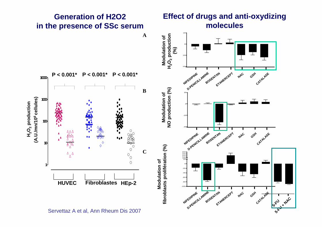
P < 0.001* P < 0.001* P < 0.001*
Servettaz A et al, Ann Rheum Dis 2007
Effect of drugs and anti-oxydizing molecules
A
B
C
- 4 0
- 2 0
0
2 0
- 1 0 0
- 8 0
- 6 0
- 4 0
- 2 0
0
2 01 0 02 0 03 0 04 0 0
Mod
ulat
ion
ofH
2O2
prod
uctio
n (%
)
Mod
ulat
ion
ofN
O p
rodu
ctio
n (%
)
Mod
ulat
ion
offib
robl
asts
pro
lifér
atio
n (%
)
- 4 0
- 2 0
0
2 0
NIFEDIPINE
D-PENIC
ILLAMINE
BOSENTAN
ETANERCEPTNAC
GSH
CATALASE
NIFEDIPINE
D-PENIC
ILLAMINE
BOSENTAN
ETANERCEPTNAC
GSH
CATALASE
NIFEDIPINE
D-PENIC
ILLAMINE
BOSENTAN
ETANERCEPTNAC
GSH
CATALASE
5-FU
5-FU +
NAC
Generation of H2O2 in the presence of SSc serum

Synthesis of TGFββββ and PDGF: activation of fibroblasts (Cotton,J Pathol, 1998)
Major dysfunction of endothelial cells (Matucci-Cerinic, Semin Arthritis Rheum. 2003)
A disease of the endothelium
Loss of physiological barrier: permeabilisation of vessels
Abnormal vascular tone regulation
Increased endothelin-1 synthesis (Mayes, Arthritis Rheum, 2003)
Defective prostacyclin synthesis
Perturbed NO synthesis (Cotton, J Pathol. 1999; Herrick, Clin Exp Rheumatol. 2001)
Apoptosis at early stages (AECA ?) (Sgonc,J Clin Invest. 1996)
Perturbed angiogenesis: VEGF decreased of not detectable (Distler O., Circ Res, 2004)
Synthesis of MCP-1 and VCAM-1: recruitment of lymphocytes (Anderegg, Arch Dermatol Res. 2000)
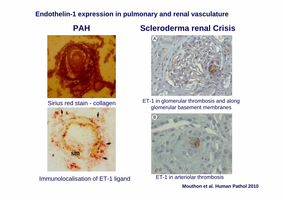
Endothelin-1 expression in pulmonary and renal vascul ature
Sirius red stain - collagen
Immunolocalisation of ET-1 ligand
PAH
ET-1 in arteriolar thrombosis
ET-1 in glomerular thrombosis and along glomerular basement membranes
Scleroderma renal Crisis
Mouthon et al. Human Pathol 2010

Endothelin 1 expression in scleroderma renal crisis
Mouthon et al. Human Pathol 2010
ET-1 in glomerular thrombosis and along glomerular basement membranes
ET-1 in arteriolar thrombosis

Pulmonary vascular remodeling in SSc-PAH
Le Pavec J et al 2010 AJRCCM

Défaut d’apoptose par Fas/Fas-ligand (Santiago B., Arthritis Rheum 2001)
FIBROBLASTES SCLÉRODERMIQUES
Acquisition d’un phénotype activé en myofibroblastes (LeRoy, E.C.. J.Clin Invest, 1974; KIRK, J Biol Chem,
1995)
α-smooth actin (Abraham, D.J. Curr. Rheumatol. Rep. 2007)
Focal Adhesion Kinase (Mimura, Y. J. Invest. Dermatol, 2005)
Activation et synthèse excessive de collagène sous le contrôle de
IL-4: prolifération (POSTLETHWAITE, J Clin Invest, 1992)
Connective Tissue Growth Factor (CTGF) (Leask, A., J. Cell Sci. 2006)
Platelet Derived Growth Factor (PDGF) (Ludwicka, A., J. Rheum. 1995)
Transforming Growth Factor-β β β β (TGF-ββββ) (Pannu, J., Curr. Opin. Rheumatol. 2004)
Défaut de synthèse des régulateurs de la MEC (métalloprotéinases) (VAN DER SLOT, J Biol Chem. 2003)
Anticorps anti fibroblastes et anti-PDGFR (Chizzolini C., Arthritis Rheum 2001, Sevgliati Baroni S., NEJM, 2006)
Formes Réactives de l’Oxygène (FRO)(Sambo P., Arthritis Rheum.,2001)

TGF-ββββ and fibroblasts in SSc
produced by EC, dermal perivascular macrophages
Activation of Smads
Activation of non-smads pathways: p21 activated kinase 2, Rho associated Kinase, …
Transcription of genes encoding for:
Type I collagenPDGF
CTGF Varga J., JCI, 2007
Regulation by Caveolin-1 (Guglielmo G., Nature Cell Biol, 2003)

(A) Early diffuse cutaneous SSc•Moderate fibrosis •Inflammatory infiltrates in the dermis and near the dermal-epidermal junction, predominantly around small blood vessels
Varga J, Abraham D. J Clin Invest 2007; 117:557–67.
(C) Established fibrosis•Dermal thickening•Loss of the microvasculature and dermal structures and the dermis-subcutaneous adipose tissue interface
(B) Early-stage diffusedisease•Profound dermal inflammation perivascular mononuclear cellular infiltrate•Perivascular fibrosis and loss of pericytes and vessel integrity
Skin inflammation and fibrosisin SSc

Elevated levels of cytokines in SSc
� Growth factors• TGF-ββββ, CTGF, VEGF, FGF, etc
� Interleukins• IL-2, IL-4, IL-6, IL-10, IL-13, etc
� Chemokines• MCP-1, IL-8 (CXCL8), TARC, fractalkine, etc
� Other cytokines• TNF-αααα, etc
CTGF = connective tissue growth factor; FGF = fibro blast growth factor; IL = interleukin; MCP = monocyte chemoattractant protein; TARC = thym us and activation-regulated chemokine; TGF = tumour growth factor; TNF = tumour necrosis factor; VEGF = vascular endothelial growth factor Slide courtesy of Kazuhiko Takehara.

CYTOKINES I
TGF-ββββ, chef d’orchestre de la régulation de la fibrogénèse, l’angiogénèse, la régulation immunitaire, prolifération et différentiation cellulaire (Blobe GC, NEJM, 2000)
TGF-ββββ, produits par CE, les monocytes, les lymphocytes T (Blobe GC, NEJM, 2000)
TGF-ββββ induits la différentiation des fibroblastes en myofibroblastes (Kawakami T, J Invest Dermatol 1998)
PDGF produits par plaquettes, macrophages, CE, fibroblastes
PDGF induit proliferation activation des fibroblastes: synthèse de collagène, fibronectine, MCP1, IL-6 (Gay S, J Invest Dermatol 1989)
TGF-ββββ
PDGF

B CD20+Plasmocyte
Infectious agent: topoisomerase 1 and cytomegalovir us
Fragmentation: hypoxia-reperfusion injury
infAAg
T helper
CD28
T CD8+
AAg
CPA
i nf B7
IL-2T helper
CD28
IL-4 (lung)
IL-4 (derma)
Anti-nuclear antibodiesNon-pathogenic Ac anti-EC, anti-fibroblasts
Pathogenic in vitro
SSc: involvement of the adaptative immune system


Identification of CXCL4 as the Major Protein Produc t of Plasmacytoid Dendritic Cells in Systemic Sclerosis.
Van Bon L et al N Engl J Med 2014

Increased Levels of Circulating CXCL4 in Systemic Scl erosis and the Association with Lung Fibrosis and PAH
Van Bon L et al N Engl J Med 2014

Changes in Endothelial Cells and Augmented Responses in Toll-Like Receptors Induced by CXCL4.
Van Bon L et al N Engl J Med 2014

Inflammatory Skin ChangesMimicking Those in Systemic Sclerosis Induced
by CXCL4 In Vivo in Mice.
Van Bon L et al N Engl J Med 2014

T cell activation in SSc
� T cell activation in blood• Soluble IL -2R level correlated with the extent of
skin fibrosis 1
• Clonal expansion of blood T cells 2
� T cell activation in skin• Oligoclonal T cell expansion in the skin 3
• Enhanced transendothelial migration of CD4 + T cells 4
� Pronounced Th17 profile in SSc; intracellular expression of TGF β and IFNg distinguishes SSc phenotypes 1. Steen VD, et al. J Rheumatol 1996; 23:646-9.
2. French LE, et al. Arch Dermatol 2001; 137:1309-13.3. Sakkas LI, et al. J Immunol 2002; 168:3649-59.
4. Stummvoll GH, et al. Ann Rheum Dis 2004; 63:569-74.Radstake, et al. Plos One 2009.

� Abnormal B cell signalling in TSK/+ mice 1
� Presence of B cells in skin 2 and in lungs from SSc patients 3
� Expanded naive B cells and diminished but activated memory B cells 4
� Presence of serum autoantibodies and elevated serum levels of cytokines such as IL-6 which correlate with skin fibrosis
� Elevated serum BAFF levels correlate with disease s everity 5
� Preliminary results from pilot studies in SSc patie nts with rituximab 2,6
1. Saito E, et al. J Clin Invest 2002; 109:1453–62.2. Bosello, et al. Arthritis Res Ther 2010; 12:R54.
3. Lafyatis R, et al. Arthritis Rheum 2007; 56:3167–8.4. Sato S, et al. Arthritis Rheum 2004; 50:1918–27.
5. Matsushita T, et al. Arthritis Rheum 2006; 54:192–201.6. Lafyatis R, et al. Arthritis Rheum 2009, 60:578-83.
SSc: involvement ofB lymphocytes

Autoantibodies in scleroderma
Gabrielli A, et al. N Engl J Med 2009
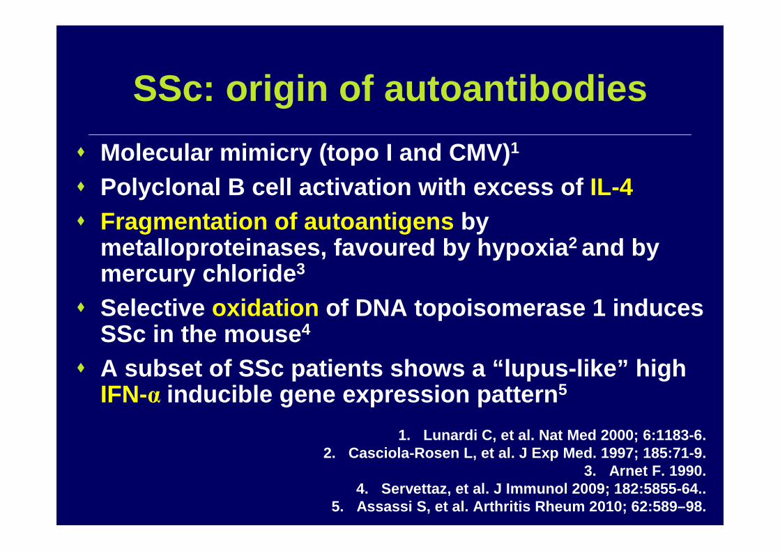
SSc: origin of autoantibodies
� Molecular mimicry (topo I and CMV) 1
� Polyclonal B cell activation with excess of IL-4� Fragmentation of autoantigens by
metalloproteinases, favoured by hypoxia 2 and by mercury chloride 3
� Selective oxidation of DNA topoisomerase 1 induces SSc in the mouse 4
� A subset of SSc patients shows a “lupus-like” high IFN-α inducible gene expression pattern 5
1. Lunardi C, et al. Nat Med 2000; 6:1183-6.2. Casciola-Rosen L, et al. J Exp Med. 1997; 185:71-9.
3. Arnet F. 1990.4. Servettaz, et al. J Immunol 2009; 182:5855-64..
5. Assassi S, et al. Arthritis Rheum 2010; 62:589–98.

• Cross-reactivity of AECA with a CMV protein 3
• Target antigens unknown except ”scleroderma specific”autoantigens 4,5
Anti-endothelial cell antibodies (AECA) in SSc
• Not disease specific• Absence of standardization• Activate EC and induce the
expression of adhesion molecules (IL-1 dependent) 1
• Induce apoptosis in the presence of NK cells 2
1. Carvalho D. Arthr Rheum 1999. 2. Bordron A. J Clin Invest 1998.3. Lunardi C, et al. Nat Med 2000. 4. Garcia de la Pena et al. Clin Immunol 2004.
5. Servettaz et al. Clin Immunol 2006. 6. Dib H, et al. Eur Resp J 2011
6. Ab: controls; cd: ssc w/o PAH; ef: SSc-PAH; gh: IPAH

• Anti-fibroblast antibodies (AFA) are present in the serum of 20 to 80% of SSc patients 1
• AFA can activate fibroblasts and induce extracellul ar matrix proteins synthesis 2
• Induce a proadhesion fibroblast phenotype byup-regulating ICAM -1 and increase fibroblast synthesis of pro-inflammatory cytokines
• AFA induce fibroblasts to produce profibrotic chemokines, with partial exploitation of TLR4 3
• Target antigens• DNA topoisomerase 1 4
• PDGF receptor 5
Anti-fibroblast Abs in SSc
1. Brentnall, 1982; Chizzolini, 2002; Alderuccio, 1 989; Ronda, 2002.2. Chizzolini C . Arthritis Rheum 2002.
3. Fineschi S. Arthritis Rheum 2008.4. Henault G. Arthritis Rheum 2004; Henault G. Arthritis Rheum 2006; Tamby MC et al. 2008.
5. Baroni S, et al. NEJM 2006; Classen, et al. 2009; Loizos, et al. 2009.

Svegliati Baroni, NEJM, 2006
ANTICORPS ANTI-PDGFR
Les IgG sériques stimulent le récepteur de PDGF, qui stabilise RAS et induit ERK1/2L’induction de ERK1/2 entraine la production de FRO (ROS)
La persistance à long terme de ROS et ERK1/2 entraîne une augmentation de l’expression du gène du collagène
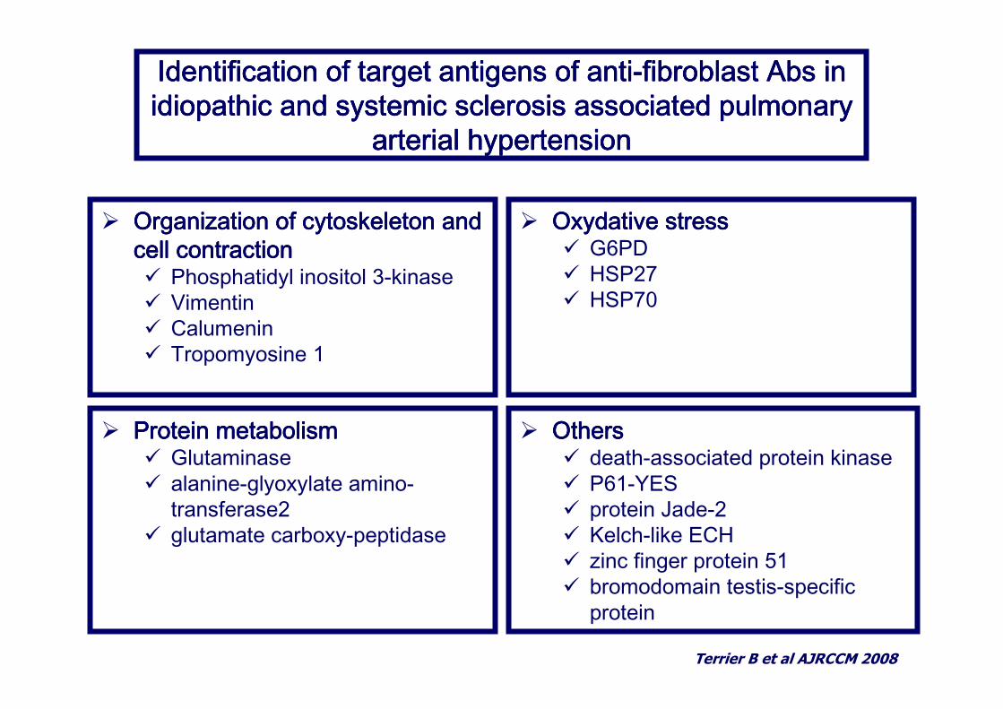
Identification of target antigens of antiIdentification of target antigens of antiIdentification of target antigens of antiIdentification of target antigens of anti----fibroblast Abs in fibroblast Abs in fibroblast Abs in fibroblast Abs in idiopathic and systemic sclerosis associated pulmonary idiopathic and systemic sclerosis associated pulmonary idiopathic and systemic sclerosis associated pulmonary idiopathic and systemic sclerosis associated pulmonary
arterial hypertensionarterial hypertensionarterial hypertensionarterial hypertension
Terrier B et al AJRCCM 2008
� OthersOthersOthersOthers� death-associated protein kinase� P61-YES� protein Jade-2� Kelch-like ECH� zinc finger protein 51� bromodomain testis-specific
protein
� Oxydative stressOxydative stressOxydative stressOxydative stress� G6PD� HSP27� HSP70
� Organization of cytoskeleton and Organization of cytoskeleton and Organization of cytoskeleton and Organization of cytoskeleton and cell contractioncell contractioncell contractioncell contraction� Phosphatidyl inositol 3-kinase� Vimentin� Calumenin� Tropomyosine 1
� Protein metabolismProtein metabolismProtein metabolismProtein metabolism� Glutaminase� alanine-glyoxylate amino-
transferase2� glutamate carboxy-peptidase

Indirect immunofluorescence on permeabilized human aortic vascular smooth muscle cells, with sera from HC or with sera from SSc-w/oPAH, SSc-PAH and iPAH.
Bussone et al.Ann Rheum Dis 2011
Inhibition of contraction

Systemic sclerosis: lesions at different stages
Gabrielli A. NEJM 2009

• Major work has been done in order to improve the understanding of SSc pathogenesis.
• A number of new experimental models have been set up, that should help to understand the disease pathogenesis and test new therapeutic targets.
• ROS represent a hallmark of the pathogenesis of SSc • Besides endothelial cells and fibroblasts, major development
has been made in the understanding of the role of B cells and autoantibodies in the pathogenesis of SSc.
• Plasmacytoid dendritic cells seem to play a major role in the pathogenesis of SSc through the secretion of CXCL4, although these data will need to be confirmed in the near future.
Conclusion
