tjms vol3.2,2016 مجلة توفيق الطبية
TRANSCRIPT

TOFIQ Journal of Medical Sciences ISSN: 2377-2808
TOFIQ Journal of Medical Sciences (TJMS)
Vol 3, Issue 2 (2016)
Issued by: TOFIQ Office, 2405 Carey lane, Vienna, VA 22181 USA
TJMS.tofiq.org

Dear TJMS readers,
It is my great pleasure to present to you the Volume 3 (2016) of our TJMS.
We would like to announce that TJMS has been accepted into DOAJ. Being
included in DOAJ will raise the profile of the journal, and the metadata we supply
to DOAJ will be searchable on their web site and via many libraries who take our
data feed..
The current issue (Issue 2, Volume 3) contains a variety of scholarly articles
which have already attracted many communications from different disciplines all
over.
We look forward to your continued support and contribution.
Thank you again,
A Hadi Al Khalili, MBChB, MPhil, FRCSE, FACS
Editor-in-Chief
TOFIQ Journal of Medical Sciences
TOFIQ Journal of Medical Sciences (TJMS) is published by TOFIQ: an NGO registered at the
State of Maryland as a non-profit organization dedicated to helping Iraq Higher Education and
Research.
TJMS is devoted to the publication of original research, commentaries on a current topic,
reviews, letters to the editor, and editorials in the field of medical sciences. The early focus of the
journal is on clinical burden of disease in Iraq: documentation of its nature and extent; clinical
patterns and epidemiology; diagnostic findings; and therapeutic strategies. Read more ...
Focus and Scope
TOFIQ Journal of Medical Sciences (TJMS) is published by TOFIQ: an NGO registered at the
State of Maryland as a non-profit organization dedicated to helping Iraq Higher Education and
Research.
TJMS is devoted to the publication of original research, commentaries on a current topic,
reviews, letters to the editor, and editorials in the field of medical sciences. The early focus of the
journal is on clinical burden of disease in Iraq: documentation of its nature and extent; clinical
patterns and epidemiology; diagnostic findings; and therapeutic strategies.
Mission:
1. TJMS is a high quality, biannually, peer-reviewed, electronic medical sciences journal,
publishing original research and scholarly review articles, letters to the editor, and
editorials.
2. TJMS focuses on the disease burden in Iraq.
3. An outlet for the current research and an academic product in Iraq in the fields of
medicine, dentistry, pharmacy, nursing and related disciplines that supports recognition
and academic advancement in Iraq and beyond.
4. TJMS will lay the groundwork for creation of sister journals in other disciplines relating
to Iraq (engineering, agriculture, science and technology, social sciences and humanities).
Structure:
1. Single editor-in-chief, working in a full-time, compensated capacity.
2. Editorial board consisting of experts in the various branches of medicine, dentistry,
pharmacy, nursing and related disciplines.
3. Open-access, electronic-only journal
4. Peer-reviewed publication supported by an online submission, review, and decision for
articles.
5. Articles can be submitted by any individual/group, or can be solicited (invited reviews
and discussions). Decisions for publication will be blinded to author or region of origin.
The criteria on which the submissions are evaluated for acceptance will be heavily
weighted on their applicability to the burden of disease in Iraq.
6. Publication will be biannual however, approved articles will be released continuously.

7. Funding: will attempt to obtain corporate sponsorship through unrestricted educational
grants. Sponsorship will be acknowledged in compliance with ACCME and ICMJE
guidelines.
8. The publication would be the official medical sciences journal of TOFIQ, and we would
encourage other medical organizations to consider collaborating with.
Editorial Team
Editor in Chief
1. Dr A Hadi Al Khalili
MBChB, FRCSE, FACS, MPhil, Professor Emeritus, Department of Surgery
(Neurosurgery) University of Baghdad, Baghdad, Iraq
Editorial Board
1. Dr. Munther Aldoori
Professor MI Aldoori ,MBChB, PhD ,FRCP, FRCS, FRCS Edin.,FRCS G. ,FACS.
Consultant in General,Vascular and Endocrine Surgery. Senior Clinical Lecturer at the
University of Leeds, UK
2. Dr. Sa'ad Al Fattal
MBChB, FRCS(Eng), FRCS(Glas), MCh Orthopedic surgeon, London, UK
3. Dr. Stephen Evans
MD, Chairman and Professor, Department of Surgery, Georgetown University,
Washington, DC Biomedical Graduate Research Organization (BGRO), Washington DC,
USA
4. Dr. Allen Dyer
MD, PhD, Professor of Psychiatry and Behavioral Sciences, George Washington
University, Washington, DC, USA
5. Dr. Adil Shamoo
Ph.D. Professor, Department of Biochemistry and Molecular Biology Professor and
former Chairman, University of Maryland School of Medicine Baltimore, MD, USA
6. Dr. Khlood Salman
PhD, Professor, School of Nursing Duquesne University Pittsburgh, PA, USA
7. Dr. Wael Khamas
BVM&S, MS, PhD Professor of Anatomy & Histology; Chair of the University Senate
College of Veterinary Medicine Western University of Health Sciences Pomona, CA,
USA
8. Dr. Amid Ismail
BDS, MPH, Dr PH, MBA, Dean, School of Dentistry, Temple University Philadelphia,
PA, USA
9. Dr. Ami Iskandrian
MD, MACC Distinguished Professor of Medicine and Radiology Section head, Non-
invasive cardiac imaging and nuclear cardiology Division of Cardiovascular diseases,
Department of Medicine University of Alabama, Birmingham, AL, USA
10. Dr. Zayd Eldadah
MD, PhD, FACC, Cardiologist, Cardiac Electrophysiology Washington Hospital Center,
Adjunct Assistant Professor, The Johns Hopkins University School of Medicine,
Washington DC, USA
11. Dr. Ali Al Attar
MD, PhD Plastic surgeon, Washington DC, USA
12. Dr. Karim Alkadhi
PhD, Professor, Department of Pharmacological and Pharmaceutical Sciences, College of
Pharmacy University of Houston, TX, USA
13. Dr. Hikmat Shaarbaf
MD, FRCP (London), FRCP (C), Prof. Emeritus Internal medicine, Former Dean of
Medical School, Baghdad University, Baghdad, Iraq
14. Dr. Sarmad Khunda
MD, FRCS, FRCOG, Professor Emeritus, College of Medicine, Baghdad University,
Baghdad, Iraq
15. Dr. Makki Fayadh
MB,ChB,MRCP UK,FRCP Ed,FRCP London, Consultant Physician, Gastroentrologist.
Former head of The Iraqi Gastroenterology Center, Baghdad, Iraq
16. Dr. Hani Haider
PhD, Prof. Director of Orthopedics Biomechanics & Advanced Surgical Technologies
Laboratory, Department of Orthopedic Surgery and Rehabilitation, University of
Nebraska Medical Center, Omaha, NE, USA
17. Dr. Alaa A. Abdulrasool
PhD, Professor Pharmaceutics, industrial Pharmacy, President of Baghdad University,
Baghdad, Iraq
18. Dr. Taghreed Hasim Al-Noor
PhD, Professor, inorganic chemistry, Chemistry Department, Ibn-Al-Haitham Education
College, Baghdad University , Baghdad,Iraq
19. Dr. Mahjoob N. Alnaddawi
MRCPUK, FRCP Lond, FRCP ED, FRCPCH, Professor of Pediatrics, Chair of Scientific
Council of Pediatric Arab Board, Baghdad, Iraq
TOFIQ Journal of Medical Sciences ISSN 2377-2808
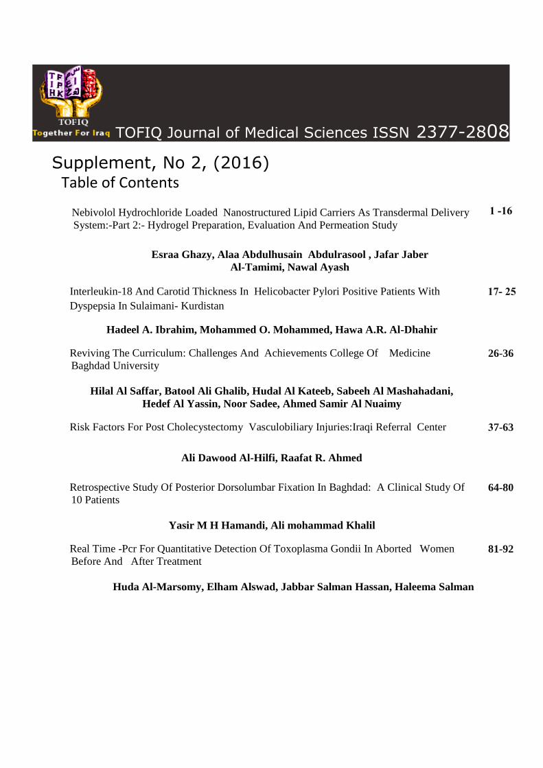
Supplement, No 2, (2016) Table of Contents
16- 1 Nebivolol Hydrochloride Loaded Nanostructured Lipid Carriers As Transdermal Delivery
System:-Part 2:- Hydrogel Preparation, Evaluation And Permeation Study
Esraa Ghazy, Alaa Abdulhusain Abdulrasool , Jafar Jaber
Al-Tamimi, Nawal Ayash
52 -11 Interleukin-18 And Carotid Thickness In Helicobacter Pylori Positive Patients With
Dyspepsia In Sulaimani- Kurdistan
Hadeel A. Ibrahim, Mohammed O. Mohammed, Hawa A.R. Al-Dhahir
26-36 Reviving The Curriculum: Challenges And Achievements College Of Medicine
Baghdad University
Hilal Al Saffar, Batool Ali Ghalib, Hudal Al Kateeb, Sabeeh Al Mashahadani,
Hedef Al Yassin, Noor Sadee, Ahmed Samir Al Nuaimy
7-633 Risk Factors For Post Cholecystectomy Vasculobiliary Injuries:Iraqi Referral Center
Ali Dawood Al-Hilfi, Raafat R. Ahmed
64-80 Retrospective Study Of Posterior Dorsolumbar Fixation In Baghdad: A Clinical Study Of
10 Patients
Yasir M H Hamandi, Ali mohammad Khalil
81-92 Real Time -Pcr For Quantitative Detection Of Toxoplasma Gondii In Aborted Women
Before And After Treatment
Huda Al-Marsomy, Elham Alswad, Jabbar Salman Hassan, Haleema Salman

TOFIQ Journal of Medical Sciences, TJMS, Vol. 3, Issue 2, (2016), 1-16 ISSN: 2377-2808
NEBIVOLOL HYDROCHLORIDE LOADED NANOSTRUCTURED LIPID
CARRIERS AS TRANSDERMAL DELIVERY SYSTEM:-PART 2:- HYDROGEL
PREPARATION, EVALUATION AND PERMEATION STUDY
Esraa Ghazy*, Alaa Abdulhusain Abdulrasool
**, Jafar Jaber Al-Tamimi
**
and Nawal Ayash *Al-Rasheed University College, Department of Pharmacy,
**Department of Pharmaceutics, College of Pharmacy, Baghdad University/Baghdad-Iraq.
Corresponding Author:
E-mail: Esraa Ghazy <[email protected]>
Abstract:
Nebivolol hydrochloride (NEB), is a third generation highly selective β1-blocker, it has
an antihypertensive properties. Its elimination half-life is around 10 hrs while its oral
bioavailability is about 12%. The objective of the current study was to develop nanostructured
lipid carriers (NLCs) for transdermal delivery of (NEB). Through the preparation,
characterization and conducting of an in vitro study for (NEB) loaded (NLCs), the formulation of
NEB-NLCs based hydrogel using different types of gelling agents was introduced. Moreover, the
incorporation of lipid nanoparticles into carbapol 934 as hydrogel base in different formulations
was described in this study. The optimized formula (350 mg Glyceryl monostearate, 150 mg
oleic acid, 2% (W/W) span 80, and 2% (W/W) Cremophor EL) was tested for entrapment
efficiency, particle size and loading capacity then incorporated into hydrogel for expedient
transdermal application. A number of measures were implemented for the NEB-NLCs based
hydrogel, the results for the optimized formula were found to be as the following: particle size
228 nm, polydispersity index 0.3, zeta potential -29mV, pH 7.05 viscosity 7210cps, spreadability
6 cm, drug content 95%, and Ex Vitro skin permeation 90.8%. The transmission electron
microscopy (TEM) and the optical microscope study revealed almost spherical shaped
nanoparticles. Nebivolol based hydrogel demonstrated no skin irritation and showed a
prolong release for up to 24 hrs. The flux for the permeation study through rat skin was found
to be (143μg/cm2/hr). Carbapol 934 was used as a gelling agent; the obtained formula gave
evidence for good spreadability, homogeneity and rheological behavior. In conclusion, the data
TOFIQ Journal of Medical Sciences, TJMS, Vol. 3, Issue 2, (2016), 1-16 2
obtained from this study illustrated a successful development of NEB-NLCs-based hydrogel in
the increase of the encapsulation efficiency of colloidal lipid carriers. The advantages of the
colloidal lipid carriers of the improved performance were in terms of stability and provides a
sustaining NEB transdermal effect.
Keywords: Nebivolol hydrochloride; Nanostructured lipid carriers, Hydrogel,
Transdermal delivery
Introduction
In the last decade, the prefix “nano” has an increasing application to various fields of
knowledge. Nanoscience, nanotechnology and nanomaterials or nanochemistry, all represent
examples of few terms that occur repeatedly in scientific reports, books as well as in daily
newspapers, it has been recognized for a wide range of audience even for non experts.
International system (IS) of units is used to indicate a reduction factor of 109
times(1)
.
Transdermal. Nebivolol hydrochloride (NEB) is a lipophilic β1-blocker, devoid of intrinsic
sympathomimetic and membrane stabilizing activity. Clinically, (NEB) is administered as a
racemic mixture of equal proportions; d-isomer ((SRRR)-nebivolol a potent cardioselective β1
adrenoceptor blocker) and L-isomer ((RSSS)-nebivolol with favorable hemodynamic profile(2,3)
.
These two enantiomers possess unequal potency regarding to β-receptor blocking activity
and nitric oxide mediated vasodilation. Moreover, the blend of (SRRR and RSSS) has a greater
antihypertensive activity than either enantiomer alone(4)
. Nebivolol hydrochloride is an official
drug in the British & Indian Pharmacopoeia(5)
. The molecular weight for NEB is 441.9 while for
the free bases are 405.4(6)
. The lipid nanoparticles, such as solid lipid nanoparticles (SLN) and
nanostructured lipid carriers (NLCs) are stable colloidal systems with notable compensations
such as drug delivery systems, i.e. biocompatibility, biodegradability, physicochemical stability,
versatility and controlled drug release. (7,8)
Furthermore, they are providing controlled release
profiles for many substances. Aqueous dispersions of lipid nanoparticles are being investigated
as drug delivery systems for different therapeutic purposes. Their most interesting characteristic
is the possibility to be used topically and transdermally, while other systems have to be
incorporated into commonly used dermal carriers, such as creams or hydrogels, in order
to have a proper semisolid consistency(7,8)
.

TOFIQ Journal of Medical Sciences, TJMS, Vol. 3, Issue 2, (2016), 1-16 3
Materials and Methods
Nebivolol (NEB) and transcutol P were purchased from ProvizerPharma, India. Oleic acid
was supplied by Riedel De Haen AGHonnover, German, Cremophore EL (Polyoxy l35 Castor
oil) was purchased from HiMedia Lab Pvt. Ltd, India. Span 80 was provided by
Hopkin&Williams LTD, England. Poloxamer 188 (Pluronic F-68) Lutrol® and Lecithin
(Phosphatidylcholine purity 72.7%) were purchased by Sigma-Aldrich, Chemie GMBH,
Germany. Potassium dihydrogen phosphate, Disodium hydrogen phosphate, Diethyl ether
Carbapol 934 and Sodium alginatewere provided by BDH Chemicals Ltd., Poole, England.
Glycerylmonostearate was supplied by BDH Chemicals Ltd. Poole, England.MyverolTM
18-04K
was provided by Gattee fosse, France. Chitosan , HPMC was purchased from Fluka AG. Chem.
1: Preparation of Nebivolol-loaded NLCs based hydrogel
According to the previous work(9)
, 100 mg NEB was incorporated into combination of
different solid and liquid lipids, surfactants and co-surfactants (Table 1). Accordingly, four
formulas were selected for hydrogel preparation. The optimized (NEB-NLCs) formula (F3) was
selected based on the evaluation of characteristics like: particle size, entrapment efficiency, and
in vitro release. Different gelling agents such as: carbopol 934, sodium alginate, chitosan, and
hydroxyl propylmethyl cellulose (HPMC) were used for the conversion of NLCs dispersion into
NLCs based hydrogel formulation (Table 2). Based on the compatibility with NLCs dispersion
and for easier spreadability, carbopol 934 as gel forming polymer was selected (F3B). The
hydrogel base was prepared by dispersing (1%w/w) carbapol 934 in distilled water, stirred for 10
mins at 1500 rpm to obtain a homogeneous gel base, and then immediately neutralized by adding
drops of triethanolamine (0.5%V/V) to promote gelation. The pH of gel was adjusted to 7.4.
Hydrogel was further allowed to stand overnight at a room temperature to remove any entrapped
air. The hydrogel was used to disperse a freshly prepared NLCs suspension. The aqueous NLCs
dispersion and hydrogel were stirred and mixed under a mechanical stirrer at a speed of 1000
rpm/15 min.
TOFIQ Journal of Medical Sciences, TJMS, Vol. 3, Issue 2, (2016), 1-16 4
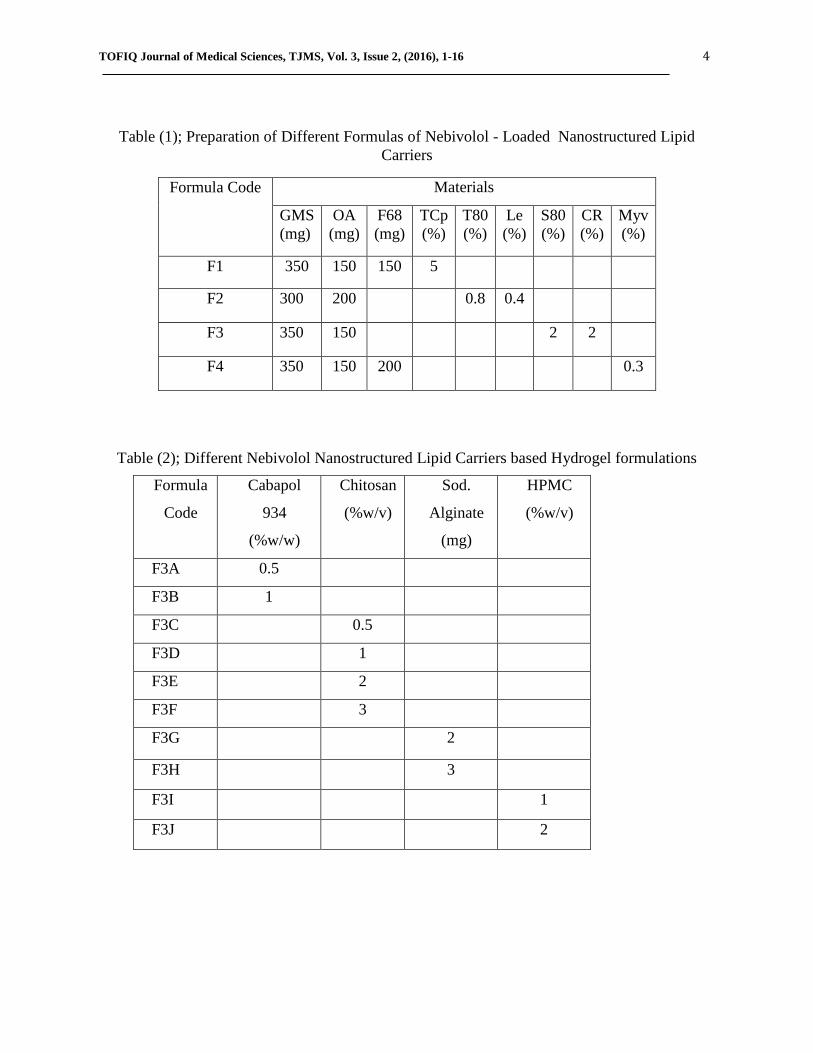
Table (1); Preparation of Different Formulas of Nebivolol - Loaded Nanostructured Lipid
Carriers
Formula Code Materials
GMS
(mg)
OA
(mg)
F68
(mg)
TCp
(%)
T80
(%)
Le
(%)
S80
(%)
CR
(%)
Myv
(%)
F1 350 150 150 5
F2 300 200 0.8 0.4
F3 350 150 2 2
F4 350 150 200 0.3
Table (2); Different Nebivolol Nanostructured Lipid Carriers based Hydrogel formulations
Formula
Code
Cabapol
934
(%w/w)
Chitosan
(%w/v)
Sod.
Alginate
(mg)
HPMC
(%w/v)
F3A 0.5
F3B 1
F3C 0.5
F3D 1
F3E 2
F3F 3
F3G 2
F3H 3
F3I 1
F3J 2

TOFIQ Journal of Medical Sciences, TJMS, Vol. 3, Issue 2, (2016), 1-16 5
2: Evaluation of the NEB-NLCs based hydrogel
1. 2. Physical Examination of NEB-NLCs based hydrogel
The prepared NEB-NLCs hydrogel formulation was inspected visually for its color,
appearance and consistency.
2.2. Determination of pH, Viscosity and Spreadability
The pH of the NLCs loaded hydrogel was determined using digital pHmeter (France and
Hanna instruments type), standardized using standard buffer solutions (pH 4.0 and 7.0). One
gram of hydrogel was dissolved in 100 mL of distilled water and stirred for 10 min then stored
for 2 hrs. Results were taken in triplicate(10)
. The viscosity of NLCs based hydrogel was
measured using Brookfield DVII+Prodigital viscometer at 25◦C. A formulation weight of 20 gm
was utilized and exposed to speed of 50 rpm. The measurements were attained in triplicate. The
rheological nature of the disperse system was assayed by plotting shear stress against shear rate
in a rheogram to determine if the systems are thixotropic. The readings were performed in
triplicate(11)
.
For determination of the spreadability of hydrogel 0.5 g of gel was placed in a circle with a
radius of 1cm premarked on a glass plate on top of another glass plate. A
weight of 500 gm was allowed to rest on the upper glass plate (10)
. The extension in the diameter
due to spreading of the hydrogels was measured with a linear scale. Experiments were done in
triplicate.
3.2. Drug Content Determination
The NEB content in the hydrogel was determined by taking required quantity of the
prepared gel which is equivalent to 10 mg of NEB and transferred to 100 ml of volumetric
flask containing phosphate buffer (pH 7.4). Then, it was sonicated and filtered. Later, it was
suitably diluted and analyzed at λmax of NEB. The content of NEB was determined using UV-
visible spectrophotometer at 281nm against blank (11)
.
4.2. Measurement of Particle Size and Polysisperisty Index of NLCs based Hydrogel
Light dynamic light scattering (LDS) was used. The aqueous NEB-NLCs was dispersed in a
fixed amount of filtered distilled water (1:50) dilution of all formulations was made and placed
in 1cm diameter disposable cuvette to yield a suitable scattering intensity. The mean particle size
and polydispersity index PDI (The measurement of the width of size of distribution) for NEB-
NLCs based hydrogel was calculated using Brookhaven Instruments Corp90 PLUS (ZetaPlus
Particle Sizing, NY, Software, Version 5.34). Experiments were carried out in triplicate, and
standard deviations (±SD) were calculated at a fixed scattering angle of 90°at (25°C)(12)
.
5.2. Zeta Potential determination of NEB- NLCs based Hydrogel
Zeta potential of NLCs based hydrogel was measured using (NanoBrookZeta PLAS)
Zetasizer. The NanoBrookZetaPALS determines zeta potential using Phase Analysis Light
Scattering technique which is up to 1000 times more sensitive than traditional light scattering
methods based on the shifted frequency spectrum. Samples were placed in disposable zeta cells
TOFIQ Journal of Medical Sciences, TJMS, Vol. 3, Issue 2, (2016), 1-16 6
and the NLC suspensions were diluted with distilled water (1:100) of all the formulations to get a
uniform dispersion prior to analysis. The conductivity of the diluted sample was measured to
choose the detection model. The whole measurement was carried out at 25°C(11)
.
6.2. Visualization by Optical Microscope
Optical microscope (BX51M Model) offered a reflected light illumination. One drop of each
prepared (NEB) dispersions was examined under the optical microscope using an (1000x)
magnifying power. Particle behavior, shape and morphology were investigated.
7.2. Visualization by transmission electron microscope (TEM)
The size and morphology of the selected formula was examined using TEM (PHILIPS CM 10),
with an accelerating voltage of 100 KVA. The work was conducted by placing one drop of the
sample on a copper grid coated with a formvar carbon film and allowed to stand at room
temperature for 90 sec to form a thin film. Excess of the solution was wicked away with the aid
of filter paper. The grid was allowed to thoroughly dry in air, the sample was viewed and ready
for analysis and photomicrographs were taken at suitable magnification
3. In-vitro Skin Permeability of NEB-NLCs based hydrogel
1.3. Preparation of Rat Skin (Diffusion Membrane)
Albino rats (4-6 week old males) were euthanized. Then the abdominal skin was shaved
lightly with an electrical clipper taking care to prevent any damage to the surface of the skin. A
rectangular section of abdominal skin several centimeters in each dimension were excised from
the animal using a sharp blade. The skin was lifted easily from the animal after incision was
made. The defatting procedure: the skin was defatted by wiping with a cotton tip soaked in
diethyl ether to remove the subcutaneous fat and scraping the dermal side to remove the muscle
and blood vessels. The skin was wiped again with a cotton tip soaked in ether to prevent any
adhering fats and kept in a phosphate buffer pH 7.4 for about 2 hrs in a water bath at constant
temperature of 37oC to allow water soluble UV absorbing material to leach out. The buffer was
change three times during this period with fresh amounts. Then the prepared skin for diffusion
study was stored in a phosphate buffer for 24hrs in the refrigerator at 2°C before use
(13).
2.3. Permeation Study of NLCs based hydrogel
Franz diffusion cell Equipment-MCF10, was used in all diffusion studies. It consists of six
Franz diffusion cells arranged in a water jacket with a heater to obtain a constant experimental
temperature of 32 ± 1oC, on magnetic stirrers at equal speed of rotation, temperature and rotation
were equilibrated electronically. All Franz diffusion cells used in the experiment consisted of
two compartments: Upper donor compartment and lower receptor compartment. Phosphate
buffer solution pH 7.4 was used as a receptor medium with volume of 30 ml for all the release
studies of NEB-NLCs to assure sink condition. Firstly, all receptor compartments were filled

TOFIQ Journal of Medical Sciences, TJMS, Vol. 3, Issue 2, (2016), 1-16 7
with phosphate buffer solution pH 7.4 and the assembled set up left in instrument to equilibrate
to experimental temperature as to get rid of air bubbles for at least half an hour. Then the rat skin
membrane with surface area of 3.97 cm2 were mounted between the two compartments of the
diffusion cells in such a way that the SC layer was facing the donor compartment and the dermis
facing the receptor compartment, and fastened with an O-ring. The solution in receptor
compartment was agitated with a magnetic stirrer at 100 rpm(14)
. After assembling the described
set up, samples of 1 ml were taken periodically through the sampling port from the receptor
compartment at pre determined time intervals (0.25, 0.5, 1, 2, 3, 4, 5, 10, 15, 20 and 24 hours),
and replaced with an equal volume of fresh receptor solution at temperature of 32 ± 1oC to
maintain a constant volume of the receptor phase. Samples were analyzed for NEB content using
UV-visible spectrophotometer. A cumulative amount of drug diffused was calculated(15)
. All
results were repeater in triplicates and presented in mean values ± standard deviations.
4. Skin irritation study
The primary skin irritation test was very important to estimate the irritancy of substances that
applied repeatedly to the skin of humans. Irritancy test was done using healthy male albino rat
weighing approximately 200 gm. Firstly, the left dorsal surface of the rat was shaved carefully
with an electrical clipper then cleaned with rectified spirit then distilled water and left for drying.
The optimized NEB-NLCs hydrogel was placed on the left dorsal surface of the rat, and skin
irritation from the formulation was determined by observations for 7 days for any skin sensitivity
and reactions such as redness, erythema, edema, and skin rash (10)
.
Results and Discussion:
1. Preparation of NEB-NLCs based Hydrogel
Based on the particle size, the entrapment efficiency and the in Vitro release profiles of NEB
based hydrogel (F 1, 2, 3 and 4) which showed optimum physicochemical properties, were
selected for the formulation of the hydrogel for transdermal NEB delivery by incorporation into
(1%W/W) carbapol 934. However, (F3B) was chosen as the selected formula as it exhibited the
best release profile, viscosity, spreadability, pH, and % drug content.
2. Evaluation of NEB-NLCs based hydrogel
1.2. Visual Inspection
The visual inspection of NEB-NLCs based hydrogel was found to be off-white in color
homogenous and showed smooth texture. Hydrogel loaded with NEB-NLCs (F43B) was stored
in a tight closed glass container, protected from light. The physical properties of the selected
formulas after two months of storage was examined, no changes in the original off-white color or
unpleasant odor were observed, which indicates no probability of microorganism growth and
hence, the physical stability of the selected formula.
TOFIQ Journal of Medical Sciences, TJMS, Vol. 3, Issue 2, (2016), 1-16 8
2.2. pH, Viscosity and Spredability Determination
The pH of the prepared NLCs loaded hydrogel was between 6.9±0.02 to 7.05±0.05 that
lies in normal range of skin pH (4.5-7) as shown in (Table 3). Viscosity of the prepared hydrogel
is mainly dependent on the gelling agent used. In the current study, when carbapol 934
(1%W/W) being under hydration condition it can form a physically bounded structure that is
crucial for providing the proper mechanical strength to the hydrogel. Accordingly there is
noticeable difference in viscosity values of the control hydrogel and NLCs based hydrogel which
can be justified as the participation of nanostructured lipid carriers on the viscosity of the
formulation. Spredability is a crucial factor to uniform and ease the application of topical
and transdermal preparations from the patient`s compliance point of view. The application of
the formulation to the inflamed skin is more comfortable if the base spreads easily, enhancing
maximum slip and drag(16)
.Spreadability was found to be 6±1 cm for the optimized NEB-NLCs
hydrogel formula (F3B) which indicates excellent spreadability as the large diameter signifies
better spreadability.
3.2. Drug Content Evaluation in the Prepared Hydrogel
Drug content of the optimized NEB-NLCs based hydrogel(F3B) was found to be
95.0%±0.6 and gave better drug loading capacity of formulation as shown in (Table 3).
Table (3); pH, Viscosity, Spreadability and % Drug Content Evaluation of the different
NEB-NLCs Based Hydrogels
Formula
Code
pH Viscosity
(cp)(50rpm)
Spredability
(cm)
%Drug
Content
Control 6.02±0.02 3521±3.2 2.9±1.5 80.3±0.6
F1B 6.10±0.03 7622±1.4 3.5±1.0 87.1±0.1
F2B 6.78±0.11 7834±2.5 4.9±0.5 86.0±0.5
F3B 7.05±0.05 7210±1.2 6.0±1.0 95.0±0.6
F4B 7.20±0.12 7243±2.3 5.2±1.5 94.1±0.7
4.2. Particle Size, Polydispersity Index and Zeta potential evaluation of NEB-NLCs based
Hydrogel
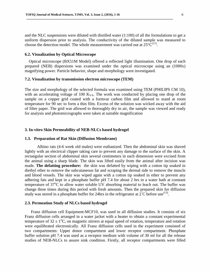
The average particle size, Polydispersity index and zeta potential of NEB-NLCs based
hydrogel (F3B) was found to be 228nm, 0.3, -29.0 respectively. Zeta potential value of the
freshly prepared hydrogel (F3B) is illustrated in figure (1). Meanwhile, figures (2 A, B, C and D)
for the NEB-NLCs based hydrogel (F3) under TEM. TEM micrographs showed nearly rounded
nanoparticle surrounded by a homogeneous shading with a rather uniform distribution and no
prominent sign of aggregate remained in the hydrogel. However, there was no drug detected in
the blank NLCs (figure 3), whose particle size was smaller than the NEB-NLCs. Such results

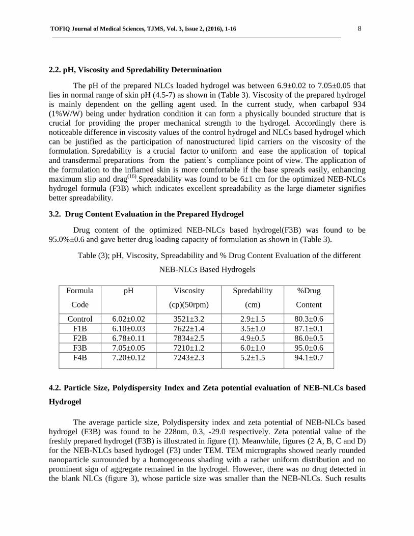
TOFIQ Journal of Medical Sciences, TJMS, Vol. 3, Issue 2, (2016), 1-16 9
were confirmed by optical microscope images (Figure 4) which suggests that the optimized
formula (F3) resembled the drug-enriched core model (i.e. the drug occupies the core of the
particles), (NEB) was dispersed well due to its miscibility in the lipid matrix. In such a model,
the core is surrounded by a practically drug-free lipid shell and homogenous hydrophilic
polymer(17,18)
.
Figure (1); Zeta Potential of the freshly prepared (NEB-NLCs) based hydrogel (F3B)
Figure (2); TEM Micrographs of A, B, C and D of NEB-NLCs, and D/Blank NEB-NLCS
TOFIQ Journal of Medical Sciences, TJMS, Vol. 3, Issue 2, (2016), 1-16 10
Figure (3); TEM image of NEB-NLCs based hydrogel (F3B)
Figure (4); NEB-NLCs based hydrogel (F3B) under optical microscope
3. Skin Permeation Study of Prepared NEB-NLCs based hydrogel
Drug permeating across the skin in the vitro status represents the amount available for skin
absorption for some anti hypertensive drugs such as carvidolol, propranolol and labetolol(19)
.
Hence, drug flux may be an indicator of the drug absorption to the targeted skin tissue. The
kinetics of drug permeation which is helpful to explain the mechanisms of drug absorption via
the skin. Release of drug from the vehicle must occur before the permeation of the drug into the
skin. In vitro permeation study was performed to compare the permeation ability of NEB to the
various NLC gel formulations (NLC-F1, NLC-F2, NLC-F3, and NLC-F4) using Franz diffusion
cells. The in vitro permeation profile is portrayed in figure (5). Cumulative permeation profile of
the different formulations revealed that the permeation of NEB from the NLCs hydrogel
formulation (NLC-F3) is significantly high (p<0.05) as compared to the permeation of NEB from
hydrogel formulations (NLC-F1, NLC-F2, and NLC-F4) which confirms the selection of (NLC-
F3) to be the best formula. It was found that 90.8% of NEB permeated within 24 hrs of the study

TOFIQ Journal of Medical Sciences, TJMS, Vol. 3, Issue 2, (2016), 1-16 11
as depicted in (Figure 5). The diffusion parameters such as lag time, permeation coefficient and
(NEB) flux of the optimized formula were: 0.5 hr, 141 (cm/hr) × 10 -3
and 143 (µg/cm2.hr),
respectively.
In this study, it has been proven that release rate of NEB is close to the flux value which
indicates that NEB can easily penetrate into the stratum corneum. This suggests that partitioning
into the skin and subsequent penetration are predominant steps in transdermal delivery of NEB.
The mechanism for this finding is not clear, however different hypotheses have been placed
forth. These include enhancement of solubility and the large surface area due to the
nanoparticulate size(19)
. Generally, intact particles are unable to permeate the stratum corneum.
Lipid nanoparticles can deliver substances by interactions of the lipids used to construct
the particles, and skin surface lipids(20)
. Since NEB shows high lipophilicity thus might have a
greater affinity for the SC. Nebivolol might be transported with lipid. A partitioning of NEB-
NLC (F3) into the stratum corneum would lead to high accumulation of the drug. A rise in the
concentration gradient leads to an elevation in the diffusion pressure of the drug into the skin. An
Increased adhesiveness to skin surfaces is a general feature of nano particles. Lipid nanoparticles
adhering to the skin form an adhesive film which leads to occluding the skin surface(18)
, thus
increasing the amount of drug reaching the site of action. This effect should be meaningful for
NEB since the therapeutic response to NEB can be increased by occlusion with a
polyethylene film. Due to the adhesion effect, hydration of the stratum corneum was raised by
reducing keratinocyte packing, and widening of the intercellular bilayers helping in facilitating
drug penetration into deeper strata(19)
. Generally, decreasing particle size results in increasing the
adhesion effect due to disruption of the permeability barrier in the skin.
Figure (5); Cumulative % of NEB-NLCs loaded hydrogel (F1, 2, and 4 B) diffused through
rat skin at 32◦C.
Cu
mm
elati
ve
% o
f N
EB
D
iffu
sed
Time (hr)
F1 48.8%
F2 56.3%
F4 67.9%
TOFIQ Journal of Medical Sciences, TJMS, Vol. 3, Issue 2, (2016), 1-16 12
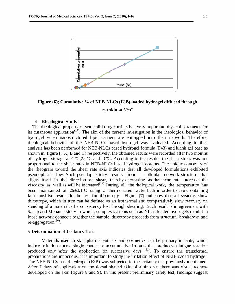
Figure (6); Cumulative % of NEB-NLCs (F3B) loaded hydrogel diffused through
rat skin at 32◦C
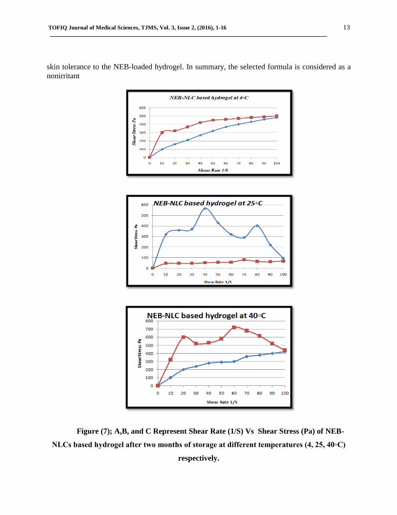
4- Rheological Study
The rheological property of semisolid drug carriers is a very important physical parameter for
its cutaneous application(17)
. The aim of the current investigation is the rheological behavior of
hydrogel when nanostructured lipid carriers are entrapped into their network. Therefore,
rheological behavior of the NEB-NLCs based hydrogel was evaluated. According to this,
analysis has been performed for NEB-NLCs based hydrogel formula (F43) and blank gel base as
shown in figure (7 A, B and C) respectively, the obtained results were recorded after two months
of hydrogel storage at 4 ºC,25 ºC and 40ºC. According to the results, the shear stress was not
proportional to the shear rates in NEB-NLCs based hydrogel systems. The unique concavity of
the rheogram toward the shear rate axis indicates that all developed formulations exhibited
pseudoplastic flow. Such pseudoplasticity results from a colloidal network structure that
aligns itself in the direction of shear, thereby decreasing as the shear rate increases the
viscosity as well as will be increased(19)
.During all the rhological work, the temperature has
been maintained at 25±0.1ºC using a thermostated water bath in order to avoid obtaining
false positive results in the test for thixotropy. Figure (7) indicates that all systems show
thixotropy, which in turn can be defined as an isothermal and comparatively slow recovery on
standing of a material, of a consistency lost through shearing. Such result is in agreement with
Sanap and Mohanta study in which, complex systems such as NLCs-loaded hydrogels exhibit a
loose network connects together the sample, thixotropy proceeds from structural breakdown and
re-aggregation(20)
.
5-Determination of Irritancy Test
Materials used in skin pharmaceuticals and cosmetics can be primary irritants, which
induce irritation after a single contact or accumulative irritants that produces a fatigue reaction
produced only after the application on successive days (21)
. To ensure the transdermal
preparations are innocuous, it is important to study the irritation effect of NEB-loaded hydrogel.
The NEB-NLCs based hydrogel (F3B) was subjected to the irritancy test previously mentioned.
After 7 days of application on the dorsal shaved skin of albino rat, there was visual redness
developed on the skin (figure 8 and 9). In this present preliminary safety test, findings suggest
Cu
mu
lati
ve a
mo
un
t o
f N
EB
time (hr)
TOFIQ Journal of Medical Sciences, TJMS, Vol. 3, Issue 2, (2016), 1-16 13
skin tolerance to the NEB-loaded hydrogel. In summary, the selected formula is considered as a
nonirritant
Figure (7); A,B, and C Represent Shear Rate (1/S) Vs Shear Stress (Pa) of NEB-
NLCs based hydrogel after two months of storage at different temperatures (4, 25, 40◦C)
respectively.

TOFIQ Journal of Medical Sciences, TJMS, Vol. 3, Issue 2, (2016), 1-16 14
Figure (8); A and B of the applied NLCs based hydrogel of NEB on the dorsal area of rat at
different magnification power
Figure (9); After 7 days of NEB-NLCs based hydrogel application
TOFIQ Journal of Medical Sciences, TJMS, Vol. 3, Issue 2, (2016), 1-16 15
Conclusion
Nebivolol hydrochloride as a model drug was used for the preparation of nanostructured
lipid carriers.The NEB-loaded NLCs could be fabricated and successfully incorporated into
hydrogel for transdermal application. The Ex -Vitro skin permeation data indicated that
NEB-NLCs bearing hydrogel provided sustained release of NEB. The results reflected the
potential of NLC as a carrier for transdermal administration of NEB and that would
demonstrate greater drug deposition into the skin. The NEB-NLCs system considered as a
promising alternative drug carriers for transdermal pharmaceutics. The data presented indicated
the successful development of NEB-NLCs-based hydrogel in increasing the encapsulation
efficiency of colloidal lipid carriers. The advantage for the colloidal lipid carriers is in the
improved performance in terms of stability and providing a sustained NEB transdermal effect.
Acknowledgement: Authors are very grateful to A. Prof. Dr. Abdul Wahab Al-Shekhly, the
head of the department of pharmacy/ Al-Rasheed University College for providing lab facilities,
A. Lecturer Nahida A. Al-Jubury and A. Lecturer Zena Qaragholi, Dept. of pharmacognosy and
Medicinal plants /college of pharmacy/ University Of Baghdad for the language editing.
References:
1- Sovan, L.; Utpal J.; P. K., Manna; G. P., Mohanta, R., Manavalan; Nanoparticle: An overview of preparation and characterization, Journal of Applied Pharmaceutical Science, 2011,l(6): 228-234.
2- Sean C.; (BPharm, FRPharmS), Martindale, the complete drug reference, thirty-sixth edition, Pharmaceutical Press 2009,: 1347.
3- Anthony, M.; David O.; Brian W.; Clarke's analysis of drugs and poisons, pharmaceutical press ,3 edition. 2005:48.
4- Budavari S.; The merck index. An encyclopedia of chemicals, drugs and biologicals. edition 3;merck& Co., Inc., whitehouse station, NJ. 200: 1152.
5- Deepak, S.; Rajesh, Y.; and Rajesh, A.; Liquid chromatographic method development and validation for assay and dissolution of nebivolol hydrochloride in tablet dosage form, Journal of Chemical and Pharmaceutical Research, 2014; 6(7): 2356-2363.
6- Shodhganga, Chapter-5, Determination of nebivolol and its impurities by RP-HPLC method, 2015. 83(4) :583–598
7- Ajay, P.; Devendra, S. .; Peeyush. K. and Jhageshwar, V. A review on novel lipid based nanocarriers. review article, inter. J. of phar. and pharm Sci. 2010;2(4).
8- Araújo, J.; Gonzalez, E.; Egea, M.; Garcia, M.; Souto E.; Nanomedicines for ocular NSAIDs: safety on drug delivery, nanomedicine, 2009;(5): 394-401.

TOFIQ Journal of Medical Sciences, TJMS, Vol. 3, Issue 2, (2016), 1-16 16
9- Ghazy E.; Abdulrasoo A.; Al-Tamimi J. J.; and Ayash N.; Nebivolol Hydrochloride Loaded Nanostructured Lipid Carriers as Transdermal Delivery System: Part 1: Preparation, Characterization and In Vitro Evaluation. AJPS, 2016;16(1):26-39.
10- Manickam B and Shyam SA, Formulation and Evaluation of Chitosan Based Bioadhesive Drug Delivery Systems of Lisinopril for Prolonged Drug Delivery. Pelagia Research Library 2013; 4(3):1-7.
11- LavKeshri and Kamla Pathak, Development of thermodynamically stable nanostructured lipid carrier system using central composite design for zero order permeation of Econazole nitrate through epidermis, Pharmaceutical Development and Technology, 2013; 18(3): 634–644.
12- Heshu Sulaiman Rahman, Abdullah Rasedee, Chee Wun How, Nazariah Allaudin Zeenathul; Zerumbone-loaded nanostructured lipid carriers: preparation, characterization, and antileukemic effect, International Journal of Nanomedicine, 2013;(8):2769-2781.
13- Jabbar G. E., Formulation and in vitro evaluation of silymarin through rat skin as topical delivery preparation, MSc thesis, College of Pharmacy, University of Baghdad, (2009).
14- KumbojiS, DR.Uma MRU, Mahala skhmiK and Shalini KB, Selection of Optimized Transdermal Patch ofAlfuzosin Hydrochloride through Different Approaches. International Journal of Inventions inPharmaceutical Sciences 2013;1(6): 515-525.
15- Arfat I, Nisar UR, ZeeshanJ, Muhammad K, Irfan A, Khizar A andTalib H, In Vitro Evaluation of Transdermal Patches of Flurbiprofen With Ethyl Cellulose. ActaPoloniaePharmaceutica-Drug Research 2014; 71(2): 287-295.
16- Elton Luiz Silva, GuilhermeCarneiro, Priscila Albuquerque Caetano, et.al., Nanostructured lipid carriers loaded with tributyrin as an alternative to improve anticancer activity of all-trans retinoic acid, Expert Rev. Anticancer Ther.15 (2), 247–256 (2015).
17- Yuhui Chen, Lizhen Pan, Ming Jiang, Dong Li, Nanostructured lipid carriers enhance the bioavailability and brain cancer inhibitory efficacy of curcuminbothin vitro and in vivo, 2015 Informa Healthcare USA.
18- Müller RH, Petersen RD, Hommoss A, Pardeike J. Nanostructured lipid carriers (NLC) in cosmetic dermal products. Adv Drug Deliv Rev. 2007;59:522–530.13. de Jong EMGJ, M.
19- Chen H, Chang X, Du D, et al. Podophyllotoxin-loaded solid lipid nanoparticles for epidermal targeting. J Control Release. 2006;110:296–306.
20- Lombardi Borgia S(1), Regehly M, Sivaramakrishnan R, Mehnert W, Korting HC, Danker K, Röder B, Kramer KD, Schäfer-Korting M.. Lipid nanoparticles for skin penetration enhancement – correlation to drug localization within the particle matrix as determined by fluorescence and parelectric spectroscopy. J Control Release. 2005;110:151–163.
21- Robert A. turner, Screening method in pharmacology, Academic press,New York and London,(1965) 742.
TOFIQ Journal of Medical Sciences, TJMS, Vol. 3, Issue 2, (2016), 17-25 ISSN: 2377-2808
INTERLEUKIN-18 AND CAROTID THICKNESS IN HELICOBACTER PYLORI
POSITIVE PATIENTS WITH DYSPEPSIA IN SULAIMANI- KURDISTAN
Hadeel A. Ibrahim*, Mohammed O. Mohammed
**, Hawa A.R.Al-Dhahir
***.
* Department of Physiology, School of Nursing, University of Sulaimani.
** Department of Medicine, School of Medicine, University of Sulaimani.
*** Department of Physiology, College of Medicine, University of Baghdad.
Corresponding author: Hadeel A. Ibrahim <[email protected]>
Abstract
Background: Helicobacter pylori (H. pylori) infection stimulates the production of
proinflammatory cytokines associated with the development of atherosclerosis. Levels of
circulating interleukin-18 (IL-18) have been positively correlated with carotid intima-media
thickness (CIMT) and coronary plaque area and have been identified as important predictors of
coronary events and cardiovascular mortality. This study aimed to examine the relationship
between serum IL-18, carotid intima thickness and H. pylori-IgG antibody as a sign of H. pylori
infection in dyspeptic patients.
Methods: This cross sectional- case control study was conducted in Kurdistan Teaching center
for Gastroenterology and Hepatology (KCGH) in Sulaimani city from January 2014 to March
2015. One hundred dyspeptic patients with positive H. pylori infection and 100 apparently
healthy asymptomatic volunteers with negative H. pylori tests were enrolled in this study. Sera
were tested for H. pylori IgG & IgA antibodies at Sulaimani Central lab., using ELISA tests.
Interleukin 18 (IL-18) was measured based on immunoenzymometric assay. All participants
TOFIQ Journal of Medical Sciences, TJMS, Vol. 3, Issue 2, (2016), 17-24 18
were evaluated for both internal carotid (IC) and common carotid (CC) arteries thickness by
using high resolution grey-scale Doppler ultrasonography.
Results: A significant difference (P<0.01) was found between patients and controls in the mean
serum IL-18. A significant correlation between H. pylori IgG level and IL-18 was found
(p < 0.01), but not with H. pylori IgA level. A significant correlation was found between IL 18
level and ICA, CCA thickness in H. pylori positive patients (p < 0.01).
Conclusion: H. pylori infection was significantly associated with higher serum IL-18. There was
significant correlation between IL-18 and Carotid intima- media thickness.
Keywords: H. pylori, IL-18, Carotid Thickness, Sulaimani.
Introduction
The immunoinflammatory response plays an important role in the development,
progression, and complication of atherosclerotic disease (1,2). Several studies have demonstrated the
association between H. pylori infection and coronary heart disease (CHD) ( 3, 4), while some studies
indicated that there was no correlation between H. pylori infection and coronary atherosclerosis
(5 , 6).
Interleukin 18, previously known as IFN-𝛾 inducing factor, is a proinflammatory member of IL-1
super family which plays role in the initiation and progression of atherosclerosis (7, 8). Carotid artery
intima-media thickness (CIMT) measured by ultrasound are predictive of cardiovascular disease in
individuals without clinically evident disease. CIMT is now widely used as an early marker for
atherosclerotic disease (9).
This study aimed to examine the relationship between serum IL-18, carotid intima thickness and
H. pylori-IgG antibody as a sign of chronic H. pylori infection in dyspeptic patients.
TOFIQ Journal of Medical Sciences, TJMS, Vol. 3, Issue 2, (2016), 17-24 19
Materials and Methods
This cross sectional case control study was conducted in Kurdistan Teaching center for
Gastroenterology and Hepatology (KCGH) in Sulaimani city during the period of January 2014 to
March 2015. One hundred dyspeptic patients with positive H. pylori infection (IgG) and 100
apparently healthy asymptomatic volunteers with negative H. pylori tests were enrolled in this study.
Both groups were comparable in age distribution and gender.
Exclusion Criteria: Pregnant women, smokers, patients previously treated for H. pylori infection and
who had received antibiotics; proton pump inhibitors or bismuth compounds in the preceding 4 weeks.
This study was approved by the Ethics Committee of Faculty of Medicine, University of Sulaimani
and Directory of Health in Sulaimani. Written informed consents were obtained from all the
participants.
A special form used to obtained demographic data (name, age, gender, history of dyspepsia, and
drug history).
After overnight fasting, 10 ml venous blood aspirated then centrifuged at 5000 r/min for 5 min. Sera
were tested for H. pylori IgG & IgA antibodies at Sulaimani Central lab., using ELISA tests (Nova
Lisa, NovaTec, Germany), according to the standard operating procedures. That has a sensitivity of
97% and a specificity of 98.8%.
Interleukin 18 (IL-18) , using RayBio_ Human IL-18 ELISA Kit (ELH-IL18BPA-001), USA was
measured based on immunoenzymometric assay. Subjects were evaluated for both internal carotid (IC)
and common carotid (CC) arteries and plaque occurrence by using high resolution grey-scale Doppler
ultrasonography ( Philips, En visor, Version C.1.3, 2007) , In a semi-dark room, the subject lay supine
with slightly hyperextended neck and rotated away from the imaging transducer. Both carotid arteries
were scanned. CIMT was defined as the distance between the leading edge of the lumen intimal
interface and the leading edge of the media adventitia interface of the far wall.
Statistical Analysis
All data were analyzed using Excel and SPSS (Version 20 software) computer program. To assess the
correlation between different variables, bivariate correlation coefficient analysis was performed. In this
analysis, the statistical significant association was determined. All p values were based on 2-sided tests
and p < 0.05 was considered statistically significant.
TOFIQ Journal of Medical Sciences, TJMS, Vol. 3, Issue 2, (2016), 17-24 20
Results
Both groups of H. pylori seropositive and seronegative groups were comparable in mean age and
gender (p>0.05).
The mean serum Interleukin 18 among patients and controls were (19.7 + 8.9) and (8.9 + 1.96)
pg/mL respectively, with highly significant difference (P< 0.01), Table 1.
Table 1. Interleukin 18 in study population.
Investigations
Patients Controls p value
Mean +SD Mean+ SD
Interleukin 18 (pg/mL) 19.7+8.9 8.9+1.96 < 0.01
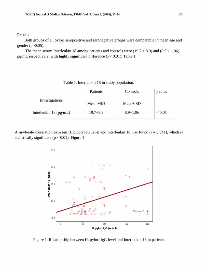
A moderate correlation between H. pylori IgG level and Interleukin 18 was found (r = 0.341), which is
statistically significant (p < 0.01), Figure 1
Figure 1. Relationship between H. pylori IgG level and Interleukin 18 in patients
TOFIQ Journal of Medical Sciences, TJMS, Vol. 3, Issue 2, (2016), 17-24 21
A negative correlation between H. pylori IgA level and Interleukin 18 was found (r = - 0.084), (p
>0.05), Table 2.
Table 2. Relationship between H. pylori IgA level and IL-18 in patients.
H. pylori IgA (Ndx) Correlation coefficient (r) p value
Interleukin 18 (pg/mL) - 0.084 >0.05
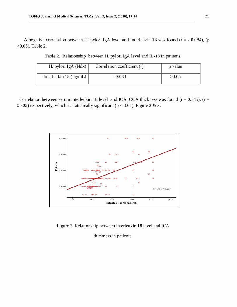
Correlation between serum interleukin 18 level and ICA, CCA thickness was found (r = 0.545), (r =
0.502) respectively, which is statistically significant (p < 0.01), Figure 2 & 3.
Figure 2. Relationship between interleukin 18 level and ICA
thickness in patients.

TOFIQ Journal of Medical Sciences, TJMS, Vol. 3, Issue 2, (2016), 17-24 22
Figure 3. Relationship between interleukin 18 level and CCA
thickness in patients.
Discussions:
In the present study the mean serum Interleukin 18 concentration was higher in H. pylori-infected
patients than in control group, this observations is consistent with findings of other studies(10, 11, 12,
13). A significant correlation between Interleukin 18 and H. pylori IgG level was found, but not with
H. pylori IgA level .Those may be explained by H. pylori infection induces IL-18 in the gastric
mucosa (11). IL-18 may play an important role in the inflammatory response and promote the chronic
and persistent inflammatory changes in the stomach. This may ultimately influence the outcome of H.
pylori -associated diseases that arise within the context of gastritis. (14). Also H. pylori have been
shown to induce a strong cytokine response in both human gastric epithelial cells and gastric epithelial
cell lines. Chronic infection with H. pylori is associated with gastric IFN- γ producing T cells and
increased mucosal IL-12, indicating a predominant Th1 response. IL-12 mRNA levels are increased in
infection with cag-positive H. pylori . This results in up-regulation of the expression of IL-18 receptors
in both Th1( T-helper 1) and NK cells. It seems that in chronic H. pylori infection, mucosal production
of IL-18, together with IL-12, would be important in promoting Th1 responses and IFN- γ secretion.
Moreover, it has been demonstrated that Th cells respond to H. pylori antigen by secreting high levels
of IFN- γ. (15).

TOFIQ Journal of Medical Sciences, TJMS, Vol. 3, Issue 2, (2016), 17-24 23
A statistically significant correlation between interleukin 18 and ICA, CCA thickness was found.
(16) found that elevated serum IL-18 levels are associated with increased carotid IMT( Intima media
thickness) as evaluated by B-mode ultrasound, suggesting their link with carotid atherosclerosis.
However IL-18 is highly expressed in human carotid atherosclerotic plaques predominantly co
localized with macrophages. Thus, increased IL-18 production from severe atherosclerotic lesions
could contribute to the higher IL-18 (7). Also, experimental studies have shown that IL-18 enhances
atherosclerosis through release of interferon- γ (17) and induces expression of IL-6 in vascular
endothelial and smooth muscle cells ( 18). Inversely, IL-18 deficiency reduces the extent of
atherosclerosis in apolipoprotein E–knockout mice. These findings are in accordance with the studies
that IL-18 plays a key role in atherogenesis, supporting the link between IL-18 and carotid
atherosclerosis ( 19).
Conclusion: H. pylori infection was significantly associated with higher serum IL-18. There was
significant correlation between IL-18 and Carotid intima- media thickness.
The auther disclose that, they have no conflict of interest.
TOFIQ Journal of Medical Sciences, TJMS, Vol. 3, Issue 2, (2016), 17-24 24
References
1-Hansson GK, Libby P. (2006):The immune response in atherosclerosis: a double-edged
sword. Nat Rev Immunol. 6(7):508–19.
2-Herbin O, Ait-Oufella H, Yu W, Fredrikson GN, Aubier B, Perez N, Gourdy P, Khallou-. (2012):
Regulatory T-cell response to apolipoprotein b100-derived peptides reduces the development
and progression of atherosclerosis in mice. Arterioscler Thromb Vasc Biol. 32(3):605–12.
3-Ayada K, Yokota K, Kobayashi K, Shoenfeld Y, Matsuura E, Oguma K.(2009):Chronic infections
and atherosclerosis. J Clin Rev Allergy Immunol. 37(1):44-8.
4-Al-Ghamdi A, Jiman-Fatani AA, El-Banna H. (2011): Role of Chlamydia pneumoniae, Helicobacter
pylori and cytomegalovirus in coronary artery disease. Pak J Pharm Sci. 24(2):95-101.
5-Ozdogru I, Kalay N, Dogan A, Inanc MT, Kaya MG, Topsakal R, Inanc MT, Kaya MG, Kalay N.
(2007):The relationship between Helicobacter pylori IgG titre and coronary atherosclerosis. Acta
Cardiol. 62:501–505.
6-Szklo M, Ding J, Tsai MY, Cushman M, Polak JF, Lima J, et al. (2009): Individual pathogens,
pathogen burden and markers of subclinical atherosclerosis: the Multi- Ethnic Study of
Atherosclerosis. J Cardiovasc Med. 10:747–751.
7-Mallat Z., Corbaz A., Scoazec A.( 2001): Expression of interleukin-18 in human atherosclerotic
plaques and relation to plaque instability. Circulation. 104:1598–1603.
8- Packard RR, Libby P.( 2008): Inflammation in atherosclerosis: from vascular biology to biomarker
discovery and risk prediction. Clin Chem. 54(1):24-38.
9-Kastelein JJ, Wiegman A, Groot E. (2003): Surrogate markers of atherosclerosis: impact of statins.
Atheroscler Suppl. 4:31-6.
10- Jafarzadeh A, Sajjadi M A. (2006): Evaluation of Serum Interleukin-18 levels in Helicobacter
Pylori-infected Peptic Ulcer Patients and its Association with Bacterial CagA Virulence Factor
Iran.J.Immunol. 3(1): 15-22.
11-Sakai K, Kita M, Sawai N, Shiomi S, Sumida Y, Kanemasa K, Shoji M, Jiro I and Yoshio
Yamaoka .( 2008): Levels of interleukin-18 are markedly increased in Helicobacter pylori-infected
gastric mucosa among patients with specific IL18 genotypes. J Infect Dis. 197(12):1752-1761.
12- Chen X, Chen YG, Chen Y, Chen Y, Chen YJ, Chen Z, Cheng A, Cheng CH, ... Dehay B,
Delbridge LM, Demarchi F, Deng YZ, Dengjel J, Dent P, Denton D,l.(2013): Relationship between

TOFIQ Journal of Medical Sciences, TJMS, Vol. 3, Issue 2, (2016), 17-24 25
Helicobacter pylori infection and serum interleukin-18 in patients with carotid atherosclerosis. J
Helicobacter, 18(2):124-8.
13-Rezaeifar A, Eskandari-Nasab E, Moghadampour M, Kharazi-Nejad E, Hasani SS, Asadi-Saghandi
A, et al.( 2013):The association of interleukin-18 promoter polymorphisms and serum levels with
duodenal ulcer, and their correlations with bacterial CagA and VacA virulence factorsScand J Infect
Dis. 45(8):584-92.
14-Bagheri N, Taghikhani A, Rahimian G, Salimzadeh L, Azadegan Dehkordi F, Zandi
F, Chaleshtori. MH, Rafieian-Kopaei M, Shirzad H.., (2013): Association between virulence factors of
helicobacter pylori and gastric mucosal interleukin-18 mRNA expression in dyspeptic patients
Microb Pathog.,65:7-13.
15- Smythies LE, Waites KB, Lindsey JR, Harris PR, Ghiara P, Smith PD.( 2000): Helicobacter
pylori-induced mucosal inflammation is Th1 mediated and exacerbated in IL-4, but not IFN-gamma,
gene-deficient mice. J Immunol. 165:1022-9.
16-Yamagami H, Kitagawa K, Hoshi T, Furukado S, Hougaku H, Nagai Y, Hori M. (2005):
Associations of serum IL-18 levels with carotid intima-media thickness. Arterioscler Thromb Vasc
Biol. 25(7):1458-1462.
17- Whitman SC, Ravisankar P, Daugherty A. ( 2002): Interleukin-18 enhances atherosclerosis in
apolipoprotein E(-/-) mice through release of interferon-gamma. Circ Res. 90(2):E34-38.
18- Gerdes N, Sukhova GK, Libby P, Reynolds RS, Young JL, Schonbeck U.( 2002): Expression of
interleukin (IL)-18 and functional IL-18 receptor on human vascular endothelial cells, smooth muscle
cells, and macrophages: implications for atherogenesis. J Exp Med. 195: 245-57.
19-Elhage R, Jawien J, Rudling M, Ljunggren HG, Takeda K, Akira S, et al. (2003): Reduced
atherosclerosis in interleukin-18 deficient apolipoprotein Eknockout mice. Cardiovasc Res. 59(1):234-
240.
TOFIQ Journal of Medical Sciences, TJMS, Vol. 3, Issue 2, (2016), 64-80 ISSN: 2377-2808
RETROSPECTIVE STUDY OF POSTERIOR DORSOLUMBAR FIXATION IN
BAGHDAD: A CLINICAL STUDY OF 100 PATIENTS
Prof. Yasir M H Hamandi * , Dr. Ali mohammad khalil
**
* Department of surgery/ neurosurgery, College of Medicine, Al-Nahrain University, Baghdad,
Iraq
** Department of surgery/ neurosurgery, Al-hilla teaching hospital, Al-Hilla, Iraq
Corresponding author:
Email: [email protected]
Abstract :
Background: Spondylolisthesis describes a condition of a forward slippage of one vertebra
over another, which may or may not be associated with demonstrable instability. Spinal fixation
is a neurosurgical procedure in which two or more vertebrae are anchored to each other through
a synthetic "vertebral fixation device"
Objective: To determine the demographic distribution of different patient factors and the most
commonly vertebra undergo fixation in the thoracolumbar instrumentation.
Patients and Methods: one hundred patients were evaluated during the period of this study in
a retrospective manner from January 2013 to January 2015 in four hospitals in Baghdad
TOFIQ Journal of Medical Sciences, TJMS, Vol. 3, Issue 2, (2016), 64-80 65
(Neurosurgical Teaching Hospital, Neuroscience hospital, Al-Kahdymia Teaching Hospital,
Medical City\Ghazy AL-Hariri Hospital). The patients' data regarding the etiology of
instability, mechanism of injury for trauma patients, gender, age, segments undergoing
instrumentation were identified.
Results: The study revealed female predominance over male: female ratio of 1:2.7, the age
distribution was highest from 3rd
to 7th
decades of life, the etiology of instability was either
degenerative or traumatic, the degenerative instability was 65% while traumatic cases was
35%. The neurological status of the patients was assessed by neurological examination and
revealed 75% with incomplete deficit and 25% with complete neurological deficit, the most
common pathologically involved vertebra was the L4, the most common vertebrae used in
fixation were the L4 and L5 levels, the most common type of fixation used was the short
segment fixation.
Conclusion: Posterior spinal fixation with pedicle screws and rods system is an effective and
safe method in maintaining the stability of spine. The intraoperative imaging is important in
maintaining safe trajectory of screws. Short segment fixation using the posterior approach with
pedicle screw-rod fixation devices achieve good stabilization. The ideal candidates for
undergoing posterior spinal fixation are patients with unstable fractures & incomplete
neurological deficit.
Recommendation: The use of intraoperative neuro-monitoring, use of navigation system, use
of fluoroscopy and the O-arm in spinal fixation surgery. Bone fusion is recommended for each
patient.
Keywords: Thoracolumbar spine, spondylolisthesis, pedicle screw fixation
Introduction:
Spinal instrumentation basically means the implantation of more or less rigid metallic or
non-metallic devices which are attached to the spine. These devices function to provide spinal
stability and thus facilitate bone healing leading to spinal fusion. [1]
Types of instrumentation[2]
1.Metallic Pedicle Screw-Rod Systems
2. Polyetheretherketone (PEEK) Rods
Goals and indications of spinal instrumentation:
1. Trauma
2. Non trauma

TOFIQ Journal of Medical Sciences, TJMS, Vol. 3, Issue 2, (2016), 64-80 66
a. Tumor
b- Infection
c. Degenerative changes and spondylolisthesis[2]
Indications for fusion fall into two broad categories:
A. Preoperative structural problems that predispose to instability after decompression:
1. Degenerative spondylolisthesis or lateral listhesis.
2. Progressive scoliosis or kyphosis.
3. Recurrent spinal stenosis requiring repeat decompression at the same level.
B. Intraoperative structural alterations that warrant consideration of a fusion:
1. Excess facet joint removal 50%
2. Pars interarticularis fracture or removal.
3. Radical disc excision with resultant destabilization of the anterior spinal column.
C. Trauma that predispose to unstable spine
Measures for Correct Screw Placement
1. Navigation , CT, and fluroscope Guidance[3,4]
2. Electromyographic Monitoring[4]
Complications:
1. Pedicle Fracture[2]
2. Cerebrospinal Fluid Fistulae[5]
3. Infection[10]
4. Hardware Failure (Screw Breakage , Screw Pull-out, Screw Loosening or Plate or Rod
Breakage, Loss of Correction, Wound Breakdown.)[5,6,7,8]
5. Nerve root or cord injury.
Free –hand technique
Free-hand pedicle screw placement relies on an intricate appreciation of the relationship
of various anatomical landmarks at each level of the thoracolumbar spine. Analogous entry sites
guided by differential anatomy are utilized for both the thoracic and lumbar spine.[9,10]
Accuracy of pedicle screw placement
Criteria of pedicle screw placement were: [11]
(1) Relation of pedicle screws to the pedicle.
(2) Relation of pedicle screws to the vertebral body.
Aim of the study:
TOFIQ Journal of Medical Sciences, TJMS, Vol. 3, Issue 2, (2016), 64-80 67
To determine the demographic distribution of different patient factors
To identify the most common vertebra levels to undergo fixation in the thoracolumbar
spine.
Patients and Methods: One hundred patients were evaluated during the period of this study in a retrospective
manner from January 2013 to January 2015 in four hospitals in Baghdad (Neurosurgical
Teaching Hospital, Neuroscience hospital, Al-Kahdimiyya Teaching Hospital, Medical
City/Ghazi AL-Hariri Hospital).
Demographic, Admission complaints and imaging were obtained for 100 patients through
chart review. On Admission the following parameters independently reviewed: gender, age, chief
complaint, etiology of instability, for the trauma patients the mechanism of injury was identified,
the level of pathology, neurological examination, type of neurological deficit (whether complete
or incomplete), level of fixation, number of screws used, outcome before discharge and a 6
months follow up.
The patients' data regarding the etiology of instability, mechanism of injury for trauma
patients, segments undergoing instrumentation were identified.
All patients' undergone hematological investigations in the form of complete blood, ESR,
C-reactive protein, renal function test, fasting blood sugar, blood group & Rh.
Only 16 patients have NCS and EMG prior to surgery, so the neuro-electro-physiological
studies can't be analyzed in correlation to other clinical factors.
The findings of the patients' pre-operative imaging including thoracolumbar X-rays, CT-
scan and MRI were reviewed.
The systems used were Medtronic and Aesculap.
TOFIQ Journal of Medical Sciences, TJMS, Vol. 3, Issue 2, (2016), 64-80 68
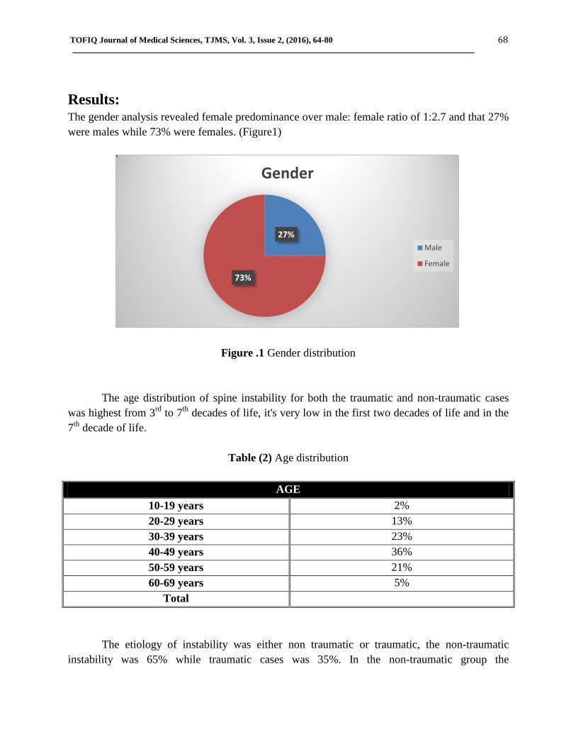
Results: The gender analysis revealed female predominance over male: female ratio of 1:2.7 and that 27%
were males while 73% were females. (Figure1)
Figure .1 Gender distribution
The age distribution of spine instability for both the traumatic and non-traumatic cases
was highest from 3rd
to 7th
decades of life, it's very low in the first two decades of life and in the
7th
decade of life.
Table (2) Age distribution
AGE
10-19 years 2%
20-29 years 13%
30-39 years 23%
40-49 years 36%
50-59 years 21%
60-69 years 5%
Total
The etiology of instability was either non traumatic or traumatic, the non-traumatic
instability was 65% while traumatic cases was 35%. In the non-traumatic group the
27%
73%
Gender
Male
Female

TOFIQ Journal of Medical Sciences, TJMS, Vol. 3, Issue 2, (2016), 64-80 69
spondylolisthesis was predominant 89.2% while the pathological fractures were 11.8% of the
pathological fractures 71.4% were due to infections while 28.6% were due to tumors. In the
traumatic patients the FFH as a cause was 51.5% while the RTA as a cause to the instability was
48.5%.
Table (3) Etiology of instability
Etiology of instability
Traumatic 35%
(35 patient)
Non-Traumatic 65%
(65 patient)
Road trrafic
accident
51.5%
(17 patient)
Fall from height
48.5%
(18 patient)
Spondylolisthesis
89.2%
(58 patient)
Pathological #
11.8%
(7 patients)
Infection
71.4%
(5 patients)
Tumor
28.6%
(2 patients)
The neurological status of the patients was assessed by neurological examination and
revealed 75% with incomplete deficit and 25 % with complete neurological deficit, of the
incomplete injury 20% were incomplete motor deficit, while 93.3% were incomplete sensory
deficit and 12% sphincter disturbance. Such deficits (motor, sensory and sphincters) occurred
either in isolation or together.(Fig.2)

TOFIQ Journal of Medical Sciences, TJMS, Vol. 3, Issue 2, (2016), 64-80 70
Figure .2 Neurological deficit
The Neurological deficit according to the etiology was reviewed and revealed a complete
neurological deficit of 23 patients in traumatic spondylolisthesis while only 2 patients with
complete neurological deficit in non-traumatic cases. The incomplete neurological deficit was
predominant in non-traumatic spondylolisthesis of 63 patients while only 12 patients having
incomplete neurological deficit in the traumatic cases.
Table (4) Linking the Neurological Deficit to Etiology
Linking the Neurological Deficit to Etiology
Etiology of Instability Complete Neurological
Deficit
Incomplete Neurological
Deficit
Traumatic 66% (23 patient) 34% (12 patient)
Non-Traumatic 3% (2 patient) 97 % (63 patient)
The most common pathologically involved vertebra was the L4, followed by L5, L3 and
L2. The least involved vertebrae were D10 and D11. Table (5) illustrates whether the
pathological vertebra involved single or multiple levels (there were 2 cases with multiple
fracture).
25%
75%
0% 0%
Neurological Deficit
Complete
Incomplete
TOFIQ Journal of Medical Sciences, TJMS, Vol. 3, Issue 2, (2016), 64-80 71
Table (5) Level of Pathology
Level of Pathology
D10 2
D11 2
D12 10
L1 5
L2 16
L3 15
L4 37
L5 15
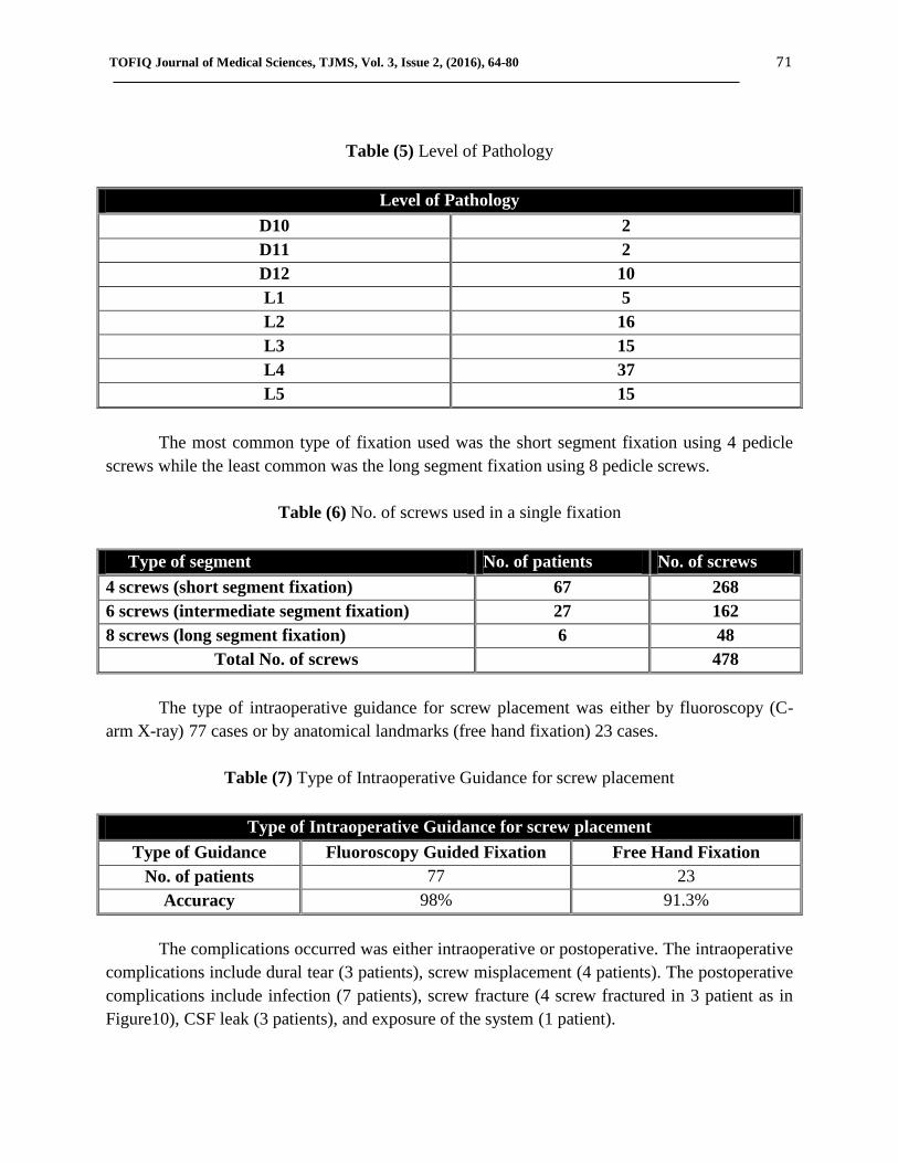
The most common type of fixation used was the short segment fixation using 4 pedicle
screws while the least common was the long segment fixation using 8 pedicle screws.
Table (6) No. of screws used in a single fixation
Type of segment No. of patients No. of screws
4 screws (short segment fixation) 67 268
6 screws (intermediate segment fixation) 27 162
8 screws (long segment fixation) 6 48
Total No. of screws 478
The type of intraoperative guidance for screw placement was either by fluoroscopy (C-
arm X-ray) 77 cases or by anatomical landmarks (free hand fixation) 23 cases.
Table (7) Type of Intraoperative Guidance for screw placement
Type of Intraoperative Guidance for screw placement
Type of Guidance Fluoroscopy Guided Fixation Free Hand Fixation
No. of patients 77 23
Accuracy 98% 91.3%
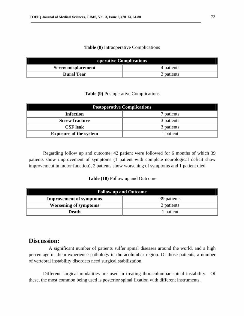
The complications occurred was either intraoperative or postoperative. The intraoperative
complications include dural tear (3 patients), screw misplacement (4 patients). The postoperative
complications include infection (7 patients), screw fracture (4 screw fractured in 3 patient as in
Figure10), CSF leak (3 patients), and exposure of the system (1 patient).
TOFIQ Journal of Medical Sciences, TJMS, Vol. 3, Issue 2, (2016), 64-80 72
Table (8) Intraoperative Complications
operative Complications
Screw misplacement 4 patients
Dural Tear 3 patients
Table (9) Postoperative Complications
Postoperative Complications
Infection 7 patients
Screw fracture 3 patients
CSF leak 3 patients
Exposure of the system 1 patient
Regarding follow up and outcome: 42 patient were followed for 6 months of which 39
patients show improvement of symptoms (1 patient with complete neurological deficit show
improvement in motor function), 2 patients show worsening of symptoms and 1 patient died.
Table (10) Follow up and Outcome
Follow up and Outcome
Improvement of symptoms 39 patients
Worsening of symptoms 2 patients
Death 1 patient
Discussion: A significant number of patients suffer spinal diseases around the world, and a high
percentage of them experience pathology in thoracolumbar region. Of those patients, a number
of vertebral instability disorders need surgical stabilization.
Different surgical modalities are used in treating thoracolumbar spinal instability. Of
these, the most common being used is posterior spinal fixation with different instruments.

TOFIQ Journal of Medical Sciences, TJMS, Vol. 3, Issue 2, (2016), 64-80 73
The most modern and commonly used fixation is the pedicle screw and rod system. Other
methods include anterior spinal fixation with cage, kyphoplasty, vertebroplasty, posterior
interbody fusion and posterior intertransverse fusion. Of the posterior spinal fixation, the various
instruments used are pedicle screws and rods system.
The neurological recovery is dependent on the degree of stability provided by fixation,
and on initial instability and neurological loss. So, if stability is provided to spine then
neurological status of some patients can improve.
Regarding the gender 73% of our study group was female and 27% was male with a
male/female ratio of 1:2.7, while in Muralidhar B.M. et al Study [12]
there was male
predominance of 80% and female of 20%, Shailendra et al study [13]
the male percentage was
76% and female was 24%. This difference is due to that 65% of our patient complaining of
instability due to non-traumatic causes of which degenerative spondylolisthesis more commonly
occurs in elderly females because of the postmenopausal osteoporosis.
About age group, in our study 36% of our patient are located in 50-59 age group which is
the commonest age group while in Muralidhar B.M.et al study most of the patient are in 50 -59
age group, in Shailendra et al study the commonest age group is 16-25 years followed by 26-35
years, this difference because the largest group of our patient had degenerative diseases other
than trauma which prevalence increased with age.[12, 13]
Concerning etiology, 35% are due to traumatic injury (17 patient RTA, 18 patient FFH),
65% are due to non-traumatic causes (58 patient spondylolisthesis, 7 patient pathological fracture
{pathological fracture 5 patient infection, 2 patient tumor}) , while in study done by Alp Ozgun
BOrcek et al [15]
the non-traumatic cases was 63.9% while the traumatic cases was 36% which is
close to our result.
The relation between etiology and neurological deficit in traumatic cases 66% had
complete neurological deficit and 34% incomplete deficit while in non-traumatic cases 3% had
complete deficit and 97% had incomplete deficit, this difference is due to two factors first is that
traumatic injury which cause vertebral fracture and instability means sever trauma while in
degenerative cases it is usually a chronic process and patient seek medical management before
complete deficit occur, the other factor is that trauma mostly cause injury to thoracolumbar
junction because the transition between the more rigid thoracic spine and the mobile lumbar
spine concentrates bending and axial loads at the thoracolumbar junction where there is spinal
cord in the spinal canal while in degenerative cases are mostly in L4 level where there is cauda
equina and no cord.
TOFIQ Journal of Medical Sciences, TJMS, Vol. 3, Issue 2, (2016), 64-80 74
Regarding the level of pathology: in 37% of cases, the pathology involving L4 followed
by 15% for L3 and L5, in Muraldhiar B.M. et al study 70 % at L1 level and 20|% at L2 level in
our study the higher percentage of L4 are due to that most of our cases are degenerative which is
more common in the lower lumbar region leading to L4 and L5 levels to be the most commonly
instrumented level. [12]
Regarding type of pedicle screw fixation, the short segment was used 67 times, while the
long segment fixation was used 6 times only because it is used in lesions involving the
thoracolumbar junction vertebrae, while in a study done by Kashif Mahmood Khan et al[16]
on 50
patient with thoracolumbar fixation, four screws with 2 rods were used in 38 patients (76%) and
8 screws were used in 12 patients (24%).
As regards the intraoperative guidance, 23% of operations done by free hand procedure
without intraoperative fluoroscope, the accuracy was 91.3%, while in Parker et al study[17]
on
free hand fixation for degenerative cases (964 patients) the accuracy was 98.3%. In Gertzbein et
al study[21]
for traumatic patient (171 patients) the accuracy was 71.9%. The accuracy of free
hand screw insertion is related to the experience of the surgeon, and possibly to change in
anatomical landmark in traumatic cases.
The intraoperative complications, screw misplacement in 4 patients 4%, while in - et al
study [38]
3 patients 3.2% had screw misplacement, dural tear occurred in 3 patients 3% in our
study, while is study done by Faraj et al 4 patients 4.4% had unintended dural tears.
The postoperative complications in our study included infection in 7 patients (7%) all of
them had removal of the system, the patients with secondary neoplasia were receiving concurrent
chemo- and radiotherapy for their malignant diseases which rendered their immunity low. In
Sanford et al study[19]
the infection rate was 3.2%. The CSF leak occurred in 3 patients 3% in our
study, while in Faraj et al study[38]
it was 2.1%. Screw fracture occurred in 4 screws in 3 patients
out of 478 screws used in our study (1.2%), while 9 screw fractures out of 296 in Ohlin et al
study.[20]
Exposure of the system occurred in 1 patient only as a complication of being bed ridden
in a complete neurological deficit, this patient died of septicemia and pneumonia.
Regarding six-month follow-up and outcome: 42 patients were followed (we couldn’t
follow all the patients because of their residency in far provinces), 39 patients showed
improvement in their neurological status (1 patient with complete neurological deficit show
improvement within 3 weeks), 2 patient show worsening of symptoms (increased pain) and 1
patient died as mentioned above . While in a study done by Roop Singh et al[21]
41% of patients
improved and 27% did not improve (all of them are paraplegic) with the difference possibly
related to severity of injury and timing of surgery.

TOFIQ Journal of Medical Sciences, TJMS, Vol. 3, Issue 2, (2016), 64-80 75
Patients who experience worsening of symptoms feel so due to misplacement of the
screw with pressure on the nerve root or due to pathology in other levels.
Conclusion:
Posterior spinal fixation with pedicle screws and rods system is an effective and safe
method in maintaining the stability of spine, by improvement in all the scores and parameters
including the pain and mobility, range of motion of spine, and deformity. 1. The intraoperative imaging in coronal and sagittal planes is important in maintaining safe
trajectory of screws, although free hand screw insertion is applicable with more
experienced hands after thorough knowledge of anatomical landmarks.
2. Short segment fixation using the posterior approach with pedicle screw-rod fixation
devices with or without bone grafting achieves good stabilization and fair enough
neurological recovery in patients with unstable thoracolumbar fractures
3. By applying the transpedicular screw fixation on the unstable fractures of the
thoracolumbar spine, a stable fracture fixation can be achieved. So early ambulation,
rapid return to work and less rehabilitation costs could be achieved. This kind of fixation
prevents secondary spine deformities.
4. The ideal candidates for undergoing posterior spinal fixation are patients with unstable
fractures & incomplete neurological deficit.
5. In patients with complete neurological deficit, spinal fixation done to aid to achieve
stabilization and easy nursing care .
6. Non-Traumatic cause in 40-49 years old females represent the most common etiology.
Recommendation:
Use of intraoperative neuro-monitoring in spinal fixation surgery.
Use of a navigation system to guide to the intraoperative manipulation.
Use of fluoroscopy and the O-arm as an intraoperative guiding tool.
Bone fusion is recommended for each patient with degenerative changes to
improve stability of the spine and to prevent fixation failure.
Use of short segment is better than long segment to decrease blood loss, timing of
surgery, risk of vascular, neural and visceral injury, lost of motion, and cost.
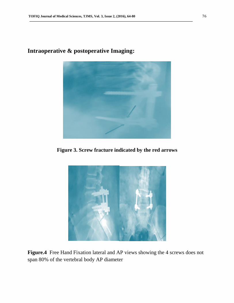
TOFIQ Journal of Medical Sciences, TJMS, Vol. 3, Issue 2, (2016), 64-80 76
Intraoperative & postoperative Imaging:
Figure 3. Screw fracture indicated by the red arrows
Figure.4 Free Hand Fixation lateral and AP views showing the 4 screws does not
span 80% of the vertebral body AP diameter
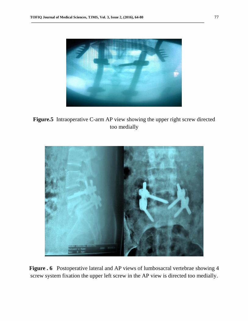
TOFIQ Journal of Medical Sciences, TJMS, Vol. 3, Issue 2, (2016), 64-80 77
Figure.5 Intraoperative C-arm AP view showing the upper right screw directed
too medially
Figure . 6 Postoperative lateral and AP views of lumbosacral vertebrae showing 4
screw system fixation the upper left screw in the AP view is directed too medially.

TOFIQ Journal of Medical Sciences, TJMS, Vol. 3, Issue 2, (2016), 64-80 78
Figure.7 Intraoperative C-arm lat.view
Figure 8 . Postoperative dorsolumbar lateral X-ray to the left and dorsal X-ray AP
view showing 6 screw fixation system
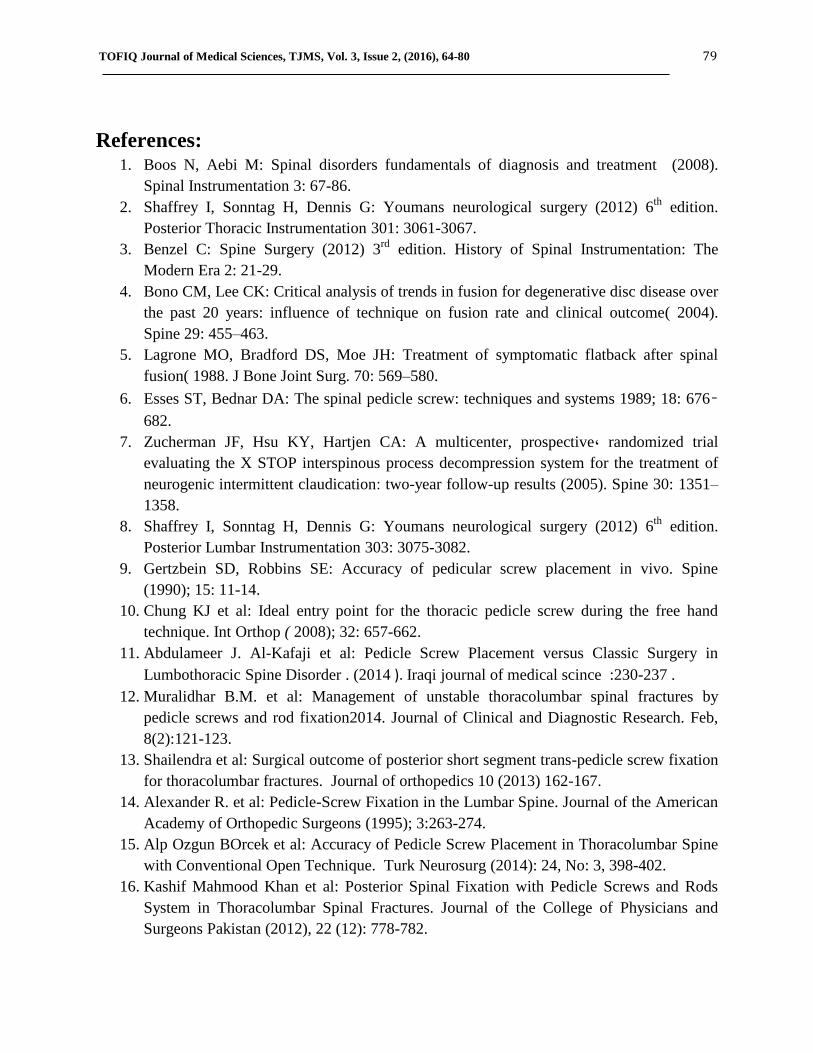
TOFIQ Journal of Medical Sciences, TJMS, Vol. 3, Issue 2, (2016), 64-80 79
References: 1. Boos N, Aebi M: Spinal disorders fundamentals of diagnosis and treatment (2008).
Spinal Instrumentation 3: 67-86.
2. Shaffrey I, Sonntag H, Dennis G: Youmans neurological surgery (2012) 6th
edition.
Posterior Thoracic Instrumentation 301: 3061-3067.
3. Benzel C: Spine Surgery (2012) 3rd
edition. History of Spinal Instrumentation: The
Modern Era 2: 21-29.
4. Bono CM, Lee CK: Critical analysis of trends in fusion for degenerative disc disease over
the past 20 years: influence of technique on fusion rate and clinical outcome( 2004).
Spine 29: 455–463.
5. Lagrone MO, Bradford DS, Moe JH: Treatment of symptomatic flatback after spinal
fusion( 1988. J Bone Joint Surg. 70: 569–580.
6. Esses ST, Bednar DA: The spinal pedicle screw: techniques and systems 1989; 18: 676–
682.
7. Zucherman JF, Hsu KY, Hartjen CA: A multicenter, prospective, randomized trial
evaluating the X STOP interspinous process decompression system for the treatment of
neurogenic intermittent claudication: two-year follow-up results (2005). Spine 30: 1351–
1358.
8. Shaffrey I, Sonntag H, Dennis G: Youmans neurological surgery (2012) 6th
edition.
Posterior Lumbar Instrumentation 303: 3075-3082.
9. Gertzbein SD, Robbins SE: Accuracy of pedicular screw placement in vivo. Spine
(1990); 15: 11-14.
10. Chung KJ et al: Ideal entry point for the thoracic pedicle screw during the free hand
technique. Int Orthop ( 2008); 32: 657-662.
11. Abdulameer J. Al-Kafaji et al: Pedicle Screw Placement versus Classic Surgery in
Lumbothoracic Spine Disorder . (2014 ). Iraqi journal of medical scince :230-237 .
12. Muralidhar B.M. et al: Management of unstable thoracolumbar spinal fractures by
pedicle screws and rod fixation2014. Journal of Clinical and Diagnostic Research. Feb,
8(2):121-123.
13. Shailendra et al: Surgical outcome of posterior short segment trans-pedicle screw fixation
for thoracolumbar fractures. Journal of orthopedics 10 (2013) 162-167.
14. Alexander R. et al: Pedicle-Screw Fixation in the Lumbar Spine. Journal of the American
Academy of Orthopedic Surgeons (1995); 3:263-274.
15. Alp Ozgun BOrcek et al: Accuracy of Pedicle Screw Placement in Thoracolumbar Spine
with Conventional Open Technique. Turk Neurosurg (2014): 24, No: 3, 398-402.
16. Kashif Mahmood Khan et al: Posterior Spinal Fixation with Pedicle Screws and Rods
System in Thoracolumbar Spinal Fractures. Journal of the College of Physicians and
Surgeons Pakistan (2012), 22 (12): 778-782.
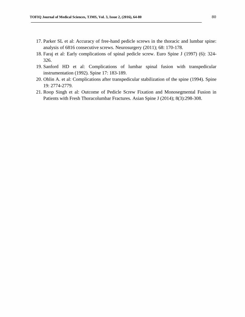
TOFIQ Journal of Medical Sciences, TJMS, Vol. 3, Issue 2, (2016), 64-80 80
17. Parker SL et al: Accuracy of free-hand pedicle screws in the thoracic and lumbar spine:
analysis of 6816 consecutive screws. Neurosurgery (2011); 68: 170-178.
18. Faraj et al: Early complications of spinal pedicle screw. Euro Spine J (1997) (6): 324-
326.
19. Sanford HD et al: Complications of lumbar spinal fusion with transpedicular
instrumentation (1992). Spine 17: 183-189.
20. Ohlin A. et al: Complications after transpedicular stabilization of the spine (1994). Spine
19: 2774-2779.
21. Roop Singh et al: Outcome of Pedicle Screw Fixation and Monosegmental Fusion in
Patients with Fresh Thoracolumbar Fractures. Asian Spine J (2014); 8(3):298-308.
TOFIQ Journal of Medical Sciences, TJMS, Vol. 3, Issue 2, (2016), 81-92 ISSN: 2377-2808
REAL TIME -PCR FOR QUANTITATIVE DETECTION OF TOXOPLASMA GONDII
IN ABORTED WOMEN BEFORE AND AFTER TREATMENT
Dr. Huda Al-Marsomy1, Dr. Elham Alswad
2, Dr. Jabbar Salman Hassan
3, Haleema Salman
4
(1, 3) Ph. D. Medical Microbiology/ College of medicine, Al-Nahrain University, Baghdad, Iraq
(2) Ph.D. Medical, Biology, College of Medicine, Al-Nahrain University, Baghdad, Iraq
(4)MSc Medical Microbiology, College of Medicine, Al-Nahrain University, Baghdad, Iraq
Corresponding Author:
E-mail: Dr. Jabbar Salman Hasan: <[email protected]>
Abstract:
Background: Human Toxoplasmosis is a parasitic disease caused by a Toxoplasma gondii.
Toxoplasma gondii was the subject of disease burden estimations as a result of the severe clinical
symptoms and lifelong implications. Primary infections with T.gondii acquired during pregnancy
are usually asymptomatic for the pregnant woman but can lead to serious neonatal complications,
isolation of parasite despite it is considered as a golden standard parameter to detect infection,
TOFIQ Journal of Medical Sciences, TJMS, Vol. 3, Issue 2, (2016), 81-92 82
the repetitive B1 gen is the most target gene used in conventional PCR for molecular detection of
T.gondii.
Objective: Detection of T. gondii DNA using Real- time PCR seems to be a reliable method in
diagnosis and follow up of patients.
Methods: Thirty patients (aborted women), their range age between (20− 46) years, were
included in this study. All patients were assessment of Real time Polymerase chain reaction from
the same patients was taken before and after spiramycin therapy to confirm the infection of the
T.gondii and to evaluate the parasite load (copy number).
Result: Data of this study revealed that there is significant difference(P=0.002) in the mean of
the copy numbers in aborted women's, the mean of parasite copy number in patients samples
before treatment was (4.24E+06) and the mean of copy number after treatment was (7.53E+05).
Conclusion: The diagnosis of toxoplasmosis is mainly based on serological tests detection of T.
gondii DNA using Real time- PCR seems to be a reliable method in diagnosis and follow up of
patients.
Keywords: Toxoplasma gondii, Real- time PCR, B1-gen
Introduction:
Toxoplasmosis is a worldwide infection that may cause severe disease and is regarded as
a serious health problem in world. It is one of the well-studied parasites because of its medical
and veterinary importance, and its suitability as a model for cell biology and molecular studies
with a unicellular organism 1. Toxoplasmosis is also a major opportunistic infection in
immunocompromised individuals, often resulting in lethal toxoplasmic encephalitis .If a woman
was infected for the first time during pregnancy the parasite may be transmitted transplacentally
to the fetus, this can result in death of the fetus, central nervous system abnormalities, or eye
disease and affecting the quality of life of the child throughout its life time 2.
Toxoplasma gondii was the subject of disease burden estimations. As a result of the
severe clinical symptoms and lifelong implications, the disease burden of T.gondii is high
.Primary infections with T.gondii acquired during pregnancy are usually asymptomatic for the
pregnant woman but can lead to serious neonatal complications 3.
Toxoplasma infection stimulates both humeral immune response characterized by
antibody production of ( IgM and IgG ) and cell mediated immunity (CMI) which are essential

TOFIQ Journal of Medical Sciences, TJMS, Vol. 3, Issue 2, (2016), 81-92 83
for the host control of intracellular infections , so the protection against Toxoplasmosis is
mediated by cellular defense 4.
The seroprevalence varies from 5% to 90% depending on geographical location, age,
habit of eating raw meat or unwashed fruit and vegetables, and general level of hygiene, the
incidence of infections is higher in warmer and humid climate and increases with age 5.
Serological tests including the detection (and quantification) of T.gondii antibodies in
serum are used to establish whether a pregnant woman has been infected and, if so, to determine
whether the infection was acquired recently or in the distant past. If serological test results
suggest a recently acquired infection, an effort is made to determine whether the infection was
likely acquired during gestation or shortly before conception. If so, the fetus is at risk 3.
In Iraq, many studies were accomplished concerning the sero prevalence of
Toxoplasmosis by using different techniques including indirect haemagglutination test (IHA),
Indirect Fluorescent Antibody Technique (IFAT), and Enzyme-linked Immunosorbent Assay
(ELISA) 6. Real time- PCR can be used as an additional diagnostic tool for the rapid detection of
T.gondii in various clinical materials 7.
The use of the macrolide antibiotic Spiramycin has been reported to decrease the
frequency of vertical transmission 8 .However, carefully designed; prospective studies that
demonstrate this effect have not been recorded. The protection has been reported to be more
distinct in women infected during their first trimester 9.
Material and methods:
Thirty women with spontaneous miscarriage were included in the study all have had
curettage operation at the Obstetrics and Gynecology Department of Al-Kadhimyia Teaching in
Baghdad and private clinic during the period from November 2012 to April 2013. Their age
ranged from (20- 45) years old. Ten healthy pregnant women with a history of a normal
pregnancy attending the outpatient clinics for routine gynecologic checking enrolled in this study
as control group, their age were ranging between (21-39) years old. From each patient two blood
samples were collected pre and post treatment with oral spiramycin , 4 capsules ( at 500mg) two
times a day , three ml. were placed in EDTA tubes for DNA extraction, and all samples were
stored at -20 °C DNA was extracted from the whole blood samples using a commercial
purification system (ExiPrepTM DNA/ RNA Prep Kit ).
The DNA concentration was determined by using the spectrophotometer (Bioneer), 10
μL of each DNA sample were added to 990 μL of D.W. and mixed well. The spectrophotometer
was used to measure the optical density (O.D) at wave length of 260 nm and 280 nm. An O.D of
one corresponds to approximately 50 μg / ml for double stranded DNA. The concentration of
DNA was calculated according to the following formula: DNA concentration (μg/ml) = O.D 260
nm x 50 x Dilution factor
TOFIQ Journal of Medical Sciences, TJMS, Vol. 3, Issue 2, (2016), 81-92 84
The spectrophotometer was used also to estimate the DNA purity ratio according to this
following formula: DNA purity ratio = O.D 260 nm / O.D 280 nm.
Amplification of B1-gene by Real-Time Polymerase chain reaction
Principle
TaqMan probes are hydrolysis probes that are designed to increase the specificity of
quantitative PCR. The principle of this technique relies on the 5´–3´ exonuclease activity of Taq
polymerase to cleave a dual-labeled probe during hybridization to the complementary target
sequence and fluorophores-based detection 10
.
Toxoplasma gondii Real-TM PCR kit uses “hot-start”, (Anatolia geneworks- Turkey)
which greatly reduces the frequency of nonspecifically primed reactions. The forward and
reverse sequences were (GCATTCCCGTCCAAACT and
AGACTGTACGGAATGGAGACGAA) respectively 11
.
The control positive DNA copy number was determined as follows: copy number (copies
/ml) =(C x Avogadro constant) / (660 x control positive DNA in bp 109) Where C (copy) is given
in ng/ml, Avogadro constant (NA) = 6.022 140 857(74) ×1023
/mole.
Preparing the PCR:
Real-time PCR assays were carried out in duplicate and performed following
manufactured protocol in a total volume of (41µl) for one reactions as follow PCR Mix (20µl),
(1.6µl) from detection Mix 1 which contains forward and reverse primers and dual-labeled
probe, (1.2µl) from detection mix 2 which contains internal control-specific forward and reverse
primers and dual-labeled probe, Internal control (0.2µl).Twenty three microliter of this master
mix was added in the PCR tube with 18µl of DNA (sample, positive or negative control). Tubes
were closed well and spin, The PCRs were performed with the Agilent Real-time PCR (Techne-
UK). After initial activation of hotStar Taq DNA polymerase at 94°C for 2 min, 40 PCR cycles
of 93°C for 10 s , 60°C for 10 s and 72 for 30s were performed.
Calculation of results:
Data were analyzed by smart-cycler software using arithmetic baseline adjustment,
T.gondii DNA copy number was estimated from the cross point threshold relative to positive
standard. The standard curve correlates each copy number with a particular Ct, the copy number
value of the unknown samples are driven from the standard curve (According to this study).
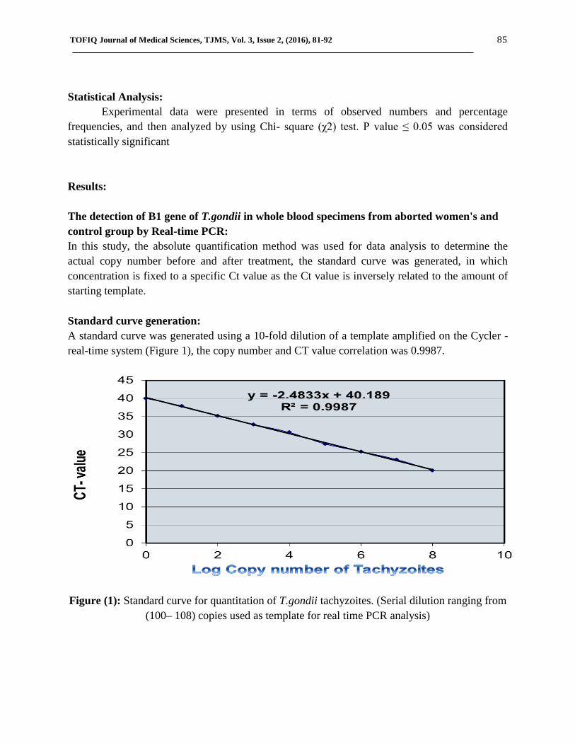
TOFIQ Journal of Medical Sciences, TJMS, Vol. 3, Issue 2, (2016), 81-92 85
Statistical Analysis:
Experimental data were presented in terms of observed numbers and percentage
frequencies, and then analyzed by using Chi- square (χ2) test. P value ≤ 0.05 was considered
statistically significant
Results:
The detection of B1 gene of T.gondii in whole blood specimens from aborted women's and
control group by Real-time PCR:
In this study, the absolute quantification method was used for data analysis to determine the
actual copy number before and after treatment, the standard curve was generated, in which
concentration is fixed to a specific Ct value as the Ct value is inversely related to the amount of
starting template.
Standard curve generation:
A standard curve was generated using a 10-fold dilution of a template amplified on the Cycler -
real-time system (Figure 1), the copy number and CT value correlation was 0.9987.
Figure (1): Standard curve for quantitation of T.gondii tachyzoites. (Serial dilution ranging from
(100– 108) copies used as template for real time PCR analysis)
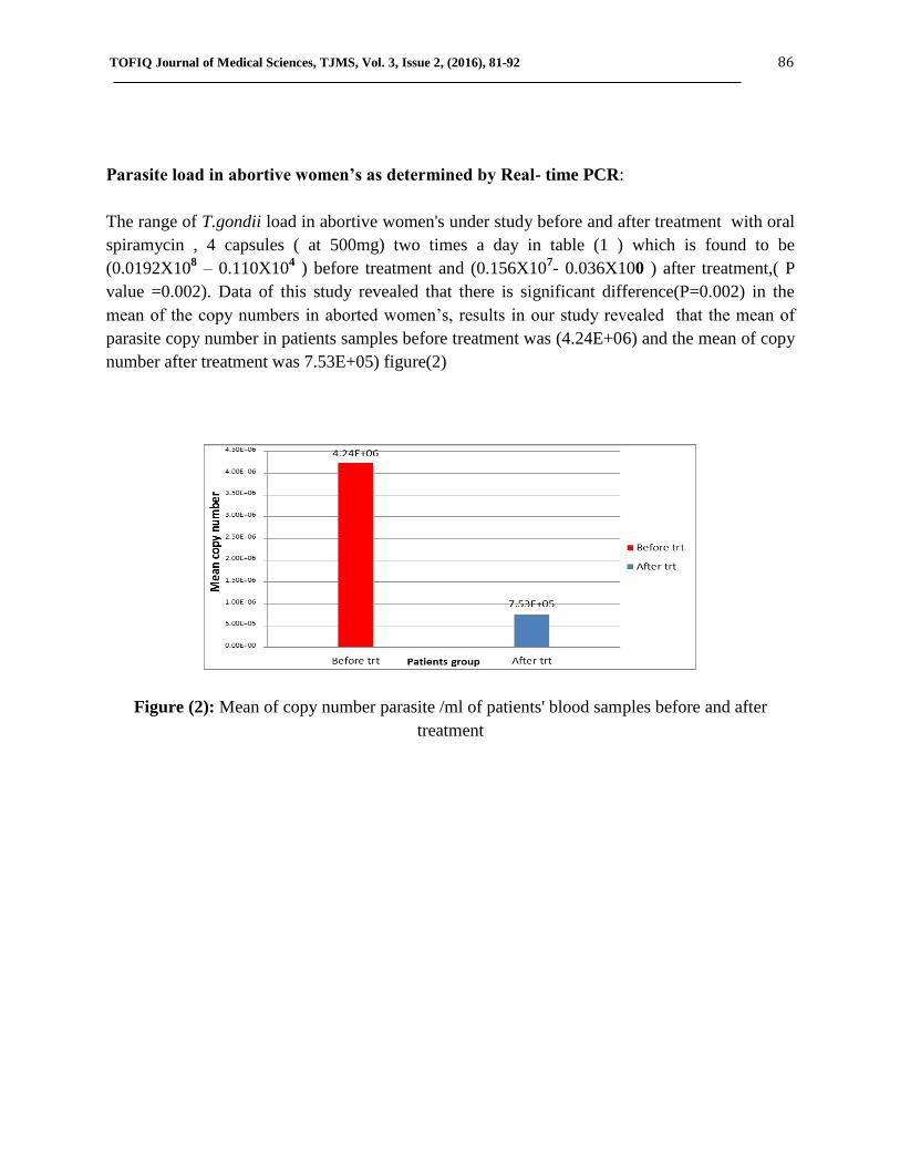
TOFIQ Journal of Medical Sciences, TJMS, Vol. 3, Issue 2, (2016), 81-92 86
Parasite load in abortive women’s as determined by Real- time PCR:
The range of T.gondii load in abortive women's under study before and after treatment with oral
spiramycin , 4 capsules ( at 500mg) two times a day in table (1 ) which is found to be
(0.0192X10⁸ – 0.110X10
4 ) before treatment and (0.156X10
7- 0.036X100 ) after treatment,( P
value =0.002). Data of this study revealed that there is significant difference(P=0.002) in the
mean of the copy numbers in aborted women’s, results in our study revealed that the mean of
parasite copy number in patients samples before treatment was (4.24E+06) and the mean of copy
number after treatment was 7.53E+05) figure(2)
Figure (2): Mean of copy number parasite /ml of patients' blood samples before and after
treatment

TOFIQ Journal of Medical Sciences, TJMS, Vol. 3, Issue 2, (2016), 81-92 87
Table (1): Toxoplasma gondii copy numbers screening in abortive women's as determined by
Real- time PCR
Sample Name CT-pre
treatment
Copy no. pre
treatment
CT-post
treatment
Copy no. post
Treatment
P 1 20.8 0.192 X 10^7 25.81 0.187X10^5
P2 27.1 0.0624X10^5 38.61 0.151X10^0
P3 20.1 0.0192X10^8 23.65 0.156X10^6
P4 21.4 0.129X10^7 24.11 0.110X10^6
P5 25.6 0.208X10^5 21.17 0.156X10^7
P6 23.7 0.151X10^6 37.12 0.057X10^1
P7 21.5 0.122X10^7 23.18 0.201X10^6
P8 19.4 0.086X10^5 22.19 0.057X10^7
P9 26.4 0.129X10^5 24.16 0.108X10^6
P10 22.7 0.007X10^7 35.55 0.206X10^1
P11 20.9 0.182X10^7 26.9 0.084X10^5
P12 20.5 0.220X10^7 37.12 0.055X10^1
P13 21.9 0.084X10^7 32.7 0.002X10^3
P14 26.4 0.129X10^5 22.41 0.036X10^7
P15 29.1 0.110X10^4 37.12 0.057X10^1
P16 22.4 0.036X10^7 33.1 0.204X10^2
P17 21.8 0.093X10^7 29.1 0.110X10^4
P18 22.5 0.028X10^7 35.1 0.009X10^2
P19 23.7 0.086X10^6 38.6 0.153X10^0
P20 22.4 0.038X10^7 32.1 0.062X10^3
P21 22.8 0.237X10^6 39.6 0.055X10^0
P22 28.6 0.158X10^4 30.1 0.014X10^4
P23 26.9 0.084X10^5 32.9 0.223X10^2
P24 25.1 0.014X10^6 39.8 0.036X10^0
P25 24.1 0.112X10^6 37.9 0.220X10^0
P26 23.5 0.170X10^6 31.8 0.088X10^3
P27 21.6 0.112X10^7 39.1 0.105X10^0
P28 26.4 0.132X10^5 31.8 0.088X10^3
P29 23.5 0.170X10^6 36.7 0.096X10^1
P30 27.9 0.225X10^4 33.7 0.146X10^2
Negative control NTC NTC
TOFIQ Journal of Medical Sciences, TJMS, Vol. 3, Issue 2, (2016), 81-92 88
Regarding response to treatments toxoplasmosis infected women in this study were divided to
the following groups:
Group I include patients had high parasite load (0.062X10⁵-0.112X10
7) which decrease
completely after completion the course of treatment (0.036X100-0.223X100).
Group II include a patients with high parasite load (0.225x10⁴ -0.036x10
7 ) at time of diagnosis
and decreases after completion the course of treatment (0.055x101 -0.223x10
1).
Group III includes patients with high parasite load at diagnosis (0.225x10⁴ -0.036x10
7) which
still with high parasite load or slightly lower (0.002x10³ – 0.201 x10
6).
Group IV includes patients with high parasite load (0.158x10⁴ – 0.129x10
5) at time of diagnosis
which showed elevated parasite load after treatment (0.014x10⁴ -0.156x10
7).
Discussion:
Firstly DNA was successfully extracted from whole blood samples for all subjects,
Menotti, et al 12
showed that the parasites are as likely to be found in the plasma or red cell
fractions, which might indicate that the most suitable sample is a whole blood extract. However,
in patient's blood T.gondii present might be intracellular in leukocytes 13
; in that case the white
cell fraction would be as appropriate as whole blood.
Parasitemia in pregnant women may be due to the toxoplasmosis reactivations occur
during immunosuppressive states in pregnancy. Tachyzoites or T.gondii DNA release from the
cyst in the body tissues may be the source of parasitemia or T.gondii DNA in blood 14
.
Quantitative detection of pathogen by real time PCR has been successfully applied to a variety of
specimens and the finding of T.gondii DNA in peripheral blood by PCR might help in the early
diagnosis of invasive diseases 15
.
The qRT-PCR that have been applied in this study to diagnose and for the determination
of copy number of T.gondii DNA in whole blood, it gives reproducible diagnostic and
quantitative results over conventional PCR with the risk of false positive due to contamination
with previously amplified products since in qRT-PCR there is no risk of hand manipulation and
the use of internal control will exclude the false negative which due to inhibiters in amplified
mixture, reproducibility of qRT-PCR quantification was tested by the testing of the 10 fold
dilution.
In the present study a qRT-PCR based B1 gene – specific Taq man assay have been
developed for quantitative detection of T.gondii which extremely sensitive to (0.168 parasite /
reaction) and its ability to quantify infection load of clinical specimen and its long linear range of
7 logs of DNA concentration with CT value as shown in figure (1) where R2=0.998 which is
indicative of a significant correlation between logarithmic value of each PCR product of copy
number amplified and CT value. These results were in agreement with Mei-huilin, (16)
. In this
assay an increase of the fluorescent signal above the preset threshold within 40 PCR cycle was
considered positive (CT< 40), Table (1) where CT for negative control was 40. Quantification of
TOFIQ Journal of Medical Sciences, TJMS, Vol. 3, Issue 2, (2016), 81-92 89
parasite load in pregnant women under study has been used to ass’s disease severity and the
treatment outcome.
The relative quantity of tachyzoites in each DNA sample was determined using the
standard curve presented in Figure (2) which showed a range between (0.110X10^4 to 0.0192
×108) before treatment and (0.036×100 to 0.156×10
7) after treatment .In addition figure (2)
revealed that there is significance difference (P<0.05) of in the mean of the copy number before
and after treatment.
According to their response to treatments, Group 1 patients which represent, 5 (16.66%)
% of patients having high parasite load at time of diagnosis with a range of (0.062X10⁵-
0.112X10⁷) copies /ml , they show complete response to treatment with decline in parasite load
to undetectable value , these results were in accordance with Costa , et al 17
who recorded that
the parasite count decreased sharply in patients having cerebral toxoplasmosis symptoms , when
treated with anti-toxoplasma therapy which was efficient and reducing the parasite load and they
conclude that Light Cycler PCR provide precise evaluation of parasite load in
immunocompromised patients and PCR is useful in monitoring of treatments .
In addition, Kupferschmidt, 18
conclude that decreasing in parasite load in patient's blood
sample demonstrated that treatment used was effective. Contini , et al 19
recorded that patient
under specific treatment did not show detectable B1 DNA levels in samples collected after many
days of therapy . So that T.gondii DNA load is a valuable tool for monitoring of toxoplasmosis.
Group Π patients which represent 10(33.33 %) of patients having high parasite load at time of
diagnosis with a range of (0.225x10⁴-0.036x10
7) copies /ml, they show lower parasite load with
a range of (0.055x101
-0.223x101) copies /ml after treatment. These results observed by Menotti,
et al 12
.
They consider that prolonged persistent detection of T. gondii DNA in blood justifies the
maintenance of anti T.gondii treatment and close follow up by PCR. These results were in
agreement with Romand, et al 20
who used quantitative Real time PCR to evaluate T. gondii
concentration in amniotic fluid and conclude that quantitative PCR may help the clinician to
form prognosis of congenital infections with regard to fetal and neonatal outcome. Other study
by Christianne, et al 21
reported that high parasite load in amniotic fluid was found in cases of
severe congenital toxoplasmosis as determined by quantitative PCR.
Group III which represent 11(36.66 %) include patients with high parasite load at time of
diagnosis with a range of (0.038x10⁷ –0.019x10
8) copies/ml, they did not show any response to
treatment, parasite load was remained with undetectable decrease. After treatment it's found to be
(0.002x10³ – 0.201 x10
6) copies/ ml. These results were in agreement with Kupferschmidt, et al
(18) .who used Tag man PCR to monitor parasite load in the blood of an immunocompromised
patients after allogenenetic bone marrow transplantation. They found that at day 199 after
therapy with pyrimethamine the detected parasite load remained unchanged and then decreased
after prolonged therapy.

TOFIQ Journal of Medical Sciences, TJMS, Vol. 3, Issue 2, (2016), 81-92 90
Regarding group IV of patients which represent 4(13.33 %) having high parasite load at
time of diagnosis with a range of (0.158x10⁴ – 0.129x10
5) copies /ml , they showed elevated
parasite load after treatment with a range of (0.014x10⁴ -0.156x10
7) copies /ml .This increase of
parasite load seems to be correlated to progression of disease or to poor response to treatment.
These results were in agreement with Menotti, et al 12
who recorded evolution in parasite load in
patients under treatments and those with slow decrease in parasite load after initiation of
treatments with pyrimethamine and sulfadiazine on day 43 post transplantation. They give
explanation to the prolonged presence of T.gondii DNA that could be due to disseminated form
of disease with a peak of parasitemia (29000 tachyzoites/ ml) of blood buffy coat.
Real time-PCR can be used as an additional diagnostic tool for the rapid detection of
T.gondii in various clinical materials. Studies have documented that Real time- PCR can actually
detect T.gondii in blood specimens of women before or during pregnancy. Nimri, et al 22
and
Khan et al 23
showed that a small numbers of parasites may be detected in circulating blood from
some patients with acute or recurrent toxoplasmosis.
Several studies have reported that, PCR could detect parasitemia a few weeks prior to the
appearance of any clinical signs or symptoms .PCR is highly sensitive and specific because a
single tachyozoite can be detected in a clinical sample 24
.
The most important records in this study is that the parasite load could be determined and
correlated with treatment. The parasite quantification in some treated subjects become lower and
in others, T.gondii load disappeared when spiramycin was given, this exhibit that the anti-
toxoplasma therapy was efficient in reducing the parasite load.
The quantitative real-time PCR assay developed in this study can be used not only to
detect the presence of T.gondii DNA but also to provide precise evaluations of the parasite load
in pregnant women’s. This PCR test should be useful for the monitoring of treatment efficacy
and should help provide an understanding of the pathogenesis of T.gondii reactivation.
TOFIQ Journal of Medical Sciences, TJMS, Vol. 3, Issue 2, (2016), 81-92 91
Reference:
1- Dubey JP, Hill DE, Jones JL, 2005 ; Prevalence of viable Toxoplasma gondii in beef, chicken, and
pork from retail meat stores in the United States: risk assessment to consumers. J Parasitol, (
91)1082–93.
2- Dubey JP, Su C, 2009; Population biology of Toxoplasma gondii: what's out and where did they
come from. Journal of MemInst Oswaldo Cruz, 104 (2) 190-1951.
3- Linguissi L, 2012; Seroprevalence of toxoplasmosis and rubella in pregnant women attending
antenatal private clinic at Ouagadougou, Burkina Faso.Asian Pac J. Trop Med, 5(10):810-813.
4- Joanna M., Violetta D., Halina K, 2009; Does Toxoplasma gondii Infection affect the Levels of
IgE and Cytokines (IL-5, IL-6, IL-10, IL-12, and TNF-alpha)? Clin Dev Immunol. (15) 374-396.
5- Evering, T. and Weiss, L.M, 2006; The immunology of parasite infections in
immunocompromised hosts. Parasite Immunolology, ( 28) 549-565.
6- Rai R., Clifford K., Cohen H., Regan L, 1995; High prospective fetal loss rate in untreated
pregnancies of women with recurrent miscarriage and antiphospholipid antibodies. Hum. Reprod,
(10)3301–3304.
7- Su C, Shwab EK, Zhou P, Zhu XQ, Dubey JP, 2010; Moving towards an integrated approach to
molecular detection and identification of Toxoplasma gondii. Parasitol J, 137(1) 1-11.
8- Rovamycine (Rhone-Poulenc Rorer), 1994; In: Vidal. 70th ed. Paris: Editions du Vidal, 1361.
9- Sebastian D., Zuhara K. andSekaran K. 2008; Influence of TORCH infections in first trimester
miscarriage in the Malabar region of Kerala. African J. Microbio. Res., ( 2) 056-059.
10- Holland PM, Abramson RD, Watson R, Gelfand DH, 1991; Detection of specific polymerase
chain reaction product by utilizing the 5’----3' exonuclease activity of Thermus aquaticus DNA
polymerase. Proc Natl Acad Sci U S A., (16):7276–80.
11- Edvinsson B, Lappalainen M, Evengård B, 2006; Real-time PCR targeting a 529-bp repeat
element for diagnosis of toxoplasmosis. Clin Microbiol Infect, ( 12) 131-136.
12- Menotti J, Garin YJ, Thulliez P, 2010; Evaluation of a new 5'-nuclease real-time PCR assay
targeting the Toxoplasma gondii AF146527 genomic repeat. Clin Microbiol. Infect, (4)363-368.
13- Ho-Yen D.O., JossA.W.,BalfourA.H., Smyth E.T.,Baird D, 1992; Use of the polymerase chain
reaction to detect Toxoplasma gondii in human blood samples. J Clin Pathol., 45(10): 910 – 913.
14- Dubey JP, Chellaiah R, Costa DG, Ferreira L, Kwok O, 2010; New Toxoplasma gondii
Genotypes Isolated from Free-Range Chickens from the Fernando De Noronha, Brazil:
Unexpected Findings. J Parasitol, ( 96)709–712.
15- Bretagne S, Costa JM, Foulet F,V Prospective study of toxoplasma reactivation by polymerase
chain reaction in allogeneic stem-cell transplant recipients. Transpl Infect Dis, ( 2)127-32.
16- Mei-Hui Lin, Tse-chingchen,Tesng Tong Kuo,chingchung Tseng, 2000; Real time PCR detection
of T.gondii .J.clin.Microbiol, 38(11):4121-4125.
17- Costa, J. M., Pautas, C., Ernault, P., Foulet, F., Cordonnier, C, 2000; Real-time PCR for diagnosis
and follow-up of Toxoplasma reactivation after allogeneic stem cell transplantation using
fluorescence resonance energy transfer hybridization probes. J Clin Microbiol, 38(8): 2929–2932.
TOFIQ Journal of Medical Sciences, TJMS, Vol. 3, Issue 2, (2016), 81-92 92
18- Kupferschmidt O, Krüger D, Held TK, Ellerbrok H, Siegert W, Janitschke K, 2001; J. Clin
Microbiol Infect, 7(3):120-4.
19- Contini C, Seraceni, Cultrea R, Incorvaia, Sebastiani A, 2005; Evaluation of a Real-time PCR-
based assay using the light cycler. International Journal for Parasitology, 35 (3):275–283.
20- Romand, S., Chosson, M., Franck, J., Wallon, M., Kieffer, F, 2004; Usefulness of quantitative
polymerase chain reaction in amniotic fluid as early prognostic marker of fetal infection with
Toxoplasma gondii. Am J Obstet Gynecol, ( 190)797-802.
21- Christianne T, Pedro A, Brasil A, Letícia G, Maria E, 2016; Performance of Polymerase Chain
Reaction Analysis of the Amniotic Fluid of Pregnant Women for Diagnosis of Congenital
Toxoplasmosis, PLOS ONE | DOI:10.1371/journal.pone , (7) 149-153
22- Nimri, L. Pelloux, H. and Elkhatib, L, 2004; Detection of Toxoplasma gondii DNA and specific
antibodies in high- risk pregnant women. Am. J. Trop. Med. Hyg. 71(6): 831-835.
23- Khan A, Jordan C, Muccioli C, 2006; Genetic divergence of Toxoplasma gondii strains associated
with ocular toxoplasmosis, Brazil. Emerg Infec Dis. (12) 942–9 45
24- Guy, E. C., Pelloux, H., Lappalainen, M., Aspock, H., Hassl, 1996; Interlaboratory comparison of
polymerase chain reaction for the detection of Toxoplasma gondii DNA added to samples of
amniotic fluid. Eur J Clin Microbiol Infect Dis, (15) 836-839.


TOFIQ Journal of Medical Sciences, TJMS, Vol. 3, Issue 2, (2016), 64-80 ISSN: 2377-2808
RETROSPECTIVE STUDY OF POSTERIOR DORSOLUMBAR FIXATION IN
BAGHDAD: A CLINICAL STUDY OF 100 PATIENTS
Prof. Yasir M H Hamandi * , Dr. Ali mohammad khalil
**
* Department of surgery/ neurosurgery, College of Medicine, Al-Nahrain University, Baghdad,
Iraq
** Department of surgery/ neurosurgery, Al-hilla teaching hospital, Al-Hilla, Iraq
Corresponding author:
Email: [email protected]

TOFIQ Journal of Medical Sciences, TJMS, Vol. 3, Issue 2, (2016), 64-80 65
Abstract :
Background: Spondylolisthesis describes a condition of a forward slippage of one vertebra
over another, which may or may not be associated with demonstrable instability. Spinal fixation
is a neurosurgical procedure in which two or more vertebrae are anchored to each other through
a synthetic "vertebral fixation device"
Objective: To determine the demographic distribution of different patient factors and the most
commonly vertebra undergo fixation in the thoracolumbar instrumentation.
Patients and Methods: one hundred patients were evaluated during the period of this study in
a retrospective manner from January 2013 to January 2015 in four hospitals in Baghdad
(Neurosurgical Teaching Hospital, Neuroscience hospital, Al-Kahdymia Teaching Hospital,
Medical City\Ghazy AL-Hariri Hospital). The patients' data regarding the etiology of
instability, mechanism of injury for trauma patients, gender, age, segments undergoing
instrumentation were identified.
Results: The study revealed female predominance over male: female ratio of 1:2.7, the age
distribution was highest from 3rd
to 7th
decades of life, the etiology of instability was either
degenerative or traumatic, the degenerative instability was 65% while traumatic cases was
35%. The neurological status of the patients was assessed by neurological examination and
revealed 75% with incomplete deficit and 25% with complete neurological deficit, the most
common pathologically involved vertebra was the L4, the most common vertebrae used in
fixation were the L4 and L5 levels, the most common type of fixation used was the short
segment fixation.
Conclusion: Posterior spinal fixation with pedicle screws and rods system is an effective and
safe method in maintaining the stability of spine. The intraoperative imaging is important in
maintaining safe trajectory of screws. Short segment fixation using the posterior approach with
pedicle screw-rod fixation devices achieve good stabilization. The ideal candidates for
undergoing posterior spinal fixation are patients with unstable fractures & incomplete
neurological deficit.
Recommendation: The use of intraoperative neuro-monitoring, use of navigation system, use
of fluoroscopy and the O-arm in spinal fixation surgery. Bone fusion is recommended for each
patient.
Keywords: Thoracolumbar spine, spondylolisthesis, pedicle screw fixation

TOFIQ Journal of Medical Sciences, TJMS, Vol. 3, Issue 2, (2016), 64-80 66
Introduction:
Spinal instrumentation basically means the implantation of more or less rigid metallic or
non-metallic devices which are attached to the spine. These devices function to provide spinal
stability and thus facilitate bone healing leading to spinal fusion. [1]
Types of instrumentation[2]
1.Metallic Pedicle Screw-Rod Systems
2. Polyetheretherketone (PEEK) Rods
Goals and indications of spinal instrumentation:
1. Trauma
2. Non trauma
a. Tumor
b- Infection
c. Degenerative changes and spondylolisthesis[2]
Indications for fusion fall into two broad categories:
A. Preoperative structural problems that predispose to instability after decompression:
1. Degenerative spondylolisthesis or lateral listhesis.
2. Progressive scoliosis or kyphosis.
3. Recurrent spinal stenosis requiring repeat decompression at the same level.
B. Intraoperative structural alterations that warrant consideration of a fusion:
1. Excess facet joint removal 50%
2. Pars interarticularis fracture or removal.
3. Radical disc excision with resultant destabilization of the anterior spinal column.
C. Trauma that predispose to unstable spine
Measures for Correct Screw Placement
1. Navigation , CT, and fluroscope Guidance[3,4]
2. Electromyographic Monitoring[4]
Complications:
1. Pedicle Fracture[2]
2. Cerebrospinal Fluid Fistulae[5]
3. Infection[10]
4. Hardware Failure (Screw Breakage , Screw Pull-out, Screw Loosening or Plate or Rod
Breakage, Loss of Correction, Wound Breakdown.)[5,6,7,8]

TOFIQ Journal of Medical Sciences, TJMS, Vol. 3, Issue 2, (2016), 64-80 67
5. Nerve root or cord injury.
Free –hand technique
Free-hand pedicle screw placement relies on an intricate appreciation of the relationship
of various anatomical landmarks at each level of the thoracolumbar spine. Analogous entry sites
guided by differential anatomy are utilized for both the thoracic and lumbar spine.[9,10]
Accuracy of pedicle screw placement
Criteria of pedicle screw placement were: [11]
(1) Relation of pedicle screws to the pedicle.
(2) Relation of pedicle screws to the vertebral body.
Aim of the study: To determine the demographic distribution of different patient factors
To identify the most common vertebra levels to undergo fixation in the thoracolumbar
spine.
Patients and Methods: One hundred patients were evaluated during the period of this study in a retrospective
manner from January 2013 to January 2015 in four hospitals in Baghdad (Neurosurgical
Teaching Hospital, Neuroscience hospital, Al-Kahdimiyya Teaching Hospital, Medical
City/Ghazi AL-Hariri Hospital).
Demographic, Admission complaints and imaging were obtained for 100 patients through
chart review. On Admission the following parameters independently reviewed: gender, age, chief
complaint, etiology of instability, for the trauma patients the mechanism of injury was identified,
the level of pathology, neurological examination, type of neurological deficit (whether complete
or incomplete), level of fixation, number of screws used, outcome before discharge and a 6
months follow up.
The patients' data regarding the etiology of instability, mechanism of injury for trauma
patients, segments undergoing instrumentation were identified.
All patients' undergone hematological investigations in the form of complete blood, ESR,
C-reactive protein, renal function test, fasting blood sugar, blood group & Rh.
Only 16 patients have NCS and EMG prior to surgery, so the neuro-electro-physiological
studies can't be analyzed in correlation to other clinical factors.
The findings of the patients' pre-operative imaging including thoracolumbar X-rays, CT-
scan and MRI were reviewed.
The systems used were Medtronic and Aesculap.

TOFIQ Journal of Medical Sciences, TJMS, Vol. 3, Issue 2, (2016), 64-80 68
Results: The gender analysis revealed female predominance over male: female ratio of 1:2.7 and that 27%
were males while 73% were females. (Fig. 1)
Fig. 1 Gender distribution
The age distribution of spine instability for both the traumatic and non-traumatic cases
was highest from 3rd
to 7th
decades of life, it's very low in the first two decades of life and in the
7th
decade of life.
Table (1) Age distribution
AGE
10-19 years 2%
20-29 years 13%
30-39 years 23%
40-49 years 36%
50-59 years 21%
60-69 years 5%
Total
The etiology of instability was either non traumatic or traumatic, the non-traumatic
instability was 65% while traumatic cases was 35%. In the non-traumatic group the
27%
73%
Gender
Male
Female
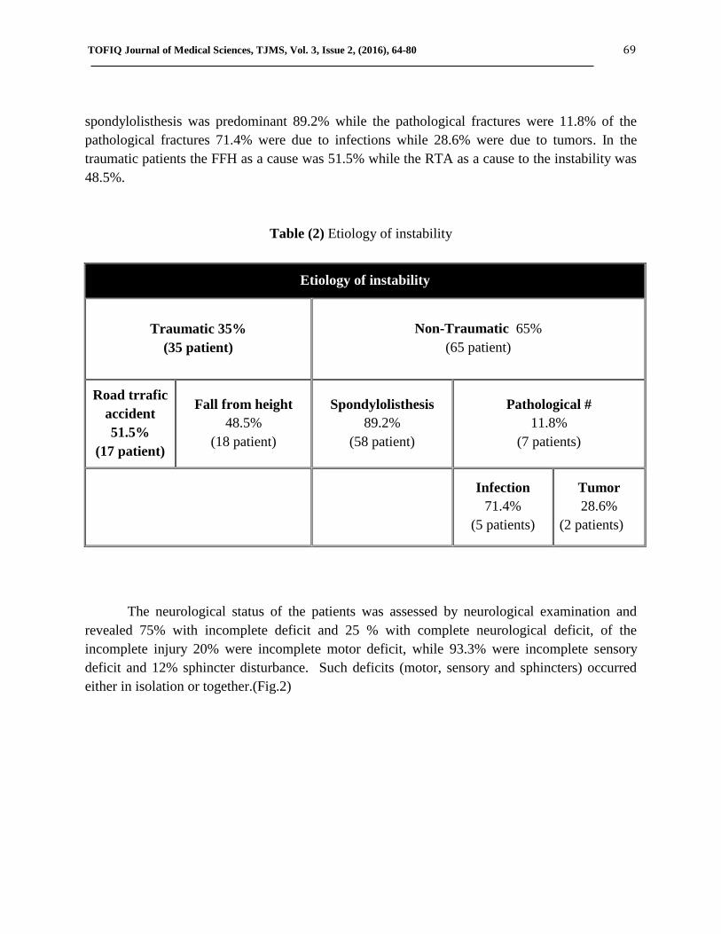
TOFIQ Journal of Medical Sciences, TJMS, Vol. 3, Issue 2, (2016), 64-80 69
spondylolisthesis was predominant 89.2% while the pathological fractures were 11.8% of the
pathological fractures 71.4% were due to infections while 28.6% were due to tumors. In the
traumatic patients the FFH as a cause was 51.5% while the RTA as a cause to the instability was
48.5%.
Table (2) Etiology of instability
Etiology of instability
Traumatic 35%
(35 patient)
Non-Traumatic 65%
(65 patient)
Road trrafic
accident
51.5%
(17 patient)
Fall from height
48.5%
(18 patient)
Spondylolisthesis
89.2%
(58 patient)
Pathological #
11.8%
(7 patients)
Infection
71.4%
(5 patients)
Tumor
28.6%
(2 patients)
The neurological status of the patients was assessed by neurological examination and
revealed 75% with incomplete deficit and 25 % with complete neurological deficit, of the
incomplete injury 20% were incomplete motor deficit, while 93.3% were incomplete sensory
deficit and 12% sphincter disturbance. Such deficits (motor, sensory and sphincters) occurred
either in isolation or together.(Fig.2)
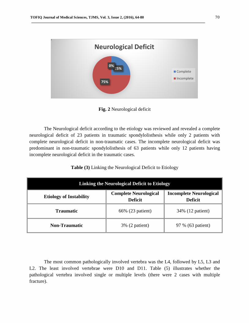
TOFIQ Journal of Medical Sciences, TJMS, Vol. 3, Issue 2, (2016), 64-80 70
Fig. 2 Neurological deficit
The Neurological deficit according to the etiology was reviewed and revealed a complete
neurological deficit of 23 patients in traumatic spondylolisthesis while only 2 patients with
complete neurological deficit in non-traumatic cases. The incomplete neurological deficit was
predominant in non-traumatic spondylolisthesis of 63 patients while only 12 patients having
incomplete neurological deficit in the traumatic cases.
Table (3) Linking the Neurological Deficit to Etiology
Linking the Neurological Deficit to Etiology
Etiology of Instability Complete Neurological
Deficit
Incomplete Neurological
Deficit
Traumatic 66% (23 patient) 34% (12 patient)
Non-Traumatic 3% (2 patient) 97 % (63 patient)
The most common pathologically involved vertebra was the L4, followed by L5, L3 and
L2. The least involved vertebrae were D10 and D11. Table (5) illustrates whether the
pathological vertebra involved single or multiple levels (there were 2 cases with multiple
fracture).
25%
75%
0% 0%
Neurological Deficit
Complete
Incomplete

TOFIQ Journal of Medical Sciences, TJMS, Vol. 3, Issue 2, (2016), 64-80 71
Table (4) Level of Pathology
Level of Pathology
D10 2
D11 2
D12 10
L1 5
L2 16
L3 15
L4 37
L5 15
The most common type of fixation used was the short segment fixation using 4 pedicle
screws while the least common was the long segment fixation using 8 pedicle screws.
Table (5) No. of screws used in a single fixation
Type of segment No. of patients No. of screws
4 screws (short segment fixation) 67 268
6 screws (intermediate segment fixation) 27 162
8 screws (long segment fixation) 6 48
Total No. of screws 478
The type of intraoperative guidance for screw placement was either by fluoroscopy (C-
arm X-ray) 77 cases or by anatomical landmarks (free hand fixation) 23 cases.
Table (6) Type of Intraoperative Guidance for screw placement
Type of Intraoperative Guidance for screw placement
Type of Guidance Fluoroscopy Guided Fixation Free Hand Fixation
No. of patients 77 23
Accuracy 98% 91.3%
The complications occurred was either intraoperative or postoperative. The intraoperative
complications include dural tear (3 patients), screw misplacement (4 patients). The postoperative
complications include infection (7 patients), screw fracture (4 screw fractured in 3 patient as in
Fig. 10), CSF leak (3 patients), and exposure of the system (1 patient).

TOFIQ Journal of Medical Sciences, TJMS, Vol. 3, Issue 2, (2016), 64-80 72
Table (7) Intraoperative Complications
operative Complications
Screw misplacement 4 patients
Dural Tear 3 patients
Table (8) Postoperative Complications
Postoperative Complications
Infection 7 patients
Screw fracture 3 patients
CSF leak 3 patients
Exposure of the system 1 patient
Regarding follow up and outcome: 42 patient were followed for 6 months of which 39
patients show improvement of symptoms (1 patient with complete neurological deficit show
improvement in motor function), 2 patients show worsening of symptoms and 1 patient died.
Table (9) Follow up and Outcome
Follow up and Outcome
Improvement of symptoms 39 patients
Worsening of symptoms 2 patients
Death 1 patient
Discussion: A significant number of patients suffer spinal diseases around the world, and a high
percentage of them experience pathology in thoracolumbar region. Of those patients, a number
of vertebral instability disorders need surgical stabilization.
Different surgical modalities are used in treating thoracolumbar spinal instability. Of
these, the most common being used is posterior spinal fixation with different instruments.
TOFIQ Journal of Medical Sciences, TJMS, Vol. 3, Issue 2, (2016), 64-80 73
The most modern and commonly used fixation is the pedicle screw and rod system. Other
methods include anterior spinal fixation with cage, kyphoplasty, vertebroplasty, posterior
interbody fusion and posterior intertransverse fusion. Of the posterior spinal fixation, the various
instruments used are pedicle screws and rods system.
The neurological recovery is dependent on the degree of stability provided by fixation,
and on initial instability and neurological loss. So, if stability is provided to spine then
neurological status of some patients can improve.
Regarding the gender 73% of our study group was female and 27% was male with a
male/female ratio of 1:2.7, while in Muralidhar B.M. et al Study [12]
there was male
predominance of 80% and female of 20%, Shailendra et al study [13]
the male percentage was
76% and female was 24%. This difference is due to that 65% of our patient complaining of
instability due to non-traumatic causes of which degenerative spondylolisthesis more commonly
occurs in elderly females because of the postmenopausal osteoporosis.
About age group, in our study 36% of our patient are located in 50-59 age group which is
the commonest age group while in Muralidhar B.M.et al study most of the patient are in 50 -59
age group, in Shailendra et al study the commonest age group is 16-25 years followed by 26-35
years, this difference because the largest group of our patient had degenerative diseases other
than trauma which prevalence increased with age.[12, 13]
Concerning etiology, 35% are due to traumatic injury (17 patient RTA, 18 patient FFH),
65% are due to non-traumatic causes (58 patient spondylolisthesis, 7 patient pathological fracture
{pathological fracture 5 patient infection, 2 patient tumor}) , while in study done by Alp Ozgun
BOrcek et al [15]
the non-traumatic cases was 63.9% while the traumatic cases was 36% which is
close to our result.
The relation between etiology and neurological deficit in traumatic cases 66% had
complete neurological deficit and 34% incomplete deficit while in non-traumatic cases 3% had
complete deficit and 97% had incomplete deficit, this difference is due to two factors first is that
traumatic injury which cause vertebral fracture and instability means sever trauma while in
degenerative cases it is usually a chronic process and patient seek medical management before
complete deficit occur, the other factor is that trauma mostly cause injury to thoracolumbar
junction because the transition between the more rigid thoracic spine and the mobile lumbar
spine concentrates bending and axial loads at the thoracolumbar junction where there is spinal
cord in the spinal canal while in degenerative cases are mostly in L4 level where there is cauda
equina and no cord.
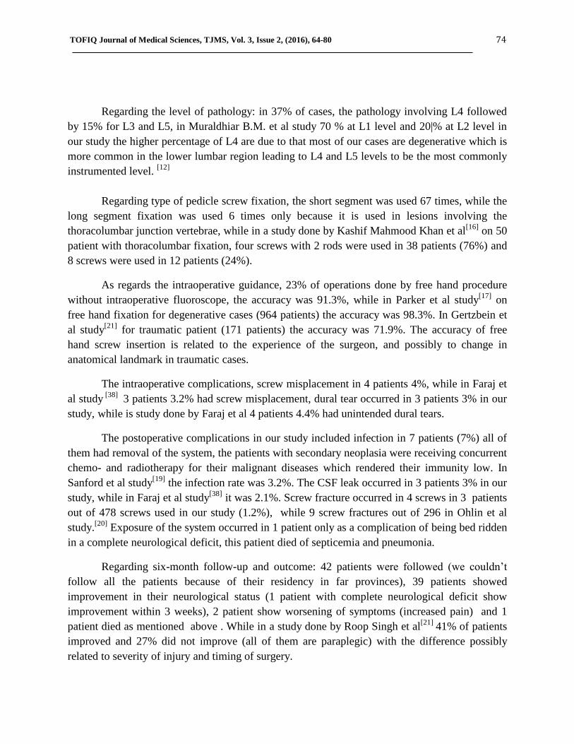
TOFIQ Journal of Medical Sciences, TJMS, Vol. 3, Issue 2, (2016), 64-80 74
Regarding the level of pathology: in 37% of cases, the pathology involving L4 followed
by 15% for L3 and L5, in Muraldhiar B.M. et al study 70 % at L1 level and 20|% at L2 level in
our study the higher percentage of L4 are due to that most of our cases are degenerative which is
more common in the lower lumbar region leading to L4 and L5 levels to be the most commonly
instrumented level. [12]
Regarding type of pedicle screw fixation, the short segment was used 67 times, while the
long segment fixation was used 6 times only because it is used in lesions involving the
thoracolumbar junction vertebrae, while in a study done by Kashif Mahmood Khan et al[16]
on 50
patient with thoracolumbar fixation, four screws with 2 rods were used in 38 patients (76%) and
8 screws were used in 12 patients (24%).
As regards the intraoperative guidance, 23% of operations done by free hand procedure
without intraoperative fluoroscope, the accuracy was 91.3%, while in Parker et al study[17]
on
free hand fixation for degenerative cases (964 patients) the accuracy was 98.3%. In Gertzbein et
al study[21]
for traumatic patient (171 patients) the accuracy was 71.9%. The accuracy of free
hand screw insertion is related to the experience of the surgeon, and possibly to change in
anatomical landmark in traumatic cases.
The intraoperative complications, screw misplacement in 4 patients 4%, while in Faraj et
al study [38]
3 patients 3.2% had screw misplacement, dural tear occurred in 3 patients 3% in our
study, while is study done by Faraj et al 4 patients 4.4% had unintended dural tears.
The postoperative complications in our study included infection in 7 patients (7%) all of
them had removal of the system, the patients with secondary neoplasia were receiving concurrent
chemo- and radiotherapy for their malignant diseases which rendered their immunity low. In
Sanford et al study[19]
the infection rate was 3.2%. The CSF leak occurred in 3 patients 3% in our
study, while in Faraj et al study[38]
it was 2.1%. Screw fracture occurred in 4 screws in 3 patients
out of 478 screws used in our study (1.2%), while 9 screw fractures out of 296 in Ohlin et al
study.[20]
Exposure of the system occurred in 1 patient only as a complication of being bed ridden
in a complete neurological deficit, this patient died of septicemia and pneumonia.
Regarding six-month follow-up and outcome: 42 patients were followed (we couldn’t
follow all the patients because of their residency in far provinces), 39 patients showed
improvement in their neurological status (1 patient with complete neurological deficit show
improvement within 3 weeks), 2 patient show worsening of symptoms (increased pain) and 1
patient died as mentioned above . While in a study done by Roop Singh et al[21]
41% of patients
improved and 27% did not improve (all of them are paraplegic) with the difference possibly
related to severity of injury and timing of surgery.
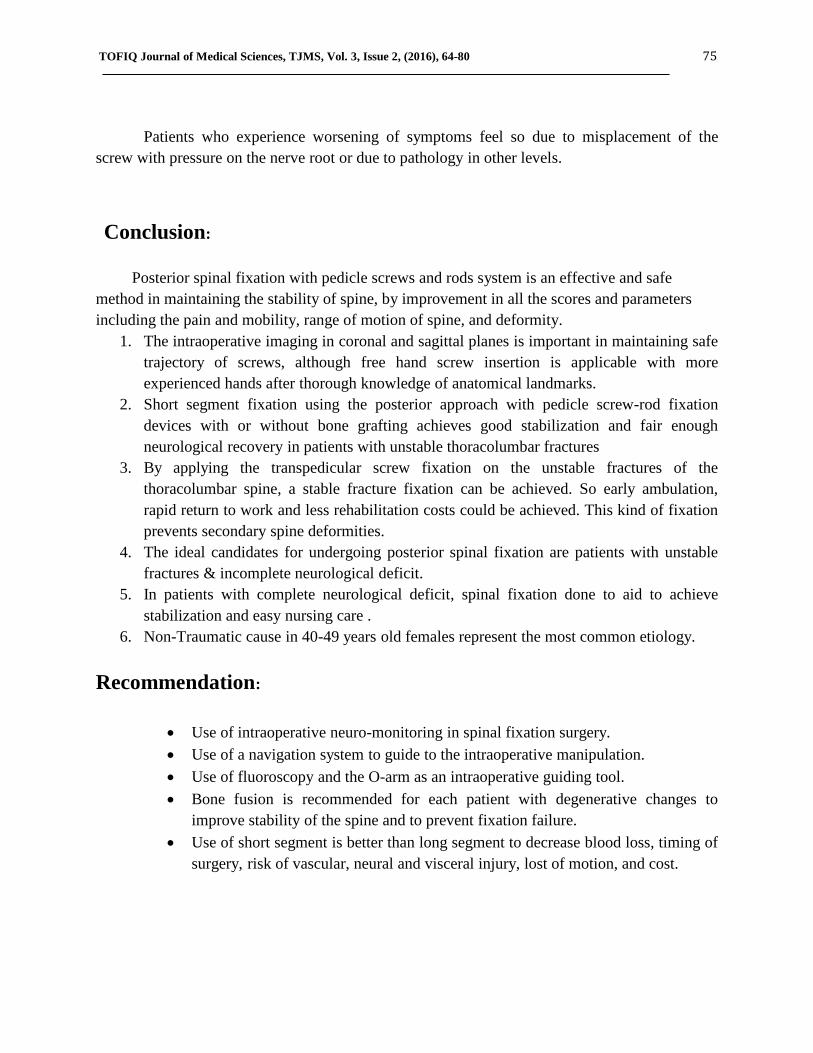
TOFIQ Journal of Medical Sciences, TJMS, Vol. 3, Issue 2, (2016), 64-80 75
Patients who experience worsening of symptoms feel so due to misplacement of the
screw with pressure on the nerve root or due to pathology in other levels.
Conclusion:
Posterior spinal fixation with pedicle screws and rods system is an effective and safe
method in maintaining the stability of spine, by improvement in all the scores and parameters
including the pain and mobility, range of motion of spine, and deformity. 1. The intraoperative imaging in coronal and sagittal planes is important in maintaining safe
trajectory of screws, although free hand screw insertion is applicable with more
experienced hands after thorough knowledge of anatomical landmarks.
2. Short segment fixation using the posterior approach with pedicle screw-rod fixation
devices with or without bone grafting achieves good stabilization and fair enough
neurological recovery in patients with unstable thoracolumbar fractures
3. By applying the transpedicular screw fixation on the unstable fractures of the
thoracolumbar spine, a stable fracture fixation can be achieved. So early ambulation,
rapid return to work and less rehabilitation costs could be achieved. This kind of fixation
prevents secondary spine deformities.
4. The ideal candidates for undergoing posterior spinal fixation are patients with unstable
fractures & incomplete neurological deficit.
5. In patients with complete neurological deficit, spinal fixation done to aid to achieve
stabilization and easy nursing care .
6. Non-Traumatic cause in 40-49 years old females represent the most common etiology.
Recommendation:
Use of intraoperative neuro-monitoring in spinal fixation surgery.
Use of a navigation system to guide to the intraoperative manipulation.
Use of fluoroscopy and the O-arm as an intraoperative guiding tool.
Bone fusion is recommended for each patient with degenerative changes to
improve stability of the spine and to prevent fixation failure.
Use of short segment is better than long segment to decrease blood loss, timing of
surgery, risk of vascular, neural and visceral injury, lost of motion, and cost.
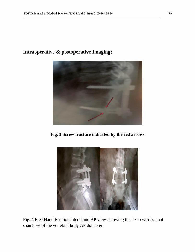
TOFIQ Journal of Medical Sciences, TJMS, Vol. 3, Issue 2, (2016), 64-80 76
Intraoperative & postoperative Imaging:
Fig. 3 Screw fracture indicated by the red arrows
Fig. 4 Free Hand Fixation lateral and AP views showing the 4 screws does not
span 80% of the vertebral body AP diameter
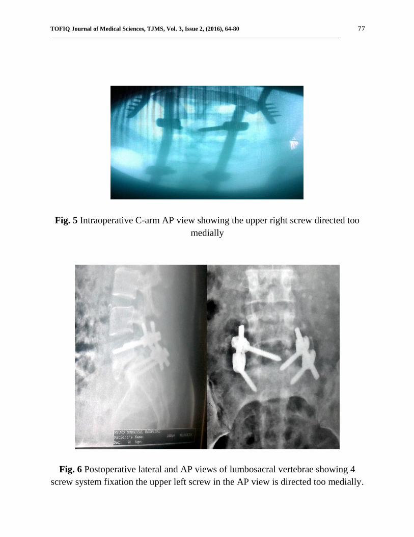
TOFIQ Journal of Medical Sciences, TJMS, Vol. 3, Issue 2, (2016), 64-80 77
Fig. 5 Intraoperative C-arm AP view showing the upper right screw directed too
medially
Fig. 6 Postoperative lateral and AP views of lumbosacral vertebrae showing 4
screw system fixation the upper left screw in the AP view is directed too medially.
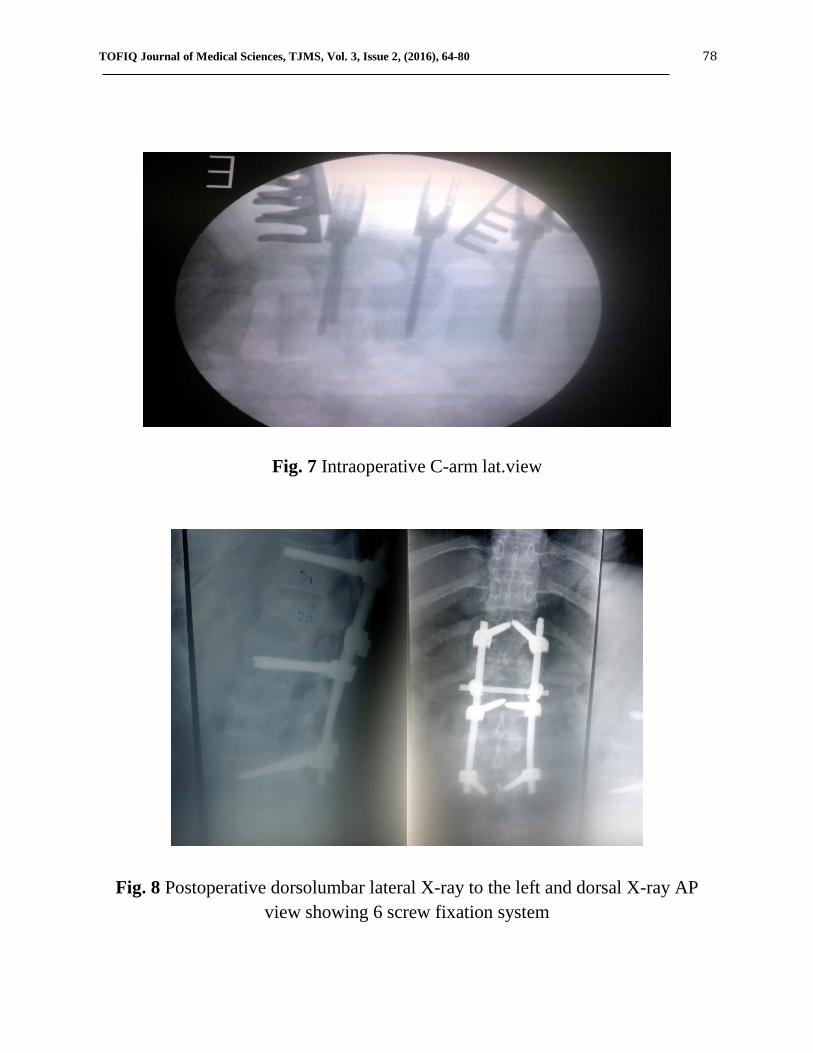
TOFIQ Journal of Medical Sciences, TJMS, Vol. 3, Issue 2, (2016), 64-80 78
Fig. 7 Intraoperative C-arm lat.view
Fig. 8 Postoperative dorsolumbar lateral X-ray to the left and dorsal X-ray AP
view showing 6 screw fixation system
TOFIQ Journal of Medical Sciences, TJMS, Vol. 3, Issue 2, (2016), 64-80 79
References: 1. Boos N, Aebi M: Spinal disorders fundamentals of diagnosis and treatment 2008. Spinal
Instrumentation 3: 67-86.
2. Shaffrey I, Sonntag H, Dennis G: Youmans neurological surgery 2012 6th
edition.
Posterior Thoracic Instrumentation 301: 3061-3067.
3. Benzel C: Spine Surgery 2012 3rd
edition. History of Spinal Instrumentation: The Modern
Era 2: 21-29.
4. Bono CM, Lee CK: Critical analysis of trends in fusion for degenerative disc disease over
the past 20 years: influence of technique on fusion rate and clinical outcome 2004. Spine
29: 455–463.
5. Lagrone MO, Bradford DS, Moe JH: Treatment of symptomatic flatback after spinal
fusion 1988. J Bone Joint Surg. 70: 569–580.
6. Esses ST, Bednar DA: The spinal pedicle screw: techniques and systems 1989; 18: 676–
682.
7. Zucherman JF, Hsu KY, Hartjen CA: A multicenter, prospective, randomized trial
evaluating the X STOP interspinous process decompression system for the treatment of
neurogenic intermittent claudication: two-year follow-up results 2005. Spine 30: 1351–
1358.
8. Shaffrey I, Sonntag H, Dennis G: Youmans neurological surgery 2012 6th
edition.
Posterior Lumbar Instrumentation 303: 3075-3082.
9. Gertzbein SD, Robbins SE: Accuracy of pedicular screw placement in vivo. Spine 1990;
15: 11-14.
10. Chung KJ et al: Ideal entry point for the thoracic pedicle screw during the free hand
technique. Int Orthop 2008; 32: 657-662.
11. Abdulameer J. Al-Kafaji et al: Pedicle Screw Placement versus Classic Surgery in
Lumbothoracic Spine Disorder . Iraqi journal of medical scince 2014 :230-237 .
12. Muralidhar B.M. et al: Management of unstable thoracolumbar spinal fractures by
pedicle screws and rod fixation. Journal of Clinical and Diagnostic Research. 2014 Feb,
Vol-8(2):121-123.
13. Shailendra et al: Surgical outcome of posterior short segment trans-pedicle screw fixation
for thoracolumbar fractures. Journal of orthopedics 10 (2013) 162-167.
14. Alexander R. et al: Pedicle-Screw Fixation in the Lumbar Spine. Journal of the American
Academy of Orthopedic Surgeons 1995; 3:263-274.
15. Alp Ozgun BOrcek et al: Accuracy of Pedicle Screw Placement in Thoracolumbar Spine
with Conventional Open Technique. Turk Neurosurg 2014, Vol: 24, No: 3, 398-402.

TOFIQ Journal of Medical Sciences, TJMS, Vol. 3, Issue 2, (2016), 64-80 80
16. Kashif Mahmood Khan et al: Posterior Spinal Fixation with Pedicle Screws and Rods
System in Thoracolumbar Spinal Fractures. Journal of the College of Physicians and
Surgeons Pakistan 2012, Vol. 22 (12): 778-782.
17. Parker SL et al: Accuracy of free-hand pedicle screws in the thoracic and lumbar spine:
analysis of 6816 consecutive screws. Neurosurgery 2011; 68: 170-178.
18. Faraj et al: Early complications of spinal pedicle screw. Euro Spine J 1997 (6): 324-326.
19. Sanford HD et al: Complications of lumbar spinal fusion with transpedicular
instrumentation 1992. Spine 17: 183-189.
20. Ohlin A. et al: Complications after transpedicular stabilization of the spine 1994. Spine
19: 2774-2779.
21. Roop Singh et al: Outcome of Pedicle Screw Fixation and Monosegmental Fusion in
Patients with Fresh Thoracolumbar Fractures. Asian Spine J 2014; 8(3):298-308.

TOFIQ Journal of Medical Sciences, TJMS, Vol. 3, Issue 2, (2016), 81-92 ISSN: 2377-2808
REAL TIME -PCR FOR QUANTITATIVE DETECTION OF TOXOPLASMA GONDII
IN ABORTED WOMEN BEFORE AND AFTER TREATMENT
Dr. Huda Al-Marsomy1, Dr. Elham Alswad
2, Dr. Jabbar Salman Hassan
3, Haleema Salman
4
(1, 3) Ph. D. Medical Microbiology/ College of medicine, Al-Nahrain University, Baghdad, Iraq
(2) Ph.D. Medical, Biology, College of Medicine, Al-Nahrain University, Baghdad, Iraq
(4)MSc Medical Microbiology, College of Medicine, Al-Nahrain University, Baghdad, Iraq
Corresponding Author:
E-mail: Dr. Jabbar Salman Hasan: <[email protected]>
Abstract:
Background: Human Toxoplasmosis is a parasitic disease caused by a Toxoplasma gondii.
Toxoplasma gondii was the subject of disease burden estimations as a result of the severe clinical
symptoms and lifelong implications. Primary infections with T.gondii acquired during pregnancy
are usually asymptomatic for the pregnant woman but can lead to serious neonatal complications,
isolation of parasite despite it is considered as a golden standard parameter to detect infection,

TOFIQ Journal of Medical Sciences, TJMS, Vol. 3, Issue 2, (2016), 81-92 82
the repetitive B1 gen is the most target gene used in conventional PCR for molecular detection of
T.gondii.
Objective: Detection of T. gondii DNA using Real- time PCR seems to be a reliable method in
diagnosis and follow up of patients.
Methods: Thirty patients (aborted women), their range age between (20− 46) years, were
included in this study. All patients were assessment of Real time Polymerase chain reaction from
the same patients was taken before and after spiramycin therapy to confirm the infection of the
T.gondii and to evaluate the parasite load (copy number).
Result: Data of this study revealed that there is significant difference(P=0.002) in the mean of
the copy numbers in aborted women's, the mean of parasite copy number in patients samples
before treatment was (4.24E+06) and the mean of copy number after treatment was (7.53E+05).
Conclusion: The diagnosis of toxoplasmosis is mainly based on serological tests detection of T.
gondii DNA using Real time- PCR seems to be a reliable method in diagnosis and follow up of
patients.
Keywords: Toxoplasma gondii, Real- time PCR, B1-gen
Introduction:
Toxoplasmosis is a worldwide infection that may cause severe disease and is regarded as
a serious health problem in world. It is one of the well-studied parasites because of its medical
and veterinary importance, and its suitability as a model for cell biology and molecular studies
with a unicellular organism 1. Toxoplasmosis is also a major opportunistic infection in
immunocompromised individuals, often resulting in lethal toxoplasmic encephalitis .If a woman
was infected for the first time during pregnancy the parasite may be transmitted transplacentally
to the fetus, this can result in death of the fetus, central nervous system abnormalities, or eye
disease and affecting the quality of life of the child throughout its life time 2.
Toxoplasma gondii was the subject of disease burden estimations. As a result of the
severe clinical symptoms and lifelong implications, the disease burden of T.gondii is high
.Primary infections with T.gondii acquired during pregnancy are usually asymptomatic for the
pregnant woman but can lead to serious neonatal complications 3.
Toxoplasma infection stimulates both humeral immune response characterized by
antibody production of ( IgM and IgG ) and cell mediated immunity (CMI) which are essential
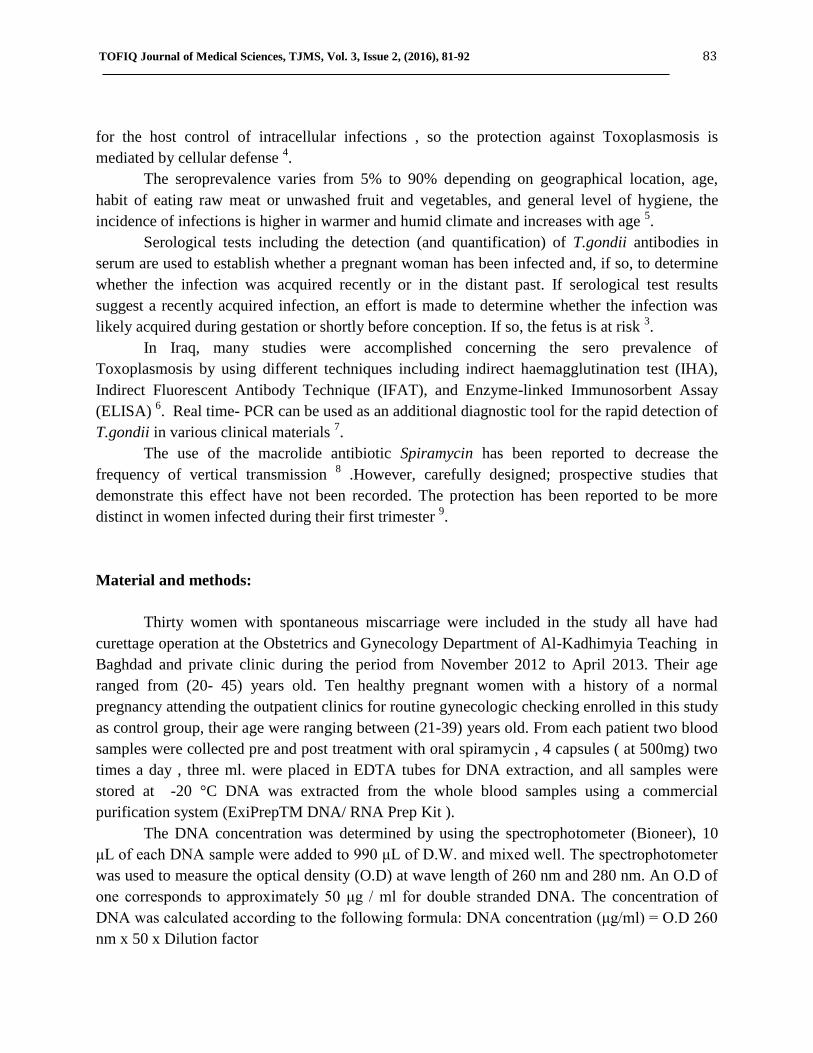
TOFIQ Journal of Medical Sciences, TJMS, Vol. 3, Issue 2, (2016), 81-92 83
for the host control of intracellular infections , so the protection against Toxoplasmosis is
mediated by cellular defense 4.
The seroprevalence varies from 5% to 90% depending on geographical location, age,
habit of eating raw meat or unwashed fruit and vegetables, and general level of hygiene, the
incidence of infections is higher in warmer and humid climate and increases with age 5.
Serological tests including the detection (and quantification) of T.gondii antibodies in
serum are used to establish whether a pregnant woman has been infected and, if so, to determine
whether the infection was acquired recently or in the distant past. If serological test results
suggest a recently acquired infection, an effort is made to determine whether the infection was
likely acquired during gestation or shortly before conception. If so, the fetus is at risk 3.
In Iraq, many studies were accomplished concerning the sero prevalence of
Toxoplasmosis by using different techniques including indirect haemagglutination test (IHA),
Indirect Fluorescent Antibody Technique (IFAT), and Enzyme-linked Immunosorbent Assay
(ELISA) 6. Real time- PCR can be used as an additional diagnostic tool for the rapid detection of
T.gondii in various clinical materials 7.
The use of the macrolide antibiotic Spiramycin has been reported to decrease the
frequency of vertical transmission 8 .However, carefully designed; prospective studies that
demonstrate this effect have not been recorded. The protection has been reported to be more
distinct in women infected during their first trimester 9.
Material and methods:
Thirty women with spontaneous miscarriage were included in the study all have had
curettage operation at the Obstetrics and Gynecology Department of Al-Kadhimyia Teaching in
Baghdad and private clinic during the period from November 2012 to April 2013. Their age
ranged from (20- 45) years old. Ten healthy pregnant women with a history of a normal
pregnancy attending the outpatient clinics for routine gynecologic checking enrolled in this study
as control group, their age were ranging between (21-39) years old. From each patient two blood
samples were collected pre and post treatment with oral spiramycin , 4 capsules ( at 500mg) two
times a day , three ml. were placed in EDTA tubes for DNA extraction, and all samples were
stored at -20 °C DNA was extracted from the whole blood samples using a commercial
purification system (ExiPrepTM DNA/ RNA Prep Kit ).
The DNA concentration was determined by using the spectrophotometer (Bioneer), 10
μL of each DNA sample were added to 990 μL of D.W. and mixed well. The spectrophotometer
was used to measure the optical density (O.D) at wave length of 260 nm and 280 nm. An O.D of
one corresponds to approximately 50 μg / ml for double stranded DNA. The concentration of
DNA was calculated according to the following formula: DNA concentration (μg/ml) = O.D 260
nm x 50 x Dilution factor

TOFIQ Journal of Medical Sciences, TJMS, Vol. 3, Issue 2, (2016), 81-92 84
The spectrophotometer was used also to estimate the DNA purity ratio according to this
following formula: DNA purity ratio = O.D 260 nm / O.D 280 nm.
Amplification of B1-gene by Real-Time Polymerase chain reaction
Principle
TaqMan probes are hydrolysis probes that are designed to increase the specificity of
quantitative PCR. The principle of this technique relies on the 5´–3´ exonuclease activity of Taq
polymerase to cleave a dual-labeled probe during hybridization to the complementary target
sequence and fluorophores-based detection 10
.
Toxoplasma gondii Real-TM PCR kit uses “hot-start”, (Anatolia geneworks- Turkey)
which greatly reduces the frequency of nonspecifically primed reactions. The forward and
reverse sequences were (GCATTCCCGTCCAAACT and
AGACTGTACGGAATGGAGACGAA) respectively 11
.
The control positive DNA copy number was determined as follows: copy number (copies
/ml) =(C x Avogadro constant) / (660 x control positive DNA in bp 109) Where C (copy) is given
in ng/ml, Avogadro constant (NA) = 6.022 140 857(74) ×1023
/mole.
Preparing the PCR:
Real-time PCR assays were carried out in duplicate and performed following
manufactured protocol in a total volume of (41µl) for one reactions as follow PCR Mix (20µl),
(1.6µl) from detection Mix 1 which contains forward and reverse primers and dual-labeled
probe, (1.2µl) from detection mix 2 which contains internal control-specific forward and reverse
primers and dual-labeled probe, Internal control (0.2µl).Twenty three microliter of this master
mix was added in the PCR tube with 18µl of DNA (sample, positive or negative control). Tubes
were closed well and spin, The PCRs were performed with the Agilent Real-time PCR (Techne-
UK). After initial activation of hotStar Taq DNA polymerase at 94°C for 2 min, 40 PCR cycles
of 93°C for 10 s , 60°C for 10 s and 72 for 30s were performed.
Calculation of results:
Data were analyzed by smart-cycler software using arithmetic baseline adjustment,
T.gondii DNA copy number was estimated from the cross point threshold relative to positive
standard. The standard curve correlates each copy number with a particular Ct, the copy number
value of the unknown samples are driven from the standard curve (According to this study).

TOFIQ Journal of Medical Sciences, TJMS, Vol. 3, Issue 2, (2016), 81-92 85
Statistical Analysis:
Experimental data were presented in terms of observed numbers and percentage
frequencies, and then analyzed by using Chi- square (χ2) test. P value ≤ 0.05 was considered
statistically significant
Results:
The detection of B1 gene of T.gondii in whole blood specimens from aborted women's and
control group by Real-time PCR:
In this study, the absolute quantification method was used for data analysis to determine the
actual copy number before and after treatment, the standard curve was generated, in which
concentration is fixed to a specific Ct value as the Ct value is inversely related to the amount of
starting template.
Standard curve generation:
A standard curve was generated using a 10-fold dilution of a template amplified on the Cycler -
real-time system (Figure 1), the copy number and CT value correlation was 0.9987.
Figure (1): Standard curve for quantitation of T.gondii tachyzoites. (Serial dilution ranging from
(100– 108) copies used as template for real time PCR analysis)

TOFIQ Journal of Medical Sciences, TJMS, Vol. 3, Issue 2, (2016), 81-92 86
Parasite load in abortive women’s as determined by Real- time PCR:
The range of T.gondii load in abortive women's under study before and after treatment with oral
spiramycin , 4 capsules ( at 500mg) two times a day in table (1 ) which is found to be
(0.0192X10⁸ – 0.110X10
4 ) before treatment and (0.156X10
7- 0.036X100 ) after treatment,( P
value =0.002). Data of this study revealed that there is significant difference(P=0.002) in the
mean of the copy numbers in aborted women’s, results in our study revealed that the mean of
parasite copy number in patients samples before treatment was (4.24E+06) and the mean of copy
number after treatment was 7.53E+05) figure(2)
Figure (2): Mean of copy number parasite /ml of patients' blood samples before and after
treatment
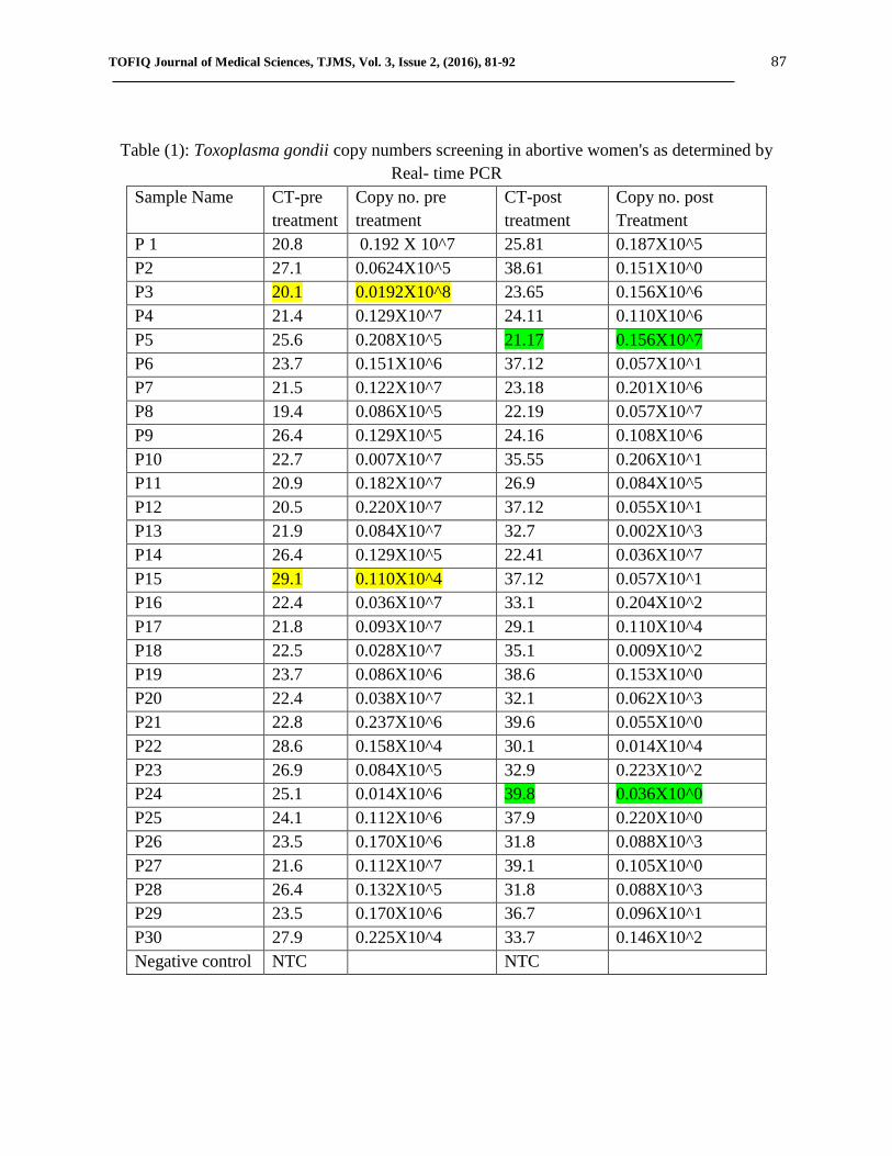
TOFIQ Journal of Medical Sciences, TJMS, Vol. 3, Issue 2, (2016), 81-92 87
Table (1): Toxoplasma gondii copy numbers screening in abortive women's as determined by
Real- time PCR
Sample Name CT-pre
treatment
Copy no. pre
treatment
CT-post
treatment
Copy no. post
Treatment
P 1 20.8 0.192 X 10^7 25.81 0.187X10^5
P2 27.1 0.0624X10^5 38.61 0.151X10^0
P3 20.1 0.0192X10^8 23.65 0.156X10^6
P4 21.4 0.129X10^7 24.11 0.110X10^6
P5 25.6 0.208X10^5 21.17 0.156X10^7
P6 23.7 0.151X10^6 37.12 0.057X10^1
P7 21.5 0.122X10^7 23.18 0.201X10^6
P8 19.4 0.086X10^5 22.19 0.057X10^7
P9 26.4 0.129X10^5 24.16 0.108X10^6
P10 22.7 0.007X10^7 35.55 0.206X10^1
P11 20.9 0.182X10^7 26.9 0.084X10^5
P12 20.5 0.220X10^7 37.12 0.055X10^1
P13 21.9 0.084X10^7 32.7 0.002X10^3
P14 26.4 0.129X10^5 22.41 0.036X10^7
P15 29.1 0.110X10^4 37.12 0.057X10^1
P16 22.4 0.036X10^7 33.1 0.204X10^2
P17 21.8 0.093X10^7 29.1 0.110X10^4
P18 22.5 0.028X10^7 35.1 0.009X10^2
P19 23.7 0.086X10^6 38.6 0.153X10^0
P20 22.4 0.038X10^7 32.1 0.062X10^3
P21 22.8 0.237X10^6 39.6 0.055X10^0
P22 28.6 0.158X10^4 30.1 0.014X10^4
P23 26.9 0.084X10^5 32.9 0.223X10^2
P24 25.1 0.014X10^6 39.8 0.036X10^0
P25 24.1 0.112X10^6 37.9 0.220X10^0
P26 23.5 0.170X10^6 31.8 0.088X10^3
P27 21.6 0.112X10^7 39.1 0.105X10^0
P28 26.4 0.132X10^5 31.8 0.088X10^3
P29 23.5 0.170X10^6 36.7 0.096X10^1
P30 27.9 0.225X10^4 33.7 0.146X10^2
Negative control NTC NTC

TOFIQ Journal of Medical Sciences, TJMS, Vol. 3, Issue 2, (2016), 81-92 88
Regarding response to treatments toxoplasmosis infected women in this study were divided to
the following groups:
Group I include patients had high parasite load (0.062X10⁵-0.112X10
7) which decrease
completely after completion the course of treatment (0.036X100-0.223X100).
Group II include a patients with high parasite load (0.225x10⁴ -0.036x10
7 ) at time of diagnosis
and decreases after completion the course of treatment (0.055x101 -0.223x10
1).
Group III includes patients with high parasite load at diagnosis (0.225x10⁴ -0.036x10
7) which
still with high parasite load or slightly lower (0.002x10³ – 0.201 x10
6).
Group IV includes patients with high parasite load (0.158x10⁴ – 0.129x10
5) at time of diagnosis
which showed elevated parasite load after treatment (0.014x10⁴ -0.156x10
7).
Discussion:
Firstly DNA was successfully extracted from whole blood samples for all subjects,
Menotti, et al 12
showed that the parasites are as likely to be found in the plasma or red cell
fractions, which might indicate that the most suitable sample is a whole blood extract. However,
in patient's blood T.gondii present might be intracellular in leukocytes 13
; in that case the white
cell fraction would be as appropriate as whole blood.
Parasitemia in pregnant women may be due to the toxoplasmosis reactivations occur
during immunosuppressive states in pregnancy. Tachyzoites or T.gondii DNA release from the
cyst in the body tissues may be the source of parasitemia or T.gondii DNA in blood 14
.
Quantitative detection of pathogen by real time PCR has been successfully applied to a variety of
specimens and the finding of T.gondii DNA in peripheral blood by PCR might help in the early
diagnosis of invasive diseases 15
.
The qRT-PCR that have been applied in this study to diagnose and for the determination
of copy number of T.gondii DNA in whole blood, it gives reproducible diagnostic and
quantitative results over conventional PCR with the risk of false positive due to contamination
with previously amplified products since in qRT-PCR there is no risk of hand manipulation and
the use of internal control will exclude the false negative which due to inhibiters in amplified
mixture, reproducibility of qRT-PCR quantification was tested by the testing of the 10 fold
dilution.
In the present study a qRT-PCR based B1 gene – specific Taq man assay have been
developed for quantitative detection of T.gondii which extremely sensitive to (0.168 parasite /
reaction) and its ability to quantify infection load of clinical specimen and its long linear range of
7 logs of DNA concentration with CT value as shown in figure (1) where R2=0.998 which is
indicative of a significant correlation between logarithmic value of each PCR product of copy
number amplified and CT value. These results were in agreement with Mei-huilin, (16)
. In this
assay an increase of the fluorescent signal above the preset threshold within 40 PCR cycle was
considered positive (CT< 40), Table (1) where CT for negative control was 40. Quantification of

TOFIQ Journal of Medical Sciences, TJMS, Vol. 3, Issue 2, (2016), 81-92 89
parasite load in pregnant women under study has been used to ass’s disease severity and the
treatment outcome.
The relative quantity of tachyzoites in each DNA sample was determined using the
standard curve presented in Figure (2) which showed a range between (0.110X10^4 to 0.0192
×108) before treatment and (0.036×100 to 0.156×10
7) after treatment .In addition figure (2)
revealed that there is significance difference (P<0.05) of in the mean of the copy number before
and after treatment.
According to their response to treatments, Group 1 patients which represent, 5 (16.66%)
% of patients having high parasite load at time of diagnosis with a range of (0.062X10⁵-
0.112X10⁷) copies /ml , they show complete response to treatment with decline in parasite load
to undetectable value , these results were in accordance with Costa , et al 17
who recorded that
the parasite count decreased sharply in patients having cerebral toxoplasmosis symptoms , when
treated with anti-toxoplasma therapy which was efficient and reducing the parasite load and they
conclude that Light Cycler PCR provide precise evaluation of parasite load in
immunocompromised patients and PCR is useful in monitoring of treatments .
In addition, Kupferschmidt, 18
conclude that decreasing in parasite load in patient's blood
sample demonstrated that treatment used was effective. Contini , et al 19
recorded that patient
under specific treatment did not show detectable B1 DNA levels in samples collected after many
days of therapy . So that T.gondii DNA load is a valuable tool for monitoring of toxoplasmosis.
Group Π patients which represent 10(33.33 %) of patients having high parasite load at time of
diagnosis with a range of (0.225x10⁴-0.036x10
7) copies /ml, they show lower parasite load with
a range of (0.055x101
-0.223x101) copies /ml after treatment. These results observed by Menotti,
et al 12
.
They consider that prolonged persistent detection of T. gondii DNA in blood justifies the
maintenance of anti T.gondii treatment and close follow up by PCR. These results were in
agreement with Romand, et al 20
who used quantitative Real time PCR to evaluate T. gondii
concentration in amniotic fluid and conclude that quantitative PCR may help the clinician to
form prognosis of congenital infections with regard to fetal and neonatal outcome. Other study
by Christianne, et al 21
reported that high parasite load in amniotic fluid was found in cases of
severe congenital toxoplasmosis as determined by quantitative PCR.
Group III which represent 11(36.66 %) include patients with high parasite load at time of
diagnosis with a range of (0.038x10⁷ –0.019x10
8) copies/ml, they did not show any response to
treatment, parasite load was remained with undetectable decrease. After treatment it's found to be
(0.002x10³ – 0.201 x10
6) copies/ ml. These results were in agreement with Kupferschmidt, et al
(18) .who used Tag man PCR to monitor parasite load in the blood of an immunocompromised
patients after allogenenetic bone marrow transplantation. They found that at day 199 after
therapy with pyrimethamine the detected parasite load remained unchanged and then decreased
after prolonged therapy.
TOFIQ Journal of Medical Sciences, TJMS, Vol. 3, Issue 2, (2016), 81-92 90
Regarding group IV of patients which represent 4(13.33 %) having high parasite load at
time of diagnosis with a range of (0.158x10⁴ – 0.129x10
5) copies /ml , they showed elevated
parasite load after treatment with a range of (0.014x10⁴ -0.156x10
7) copies /ml .This increase of
parasite load seems to be correlated to progression of disease or to poor response to treatment.
These results were in agreement with Menotti, et al 12
who recorded evolution in parasite load in
patients under treatments and those with slow decrease in parasite load after initiation of
treatments with pyrimethamine and sulfadiazine on day 43 post transplantation. They give
explanation to the prolonged presence of T.gondii DNA that could be due to disseminated form
of disease with a peak of parasitemia (29000 tachyzoites/ ml) of blood buffy coat.
Real time-PCR can be used as an additional diagnostic tool for the rapid detection of
T.gondii in various clinical materials. Studies have documented that Real time- PCR can actually
detect T.gondii in blood specimens of women before or during pregnancy. Nimri, et al 22
and
Khan et al 23
showed that a small numbers of parasites may be detected in circulating blood from
some patients with acute or recurrent toxoplasmosis.
Several studies have reported that, PCR could detect parasitemia a few weeks prior to the
appearance of any clinical signs or symptoms .PCR is highly sensitive and specific because a
single tachyozoite can be detected in a clinical sample 24
.
The most important records in this study is that the parasite load could be determined and
correlated with treatment. The parasite quantification in some treated subjects become lower and
in others, T.gondii load disappeared when spiramycin was given, this exhibit that the anti-
toxoplasma therapy was efficient in reducing the parasite load.
The quantitative real-time PCR assay developed in this study can be used not only to
detect the presence of T.gondii DNA but also to provide precise evaluations of the parasite load
in pregnant women’s. This PCR test should be useful for the monitoring of treatment efficacy
and should help provide an understanding of the pathogenesis of T.gondii reactivation.

TOFIQ Journal of Medical Sciences, TJMS, Vol. 3, Issue 2, (2016), 81-92 91
Reference:
1- Dubey JP, Hill DE, Jones JL, 2005 ; Prevalence of viable Toxoplasma gondii in beef, chicken, and
pork from retail meat stores in the United States: risk assessment to consumers. J Parasitol, (
91)1082–93.
2- Dubey JP, Su C, 2009; Population biology of Toxoplasma gondii: what's out and where did they
come from. Journal of MemInst Oswaldo Cruz, 104 (2) 190-1951.
3- Linguissi L, 2012; Seroprevalence of toxoplasmosis and rubella in pregnant women attending
antenatal private clinic at Ouagadougou, Burkina Faso.Asian Pac J. Trop Med, 5(10):810-813.
4- Joanna M., Violetta D., Halina K, 2009; Does Toxoplasma gondii Infection affect the Levels of
IgE and Cytokines (IL-5, IL-6, IL-10, IL-12, and TNF-alpha)? Clin Dev Immunol. (15) 374-396.
5- Evering, T. and Weiss, L.M, 2006; The immunology of parasite infections in
immunocompromised hosts. Parasite Immunolology, ( 28) 549-565.
6- Rai R., Clifford K., Cohen H., Regan L, 1995; High prospective fetal loss rate in untreated
pregnancies of women with recurrent miscarriage and antiphospholipid antibodies. Hum. Reprod,
(10)3301–3304.
7- Su C, Shwab EK, Zhou P, Zhu XQ, Dubey JP, 2010; Moving towards an integrated approach to
molecular detection and identification of Toxoplasma gondii. Parasitol J, 137(1) 1-11.
8- Rovamycine (Rhone-Poulenc Rorer), 1994; In: Vidal. 70th ed. Paris: Editions du Vidal, 1361.
9- Sebastian D., Zuhara K. andSekaran K. 2008; Influence of TORCH infections in first trimester
miscarriage in the Malabar region of Kerala. African J. Microbio. Res., ( 2) 056-059.
10- Holland PM, Abramson RD, Watson R, Gelfand DH, 1991; Detection of specific polymerase
chain reaction product by utilizing the 5’----3' exonuclease activity of Thermus aquaticus DNA
polymerase. Proc Natl Acad Sci U S A., (16):7276–80.
11- Edvinsson B, Lappalainen M, Evengård B, 2006; Real-time PCR targeting a 529-bp repeat
element for diagnosis of toxoplasmosis. Clin Microbiol Infect, ( 12) 131-136.
12- Menotti J, Garin YJ, Thulliez P, 2010; Evaluation of a new 5'-nuclease real-time PCR assay
targeting the Toxoplasma gondii AF146527 genomic repeat. Clin Microbiol. Infect, (4)363-368.
13- Ho-Yen D.O., JossA.W.,BalfourA.H., Smyth E.T.,Baird D, 1992; Use of the polymerase chain
reaction to detect Toxoplasma gondii in human blood samples. J Clin Pathol., 45(10): 910 – 913.
14- Dubey JP, Chellaiah R, Costa DG, Ferreira L, Kwok O, 2010; New Toxoplasma gondii
Genotypes Isolated from Free-Range Chickens from the Fernando De Noronha, Brazil:
Unexpected Findings. J Parasitol, ( 96)709–712.
15- Bretagne S, Costa JM, Foulet F,V Prospective study of toxoplasma reactivation by polymerase
chain reaction in allogeneic stem-cell transplant recipients. Transpl Infect Dis, ( 2)127-32.
16- Mei-Hui Lin, Tse-chingchen,Tesng Tong Kuo,chingchung Tseng, 2000; Real time PCR detection
of T.gondii .J.clin.Microbiol, 38(11):4121-4125.
17- Costa, J. M., Pautas, C., Ernault, P., Foulet, F., Cordonnier, C, 2000; Real-time PCR for diagnosis
and follow-up of Toxoplasma reactivation after allogeneic stem cell transplantation using
fluorescence resonance energy transfer hybridization probes. J Clin Microbiol, 38(8): 2929–2932.

TOFIQ Journal of Medical Sciences, TJMS, Vol. 3, Issue 2, (2016), 81-92 92
18- Kupferschmidt O, Krüger D, Held TK, Ellerbrok H, Siegert W, Janitschke K, 2001; J. Clin
Microbiol Infect, 7(3):120-4.
19- Contini C, Seraceni, Cultrea R, Incorvaia, Sebastiani A, 2005; Evaluation of a Real-time PCR-
based assay using the light cycler. International Journal for Parasitology, 35 (3):275–283.
20- Romand, S., Chosson, M., Franck, J., Wallon, M., Kieffer, F, 2004; Usefulness of quantitative
polymerase chain reaction in amniotic fluid as early prognostic marker of fetal infection with
Toxoplasma gondii. Am J Obstet Gynecol, ( 190)797-802.
21- Christianne T, Pedro A, Brasil A, Letícia G, Maria E, 2016; Performance of Polymerase Chain
Reaction Analysis of the Amniotic Fluid of Pregnant Women for Diagnosis of Congenital
Toxoplasmosis, PLOS ONE | DOI:10.1371/journal.pone , (7) 149-153
22- Nimri, L. Pelloux, H. and Elkhatib, L, 2004; Detection of Toxoplasma gondii DNA and specific
antibodies in high- risk pregnant women. Am. J. Trop. Med. Hyg. 71(6): 831-835.
23- Khan A, Jordan C, Muccioli C, 2006; Genetic divergence of Toxoplasma gondii strains associated
with ocular toxoplasmosis, Brazil. Emerg Infec Dis. (12) 942–9 45
24- Guy, E. C., Pelloux, H., Lappalainen, M., Aspock, H., Hassl, 1996; Interlaboratory comparison of
polymerase chain reaction for the detection of Toxoplasma gondii DNA added to samples of
amniotic fluid. Eur J Clin Microbiol Infect Dis, (15) 836-839.
