ventricular septal defect
DESCRIPTION
VSDTRANSCRIPT

7/16/2019 Ventricular Septal Defect
http://slidepdf.com/reader/full/ventricular-septal-defect-5634f8f73efc5 1/12
CHAPTER I
INTRODUCTION
Sistem kardiovaskular menjalankan fungsinya melalui organ jantung dan pembuluhdarah. Dimana organ yang memiliki peranan penting dalam hal ini adalah jantung yang juga
merupakan organ besar dalam tubuh. Jantung adalah organ berupa otot berbentuk kerucut.
Fungsi utama jantung adalah untuk memompakan darah ke seluruh tubuh dengan cara
mengembang dan menguncup yang disebabkan oleh karena adanya rangsangan yang berasal dari
susunan saraf otonom. Seperti pada organ-organ yang lain, jantung juga dapat mengalami
kelainan ataupun disfungsi.
Penyakit jantung dapat dibedakan dalam dua kelompok, yaitu penyakit jantung didapat
dan penyakit jantung bawaan. Penyakit jantung bawaan adalah kelainan struktural jantung yang
kemungkinan terjadi sejak lahir dan beberapa waktu setelah bayi dilahirkan. Salah satu jenis
penyakit jantung yang tergolong penyakit jantung bawaan adalah VSD yang paling sering
ditemukan, yaitu 30% dari semua jenis penyakit jantung bawaan. Pada sebagian kasus, diagnosis
kelainan ini ditegakkan setelah melewati masa neonates, karena pada minggu-minggu pertama
bising yang bermakna biasanya belum terdengar oleh karena resistensi vascular paru masih
tinggi dan akan menurun setelah 8-10 minggu.

7/16/2019 Ventricular Septal Defect
http://slidepdf.com/reader/full/ventricular-septal-defect-5634f8f73efc5 2/12
CHAPTER II
CONTENT
2.1 Definition
Supracristal (or doubly committed) ventricular septal defect (VSD) is the least common type of
VSD in the Western Hemisphere, accounting for approximately 5-7% of such defects in this part
of the world, including in the United States. The location of the supracristal VSD, with its close
proximity to the aortic root, accounts for the common development of aortic insufficiency with
this defect. Left-to-right shunting of blood through the defect is believed to progressively pull
aortic valve tissue (especially the right coronary cusp) through a Venturi effect (see the image
below).
Parasternal long-axis echocardiogram view showing supracristal
ventricular septal defect (arrow) with buckling and prolapse (***) of the right coronary leaflet of
the aortic valve.
The crista supraventricularis can be considered synonymous with the infundibular (or conus)
ventricular septum. It is the portion of the septum that separates the tricuspid and pulmonary
valves. Defects above this part of the septum are referred to as supracristal defects. The term is
generally reserved for defects lying immediately under the pulmonary valve. A defect, usually
small, may occur within the conus septum, and, technically can be termed supracristal. The
spiraling course of the ventricular septum may make diagnosis of supracristal VSD more
difficult
7/16/2019 Ventricular Septal Defect
http://slidepdf.com/reader/full/ventricular-septal-defect-5634f8f73efc5 3/12
The patient’s risk of developing infective endocarditis is higher for supracristal VSD with aortic
insufficiency than it is for an isolated VSD. Patients and families should be educated on the
importance of good oral and dental hygiene. Routine prophylaxis for dental or surgical
procedures is no longer recommended unless there has been a prior episode of endocarditis.
2.2 Anatomy
The infundibular (or conus) septum separates the tricuspid and pulmonary valves and
accounts for the more superior placement of the pulmonary valve relative to the aortic valve.
This portion of the septum also provides fairly rigid, muscular support for the aortic valve,
especially the right coronary cusp. Numerous synonyms indicate the confusion often associated
with describing supracristal ventricular septal defects (VSDs). The term supracristal may bemisleading because the entire conus septum (or a major portion of the septum) may be missing.
The term is commonly used, however, emphasizing the superior location of the defect along with
its close approximation to the aortic and pulmonary valve leaflets. Lack of support for the right
aortic leaflet is crucial to the natural history of this type of VSD.
The plane of the conus septum in the right ventricular outflow tract lies almost
perpendicular to that of the remainder of the septum. From a surgical perspective, a defect lying
in the conus septum may not be visualized from the standard right atriotomy approach, looking
through the tricuspid valve.Unlike the more common perimembranous type of VSD, the
supracristal VSD does not lie near the tricuspid valve. Unless the supracristal defect is large,
extending inferiorly to the perimembranous septum, the tricuspid valve is not involved in partial
closure of the defect. Conduction system tissue lies inferior to the supracristal VSD. The
conduction system may lie closer to a larger defect that crosses from the outlet septum into the
perimembranous area.
2.3 Pathophysiology
The natural history of supracristal ventricular septal defects (VSDs) depends on the
location and size of the defect. Patients with small, isolated supracristal VSDs may have no
symptoms or signs of congestive failure such as might be observed with a large shunt.

7/16/2019 Ventricular Septal Defect
http://slidepdf.com/reader/full/ventricular-septal-defect-5634f8f73efc5 4/12
Progressive aortic insufficiency may develop late in the first decade of life. Larger defects of the
outlet septum frequently are associated with forms of aortic outflow obstruction (eg, coarctation,
interrupted aortic arch). In such cases, symptoms of congestive heart failure and possible
circulatory collapse appear early. Patients with larger, isolated supracristal VSDs may develop
congestive heart failure early in infancy due to a large left-to-right shunt. While spontaneous
closure is not common, a decrease in the magnitude of the left-to-right shunt may occur due to
progressive prolapse into the defect of aortic valve tissue (the right coronary cusp or, possibly,
the right sinus of Valsalva. This valve leaflet prolapse is believed to result from the Venturi
effect, as the high-velocity shunt flow produces negative pressure. Progressive distortion of the
aortic leaflet or sinus may lead to increasing aortic valve insufficiency.
2.4 Etiology
The muscular outlet septum is primarily formed from the proximal endocardial ridges
(similar to endocardial cushion tissue). Semilunar valve tissue and the actual connection between
the septum and the arteries are formed by the more distal endocardial ridges. Extracardiac
mesenchyme, derived from neural crest tissue, condenses as prongs (which act as a welding
agent) with the most superior portion of the distal cushions to form the aortopulmonary septum.
By exposing neural crest tissue to homocysteine, supracristal ventricular septal defects (VSDs)
have been induced in a high percentage of chick embryos. Disruption of apoptosis and
myocardialization has been proposed to explain these findings. The frequent association between
arch abnormalities and significant conal VSDs suggests a common mechanism involving a
chromosome band 22q11 microdeletion. Deletions in this area have not been linked with isolated
supracristal VSDs.
2.5 Epidemology
As stated above, supracristal ventricular septal defect (VSD) accounts for approximately
5-7% of VSDs in the Western Hemisphere. In the Eastern Hemisphere, however, the incidence of
this condition is much higher, reaching 25% of all VSDs in patients from this part of the world,
as supracristal VSDs are much more common in persons of Asian descent than in individuals of
other races. Although the overall incidence of VSDs is no greater in Asians than in other groups,

7/16/2019 Ventricular Septal Defect
http://slidepdf.com/reader/full/ventricular-septal-defect-5634f8f73efc5 5/12
supracristal VSDs account for approximately 30% of VSDs in Asians. Higher occurrence of the
condition in this population has not been adequately explained, but one may assume that it is
genetically determined.
2.6 Clinical Presentation
In patients with supracristal ventricular septal defects (VSDs), symptoms and severity are
a function of the size and location of the defect, the relative systemic and pulmonary vascular
resistances, and the presence of associated abnormalities. Symptoms may range from severe
congestive failure and cardiogenic shock in patients with large conal defects and left heart
obstruction to complete absence of symptoms in patients with small, isolated defects.Exercise
intolerance and dyspnea suggest progressive aortic insufficiency, although early detection and
treatment for valve insufficiency should obviate any significant symptoms.
2.7 Physical Examination
Congestive heart failure does not occur in the patient with an isolated, small supracristal
ventricular septal defect (VSD). General examination findings remain normal, with no signs of
respiratory distress or growth failure. Infants with larger defects, especially those associated with
significant left ventricular outflow obstruction (eg, doubly committed subarterial defect withinterrupted aortic arch), may present as early as the first week of life with profound congestive
heart failure and cardiogenic shock. Infants with only a large left-to-right shunt usually develop
symptoms in the second month of life. The murmur of an isolated, small supracristal VSD is
similar to that of other types of small VSDs. While it may be loudest in the third left intercostal
space (ie, more superior than other VSDs), it begins with the first heart sound and has a similar
harsh, noisy quality. As with other types, a large defect may produce no murmur from the defect
itself. In this case, a murmur may result from turbulent flow through the pulmonic valve, thus
becoming crescendo-decrescendo in character. This murmur may radiate laterally and posteriorly
because of shunt flow directed into the branch pulmonary arteries.
Second heart sound findings depend on volume of shunt flow as well as pulmonary artery
pressure and resistance. With a small shunt, the second heart sound splits and varies normally
7/16/2019 Ventricular Septal Defect
http://slidepdf.com/reader/full/ventricular-septal-defect-5634f8f73efc5 6/12
with respiration, and the pulmonary component is normal in intensity. With a large shunt, and
elevated pulmonary artery pressure, the pulmonary component of S2 increases in intensity.
Intensity of this sound is further increased if pulmonary resistance is increased, in which case the
splitting interval of S2 is decreased. With a large left-to-right shunt, one should hear a short, low-
frequency, middiastolic apical murmur due to enhanced, rapid (passive) filling of the left
ventricle. With significantly elevated pulmonary vascular resistance, shunt flow decreases and
this diastolic murmur does not occur. When a patient is known to have a supracristal VSD,
physical examination should focus on whether aortic insufficiency is present. Blood pressure
must be carefully evaluated for pulse pressure (ie, the difference between systolic and diastolic
blood pressures) and pulse amplitude, as these increase with increasing aortic insufficiency
unless heart failure also occurs. With significant aortic insufficiency, the aortic component of S2
decreases in intensity. If left ventricular end diastolic pressure increases, left atrial pressure
increases, thus causing an increase in intensity of the pulmonic component of S2. Aortic
insufficiency causes a high-pitched diastolic murmur beginning with the aortic component of the
second heart sound. It is best heard along the left sternal border, usually in the third left
intercostal space at the sternal edge.
The combined systolic and diastolic murmurs of supracristal VSD with aortic
insufficiency may be likened to the sound of sawing wood. This systolic-diastolic murmur
combination should not be misinterpreted as a continuous murmur (eg, patent ductus arteriosus,
arteriovenous malformation or fistula). Significant aortic insufficiency may cause a late diastolic
murmur at the apex resulting from atrial contraction augmenting late ventricular filling. This is
the Austin Flint murmur.
2.8 Supporting Examination
The recurrence risk for the offspring of mothers with supracristal ventricular septal defect(VSD) is estimated at 4-5%; the recurrence risk for the offspring of fathers with the condition is
approximately 2-3%. Detailed prenatal fetal echocardiography (ECHO) may be indicated.
Supracristal VSD cannot be identified from a routine prenatal 4-chamber view.

7/16/2019 Ventricular Septal Defect
http://slidepdf.com/reader/full/ventricular-septal-defect-5634f8f73efc5 7/12
Electrocardiographic findings may be normal in infancy, because the defect may not be large
enough to cause a significant left-to-right shunt and ventricular hypertrophy. With larger defects,
the electrocardiogram (ECG) may show left atrial enlargement, as well as both left and right
ventricular hypertrophy. With progressive aortic insufficiency in the older child or adult,
electrocardiography usually reveals evidence of left heart enlargement from volume overload (ie,
left atrial enlargement and left ventricular hypertrophy [tall R waves in the left precordium with
or without ST-T changes]). A diagnostic pitfall associated with supracristal VSD is the failure to
diagnose the condition adequately and, therefore, failure to recognize the potential for aortic
valve involvement.
2.8.1 Imaging Studies
Chest radiography
Chest radiography is normal in infancy if the left-to-right shunt is small. If a large shunt is
present, cardiomegaly (left heart enlargement, both the left atrium and the left ventricle) with
increased pulmonary vascularity from increased pulmonary blood flow may be observed.
Radiography in the older child or adult with progressive aortic insufficiency may reveal left heart
enlargement (particularly left ventricular enlargement) and prominence of the ascending aorta.Shunt volume is generally smaller, thus pulmonary arterial vascularity is generally normal.
Advanced left heart failure produces pulmonary edema.
Two-dimensional transthoracic echocardiography
Echocardiography (ECHO) provides the most efficient means to diagnose supracristal ventricular
septal defect (VSD) accurately (see the image below) and the most effective means to monitor
progressive aortic insufficiency.[7]
An accurate diagnosis can generally be made in infants and
children with standard transthoracic ECHO examination findings. In the older child and adult,
transthoracic ECHO findings may be inconclusive; in such cases, transesophageal ECHO may be
extremely helpful.

7/16/2019 Ventricular Septal Defect
http://slidepdf.com/reader/full/ventricular-septal-defect-5634f8f73efc5 8/12
Parasternal long-axis echocardiogram view showing supracristal
ventricular septal defect (arrow) with buckling and prolapse (***) of the right coronary leaflet of
the aortic valve.
Two-dimensional (2D) imaging reveals the supracristal VSD in the parasternal short-axis view or
the modified apical 3-chamber view (ie, left atrium, left ventricle, aortic root, and pulmonaryroot, equivalent to the transesophageal view with transducer at 90°). The defect can also be
observed well in the subcostal parasagittal view (ie, visualizing the pulmonary and aortic outflow
tracts).
A supracristal VSD cannot be imaged from the apical 4-chamber view because of the orientation
of the outlet septum. Distortion of the right aortic leaflet may be the only clue to the presence of
a significant supracristal VSD, because the aortic leaflet may obstruct the defect.
Color Doppler echocardiography
Color Doppler examination using the parasternal short-axis view reveals left-to-right shunting
with turbulent flow directed into the pulmonary outflow tract and often across the pulmonary
valve. This turbulence may be confused with pulmonary stenosis; however, careful, slow-motion
review of color flow results (with electrocardiographic timing) may reveal the early appearance
of turbulent flow below the pulmonary valve.
7/16/2019 Ventricular Septal Defect
http://slidepdf.com/reader/full/ventricular-septal-defect-5634f8f73efc5 9/12
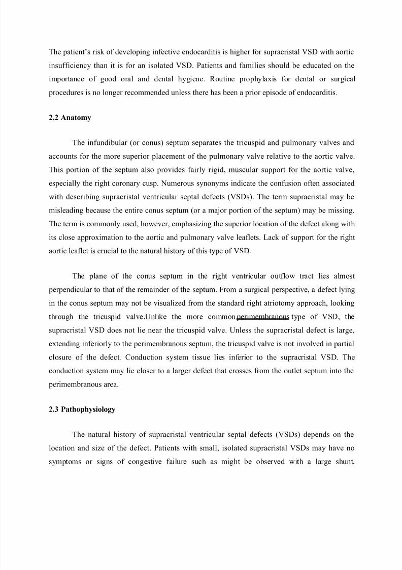
Parasternal short-axis echocardiogram view with color Doppler
showing proximity of ventricular septal defect jet to the aortic and pulmonic valves. The patient
is an infant with neither aortic valve prolapse nor aortic insufficiency.
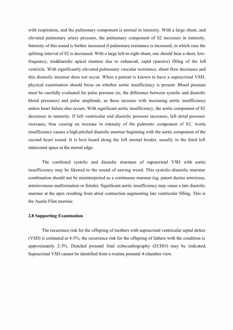
Subcostal "right ventricular inflow/outflow" view showing the
close relationship between the aortic and pulmonic valves in the presence of supracristal
ventricular septal defect. Turbulent shunt flow is shown directed into the main pulmonary artery.
The patient is an infant with neither aortic valve prolapse nor insufficiency.
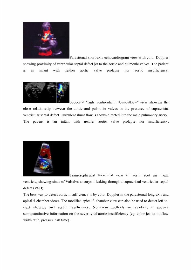
Transesophageal horizontal view of aortic root and right
ventricle, showing sinus of Valsalva aneurysm leaking through a supracristal ventricular septal
defect (VSD)The best way to detect aortic insufficiency is by color Doppler in the parasternal long-axis and
apical 5-chamber views. The modified apical 3-chamber view can also be used to detect left-to-
right shunting and aortic insufficiency. Numerous methods are available to provide
semiquantitative information on the severity of aortic insufficiency (eg, color jet–to–outflow
width ratio, pressure half time).

7/16/2019 Ventricular Septal Defect
http://slidepdf.com/reader/full/ventricular-septal-defect-5634f8f73efc5 10/12
The best way to identify progression of aortic insufficiency by echocardiography is by serial
comparison of left ventricular systolic and diastolic dimensions and ventricular function
(shortening fraction or ejection fraction). Progressive left atrial enlargement can be a sign of
ventricular diastolic dysfunction.
Three-dimensional echocardiography
Three-dimensional (3D) echocardiographic imaging of VSDs closely correlates with surgical
findings, although specific findings with supracristal defects have not been reported.Three-
dimensional echocardiography may prove useful in differentiating supracristal VSD from
unruptured sinus of Valsalva aneurysm.
Angiography
Supracristal VSD is best defined in the right anterior oblique projection or in the cranially tilted
left anterior oblique projection. Small supracristal defects may not be identified in the standard
long-axial oblique projection because of rotation of the septum. Distortion of an aortic valve
cusp may be the only clue to a supracristal VSD of significant size, even though the apparent
volume of the left-to-right shunt may be small.
Magnetic resonance imaging
Magnetic resonance imaging (MRI) may be used with appropriate projections and alignment to
show the pulmonary outflow tract. Serial MRI studies can be helpful in that they do not expose
the patient to ionizing radiation. Blood flow studies can be used to provide quantitative
information on regurgitant volume in the assessment of aortic insufficiency.
2.8.2 Catherization
Cardiac catheterization can quantify shunt volume and pulmonary arterial
resistance. Step-up in oxygen saturation may be detected in the pulmonary artery rather than in
the right ventricular cavity because of streaming of the shunted blood into the pulmonic trunk. If

7/16/2019 Ventricular Septal Defect
http://slidepdf.com/reader/full/ventricular-septal-defect-5634f8f73efc5 11/12
aortic valve prolapse is significant, left-to-right shunting by oximetry may be fairly
unremarkable, because the ventricular septal defect (VSD) in such cases is partially obstructed.
Postcatheterization concerns include hemorrhage, vascular disruption after balloon dilation, pain,
nausea and vomiting, and arterial or venous obstruction from thrombosis or spasm. Possible
complications also include blood vessel rupture, tachyarrhythmias, bradyarrhythmias, and
vascular occlusion.
2.9.1 Treatment
Once the diagnosis of supracristal ventricular septal defect (VSD) has been made,
carefully monitor patients for the development of aortic insufficiency. This necessitates not only
periodic physical examination with auscultation but also serial and Doppler echocardiograms,
because these diagnostic studies are more sensitive than auscultation in detecting valve
regurgitation. Because spontaneous closure is uncommon in supracristal VSDs and aortic
insufficiency is a common complication, surgical closure is recommended in most cases. Aortic
insufficiency in supracristal VSD is usually progressive and warrants an aggressive approach
with early intervention to avoid aortic valve deformity and replacement.
Aortic insufficiency caused by supracristal VSD must be differentiated from that caused
by an abnormal aortic valve (usually a bicuspid valve). Surgical intervention is usually delayed
in the latter disorder, because the abnormal aortic valve typically requires replacement rather
than repair in cases of aortic insufficiency.
2.9.2 Surgical Treatment
Because of the orientation of the right ventricular outflow tract, a surgical approach from
the right atrium may not allow adequate visualization of the ventricular septal defect (VSD).
Incision into the main pulmonary artery, which exposes the defect through the pulmonic valve,
has proved successful. Repair may be achieved with patch or suture closure, depending on the
size of the defect. Aortic valvuloplasty is often, but not always, necessary, and incision through
the aortic root can allow adequate visualization for valve repair (Trusler technique). The

7/16/2019 Ventricular Septal Defect
http://slidepdf.com/reader/full/ventricular-septal-defect-5634f8f73efc5 12/12
approach through the main pulmonary artery avoids the need for incision into the right ventricle.
Care should be taken to avoid capturing the aortic cusp into one of the patch sutures.
Intraoperative transesophageal echocardiographic monitoring can be extremely helpful in
precisely defining aortic valve prolapse and the severity of valve insufficiency, which determine
the necessity of valvuloplasty. Follow-up intraoperative assessment should be used to rule out
residual insufficiency. More extensive damage to the aortic valve from long-standing prolapse
and distortion may require valve replacement. Follow-up care after supracristal ventricular septal
defect (VSD) repair and aortic valvuloplasty is essential to ensure that the aortic insufficiency
has been corrected completely.
2.10 Prognosis
The prognosis in patients with supracristal ventricular septal defect (VSD) should be
considered good to excellent when the potential complication of aortic valve insufficiency is
recognized and aggressively treated. Delayed recognition of or surgical treatment for progressive
aortic valve insufficiency may lead to severe distortion of the aortic valve leaflet, making
eventual valve replacement more likely. Morbidity or mortality in supracristal VSD is generally
not the result of a large left-to-right shunt. Rather, it is caused by the development of aortic valve
insufficiency. When progressive and severe, this results in left ventricular enlargement and
eventual congestive heart failure, hence the admonition to address this problem early. The
appearance of aortic insufficiency as a complication of supracristal VSD is related to age. Young
infants and toddlers presenting with supracristal VSDs are more likely to have findings of left-to-
right shunt only. While it may occur earlier in infancy, onset of aortic valve prolapse and
progressive aortic insufficiency generally begins in children aged 6-10 years.Patients with
supracristal ventricular septal defect (VSD) are at increased risk of infective endocarditis. The
risk is higher if aortic valve insufficiency is present.