application of metabolomics approaches to the study of...
TRANSCRIPT

2265ISSN 1757-6180Bioanalysis (2012) 4(18), 2265–229010.4155/BIO.12.218 © 2012 Future Science Ltd
Review
Special FocuS iSSue: MetaboloMicS
Respiratory diseases: the unmet need for new biomarkers & increased understanding of pathobiologyRespiratory diseases are a major cause of global morbidity and mortality, affecting all age groups of the population [1–3]. The principal lung diseases are asthma, chronic obstructive pulmo-nary disorder (COPD), pulmonary arterial hypertension, cystic fibrosis and, in particular, a range of infectious diseases including TB. Sarcoidosis is an example of a rare immunological disease that shares certain features both with TB and autoimmune reactions. There are in fact many different immunological and inflammatory lung diseases and they are often severe and progressive, as in the case of interstitial pulmonary fibrosis and several occupational diseases, including asbestosis. There is a wealth of published work considering these diseases in detail, and accordingly, the pathology of individual respiratory diseases will not be discussed in this review. Interested readers are directed to a number of review articles that deal with these topics in detail [4–7].
Lung diseases are very prevalent as demonstrated by the following two examples. First, asthma is in fact the greatest cause of handicap among children. The lifetime risk of developing asthma is as high as for cancer and diabetes, but in contrast to the latter diseases, asthma is a burden to the health system throughout adult life, making it a major reason for diseaserelated work absenteeism among adults [8]. Second, COPD
alone is estimated to have an annual global mortality of approximately 2.7 million people, cause significant morbidity in >200 million people [9], and is on target to be the third largest cause of global mortality by 2020 [10].
While the exact mechanism(s) of disease onset are still unknown, environmental exposures are directly linked to the development of lung diseases. In addition to triggering of allergic asthma by a range of environmental allergens, including grass pollen [11], birch pollen [12,13] dust mites [14] or cat allergens [14], there are many other environmental factors that can exacerbate existing pulmonary conditions; for example, exposure to air pollution in urban areas [15–18], diesel exhaust [19], smoke from heating fires in rural areas [20], and smoke from indoor cooking fires [21,22]. Air pollution in urban areas has been shown to have a detrimental effect on the health of individuals with asthma, especially children [23–25]. Long and shortterm exposures have been shown to induce a range of atopic conditions, including wheezing [26], eczema and allergies [27], and even hospitalization [28], all of which lead to a worse quality of life for many children. In developing countries millions of people are exposed to high levels of air pollution, due to smoke from inefficient and poorly ventilated solid fuel fires (biomass or coal) [21,22,29]. The smoke from these fires has been shown to have a similar composition to tobacco smoke [30] and cause health problems including COPD [22,31], strokes [32], ophthalmic disorders [33], TB [34] and cancer [31].
Application of metabolomics approaches to the study of respiratory diseases
Metabolomics is the global unbiased ana lysis of all the small-molecule metabolites within a biological system, under a given set of conditions. These methods offer the potential for a holistic approach to clinical medicine, as well as improving disease diagnosis and understanding of pathological mechanisms. Respiratory diseases including asthma and chronic obstructive pulmonary disorder are increasing globally, with the latter predicted to become the third leading cause of global mortality by 2020. The root causes for disease onset remain poorly understood and no cures are available. This review presents an overview of metabolomics followed by in-depth discussion of its application to the study of respiratory diseases, including the design of metabolomics experiments, choice of clinical material collected and potentially confounding experimental factors. Particular challenges in the field are presented and placed within the context of the future of the applications of metabolomics approaches to the study of respiratory diseases.
Stuart Snowden1,2, Sven-Erik Dahlén2,3 & Craig E Wheelock*1,2
1Department of Medical Biochemistry & Biophysics, Division of Physiological Chemistry II, Karolinska Institutet, Stockholm, Sweden 2Centre for Allergy Research, Karolinska Institutet, Stockholm, Sweden 3Unit for Experimental Asthma & Allergy Research, Karolinska Institutet, Stockholm, Sweden *Author for correspondence: Tel.: +46 8 5248 7630 E-mail: [email protected]
For reprint orders, please contact [email protected]

Review | Snowden, Dahlén & Wheelock
Bioanalysis (2012) 4(18)2266 future science group
Current clinical diagnosis of respiratory diseases relies on a trained clinician making a decision based upon patient medical history and presentation of symptoms. A range of quanti tative and semiquantitative tests, including radiological examinations, spirometry [35], sputum analysis [36] and, more recently, exhaled nitric oxide [36,37], have been used to improve clinical diagnosis of respiratory diseases. Carbon monoxide testing has been shown to be of utility in assessing the smoking status of patients with COPD [38]. There are also numerous chemical tests that can be used to improve clinical diagnosis of respiratory diseases, which quantify the concentrations of a range of markers [39]. Whilst tests that utilize a single biomarker can provide valuable information for clinical diagnosis, they tend to have relatively low specificity. In addition, they are often incapable of identifying and diagnosing specific disease subphenotypes [39,40]. This means that it is often challenging to accurately phenotype patients in terms of the diagnostic subphenotype [39] (e.g., aspirinintolerant asthma [41]). However, MSbased techniques have been used to measure leukotriene B4 (LTB
4), a marker of
inflammation [42], and 8isoprostane, a marker of oxidative stress [43], in exhaledbreath condensate [44,45]. Nonetheless, disease subphenotypes are poorly described by existing diagnostic criteria and it is expected that approaches employing multiple chemical biomarkers will improve the accuracy and specificity of clinical diagnoses [39,46]. For example, different biomarkers of the same clinical condition are often only weakly, if at all, correlated [47,48], indicating that these markers do not provide a comprehensive picture of disease status.
There are a number of challenges that need to be addressed to improve diagnosis and treatment of respiratory diseases: disease subphenotypes are poorly described using existing diagnostic criteria [39], which means that there is a need for rigorous phenotyping to characterize specific disease states [48]; many current markers are not specific and only enable the generic diagnosis of the disease (e.g., asthma), which makes it necessary to identify sets of new biomarkers capable of diagnosing specific disease subphenotypes; and understanding of the disease processes is still poor, particularly at the metabolic level, and especially in terms of the onset of disease and the response to therapeutic intervention. One major unmet need in respiratory medicine is, therefore, to identify biomarkers that reflect specific pathobiologies. It is expected that the application of
metabolomics approaches will provide partial solutions to these challenges [46,49–51]. Whereas the use of other ‘omics’ technologies is well established in studying respiratory disease [52], the application of metabolomics techniques is still in its infancy and is trailing behind their use in other diseases such as cancer [53–56], cardiovascular disease [57–59] and diabetes [60–63]. Much of the metabolomics work carried out to date examining respiratory diseases has focused on the development and validation of analytical approaches that can provide robust and reproducible data from a range of different bio fluids [64–67]. The next phase will be to apply the wealth of metabolomics experience acquired in other fields to the study of respiratory diseases.
Introduction to metabolomicsMetabolomics is “the ana lysis of the whole of the metabolome under a given set of conditions” [68]. The exact definition of the metabolome is subject to some debate, but generally can be thought of as the complete compliment of all of the lowmolecular weight molecules (<1500 amu) present in the biological compartment in a particular physiological state under a given set of environmental conditions (adapted from [69]). This definition of metabolomics requires that the entire chemical diversity of the metabolome is captured simultaneously; however, this is not possible with currently available technologies [70]. For example, detected metabolite composition in a given matrix is dependent upon multiple parameters, including extractionsolvent polarity [71], choice of chromatographic column (e.g., C
18, ion exchange, hydrophilicinter action
LC) [72] and detector (e.g., UV, Raman, mass spectrometer). Accordingly, a single analytical approach will only provide a snapshot of the system. The obstacles faced in analyzing the metabolome were well demonstrated in a recent study by Psychogios et al., who used a multi platform approach to examine the human serum metabolome [73]. In order to achieve a coverage of 4229 metabolites, six distinct analytical platforms were required, including highresolution NMR, GC–MS, LC–MS and direct flow injection–MS. While representing one of the most comprehensive studies to date on the human serum metabolome, the identified metabolites most likely do not cover the entire potential metabolome, with the Human Metabolome Database containing over 7900 distinct metabolites to date [74,301]. In addition, it is not feasible to routinely apply six different
Key Terms
Asthma: Disease characterized by reversible airway narrowing in response to nonspecific stimuli, such as allergens, irritants and environmental pollutants.
Chronic obstructive pulmonary disorder: Heterogeneous disease characterized by airflow obstruction associated with chronic bronchitis, bronchiolitis, emphysema or fibrosis.
Cystic fibrosis: Autosomal recessive genetic disorder that causes the body to produce abnormally thick and sticky mucus. The disease particularly affects the lung and digestive system and is characterized by abnormal transport of ions (sodium and chloride) across epithelia, resulting in viscous secretions.
Metabolomics: Analysis of the whole of the metabolome under a given set of conditions.
Metabolome: Complete compliment of all of the low-molecular weight molecules (<1500 amu) present in the biological compartment in a particular physiological state under a given set of environmental conditions.
Metabolite profiling: Targeted quantification of a predefined subset of metabolite components of the metabolome that usually are of related chemical structure and/or biological activity.
Application of metabolomics approaches to the study of respiratory diseases | Review
www.future-science.com 2267future science group
analytical platforms to every sample, which would be both expensive and time consuming.
Partially as a result of the difficulty associated with capturing the composition of the metabolome and the challenges posed by working with highly dimensional datasets, different levels of metabolic ana lysis have been used. These approaches can largely be split into two categories: metabolomics [66,75] and metabolite pro-filing [64,65,76], although the use of these terms differs greatly in the literature. For the purposes of this review, we will employ the following definitions: metabolomics experiments are defined as the “global, unbiased ana lysis of the metabolite composition of the biological compartment in a spe-cific physiological state under given environmental conditions” [77]; metabolite profiling is defined as the “targeted quantification of a predefined subset of metabolite components of the metabolome that usually are of related chemical structure and/or biological activity” [77]; and metabolite fingerprinting approaches are “rapid high-throughput techniques that group data according to shared biochemical characteristics, distinguishing these features from background variation without iden-tifying individual metabolite annotations” [78]. In a metaboliteprofiling experiment, authentic analytical standards for each metabolite being analyzed are employed, enabling exact quantitation [79]. This is not feasible in global metabolomics due to the large number of variables, the exact metabolite identity of which is often unknown [80–82]. Of course, in a metabolite profiling experiment only changes in the focused set of metabolites will be observed, whereas the global nature of metabolomics experiments enables novel areas of metabolism to be identified [83,84].
Numerous analytical techniques can be used in metabolomics ana lysis; however, they can largely be split into two categories: NMR [54,85] and MS [86,87] (although applications with capillary electrophoresis [88] and other spectral approaches [UV, IR and Raman] are employed for selected applications). While it is generally less sensitive than MS [89], NMR requires minimal sample preparation prior to ana lysis and offers relatively short analytical run times [89], making it a robust highthroughput technology capable of rapidly analyzing large numbers of samples. NMR techniques are also non destructive, allowing intact metabolites to be analyzed [90], which can simplify metabolite identification and enables the retention of samples for repeat or further indepth followup analyses. These advantages, as well as relative
ease of data interpretation, may explain why NMR techniques have been more widely utilized in the study of respiratory diseases [3,64,65,67,91–93] relative to MS [66,94].
There are multiple approaches to applying MS; however, they can be approximately categorized into two principle types: chromatographycoupled MS and direct or flowinfusion MS. Chromatographycoupled techniques attempt to simultaneously detect and quantify metabolite peaks, following separation of the sample on a chromatographic column. Chromatographic separation can be achieved in either the gas or liquid phase, depending upon the target analytes. GC–MS techniques are routinely capable of resolving hundreds of metabolite peaks, with metabolite identifications commonly performed by matching electron impact fragmentation spectra and retention indices to established libraries [95]. 2D GC (GC × GC–MS) approaches have also been used in metabolomics [96–99], and because these techniques employ two orthogonal GC columns, an increased number of metabolites can be separated in a single run [100]. GC–MS techniques are limited to the analysis of volatile, thermally stable and relatively nonpolar compounds. Compound volatility can be increased using derivatization; however, this step is laborious and can potentially increase annotation complexity [82]. LC–MS systems are capable of analyzing a wider range of chemical species, including polar and nonvolatile compounds, over a greater mass range than GC–MS approaches and do not require sample derivatization [101]. A significant obstacle in LC–MS approaches is the lack of established spectral libraries, but efforts such as the METLIN [102] and FiehnLib [103] databases represent significant advances. Interested readers are directed to a number recent reviews for more information on the theory and application of MS [104–106].
Rather than performing timeconsuming sample separation prior to ana lysis, it is also possible to utilize directinfusion MS techniques, in which metabolites are represented simply as a mass variable (m/z) [75,107]. Directinfusion metabolite fingerprints can be generated in two ways: first, by dissolving the sample in an appropriate solvent and injecting it directly into the ionization chamber (directinjection MS); or second, the sample can be infused into the ionization chamber in a plug of solvent (flow infusion electrospray ionizationMS [FIEMS]). Those methods that do not separate sample components prior to ana lysis are particularly susceptible to
Review | Snowden, Dahlén & Wheelock
Bioanalysis (2012) 4(18)2268 future science group
ion suppression, which is caused by changes in the ionizationspray droplet due to the presence of high abundances of less or nonvolatile compounds [107]. The detailed comparison of different metabolomics technologies falls outside the scope of this review, although numerous review articles have addressed these topics specifically [85,107–110].
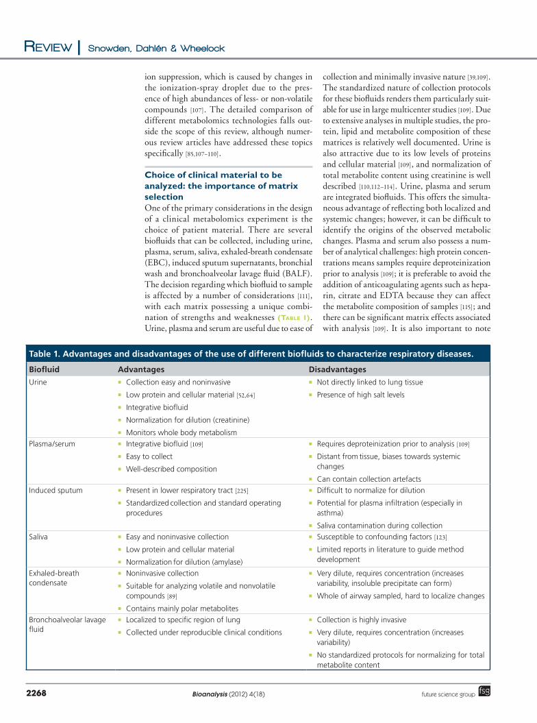
Choice of clinical material to be analyzed: the importance of matrix selectionOne of the primary considerations in the design of a clinical metabolomics experiment is the choice of patient material. There are several biofluids that can be collected, including urine, plasma, serum, saliva, exhaledbreath condensate (EBC), induced sputum supernatants, bronchial wash and bronchoalveolar lavage fluid (BALF). The decision regarding which biofluid to sample is affected by a number of considerations [111], with each matrix possessing a unique combination of strengths and weaknesses (Table 1). Urine, plasma and serum are useful due to ease of
collection and minimally invasive nature [39,109]. The standardized nature of collection protocols for these biofluids renders them particularly suitable for use in large multicenter studies [109]. Due to extensive analyses in multiple studies, the protein, lipid and metabolite composition of these matrices is relatively well documented. Urine is also attractive due to its low levels of proteins and cellular material [109], and normalization of total metabolite content using creatinine is well described [110,112–114]. Urine, plasma and serum are integrated bio fluids. This offers the simultaneous advantage of reflecting both localized and systemic changes; however, it can be difficult to identify the origins of the observed metabolic changes. Plasma and serum also possess a number of analytical challenges: high protein concentrations means samples require deproteinization prior to ana lysis [109]; it is preferable to avoid the addition of anticoagulating agents such as heparin, citrate and EDTA because they can affect the metabolite composition of samples [115]; and there can be significant matrix effects associated with ana lysis [109]. It is also important to note
Table 1. Advantages and disadvantages of the use of different biofluids to characterize respiratory diseases.
Biofluid Advantages Disadvantages
Urine � Collection easy and noninvasive
� Low protein and cellular material [52,64]
� Integrative biofluid
� Normalization for dilution (creatinine)
� Monitors whole body metabolism
� Not directly linked to lung tissue
� Presence of high salt levels
Plasma/serum � Integrative biofluid [109]
� Easy to collect
� Well-described composition
� Requires deproteinization prior to ana lysis [109]
� Distant from tissue, biases towards systemic changes
� Can contain collection artefacts
Induced sputum � Present in lower respiratory tract [225]
� Standardized collection and standard operating procedures
� Difficult to normalize for dilution
� Potential for plasma infiltration (especially in asthma)
� Saliva contamination during collection
Saliva � Easy and noninvasive collection
� Low protein and cellular material
� Normalization for dilution (amylase)
� Susceptible to confounding factors [123]
� Limited reports in literature to guide method development
Exhaled-breath condensate
� Noninvasive collection
� Suitable for analyzing volatile and nonvolatile compounds [89]
� Contains mainly polar metabolites
� Very dilute, requires concentration (increases variability, insoluble precipitate can form)
� Whole of airway sampled, hard to localize changes
Bronchoalveolar lavage fluid
� Localized to specific region of lung
� Collected under reproducible clinical conditions
� Collection is highly invasive
� Very dilute, requires concentration (increases variability)
� No standardized protocols for normalizing for total metabolite content

Application of metabolomics approaches to the study of respiratory diseases | Review
www.future-science.com 2269future science group
that while working with plasma and serum is similar, the metabolite composition of these two biofluids is distinct [116]. In addition, the generation of plasma can result in the formation of artefacts and commensurate shifts in metabolite levels that are solely a result of the sample collection process [117].
EBC and saliva are utilized in metabolomics studies due to the ease and noninvasive nature of collection [2,42,44,89,92,93,118,119]. However, because they have yet to be widely employed, protocols for their collection are not widely standardized, leading to potentially high variability between laboratories. EBC is dilute and often requires concentration prior to ana lysis, which can lead to increased intersample variability. It has been suggested that EBC can be normalized for solute concentration using the sum of the sodium and potassium ions [120]. Saliva has been normalized using amylase concentrations [121]; however, saliva is susceptible to a range of confounding factors that affect the composition of the metabolome (a confounding factor is an internal or external source of metabolic variability that can obfuscate the detection of metabolic changes resulting from the factor being studied). Saliva is especially vulnerable to the effects of diet [122] and smoking [123], but changes resulting from factors including gender [123] and sampling time [67] have also been reported. A small number of metabolomics studies have analyzed BALF [124,125]; however, the highly invasive and expensive nature of sample collection renders it a lessattractive biofluid with which to work. Bronchoscopy can only be performed by welltrained specialists on a limited number of patients [126] and is, therefore, generally not suitable for the collection of temporally associated longitudinal studies. Variability in the volume of saline recovered during collection is a concern; however, normalizing the data using the ratio of instilled versus recovered volume has been shown to slightly reduce variability [127]. BALF samples are also dilute and require concentraare also dilute and require concentration prior to ana lysis, which can introduce further variability. Whilst some metabolite profiling work has been performed on sputum [128], global metabolomics techniques have yet to be applied, which may be due to a number of challenges faced in working with this matrix (Table 1).
Following the choice of clinical material, it is important to design a sampling strategy that will minimize variability and remove sampling bias. A number of factors present in a daily routine significantly affect the metabolome composition (Table 2), including gender [123], smoking
[123], sampling time [67], diet [122], environment [15,16], age [129,130], exercise [131,132] and associated comorbidities. Some reported studies chose not to control these confounding factors, under the rationale that “the metabolites of interest would be altered sufficiently between disease and nondisease groups that such intrapersonal variability would be superseded” [64,65]. Saude et al. were able to claswere able to classify stable asthmatic patients with an accuracy of 94%, yet only matched the age and gender composition. Numerous strategies have been used to correct for intra and interpersonal variability: �Fasting prior to blood or urine collection
can reduce the impact of dietrelated effects [104,133,134];
�Food frequency questionnaires have been widely used in nutrition studies, and allow compositional changes to be linked to changes in food intake [135]; however, food frequency questionnaire compliance has been shown to be a problem [136];
�Standardization of urine collection protocols is important with different strategies required depending upon the study aim (e.g., first evacuation of the day, collection in the clinic, collection in the home, collection and combination of multiple samples).
Table 2. Metabolites reported as changing in abundance in response to environmental factors in healthy patients.
Metabolite Comment Confounding factor
Citrate Increased in smokers [123] Smoking
Formate Decreased in smokers [123] Smoking
Lactate Increased in smokers [123] Smoking
Pyruvate Increased in smokers[123] Smoking
Sucrose Increased in smokers[123] Smoking
Acetate Higher in men [123] Gender
Formate Higher in men [123] Gender
Glycine Higher in men [123] Gender
Lactate Higher in men [123] Gender
Methanol Higher in men [123] Gender
Propionate Higher in men [123] Gender
Propylene glycol Higher in men [123] Gender
Pyruvate Higher in men [123] Gender
Succinate Higher in men [123] Gender
Taurine Higher in men [123] Gender
Alanine Lower at night [67] Sampling time
Choline Lower at night [67] Sampling time
Methanol Higher at night [67] Sampling time
N-acetyl groups Higher at night [67] Sampling time
Propionate Lower at night [67] Sampling time
Trimethyamine oxide Lower at night [67] Sampling time
Review | Snowden, Dahlén & Wheelock
Bioanalysis (2012) 4(18)2270 future science group
Achievements of metabolomics in the study of respiratory disease: progress in the field to dateInitial applications of metabolomics have been promising and have successfully classified several respiratory diseases, including asthma [2,64,65,94,99,137], COPD [3,91,93,138] and cystic fibrosis [66,92,125,137] with a high degree of accuracy. No known studies have, to date, investigated sarcoidosis or other respiratory diseases. The high classification accuracy of these models, generated from sample material that was collected noninvasively (e.g., urine and EBC) suggests that metabolomics approaches can play a central role in diagnosing/characterizing respiratory diseases. Metabolomics approaches have also identified the individual candidate
metabolites that are responsible for discriminating respiratory disease patients and healthy controls within these experiments (Tables 3–6). These lists of metabolites render it possible to tentatively identify distinct areas of metabolism and the pathways that characterize the individual disease metabolic phenotypes. We present these studies on a diseasespecific basis below.
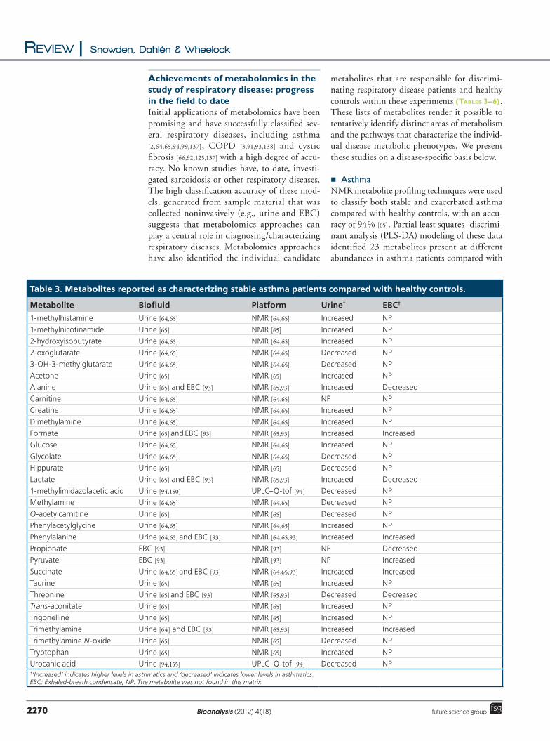
� AsthmaNMR metabolite profiling techniques were used to classify both stable and exacerbated asthma compared with healthy controls, with an accuracy of 94% [65]. Partial least squares–discriminant ana lysis (PLSDA) modeling of these data identified 23 metabolites present at different abundances in asthma patients compared with
Table 3. Metabolites reported as characterizing stable asthma patients compared with healthy controls.
Metabolite Biofluid Platform Urine† EBC†
1-methylhistamine Urine [64,65] NMR [64,65] Increased NP
1-methylnicotinamide Urine [65] NMR [65] Increased NP
2-hydroxyisobutyrate Urine [64,65] NMR [64,65] Increased NP
2-oxoglutarate Urine [64,65] NMR [64,65] Decreased NP
3-OH-3-methylglutarate Urine [64,65] NMR [64,65] Decreased NP
Acetone Urine [65] NMR [65] Increased NP
Alanine Urine [65] and EBC [93] NMR [65,93] Increased Decreased
Carnitine Urine [64,65] NMR [64,65] NP NP
Creatine Urine [64,65] NMR [64,65] Increased NP
Dimethylamine Urine [64,65] NMR [64,65] Increased NP
Formate Urine [65] and EBC [93] NMR [65,93] Increased Increased
Glucose Urine [64,65] NMR [64,65] Increased NP
Glycolate Urine [64,65] NMR [64,65] Decreased NP
Hippurate Urine [65] NMR [65] Decreased NP
Lactate Urine [65] and EBC [93] NMR [65,93] Increased Decreased
1-methylimidazolacetic acid Urine [94,150] UPLC–Q-tof [94] Decreased NP
Methylamine Urine [64,65] NMR [64,65] Decreased NP
O-acetylcarnitine Urine [65] NMR [65] Decreased NP
Phenylacetylglycine Urine [64,65] NMR [64,65] Increased NP
Phenylalanine Urine [64,65] and EBC [93] NMR [64,65,93] Increased Increased
Propionate EBC [93] NMR [93] NP Decreased
Pyruvate EBC [93] NMR [93] NP Increased
Succinate Urine [64,65] and EBC [93] NMR [64,65,93] Increased Increased
Taurine Urine [65] NMR [65] Increased NP
Threonine Urine [65] and EBC [93] NMR [65,93] Decreased Decreased
Trans-aconitate Urine [65] NMR [65] Increased NP
Trigonelline Urine [65] NMR [65] Increased NP
Trimethylamine Urine [64] and EBC [93] NMR [65,93] Increased Increased
Trimethylamine N-oxide Urine [65] NMR [65] Decreased NP
Tryptophan Urine [65] NMR [65] Increased NP
Urocanic acid Urine [94,155] UPLC–Q-tof [94] Decreased NP†‘Increased’ indicates higher levels in asthmatics and ‘decreased’ indicates lower levels in asthmatics.EBC: Exhaled-breath condensate; NP: The metabolite was not found in this matrix.
Application of metabolomics approaches to the study of respiratory diseases | Review
www.future-science.com 2271future science group
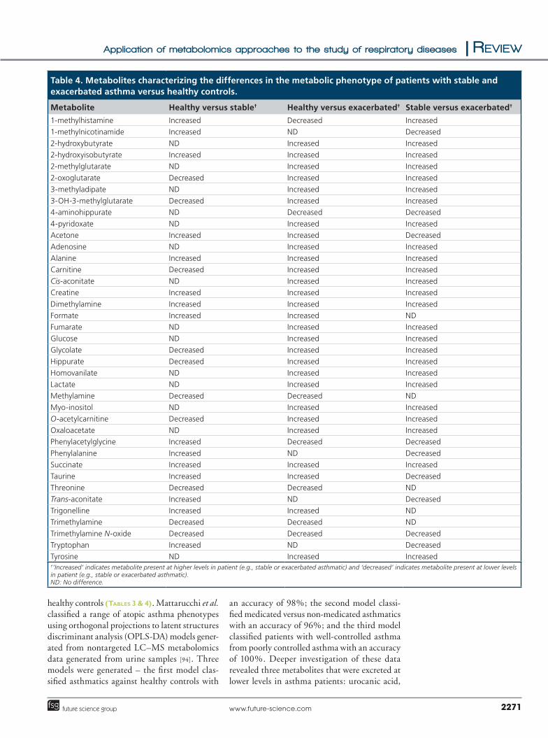
healthy controls (Tables 3 & 4). Mattarucchi et al. classified a range of atopic asthma pheno types using orthogonal projections to latent structures discriminant ana lysis (OPLSDA) models generated from nontargeted LC–MS metabolomics data generated from urine samples [94]. Three models were generated – the first model classified asthmatics against healthy controls with
an accuracy of 98%; the second model classified medicated versus nonmedicated asthmatics with an accuracy of 96%; and the third model classified patients with wellcontrolled asthma from poorly controlled asthma with an accuracy of 100%. Deeper investigation of these data revealed three metabolites that were excreted at lower levels in asthma patients: urocanic acid,
Table 4. Metabolites characterizing the differences in the metabolic phenotype of patients with stable and exacerbated asthma versus healthy controls.
Metabolite Healthy versus stable† Healthy versus exacerbated† Stable versus exacerbated†
1-methylhistamine Increased Decreased Increased
1-methylnicotinamide Increased ND Decreased
2-hydroxybutyrate ND Increased Increased
2-hydroxyisobutyrate Increased Increased Increased
2-methylglutarate ND Increased Increased
2-oxoglutarate Decreased Increased Increased
3-methyladipate ND Increased Increased
3-OH-3-methylglutarate Decreased Increased Increased
4-aminohippurate ND Decreased Decreased
4-pyridoxate ND Increased Increased
Acetone Increased Increased Decreased
Adenosine ND Increased Increased
Alanine Increased Increased Increased
Carnitine Decreased Increased Increased
Cis-aconitate ND Increased Increased
Creatine Increased Increased Increased
Dimethylamine Increased Increased Increased
Formate Increased Increased ND
Fumarate ND Increased Increased
Glucose ND Increased Increased
Glycolate Decreased Increased Increased
Hippurate Decreased Increased Increased
Homovanilate ND Increased Increased
Lactate ND Increased Increased
Methylamine Decreased Decreased ND
Myo-inositol ND Increased Increased
O-acetylcarnitine Decreased Increased Increased
Oxaloacetate ND Increased Increased
Phenylacetylglycine Increased Decreased Decreased
Phenylalanine Increased ND Decreased
Succinate Increased Increased Increased
Taurine Increased Increased Decreased
Threonine Decreased Decreased ND
Trans-aconitate Increased ND Decreased
Trigonelline Increased Increased ND
Trimethylamine Decreased Decreased ND
Trimethylamine N-oxide Decreased Decreased Decreased
Tryptophan Increased ND Decreased
Tyrosine ND Increased Increased†‘Increased’ indicates metabolite present at higher levels in patient (e.g., stable or exacerbated asthmatic) and ‘decreased’ indicates metabolite present at lower levels in patient (e.g., stable or exacerbated asthmatic).ND: No difference.
Review | Snowden, Dahlén & Wheelock
Bioanalysis (2012) 4(18)2272 future science group
methylimidazolacetic acid and a chemical species resembling an isoleucineproline fragment. Metabolomics techniques are capable of improving asthma classification compared with traditional diagnostic techniques. Carraro et al. used NMR ana lysis of EBC to classify stable asthma in pediatric patients with an accuracy of 86%, which was an improvement over the 81% accuracy achieved using exhaled nitric oxide and forced expiration volume in 1 s (FEV
1) [2].
In individuals with asthma who have recently suffered an exacerbation, five metabolites acting in the TCA cycle, succinate, fumarate, oxaloacetate, cisaconitate and 2oxoglutarate have all been reported as being present at higher abundances in urine compared with controls (Figure 1) [65]. This increase in the abundance of TCA cycle intermediates suggests an up regulation in TCAcycle metabolism as the result of a greater effort to breathe during exacerbation and/or hypoxic stress due to poor oxygenation as a result of exacerbation. Similar shifts in TCAcycle metabolism have also been
reported during exercise [139,140], supporting the hypothesis that increased abundances of these metabolites during exacerbation is a result of the effort to breath and hypoxic stress. Higher levels of lactate were also reported during exacerbation (Table 4), further supporting the idea that patients are undergoing hypoxic stress, because increased levels of this metabolite have been reported during periods of anaerobic exercise [141]. However, during these periods of exercise, increases in the abundance of lactate have been reported as occurring in conjunction with a decrease in the abundance of citrate [140], which has not been observed in individuals with asthma. Nicholson et al. hypothesized that this increase in the abundance of lactate would lead to lactic acidosis, which would in turn cause renal tubular acidosis leading to lower levels of glycine and hippurate in urine [142], as both of these compounds have been described as biomarkers of reversible renal malfunction [143]. Neither of these metabolites has been reported as being reduced in abundance in urine after
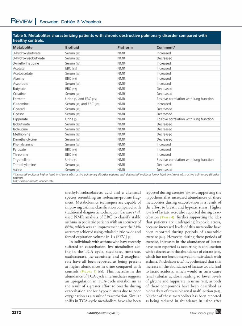
Table 5. Metabolites characterizing patients with chronic obstructive pulmonary disorder compared with healthy controls.
Metabolite Biofluid Platform Comment†
3-hydroxybutyrate Serum [91] NMR Increased
3-hydroxyisobutyrate Serum [91] NMR Decreased
3-methylhistidine Serum [91] NMR Increased
Acetate EBC [89] NMR Increased
Acetoacetate Serum [91] NMR Increased
Alanine EBC [93] NMR Increased
Ascorbate Serum [91] NMR Increased
Butyrate EBC [93] NMR Decreased
Creatine Serum [91] NMR Decreased
Formate Urine [3] and EBC [93] NMR Positive correlation with lung function
Glutamine Serum [91] and EBC [89] NMR Increased
Glycerol Serum [91] NMR Decreased
Glycine Serum [91] NMR Decreased
Hippurate Urine [3] NMR Positive correlation with lung function
Isobutyrate Serum [91] NMR Decreased
Isoleucine Serum [91] NMR Decreased
Methionine Serum [91] NMR Decreased
Dimethylglycine Serum [91] NMR Decreased
Phenylalanine Serum [91] NMR Increased
Pyruvate EBC [93] NMR Increased
Threonine EBC [93] NMR Increased
Trigonelline Urine [3] NMR Positive correlation with lung function
Trimethylamine Serum [91] NMR Decreased
Valine Serum [91] NMR Decreased†‘Increased’ indicates higher levels in chronic obstructive pulmonary disorder patients and ‘decreased’ indicates lower levels in chronic obstructive pulmonary disorder patients. EBC: Exhaled-breath condensate.
Application of metabolomics approaches to the study of respiratory diseases | Review
www.future-science.com 2273future science group
exacerbation, conversely, hippurate is reported as increasing in abundance (Table 4). This suggests that if hypoxic stress is occurring, then it is not at sufficient levels to cause renal malfunction. Hippurate is reported at lower concentrations in the urine of stable asthmatics, in conjunction with increased levels of lactate and some intermediates of the TCA cycle (e.g., succinate). These data might potentially suggest that individuals with asthma may permanently suffer from some degree of hypoxic stress leading to lowlevel renal malfunction; however, this is highly speculative and there are no reports in the literature to support this theory.
1methylhistamine is a major downstream metabolite of histamine, a significant proinflammatory agent [144,145] and mediator of inf lammation [146,147], as well as an impor as well as an important component of allergic metabolism.
1methyl histamine is produced via the methylation of histamine by histamine methyltransferase [148,149]. 1methylimidazolacetic acid is a downstream product of the histamine methylation pathway (Figure 2), with both 1methylhistamine and 1mehtylimidazolacetic acid occurring at modified abundances in asthma patients (Table 3). 1methylhistamine is reported at higher abundances in asthma patients [65], with lower levels of 1methylimidazolacetic observed in the urine of individuals with asthma [94,150]. In experiments using 14Clabeled histamine, methylation was the primary route of histamine metabolism in humans [123,151], suggesting that 1methylhistamine and 1methylimidazolacetic acid are appropriate urinary markers of histamine metabolism [152] and potential biomarkers for the diagnosis of pathological inflammation. Histamine release has been reported
Table 6. Metabolites characterizing patients with cystic fibrosis compared with healthy controls.
Metabolite Biofluid Platform Comment†
1-methylnicotinamide PHAECC [66] UHPLC–MS [66] Increased
2-propanol EBC [92] NMR [92] Discriminate CF and HC
Acetate EBC [92] NMR [92] Discriminate CF and HC
Acetone EBC [92] NMR [92] Discriminate CF and HC
Adenosine PHAECC [66] UHPLC–MS [66] Decreased
Anthranilate PHAECC [66] UHPLC–MS [66] Increased
Cytidine PHAECC [66] UHPLC–MS [66] Decreased
Formate EBC [92] NMR [92] Discriminate CF and HC
Fructose PHAECC [66] UHPLC–MS [66] Decreased
Fructose-6-phosphate PHAECC [66] UHPLC–MS [66] Decreased
Glucose PHAECC [66] UHPLC–MS [66] Decreased
Glucose-6-phosphate PHAECC [66] UHPLC–MS [66] Decreased
Glutamate EBC [92] NMR [92] Discriminate CF and HC
Glutamine EBC [92] NMR [92] Discriminate CF and HC
Glutathione (oxidized GSSG) PHAECC [66] UHPLC–MS [66] Decreased
Glutathione (reduced GSH) PHAECC [66] UHPLC–MS [66] Decreased
Glycerophosphorylcholine PHAECC [66] UHPLC–MS [66] Decreased
Guanosine PHAECC [66] UHPLC–MS [66] Decreased
Hypoxanthine PHAECC [66] UHPLC–MS [66] Decreased
Inosine PHAECC [66] UHPLC–MS [66] Decreased
Kynurenine PHAECC [66] UHPLC–MS [66] Increased
Lactate PHAECC [66] and EBC [92] UHPLC–MS [66] and NMR [92] Decreased
Malate PHAECC [66] UHPLC–MS [66] Decreased
Mannose-6-phosphate PHAECC [66] UHPLC–MS [66] Decreased
Nicotinamide PHAECC [66] UHPLC–MS [66] Decreased
Opthalmate PHAECC [66] UHPLC–MS [66] Decreased
Propionate EBC [92] NMR [92] Discriminate CF and HC
Ribulose-5-phosphate PHAECC [66] UHPLC–MS [66] Decreased
S-lactoylglutathione PHAECC [66] UHPLC–MS [66] Decreased
Sorbitol PHAECC [66] UHPLC–MS [66] Decreased†‘Increased’ indicates higher levels in cystic fibrosis patients and ‘decreased’ indicates lower levels in cystic fibrosis patients.CF: Cystic fibrosis, EBC: Exhaled-breath condensate; HC: Healthy controls; PHAECC: Primary human airway epithelial cell cultures.

Review | Snowden, Dahlén & Wheelock
Bioanalysis (2012) 4(18)2274 future science group
during mastcell activation in individuals with allergic asthma [153,154]. This might suggest that the changes in the abundance of 1methylhistamine, 1methylimidazolacetic and urocanic acid observed are also linked to mastcell activation. The change in abundance of these two metabolites along with decreased levels of urocanic acid in individuals with asthma [94,155] is consistent with a reduction in the metabolism of free histamine [150]. Kerr [150] suggested that the methylation step is the most likely location for the blockage in histamine metabolism; however, the reported increase in the levels of 1methylhistamine (Table 3) in asthmatics makes this unlikely because if the blockage was upstream of this metabolite, it would not accumulate.
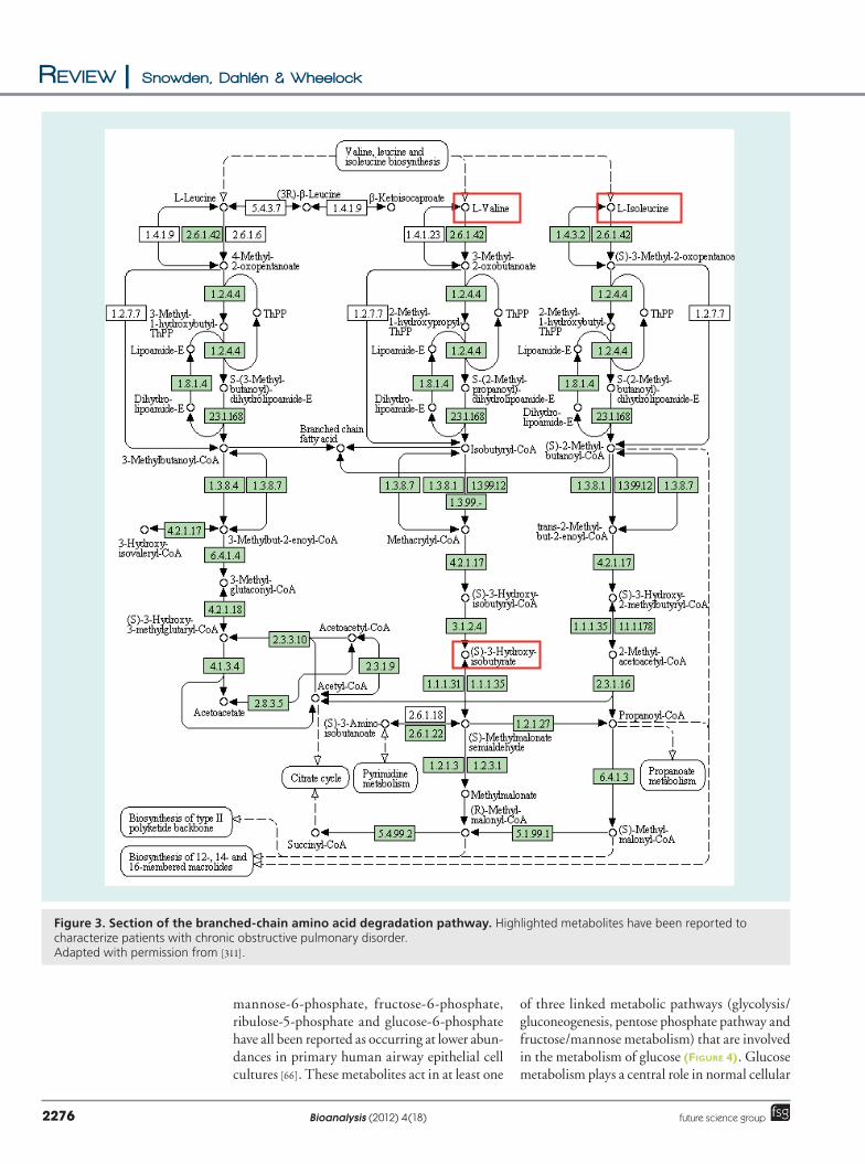
� COPDAnalysis of serum samples using NMR has been successfully used to classify severe COPD (Global Initiative for Chronic Obstructive Lung Disease [GOLD] stage III [156]) and very severe
COPD (GOLD stage IV) patients with an accuracy of 82% [91], where decreased levels of branchedchain amino acids (BCAA) and their associated metabolites are responsible for the discrimination of COPD patients and healthy controls [91]. LC–MS ana lysis of plasma samples successfully classified emphysematous COPD and nonemphysematous COPD patients with an accuracy of 64% using hierarchical clustering [138]. This classification accuracy is relatively low; however, when using linear discriminant ana lysis on a subset of biomarkers classification accuracy was improved. When using the seven optimal biomarkers, classification accuracy was improved to 97% [138], although the biomarkers were not identified.
The most significant area of metabolism identified as discriminating COPD patients from healthy controls is the metabolism of BCAA (Table 5) [91,157], with valine, isoleucine and their degradation product 3hydroxyisobutyrate reported at lower abundances in
Figure 1. Section of the TCA cycle. Highlighted metabolites have been reported to increase in abundance shortly after asthma exacerbation. Adapted with permission from [309].

Application of metabolomics approaches to the study of respiratory diseases | Review
www.future-science.com 2275future science group
patients suffering from COPD (Figure 3). This modification of BCAA metabolism might be the result of cachexia (a wasting syndrome characterized by muscle atrophy, fatigue, weakness and significant weight loss) in COPD patients. During extended periods of fasting the proteolysis of skeletal muscle and the transamination of BCAA by branchedchain aminotransferase (BCKHD) is an important metabolic response providing a resource for gluconeogenesis [91]. Increased levels of gluconeogenesis that are not suppressed by glucose have been reported in patients suffering from cachexic weight loss [158]. This hypothesis is consistent with the results reported in Ubhi et al., which showed that the reduction in BCAA’s correlated with the patient body mass index [91]. Increased levels of BCAA catabolism have also been reported in urine samples collected after exercise [140], where the increase in BCAA catabolism is thought to be the result of proteolysis in skeletal muscle and the visceral region [159]. This might be the result of increased gluconeogenesis due to increased demand for energy during exercise. If accurate, these findings would support the hypothesis that the lower levels of BCAAs in COPD patients are the result of proteolysis, potentially due to wasting.
� Cystic fibrosisThe ana lysis of NMR spectra generated from EBC has been used to classify patients with cystic fibrosis relative to healthy controls with a classification accuracy of 96%. Validation of this model showed that it had a sensitivity of 91% and a specificity of 96% [92]. A second model was generated from these data, classifying stable versus unstable cystic fibrosis patients with an accuracy of 95%. Validation showed that this model had a sensitivity of 86% and a specificity of 94% (the metabolites responsible for the discrimination in both of these models are provided in Table 6) [92]. Preliminary metabolomics ana lysis has been performed looking at BALF collected from pediatric cystic fibrosis patients, which could be classified into high and low inflammation groups [125]. An OPLSDA model of NMR spectra was generated that was able to discriminate the two groups of patients. This model had an R2Y of 0.96 (i.e., the model explained 96% of the variation observed within the data) and a Q2 of 0.80, indicating that the model had good predictive power.
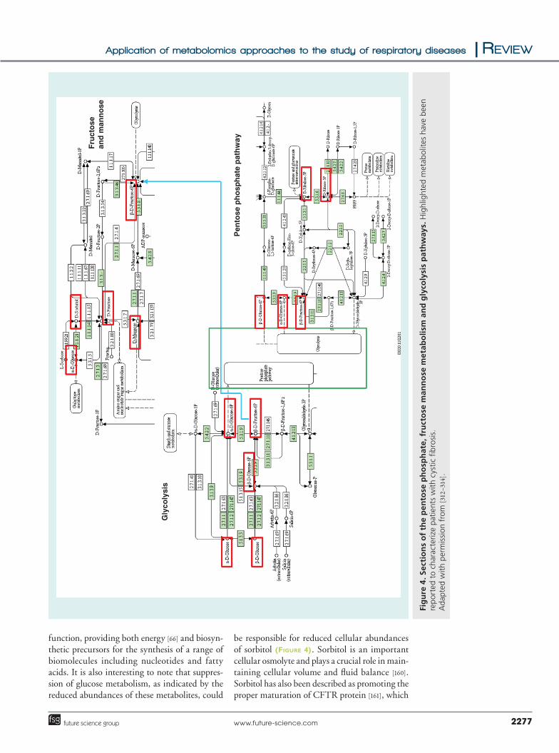
As with asthma and COPD, the metabolomics studies looking at cystic fibrosis performed to date have successfully identified numerous metabolites and areas of metabolism that characterize the disease state (Table 6). Sorbitol, fructose, glucose,
Figure 2. Section of the histidine metabolism pathway. Highlighted metabolites have been reported to characterize patients with stable asthma. Adapted with permission from [310].
Review | Snowden, Dahlén & Wheelock
Bioanalysis (2012) 4(18)2276 future science group
mannose6phosphate, fructose6phosphate, ribulose5phosphate and glucose6phosphate have all been reported as occurring at lower abundances in primary human airway epithelial cell cultures [66]. These metabolites act in at least one
of three linked metabolic pathways (glycolysis/gluconeogenesis, pentose phosphate pathway and fructose/mannose metabolism) that are involved in the metabolism of glucose (Figure 4). Glucose metabolism plays a central role in normal cellular
Figure 3. Section of the branched-chain amino acid degradation pathway. Highlighted metabolites have been reported to characterize patients with chronic obstructive pulmonary disorder. Adapted with permission from [311].
Application of metabolomics approaches to the study of respiratory diseases | Review
www.future-science.com 2277future science group
function, providing both energy [66] and biosynthetic precursors for the synthesis of a range of biomolecules including nucleotides and fatty acids. It is also interesting to note that suppression of glucose metabolism, as indicated by the reduced abundances of these metabolites, could
be responsible for reduced cellular abundances of sorbitol (Figure 4). Sorbitol is an important cellular osmolyte and plays a crucial role in maintaining cellular volume and fluid balance [160]. Sorbitol has also been described as promoting the proper maturation of CFTR protein [161], which
Pen
tose
ph
osp
hat
e p
ath
way
Fru
cto
se
and
man
no
se
Gly
coly
sis
Fig
ure
4. S
ecti
on
s o
f th
e p
ento
se p
ho
sph
ate,
fru
cto
se m
ann
ose
met
abo
lism
an
d g
lyco
lysi
s p
ath
way
s. H
ighl
ight
ed m
etab
olit
es h
ave
bee
n re
por
ted
to c
hara
cter
ize
pati
ents
wit
h cy
stic
fibr
osi
s.
Ada
pted
wit
h p
erm
issi
on f
rom
[312
–314
].

Review | Snowden, Dahlén & Wheelock
Bioanalysis (2012) 4(18)2278 future science group
is an ATPgated anionselective channel that transports chloride ions across the epithelial cell membrane [162]. The transport of chloride ions plays a crucial role in maintaining airway surface liquid volume. Accelerated Na+ absorption and defective Cl secretion in airway epithelial cells is the underlying cause of the pathogenic thickening of airway mucus observed in cystic fibrosis patients [163].
The abundances of five purines and purine metabolites (adenosine, guanosine, inosine, hypoxanthine and adenine) have also been reported to be lower in cystic fibrosis patients (Table 6), suggesting that these patients exhibit lower levels of purine synthesis relative to healthy controls (Figure 5). This is biologically important because purines have been shown to act as signaling molecules [164,165]. Adenosine is especially relevant to cystic fibrosis, because it has been reported to play a role in controlling the viscosity of airway mucus by regulating airway surface liquid volume through activation of CFTR receptors [166,167]. In studies of rat lung tissue, the addition of adenosine directly increased chloride ion flux through CFTR ion channels [168]. Three metabolites relating to glutathione – reduced glutathione, oxidized glutathione and Slactoylglutathione – have been reported at lower concentrations in primary human airway epithelial cell cultures (Table 6). A number of metabolites related to glutathione, including Snitrosoglutathione and Snitrosylating compounds act to promote CFTR cellsurface expression and channel activation in cystic fibrosis epithelial cells [169,170]. As well as these global metabolomics efforts, metabolite profiling of oxylipins has also been used to study cystic fibrosis [171,172].
Challenges in applying metabolomics to the study of respiratory diseasesThe application of metabolomics techniques to the study of respiratory diseases faces a number of significant challenges, some of which are common to all metabolomics experiments and some of which are more specific to clinical studies. The most significant of these are discussed below.
� Biobanks The generation of biobanks is part of the growing trend towards the use of largescale biology (such as ‘omics’ and systems biology) to address questions in clinical science. The term biobank generally refers to repositories of biological material that are relevant to the study of disease [173]. Biobanks are likely to play an integral part in the
future of biomedicine because they can supply sufficient numbers of samples to power largescale systemsbased studies. Biobanks will also provide the opportunity to study the etio logy of diseases from large populations in a longitudinal fashion, greatly increasing the sensitivity for detecting shifts relating to the onset of disease. For example, the ‘LifeGene’ biobank aims to collect samples from 500,000 patients, which represents approximately 5% of the Swedish population, thereby enabling diseases to be studied at the population level [174]. However, a number of important ethical issues with using biobanks have been raised [175–177]. There are also a range of analytical challenges faced in using metabolomics techniques to analyze biobank samples. One of the foremost issues is the reproducibility of sample collection strategy employed in the studies, making the development of a standardized collection protocol important [178]. A second major obstacle is the effect of longterm storage on the sample metabolite composition. Even samples stored at 80°C have been demonstrated to exhibit metabolite loss during storage [71,179], with the rate of loss dependent upon the matrix and analyte. For these vast libraries of samples that are being collected in biobanks to be useful in the metabolomics study of clinical diseases, it is necessary to develop and implement a range of standard operating procedures for sample collection [178] and prestorage processing [180]. It is also important to assess the effects of different storage strategies on the metabolite compositions of samples to identify the strategy that best preserves the true metabolic phenotype of the sample, and to identify the limits of viable storage so that time and resources are not wasted on analyzing poorquality samples. Towards this end, reliable degradation markers should be identified to monitor sample integrity. Another issue involves sample ownership, with potential conflicts between researchers who deposit samples in biobanks and the clinical entities that maintain the biobanks. Interested readers are directed to the Biobanking and Biomolecular Resources Research Infrastructure Initiative [302]. It has also been suggested that the interpretation of results generated from biobank samples may be problematic. This is due to the varied nature of respiratory diseases, which exhibit large deviations in pathological phenotype. These differences will make it potentially difficult to link experimental results to clinical aspects of the disease [173] – further illustrating the need for rigorous and standardized clinical phenotyping of patients.

Application of metabolomics approaches to the study of respiratory diseases | Review
www.future-science.com 2279future science group
� Experimental designThe high intra and interindividual variability need to be addressed in clinical studies
[181–183]. Strategies for controlling confounding factors (e.g., diet [122] and smoking [123]) have been described, including matching the age
Fig
ure
5. P
uri
ne
met
abo
lism
pat
hw
ay h
igh
ligh
tin
g m
etab
olit
es m
od
ified
in p
atie
nts
wit
h c
ysti
c fi
bro
sis.
Ada
pted
wit
h p
erm
issi
on f
rom
[315
].

Review | Snowden, Dahlén & Wheelock
Bioanalysis (2012) 4(18)2280 future science group
and gender composition of sample groups [65], smoking history of patients [91], and patient fasting prior to sample collection [104,133,134,184]. However, these approaches only reduce the variability introduced by these confounding factors and do not completely remove it. These strategies also introduce problems related to patient conformity (i.e., patient compliance with study protocols regarding dietary and behavioral restrictions). It would be advantageous to develop analytical strategies to account for this variability; for example, metabolites that are modified in abundance as the result of disease should correlate across all patients with the diseased state, and metabolite abundances that are modified as the result of confounding factors should be excluded.
Metabolomics experiments also suffer from problems of poor laboratorytolaboratory reproducibility, this is especially true for nontargeted MSbased techniques. To facilitate collaborative research between multiple laboratories, strategies to reduce this variability should be implemented. Initially, mRNA microarray experiments suffered from similar limitations, although studies have now shown that interlaboratory variability can be reduced via use of common platforms and procedures [185,186]. It has also been shown (for microarray ana lysis) that data normalization is a vital component in reducing variability [185]. The ana lysis of standard reference materials is crucial in reducing interlaboratory variability [187]. The inclusion of reference materials also enables comparison of instrumental accuracy/precision between sample batches, experiments and analytical platforms [187]. One strategy to address this variability is to normalize metabolite abundance by total metabolite composition, which can remove effects of sample dilution in biofluids. Methods for normalizing for sample dilution are well described for saliva using amylase abundance [121], and using creatinine in urine [110,112–114]. It is especially important for urine because its concentration can vary widely [114]; however, it is less of an issue in plasma and serum as solute concentration is tightly controlled [114]. For NMR spectroscopy it is possible to normalize data for total metabolite content by reference to the total spectral area of each sample [119]. More detailed descriptions of stratMore detailed descriptions of strategies for the normalization of metabolite concentration for total metabolite content [114,188], and general discussions of metabolomics and metabolomics workflows [77,109,189], are widely available. While a separate subject in and of
itself, the field of design of experiments (DOE) is extremely important, but is unfortunately rarely discussed in either clinical or analytical papers. DOE selects a diverse and representative set of experiments in which all factors are independent of each other despite being varied simultaneously. The result is a causal predictive model showing the importance of all factors and their interactions [303]. Increased effort placed on appropriate DOE will enable the use of decreased sample numbers while maintaining experimental power and will be a useful tool in clinical metabolomics [190].
� Data ana lysis Data ana lysis is a critical part of all metabolomics experiments, regardless of the biological system or analytical instrument in question, and presents a major challenge to the development and application of metabolomics techniques. The statistical ana lysis of metabolomics data is challenging for multiple reasons, with detailed discussion of these problems being outside the scope of this review. A number of articles have dealt with these problems in detail [191–194] and only a few of these issues will be highlighted here. Metabolomics experiments, along with most largescale biology studies in general, have few degrees of freedom, consisting of low sample number combined with highdimensionality datasets. It is accordingly challenging to derive meaningful biological knowledge via visual analysis of the dataset [195]. Identifying the appropriate statistical approaches is therefore one of the foremost challenges in data ana lysis. Univariate statistics, such as ana lysis of variance (ANOVA) and Student’s ttest, identify variables as being significant at a given probability (i.e., a = 0.05, which means that there is a 5% chance that any given variable is identified as being significant by chance, a socalled falsepositive or type I error.) Within large metabolomics datasets containing thousands of variables [189,196,197], there are potentially hundreds of falsepositives, which can be referred to as the falsediscovery rate (FDR) [198,199]. Many studies within the ‘omics’ paradigm employ various approaches to data ana lysis in addressing the FDR issue, including Tukey’s honestly significant difference [200], the Bonferroni correction [201], HolmBonferroni [202], the Šidák correction [203,204] and Benjamini and Hochberg [198]. The drawback to many of these approaches for correcting for multiple hypothesis testing relates to the issue of false negatives (type II errors). There is a strong
Application of metabolomics approaches to the study of respiratory diseases | Review
www.future-science.com 2281future science group
likelihood that application of an FDRbased correction will result in the generation of false negatives, which is particularly problematic in a hypothesisgenerating experiment. Accordingly, any statistical treatment of the data will have to balance type I and II errors appropriately to address the aim of the study.
Another approach that has been particularly employed in the metabolomics community is the use of multivariate statistical ana lysis. There are numerous multivariate statistical techniques that can be applied to metabolomics data, including unsupervised techniques (e.g., principal component ana lysis [51,205]), supervised techniques (e.g., PLSDA [143,205], OPLSDA [206,207], bi directional OPLS [O2PLS] [208,209]) and more specialized approaches such as ensemble classifiers (e.g., random forest [210]). For any multivariate ana lysis, it is important to report the appropriate model statistics. At the minimum, the R2 and Q2 values should be provided for principal component ana lysis, PLS, OPLSDA and O2PLS models, and eigen values for linear discriminant ana lysis. In addition, the number of components, and CVANOVA pvalues, should be provided where appropriate, such as in discriminant ana lysis. The values of R2 and Q2 are commonly utilized model statistics to evaluate a multivariate model. The R2 represents the percentage of variation within a dataset that can be explained by the model, which is often referred to as a measure of fit. The Q2 is the percentage variation of the response predicted by the model according to the cross validation, in other words, how accurately the model can predict new data. Unfortunately, many papers omit these values, making it impossible for the reader to assess the quality of the model. Visual inspection of the model does not give evidence of significant separation, especially for supervised methods.
To improve the reporting of results in metabolomics experiments it would be advantageous to develop a set of ‘minimum reporting standards’ for metabolomics data. Ideally, these standards would apply to both the analytical method ology as well as the statistical analyses. Goodacre et al. proposed a set of reporting standards aimed at addressing two main issues: formalize a ‘reporting’ scheme to prevent confusion over the use of termin ology; and define a set of ‘minimum reporting standards’ for all stages of ana lysis from data preprocessing to validation of initial hypothesis [195]. These reporting standards represent a starting point; however, they are unfortunately not yet widely adhered to in the scientific
literature. The development and implementation of datareporting standards to enable the exchange of information is an important goal in metabolomics [184]. The Metabolomics Society has initiated the metabolomics standards initiative [304]. These reporting standards outline the minimum information content that should be reported for a metabolomics study, including clear and accurate description of the design and implementation of the study, the subjects involved, biological material sampled and the data collected. This standard ization will increase the accessibility of the information and enable the maximum amount of knowledge to be extracted from a dataset [211].
� Data interpretation A significant bottleneck in an ‘omics’ experiment is interpretation of the acquired data. It is useful to map the metabolites that describe respiratory diseases on to metabolic pathways to identify both areas of metabolism and specifi c metabolic pathmetabolism and specific metabolic pathways to develop an improved understanding of the underlying biological perturbation [212]. One of the most powerful tools is the Kyoto Encyclopedia of Genes and Genomes (KEGG) [213,305]; however, there are others, including WikiPathways [306], Ingenuity Pathway Analysis, MetaCyc [307] and the Human Metabolome Database [214,301]. Metabolomics data can be mapped onto KEGG pathways either manually or using the KegArray tool [215], which is a Javabased application that enables metabolomics data to be considered in isolation or in conjunction with both transcriptomics and proteomics data. A relatively new function in the KEGG suite is the DISEASE utility, which highlights known metabolic pathways in a range of diseases including asthma (H00079 [308]). This functionality could play a useful role in interpretation of metabolomics data generated for asthma; however, a closer look at the asthma pathway reveals that there is a significant need for expansion of the mechanistic information to be of utility in data interpretation [127].
Future perspective The use of metabolomics approaches in the study of respiratory diseases is still in its infancy and lags behind applications in other diseases. To date both NMR [3,64,65,67,91–93] and MSbased [66,94] metabolomics techniques have been used to analyze respiratory diseases including asthma [2,64,65,94], COPD [3,91,93,138] and cystic fibrosis [66,92,125] in biofluids including urine [3,64,65,94], EBC [2,92,93], serum [91], plasma [138], BALF [125]

Review | Snowden, Dahlén & Wheelock
Bioanalysis (2012) 4(18)2282 future science group
and sputum supernatants [128]. These studies have demonstrated the power of metabolomics techniques to classify and potentially diagnose patients suffering from multiple respiratory diseases. In particular, metabol omics approaches have the potential to diagnose respiratory diseases with a higher degree of accuracy than traditional diagnostic methods [2]. Metabolomics approaches have identified several areas of metabolism and metabolic pathways that describe the metabolic phenotype of respiratory diseases. The TCA cycle and histamine metabolism can characterize asthma, with COPD charac terized by BCAA catabolism and cystic fibrosis by glucose, purine and glutathione metabolism. Examining the metabolites that are remodeled in each of these respiratory diseases, it can be seen that whilst some metabolites are remodeled in multiple respiratory diseases compared with controls, the metabolic phenotype of each of the three diseases is unique. It is also interesting to note that the metabolic phenotype of an asthma exacerbation is unique to that of stable asthma [65], rather than it being a simple worsening of the underlying asthma pheno type. This is also true for the comparison of the metabolic phenotype of stable and unstable cystic fibrosis, with unstable cystic fibrosis possessing a unique phenotype rather than being a worsening of the stable phenotype. Finally, in the context of studies in asthmatics, the influence of treatments, and in particular the widely used glucocorticosteriods, must be determined in future studies.
The use of metabolomics to study respiratory diseases has produced some promising results and its future applications can largely be split into two areas. The first is to identify and describe the shifts in metabolite composition and metabolism associated with different respiratory diseases. Initial efforts in applying metabolomics approaches should focus on gathering as much information as possible to provide an overview of diseasespecific biochemistry. This should be done using both global metabolomics approaches and targeted metabolicprofiling techniques, because global metabolomics methods often lack the sensitivity of metabolicprofiling methods. Accordingly, in order to examine the complete underlying metabolic picture, there is a need for targeted metabolite profiling methods to be performed in parallel to detect lowabundance compounds (e.g., eicosanoids and other oxylipins) [216]. Targetedprofiling methods have identified numerous lipid mediators that would not otherwise be detected using global,
nontargetedmetabolomics methods [79]. These mediators play vital roles in the pathology of a range of respiratory diseases including asthma [25,79,217], COPD [218,219] and cystic fibrosis [219–221]. Once sufficient information has been gathered to accurately describe respiratory diseases, it will be necessary to develop targeted methods that are both quantitative and highthroughput to process large numbers of samples to further refine disease models. The second area to which metabolomics can contribute to improved disease management is improving our understanding of the metabolic mechanisms of disease pathology and how this underlying metabolic phenotype responds to therapeutic intervention. For example, it is unlikely that pharmacological treatment shifts the metabolic phenotype of diseased state patients back to that of healthy controls, instead, it is much more probable that the phenotype is shifted to a third state that could be considered as the pharmacological phenotype [222–224]. Accordingly, the true power of metabolomics in identifying and quantifying phenotypes will come into play for classifying subphenotypes of disease and their homeo dynamic shifts between disease and pharmacological phenotypes relative to healthy individuals. The acquisition of flux data will enable the development of quantitative models for these dynamic interactions, greatly increasing our ability to monitor disease and predict patient response to interventions. Metabolites are the end products of cellular processes and can be thought of as the ultimate response of a system to genetics and environment [77]. Studying the metabolite composition of systems using metabol omics approaches will provide a powerful tool in understanding the pathological mechanisms of respiratory diseases and for developing new therapeutic strategies for their treatment.
The next 5–10 years should see some exciting developments in the field of metabolomics. It is expected that the use of metabolomics techniques will become more routine, both in general and in applications to respiratory disease. One of the greatest challenges will remain obtaining comprehensive coverage of the metabolome, especially in determining endogenous from exo genous metabolites, in particular those related to diet or microbiota production. While it is not expected that a single analytical platform will evolve in the near future to address this need, the ability of MS to acquire a greater portion of the metabolome will increase. These efforts will most likely involve multi dimensional chromatography equipped
Application of metabolomics approaches to the study of respiratory diseases | Review
www.future-science.com 2283future science group
with the ability to analyze several stationary phases in a single ana lysis (e.g., hydrophilic interaction LC, C
18 and ion exchange). These complex
multistationary phase instruments in combination with extremely high pressures, long analytical columns and potentially microfluidics (i.e., nanoflow) will provide the resolution necessary to chromatographically separate a metabolome. These instrument configurations will require highresolution mass spectrometers with very fast scanning rates and the ability to perform multiple MS/MS experiments without a significant loss in signal. There will also be a concomitant increased emphasis placed on metabolomics kits (e.g., Biocrates and their MetaDisIDQ® Kit) and chipbased technologies with, for example, a lipidomicsbased chip designed to capture the lipidome (e.g., Agilent’s HPLCChip/MS system). One can envision the production of a respiratory kit or an asthma kit capable of identifying or classifying subphenotypes, such as poor glucocorticosteriod responders. One of the major challenges will continue to be the quantitative acquisition of a global metabolome. Given that it is not feasible to have internal standards for all potential metabolites, these efforts will require other novel approaches besides the use of stable isotope dilution or external standards. The field also requires the development of a comprehensive set of analytical standards available as a commercial kit. Such a product could be used for interlaboratory comparisons to produce quantitative
data and for routine quality assurance/quality control. An additional need is for standardized reference material, such as the NIST SRM 1950 plasma. This material consists of a plasma pool collected from an equal number of men and women and with a racial distribution that reflects the US population. The inclusion of this material in each published dataset would provide a standardized metric for data normalization. There is a distinct need for a public repository or metabolomics data along the lines of Gene Expression Omnibus; however, to make these data useful, standardization will be necessary. Accordingly, there are multiple challenges remaining in the widespread application of metabolomics methods, but the future is very bright and the field of metabolomics should continue to grow as well as its application to the study of respiratory diseases.
Financial & competing interests disclosureThis work was funded by the Swedish Medical Research Council, the Swedish Heart–Lung Foundation, Vinnova Chronic Inflammation – Diagnostic and Therapy, the Swedish Foundation for Strategic Research, the Stockholm County Council Research Funds, IMI (U-BIOPRED) and Karolinska Institutet. The authors have no other relevant affiliations or financial involvement with any organization or entity with a financial interest in or financial conflict with the subject matter or materials discussed in the manuscript apart from those disclosed.
No writing assistance was utilized in the production of this manuscript.
Executive summary
Respiratory diseases: the unmet need for new biomarkers & increased understanding of pathobiology
� Respiratory diseases are a major cause of global morbidity and mortality.
� Metabolomics has yet to be extensively applied to the field of respiratory disease.
� Applications of metabolomics methodologies represent an opportunity to gain insight into the pathobiology of respiratory diseases, characterize disease subphenotypes and develop new diagnostic tools.
Choice of clinical material to be analyzed: the importance of matrix selection
� Clinical samples have matrix-specific strengths and weaknesses. The appropriate choice depends on the biological question and analytical method being applied. The selection of matrix should be carefully considered during the experimental design phase of any clinical study.
Achievements of metabolomics in the study of respiratory disease: progress in the field to date
� Metabolomics approaches can differentiate healthy controls from individuals with a variety of respiratory diseases, including asthma, chronic obstructive pulmonary disorder and cystic fibrosis. These results suggest that there is potential for diagnostic applications.
� Metabolomics has implicated numerous metabolites that associate with a range of respiratory diseases, providing insight into potential areas of metabolism responsible for disease pathology.
Challenges in applying metabolomics to the study of respiratory diseases
� The most important challenges in a metabolomics experiment include metabolome coverage, data ana lysis, annotation and subsequent interpretation.
� The primary clinical challenges for respiratory diseases include rigorous clinical phenotyping of patients, identification of disease-specific subphenotypes and normalization of biofluids (e.g., saliva, sputum, bronchoalveolar lavage fluid and exhaled-breath condensate).

Review | Snowden, Dahlén & Wheelock
Bioanalysis (2012) 4(18)2284 future science group
References1 Mukherjee AB, Zhang Z. Allergic asthma:
influence of genetic and environmental factors. J. Biol. Chem. 286(38), 32883–32889 (2011).
2 Carraro S, Rezzi S, Reniero F et al. Metabolomics applied to exhaled breath condensate in childhood asthma. Am. J. Respir. Crit. Care Med. 175(10), 986–990 (2007).
3 Mcclay JL, Adkins DE, Isern NG et al. 1H nuclear magnetic resonance metabolomics analysis identifies novel urinary biomarkers for lung function. J. Proteome Res. 9(6), 3083–3090 (2010).
4 Mastrangelo G, Tartari M, Fedeli U, Fadda E, Saia B. Ascertaining the risk of chronic obstructive pulmonary disease in relation to occupation using a case–control design. Occup. Med. 53(3), 165–172 (2003).
5 Holgate ST, Polosa R. The mechanisms, diagnosis, and management of severe asthma in adults. Lancet 368(9537), 780–793 (2006).
6 Fireman P. Understanding asthma pathophysiology. Allergy Asthma Proc. 24(2), 79–83 (2003).
7 Gibson RL, Burns JL, Ramsey BW. Pathophysiology and management of pulmonary infections in cystic fibrosis. Am. J. Respir. Crit. Care Med. 168(8), 918–951 (2003).
8 To T, Wang C, Guan J, Mclimont S, Gershon AS. what is the lifetime risk of physiciandiagnosed asthma in Ontario, Canada? Am. J. Respir. Crit. Care Med. 181(4), 337–343 (2010).
9 Lopez AD, Shibuya K, Rao C et al. Chronic obstructive pulmonary disease: current burden and future projections. Eur. Resp. J. 27(2), 397–412 (2006).
10 Rubinsztajn R, Chazan R. An ana lysis of the causes of mortality and comorbidities in hospitalised patients with chronic obstructive pulmonary disease. Pneumonol. Alergol. Pol. 79(5), (2011).
11 Davies JM, Li H, Green M, Towers M, Upham JW. Subtropical grass pollen allergens are important for allergic respiratory diseases in subtropical regions. Clin. Transl Allergy 2(1), 4 (2012).
12 Dreborg S, Foucard T. Allergy to apple, carrot and potato in children with birch pollen allergy. Allergy 38, 167–172 (1983).
13 De Swert LF, Gadisseur R, Sjolander S, Raes M, Leus J, Van Hoeyveld E. Secondary soy allergy in children with birch pollen allergy may cause both chronic and acute symptoms. Pediatr. Allergy Immunol. 23(2), 118–124 (2012).
14 Lau S, Illi S, Sommerfeld C et al. Early exposure to housedust mite and cat allergens and development of childhood asthma: a cohort study. Lancet 356(9239), 1392–1397 (2000).
15 Sunyer J, Spix C, Quénel P et al. Urban air pollution and emergency admissions for asthma in four European cities: the APHEA Project. Thorax 52(9), 760–765 (1997).
16 Halonen JI, Lanki T, YliTuomi T, Kulmala M, Tiittanen P, Pekkanen J. Urban air pollution, and asthma and COPD hospital emergency room visits. Thorax 63(7), 635–641 (2008).
17 Larsson BM, Grunewald J, Sköld CM et al. Limited airway effects in mild asthmatics after exposure to air pollution in a road tunnel. Resp. Med. 104(12), 1912–1918 (2010).
18 Stenfors N, Bosson J, Helleday R et al. Ozone exposure enhances mastcell inflammation in asthmatic airways despite inhaled corticosteroid therapy. Inhal. Toxicol. 22(2), 133–139 (2010).
19 Sehlstedt M, Behndig AF, Boman C et al. Airway inflammatory response to diesel exhaust generated at urban cycle running conditions. Inhal. Toxicol. 22(14), 1144–1150 (2010).
20 Sehlstedt M, Dove R, Boman C et al. Antioxidant airway responses following experimental exposure to wood smoke in man. Part. Fibre Toxicol. 7, 21 (2010).
21 Agrawal S. Effect of indoor air pollution from biomass and solid fuel combustion on prevalence of selfreported asthma among adult men and women in India: findings from a nationwide largescale crosssectional survey. J. Asthma 49(4), 355–365 (2012).
22 Salvi S, Barnes P. Is exposure to biomass smoke the biggest risk factor for COPD globally? Chest 138(1), 3–6 (2010).
23 Tzivian L. Outdoor air pollution and asthma in children. J. Asthma 48(5), 470–481 (2011).
24 Trasande L, Thurston GD. The role of air pollution in asthma and other pediatric morbidities. J. Allergy Clin. Immunol. 115(4), 689–699 (2005).
25 Kuehl FA, Zanetti ME, Soderman DD, Miller DK, Ham EA. Cyclic AMPdependent regulation of lipid mediators in white cells: a unifying concept for explaining the efficacy of theophylline in asthma. Am. J. Respir. Crit. Care Med. 136(1), 210–213 (1987).
26 Bernstein DI. Trafficrelated pollutants and wheezing in children. J. Asthma 49(1), 5–7 (2012).
27 PénardMorand C, Raherison C, Charpin D et al. Longterm exposure to closeproximity air pollution and asthma and allergies in urban children. Eur. Resp. J. 36(1), 33–40 (2010).
28 Iskandar A, Andersen ZJ, Bønnelykke K, Ellermann T, Andersen KK, Bisgaard H. Coarse and fine particles but not ultrafine particles in urban air trigger hospital admission for asthma in children. Thorax 67(3), 252–257 (2012).
29 Harris SA, Weeks JB, Chen JP, Layde P. Health effects of an efficient vented stove in the highlands of Guatemala. Glob. Public Health 6(4), 421–432 (2010).
30 PerezPadilla R, Schilmann A, RiojasRodriguez H. Respiratory health effects of indoor air pollution. Int. J. Tuberc. Lung Dis. 14(9), 1079–1086 (2010).
31 Kurmi OP, Lam KBH, Ayres JG. Indoor air pollution and the lung in low and medium income countries. Eur. Resp. J. 40(1), 239–254 (2012).
32 Hong YC, Lee J, Kim H, Ha EH, Schwartz J, Christiani DC. Effects of air pollutants on acute stroke mortality. Environ. Health Persp. 110(2), 187–191 (2002).
33 Pokhrel AK, Smith KR, Khalakdina A, Deuja A, Bates MN. Case–control study of indoor cooking smoke exposure and cataract in Nepal and India. Int. J. Epidemiol. 34(3), 702–708 (2005).
34 Lin HH, Ezzati M, Murray M. Tobacco smoke, indoor air pollution and tuberculosis: a systematic review and metaanalysis. PLoS Med. 4(1), e20 (2007).
35 Celli BR. The importance of spirometry in COPD and asthma: effect on approach to management. Chest 117(90020), 15S–19S (2000).
36 Smith AD, Cowan JO, Filsell S et al. Diagnosing asthma: comparisons between exhaled nitric oxide measurements and conventional tests. Am. J. Respir. Crit. Care Med. 169(4), 473–478 (2004).
37 Dupont LJ. Prospective evaluation of the validity of exhaled nitric oxide for the diagnosis of asthma. Chest 123(3), 751–756 (2003).
38 Sandberg A, Sköld CM, Grunewald J, Eklund A, Wheelock ÅM. Assessing recent smoking status by measuring exhaled carbon monoxide levels. PLoS ONE 6(12), e28864 (2011).
39 Atzei A, Atzori L, Moretti C et al. Metabolomics in paediatric respiratory diseases and bronchiolitis. J. Matern. Fetal Neonatal Med. 24(Suppl. 2), 59–62 (2011).
40 Slupsky CM, Rankin KN, Fu H et al. Pneumococcal pneumonia: potential for
Application of metabolomics approaches to the study of respiratory diseases | Review
www.future-science.com 2285future science group
diagnosis through a urinary metabolic profile. J. Proteome Res. 8(12), 5550–5558 (2009).
41 Kim SH, Hur GY, Choi JH, Park HS. Pharmacogenetics of aspirinintolerant asthma. Pharmacogenomics 9(1), 85–91 (2007).
42 Montuschi P, Ragazzoni E, Valente S et al. Validation of leukotriene B4 measurements in exhaled breath condensate. Inflamm. Res. 52(2), 69–73 (2003).
43 Montuschi P, Currò D, Ragazzoni E, Preziosi P, Ciabattoni G. Anaphylaxis increases 8isoprostaglandin F2a release from guineapig lung in vitro. Eur. J. Pharmacol. 365(1), 59–64 (1999).
44 Montuschi P, Martello S, Felli M, Mondino C, Chiarotti M. Ion trap liquid chromatography/tandem mass spectrometry ana lysis of leukotriene B4 in exhaled breath condensate. Rapid Commun. Mass Spectrom. 18(22), 2723–2729 (2004).
45 Carpenter CT, Price PV, Christman BW. Exhaled breath condensate isoprostanes are elevated in patients with acute lung injury or ARDS. Chest 114(6), 1653–1659 (1998).
46 Madsen R, Lundstedt T, Trygg J. Chemometrics in metabolomics – a review in human disease diagnosis. Anal. Chim. Acta 659(1–2), 23–33 (2010).
47 Lemiere C, Ernst P, Olivenstein R et al. Airway inflammation assessed by invasive and noninvasive means in severe asthma: eosinophilic and noneosinophilic phenotypes. J. Allergy Clin. Immunol. 118(5), 1033–1039 (2006).
48 Bel EH, Sousa A, Fleming L et al. Diagnosis and definition of severe refractory asthma: an international consensus statement from the Innovative Medicine Initiative (IMI). Thorax 66(10), 910–917 (2011).
49 Ellis DI, Goodacre R. Metabolic fingerprinting in disease diagnosis: biomedical applications of infrared and Raman spectroscopy. Analyst 131(8), 875–885 (2006).
50 Shyur LF, Yang NS. Metabolomics for phytomedicine research and drug development. Curr. Opin. Chem. Biol. 12(1), 66–71 (2008).
51 Griffin J. Metabonomics: NMR spectroscopy and pattern recognition ana lysis of body fluids and tissues for characterisation of xenobiotic toxicity and disease diagnosis. Curr. Opin. Chem. Biol. 7(5), 648–654 (2003).
52 Wheelock Ce, Goss V, Balgoma D et al. Application of ‘omics technologies to biomarker discovery in inflammatory lung diseases. Eur. Resp. J. (2012) (In press).
53 Claudino WM, Quattrone A, Biganzoli L, Pestrin M, Bertini I, Di Leo A. Metabolomics: available results, current research projects in breast cancer, and future applications. J. Clin. Oncol. 25(19), 2840–2846 (2007).
54 Kim K, Aronov P, Zakharkin SO et al. Urine metabolomics ana lysis for kidney cancer detection and biomarker discovery. Mol. Cell. Proteomics 8(3), 558–570 (2009).
55 Fan TW, Lane AN, Higashi RM et al. Altered regulation of metabolic pathways in human lung cancer discerned by (13)C stable isotoperesolved metabolomics (SIRM). Mol. Cancer 8, 41 (2009).
56 Sugimoto M, Wong D, Hirayama A, Soga T, Tomita M. Capillary electrophoresis mass spectrometrybased saliva metabolomics identified oral, breast and pancreatic cancerspecific profiles. Metabolomics 6(1), 78–95 (2010).
57 Lewis GD, Asnani A, Gerszten RE. Application of metabolomics to cardiovascular biomarker and pathway discovery. J. Am. Coll. Cardiol. 52(2), 117–123 (2008).
58 Sabatine MS, Liu E, Morrow DA et al. Metabolomic identification of novel biomarkers of myocardial ischemia. Circulation 112(25), 3868–3875 (2005).
59 Brindle JT, Antti H, Holmes E et al. Rapid and noninvasive diagnosis of the presence and severity of coronary heart disease using 1HNMRbased metabonomics. Nat. Med. 8(12), 1439–1444 (2002).
60 Suhre K, Meisinger C, Döring A et al. Metabolic footprint of diabetes: a multiplatform metabolomics study in an epidemiological setting. PLoS ONE 5(11), e13953 (2010).
61 Bain JR, Stevens RD, Wenner BR, Ilkayeva O, Muoio DM, Newgard CB. Metabolomics applied to diabetes research: moving from information to knowledge. Diabetes 58(11), 2429–2443 (2009).
62 Lanza IR, Zhang S, Ward LE, Karakelides H, Raftery D, Nair KS. Quantitative metabolomics by 1HNMR and LC–MS/MS confirms altered metabolic pathways in diabetes. PLoS ONE 5(5), e10538 (2010).
63 Patterson AD, Bonzo JA, Li F et al. Metabolomics reveals attenuation of the SLC6A20 kidney transporter in nonhuman primate and mouse models of Type 2 diabetes mellitus. J. Biol. Chem. 286(22), 19511–19522 (2011).
64 Saude EJ, Obiefuna IP, Somorjai RL et al. Metabolomic biomarkers in a model of asthma exacerbation: urine nuclear magnetic resonance. Am. J. Respir. Crit. Care Med. 179(1), 25–34 (2009).
65 Saude EJ, Skappak CD, Regush S et al. Metabolomic profiling of asthma: diagnostic utility of urine nuclear magnetic resonance spectroscopy. J. Allergy Clin. Immunol. 127(3), 757–764 (2011).
66 Wetmore DR, Joseloff E, Pilewski J et al. Metabolomic profiling reveals biochemical pathways and biomarkers associated with pathogenesis in cystic fibrosis cells. J. Biol. Chem. 285(40), 30516–30522 (2010).
67 Bertram HC, Eggers N, Eller N. Potential of human saliva for nuclear magnetic resonancebased metabolomics and for healthrelated biomarker identification. Anal. Chem. 81(21), 9188–9193 (2009).
68 Fiehn O. Combining genomics, metabolome ana lysis, and biochemical modelling to understand metabolic networks. Comp. Funct. Genomics 2(3), 155–168 (2001).
69 Oliver SG, Winson MK, Kell DB, Baganz F. Systematic functional ana lysis of the yeast genome. Trends Biotechnol. 16(9), 373–378 (1998).
70 Dunn WB, Goodacre R, Neyses L, Mamas M. Integration of metabolomics in heart disease and diabetes research: current achievements and future outlook. Bioana lysis 3(19), 2205–2222 (2011).
71 Dunn WB, Ellis DI. Metabolomics: current analytical platforms and methodologies. Trends Analyt. Chem. 24(4), 285–294 (2005).
72 Gika HG, Theodoridis GA, Wilson ID. Hydrophilic interaction and reversedphase ultraperformance liquid chromatography TOFMS for metabonomic ana lysis of Zucker rat urine. J. Sep. Sci. 31(9), 1598–1608 (2008).
73 Psychogios N, Hau DD, Peng J et al. The human serum metabolome. PLoS ONE 6(2), e16957 (2011).
74 Wishart DS, Knox C, Guo AC et al. HMDB: a knowledgebase for the human metabolome. Nucleic Acids Res. 37(Suppl. 1), D603–D610 (2009).
75 Parker D, Beckmann M, Zubair H et al. Metabolomic ana lysis reveals a common pattern of metabolic reprogramming during invasion of three host plant species by Magnaporthe grisea. Plant J. 59(5), 723–737 (2009).
76 Fiehn O, Kopka J, Dörmann P, Altmann T, Trethewey RN, Willmitzer L. Metabolite profiling forplant functional genomics. Nat. Biotechnol. 18, 1157–1161 (2000).
77 Fiehn O. Metabolomics – the link between genotypes and phenotypes. Plant Mol. Biol. 48(1), 155–171 (2002).
78 Scholz M, Gatzek S, Sterling A, Fiehn O, Selbig J. Metabolite fingerprinting: detecting biological features by independent component
Review | Snowden, Dahlén & Wheelock
Bioanalysis (2012) 4(18)2286 future science group
ana lysis. Bioinformatics 20(15), 2447–2454 (2004).
79 Lundström SL, Levänen B, Nording M et al. asthmatics exhibit altered oxylipin profiles compared to healthy individuals after subway air exposure. PLoS ONE 6(8), e23864 (2011).
80 Sana TR, Roark J, Li X, Waddell K, Fischer SM. Molecular formula and metlin personal metabolite database matching applied to the identification of compounds generated by LC–TOFMS. J. Biomol. Tech. 9, 258–256 (2008).
81 Kind T, Fiehn O. Metabolomic database annotations via query of elemental compositions: mass accuracy is insufficient even at less than 1 ppm. BMC Bioinformatics 7, 234 (2006).
82 Draper J, Enot DP, Parker D et al. Metabolite signal identification in accurate mass metabolomics data with MZedDB, an interactive m/z annotation tool utilising predicted ionisation behaviour ‘rules’. BMC Bioinformatics 10, 227 (2009).
83 Chen C, Krausz KW, Idle JR, Gonzalez FJ. Identification of novel toxicityassociated metabolites by metabolomics and mass isotopomer ana lysis of acetaminophen metabolism in wildtype and Cyp2e1null mice. J. Biol. Chem. 283(8), 4543–4559 (2008).
84 Viant MR, Rosenblum ES, Tjeerdema RS. NMRBased Metabolomics: a powerful approach for characterizing the effects of environmental stressors on organism health. Environ. Sci. Technol. 37(21), 4982–4989 (2003).
85 Reo NV. NMRbased metabolomics. Drug Chem. Toxicol. 25(4), 375–382 (2002).
86 Dettmer K, Aronov PA, Hammock BD. Mass spectrometrybased metabolomics. Mass Spectrom. Rev. 26(1), 51–78 (2007).
87 Katajamaa M, Oresic M. Data processing for mass spectrometrybased metabolomics. J. Chromatogr. A 1158(1–2), 318–328 (2007).
88 Soga T, Ohashi Y, Ueno Y, Naraoka H, Tomita M, Nishioka T. Quantitative metabolome analysis using capillary electrophoresis mass spectrometry. J. Proteome Res. 2(5), 488–494 (2003).
89 De Laurentiis G, Paris D, Melck D et al. Metabonomic ana lysis of exhaled breath condensate in adults by nuclear magnetic resonance spectroscopy. Eur. Resp. J. 32(5), 1175–1183 (2008).
90 Wishart DS. Quantitative metabolomics using NMR. Trends Analyt. Chem. 27(3), 228–237 (2008).
91 Ubhi BK, Riley JH, Shaw PA et al. Metabolic profiling detects biomarkers of protein
degradation in COPD patients. Eur. Resp. J. 40(2), 345–355 (2011).
92 Montuschi P, Paris D, Melck D et al. NMR spectroscopy metabolomic profiling of exhaled breath condensate in patients with stable and unstable cystic fibrosis. Thorax 67(3), 222–228 (2012).
93 Sofia M, Maniscalco M, De Laurentiis G, Paris D, Melck D, Motta A. Exploring airway diseases by NMRbased metabonomics: a review of application to exhaled breath condensate. J. Biomed. Biotechnol. 2011, 403260 (2011).
94 Mattarucchi E, Baraldi E, Guillou C. Metabolomics applied to urine samples in childhood asthma; differentiation between asthma phenotypes and identification of relevant metabolites. Biomed. Chromatogr. 26(1), 89–94 (2012).
95 Lisec J, Schauer N, Kopka J, Willmitzer L, Fernie AR. Gas chromatography mass spectrometrybased metabolite profiling in plants. Nat. Protocols 1(1), 387–396 (2006).
96 Almstetter M, Oefner P, Dettmer K. Comprehensive twodimensional gas chromatography in metabolomics. Anal. Bioanal. Chem. 402(6), 1993–2013 (2012).
97 Lankinen M, Schwab U, SeppänenLaakso T et al. Metabolomic analysis of plasma metabolites that may mediate effects of rye bread on satiety and weight maintenance in postmenopausal women. J. Nutr. 141(1), 31–36 (2011).
98 RalstonHooper K, Hopf A, Oh C, Zhang X, Adamec J, Sepúlveda MS. Development of GC × GC/TOF–MS metabolomics for use in ecotoxicological studies with invertebrates. Aquat. Toxicol. 88(1), 48–52 (2008).
99 Caldeira M, Perestrelo R, Barros AS et al. Allergic asthma exhaled breath metabolome: a challenge for comprehensive twodimensional gas chromatography. J. Chromatogr. A 1254, 87–97 (2012).
100 Van Mispelaar VG, Tas AC, Smilde AK, Schoenmakers PJ, Van Asten AC. Quantitative ana lysis of target components by comprehensive twodimensional gas chromatography. J. Chromatogr. A 1019(1–2), 15–29 (2003).
101 De Vos RC, Moco S, Lommen A, Keurentjes JJ, Bino RJ, Hall RD. Untargeted largescale plant metabolomics using liquid chromatography coupled to mass spectrometry. Nat. Protoc. 2(4), 778–791 (2007).
102 Smith CA, Maille GO, Want EJ et al. METLIN: a metabolite mass spectral database. Ther. Drug Monit. 27(6), 747–751 (2005).
103 Kind T, Wohlgemuth G, Lee DY et al. FiehnLib: mass spectral and retention index libraries for metabolomics based on quadrupole and timeofflight gas chromatography/mass spectrometry. Anal. Chem. 81(24), 10038–10048 (2009).
104 Scalbert A, Brennan L, Fiehn O et al. Massspectrometrybased metabolomics: limitations and recommendations for future progress with particular focus on nutrition research. Metabolomics 5(4), 435–458 (2009).
105 Koulman A, Cao M, Faville M, Lane G, Mace W, Rasmussen S. Semiquantitative and structural metabolic phenotyping by direct infusion ion trap mass spectrometry and its application in genetical metabolomics. Rapid Commun. Mass Spectrom. 23(15), 2253–2263 (2009).
106 Lee DY, Bowen BP, Northen TR. Mass spectrometrybased metabolomics, ana lysis of metaboliteprotein interactions, and imaging. Biotechniques 49(2), 557–565 (2010).
107 Beckmann M, Parker D, Enot DP, Duval E, Draper J. Highthroughput, nontargeted metabolite fingerprinting using nominal mass flow injection electrospray mass spectrometry. Nat. Protoc. 3(3), 486–504 (2008).
108 Oldiges M, Lutz S, Pflug S, Schroer K, Stein N, Wiendahl C. Metabolomics: current state and evolving methodologies and tools. Appl. Microbiol. Biotechnol. 76(3), 495–511 (2007).
109 Dunn WB, Broadhurst D, Begley P et al. Procedures for largescale metabolic profiling of serum and plasma using gas chromatography and liquid chromatography coupled to mass spectrometry. Nat. Protoc. 6(7), 1060–1083 (2011).
110 Viau C, Lafontaine M, Payan JP. Creatinine normalization in biological monitoring revisited: the case of 1hydroxypyrene. Int. Arch. Occup. Environ. Health 77(3), 177–185 (2004).
111 Bos LDJ, Sterk PJ, Schultz MJ. Measuring metabolomics in acute lung injury: choosing the correct compartment? Am. J. Respir. Crit. Care Med. 185(7), 789 (2012).
112 Wagner S, Scholz K, Sieber M, Kellert M, Voelkel W. Tools in metabonomics: an integrated validation approach for LC–MS metabolic profiling of mercapturic acids in human urine. Anal. Chem. 79(7), 2918–2926 (2007).
113 Jentzmik F, Stephan C, Miller K et al. Sarcosine in urine after digital rectal examination fails as a marker in prostate cancer detection and identification of aggressive tumours. Eur. Urol. 58(1), 12–18; discussion 20–11 (2010).
114 Warrack BM, Hnatyshyn S, Ott KH et al. Normalization strategies for metabonomic

Application of metabolomics approaches to the study of respiratory diseases | Review
www.future-science.com 2287future science group
ana lysis of urine samples. J. Chromatogr. B 877(5–6), 547–552 (2009).
115 Boernsen KO, Gatzek S, Imbert G. Controlled protein precipitation in combination with chipbased nanospray infusion mass spectrometry. an approach for metabolomics profiling of plasma. Anal. Chem. 77(22), 7255–7264 (2005).
116 Yu Z, Kastenmüller G, He Y et al. Differences between human plasma and serum metabolite profiles. PLoS ONE 6(7), e21230 (2011).
117 Goodfriend TL, Pedersen TL, Grekin RJ, Hammock BD, Ball DL, Vollmer A. Heparin, lipoproteins, and oxygenated fatty acids in blood: a cautionary note. Prostaglandins Leukot. Essent. Fatty Acids 77(5–6), 363–366 (2007).
118 Battaglia S, Den Hertog H, Timmers MC et al. Small airways function and molecular markers in exhaled air in mild asthma. Thorax 60(8), 639–644 (2005).
119 Motta A, Paris D, Melck D et al. Nuclear magnetic resonancebased metabolomics of exhaled breath condensate: methodological aspects. Eur. Resp. J. 39(2), 498–500 (2012).
120 Effros RM. Saving the breath condensate approach. Am. J. Respir. Crit. Care Med. 168(9), 1129–1132 (2003).
121 Voho A, Chen J, Kumar M, Rao M, Wetmur J. lipase expression and activity in saliva in a healthy population. Epidemiology 17(6), S336 (2006).
122 Walsh MC, Brennan L, Malthouse JPG, Roche HM, Gibney MJ. Effect of acute dietary standardization on the urinary, plasma, and salivary metabolomic profiles of healthy humans. Am. J. Clin. Nutr. 84(3), 531–539 (2006).
123 Takeda I, Stretch C, Barnaby P et al. Understanding the human salivary metabolome. NMR Biomed. 22(6), 577–584 (2009).
124 Fabiano A, Gazzolo D, Zimmermann LJ et al. Metabolomic ana lysis of bronchoalveolar lavage fluid in preterm infants complicated by respiratory distress syndrome: preliminary results. J. Matern. Fetal Neonatal Med. 24(Suppl. 2), 55–58 (2011).
125 Wolak JE, Esther CR, Jr., O’Connell TM. Metabolomic ana lysis of bronchoalveolar lavage fluid from cystic fibrosis patients. Biomarkers 14(1), 55–60 (2009).
126 Montravers P, Gauzit R, Dombret MC, Blanchet F, Desmonts JM. Cardiopulmonary effects of bronchoalveolar lavage in critically ill patients. Chest 104(5), 1541–1547 (1993).
127 Lundstrom S, Balgoma D, Wheelock AM, Haeggstrom JX, Dahlen SE, Wheelock CE. Lipid mediator profiling in pulmonary
disease. Curr. Pharm. Biotechnol. 12(7), 1026–1052 (2011).
128 Saude EJ, Lacy P, MusatMarcu S et al. NMR ana lysis of neutrophil activation in sputum samples from patients with cystic fibrosis. Magn. Resonan. Med. 52(4), 807–814 (2004).
129 Holm G, Jacobsson B, Björntorp P, Smith U. Effects of age and cell size on rat adipose tissue metabolism. J. Lipid Res. 16(6), 461–464 (1975).
130 Salmon E, Collette F, Degueldre C, Lemaire C, Franck G. Voxelbased ana lysis of confounding effects of age and dementia severity on cerebral metabolism in Alzheimer’s disease. Hum. Brain Mapp. 10(1), 39–48 (2000).
131 Krug S, Kastenmuller G, Stuckler F et al. The dynamic range of the human metabolome revealed by challenges. FASEB J. 26(6), 2607–2619 (2012).
132 Chorell E, Svensson MB, Moritz T, Antti H. Physical fitness level is reflected by alterations in the human plasma metabolome. Mol. Biosyst. 8(4), 1187–1196 (2012).
133 Lloyd AJ, Beckmann M, Fave G, Mathers JC, Draper J. Proline betaine and its biotransformation products in fasting urine samples are potential biomarkers of habitual citrus fruit consumption. Br. J. Nutr. 106(6), 812–824 (2011).
134 Lloyd AJ, Favé G, Beckmann M et al. Use of mass spectrometry fingerprinting to identify urinary metabolites after consumption of specific foods. Am. J. Clin. Nutr. 94(4), 981–991 (2011).
135 Wishart DS. Metabolomics: applications to food science and nutrition research. Trends Food Sci. Technol. 19(9), 482–493 (2008).
136 Brantsaeter AL, Haugen M, Rasmussen SE, Alexander J, Samuelsen SO, Meltzer HM. Urine flavonoids and plasma carotenoids in the validation of fruit, vegetable and tea intake during pregnancy in the Norwegian Mother and Child Cohort Study (MoBa). Public Health Nutr 10(8), 838–847 (2007).
137 Adamko DJ, Sykes BD, Rowe BH. The metabolomics of asthma. Chest 141(5), 1295–1302 (2012).
138 Paige M, Burdick MD, Kim S, Xu J, Lee JK, Michael Shim Y. Pilot ana lysis of the plasma metabolite profiles associated with emphysematous chronic obstructive pulmonary disease phenotype. Biochem. Biophys. Res. Commun. 413(4), 588–593 (2011).
139 Enea C, Seguin F, PetitpasMulliez J et al. (1)H NMRbased metabolomics approach for exploring urinary metabolome modifications after acute and chronic physical exercise.
Anal. Bioanal. Chem. 396(3), 1167–1176 (2010).
140 Pechlivanis A, Kostidis S, Saraslanidis P et al. 1H NMRbased metabonomic investigation of the effect of two different exercise sessions on the metabolic fingerprint of human urine. J. Proteome Res. 9(12), 6405–6416 (2010).
141 Nelson DL, Cox MM. Lehninger Principles of Biochemistry (5th Edition). WH Freeman, NY, USA (2008).
142 Nicholson JK, Timbrell JA, Sadler PJ. Proton NMR spectra of urine as indicators of renal damage. Mercuryinduced nephrotoxicity in rats. Mol. Pharmacol. 27(6), 644–651 (1985).
143 Bairaktari E, Seferiadis K, Liamis G, Psihogios N, Tsolas O, Elisaf M. Rhabdomyolysisrelated renal tubular damage studied by proton nuclear magnetic resonance spectroscopy of urine. Clin. Chem. 48(7), 1106–1109 (2002).
144 Bachert C. The role of histamine in allergic disease: reappraisal of its inflammatory potential. Allergy 57(4), 287–296 (2002).
145 Sharif NA, Xu SX, Magnino PE, Pang IH. Human conjunctival epithelial cells express histamine1 receptors coupled to phosphoinositide turnover and intracellular calcium mobilization: role in ocular allergic and inflammatory diseases. Exp. Eye Res. 63(2), 169–178 (1996).
146 Singh TS, Lee S, Kim HH, Choi JK, Kim SH. Perfluorooctanoic acid induces mast cellmediated allergic inflammation by the release of histamine and inflammatory mediators. Toxicol. Lett. 210(1), 64–70 (2012).
147 Macglashan D. Histamine: a mediator of inflammation. J. Allergy Clin. Immunol. 112(Suppl. 4), S53–S59 (2003).
148 Furukawa F, Yoshimasu T, Yamamoto Y, Kanazawa N, Tachibana T. Mast cells and histamine metabolism in skin lesions from MRL/MPlpr/lpr mice. Autoimmun. Rev. 8(6), 495–499 (2009).
149 Ogasawara M, Yamauchi K, Satoh YI et al. Recent advances in molecular pharmacology of the histamine systems: organic cation transporters as a histamine transporter and histamine metabolism. J. Pharmacol. Sci. 101(1), 24–30 (2006).
150 Kerr JW. Identification of histamine metabolite 1methyl,4imidazole acetic acid in human urine and its absence in status asthmaticus. Br. Med. J. 2, 3 (1964).
151 Schayer RW, Cooper JAD. Metabolism of C14 histamine in man. J. Appl. Physiol. 9(3), 481–483 (1956).
152 Winterkamp WM, Otte P, Stolper J, Schwab D, Hahn EG, Raithel M. Urinary excretion of
Review | Snowden, Dahlén & Wheelock
Bioanalysis (2012) 4(18)2288 future science group
Nmethylhistamine as a marker of disease activity in inflammatory bowel disease. Am. J. Gastroenterol. 97(12), 3071–3077 (2002).
153 Wenzel SE, Fowler AA, Schwartz LB. Activation of pulmonary mast cells by bronchoalveolar allergen challenge: in vivo release of histamine and tryptase in atopic subjects with and without asthma. Am. J. Respir. Crit. Care Med. 137(5), 1002–1008 (1988).
154 Brown MJ, Ind PW, Causon R, Lee TH. A novel doubleisotope technique for the enzymatic assay of plasma histamine: application to estimation of mast cell activation assessed by antigen challenge in asthmatics. J. Allergy Clin. Immunol. 69(1, Pt 1), 20–24 (1982).
155 Kerr J. Identification of urocanic acid in the urine in status asthmaticus. Lancet 282(7310), 709–710 (1963).
156 Pauwels RA, Buist AS, Calverley PMA, Jenkins CR, Hurd SS. Global strategy for the diagnosis, management, and prevention of chronic obstructive pulmonary disease. Am. J. Respir. Crit. Care Med. 163(5), 1256–1276 (2001).
157 Hofford JM, Milakofsky L, Vogel WH, Sacher RS, Savage GJ, Pell S. The nutritional status in advanced emphysema associated with chronic bronchitis: a study of amino acid and catecholamine levels. Am. J. Respir. Crit. Care Med. 141(4, Pt 1), 902–908 (1990).
158 Baracos VE, Mackenzie ML. Investigations of branchedchain amino acids and their metabolites in animal models of cancer. J. Nutr. 136(1), 237S242S (2006).
159 Poortman JR. Chapter 8. In: Principles of Exercise Biochemistry. Karger Publishers, Basel, Switzerland, 227–278 (2004).
160 Zhang XM, Wang XT, Yue H et al. Organic solutes rescue the functional defect in delta F508 cystic fibrosis transmembrane conductance regulator. J. Biol. Chem. 278(51), 51232–51242 (2003).
161 Lang F. Mechanisms and significance of cell volume regulation. J. Am. Coll. Nutr. 26(Suppl. 5), S613–S623 (2007).
162 Miller C. CFTR: break a pump, make a channel. Proc. Natl Acad. Sci. USA 107(3), 959–960 (2010).
163 Yancey PH. Organic osmolytes as compatible, metabolic and counteracting cytoprotectants in high osmolarity and other stresses. J. Exp. Biol. 208(Pt 15), 2819–2830 (2005).
164 Haskó G. Adenosine: an endogenous regulator of innate immunity. Trends Immunol. 25(1), 33–39 (2004).
165 Blackburn MR. Too much of a good thing: adenosine overload in adenosinedeaminase
deficient mice. Trends Pharmacol. Sci. 24(2), 66–70 (2003).
166 Com G, Clancy JP. Adenosine Receptors, Cystic Fibrosis, and Airway Hydration Adenosine Receptors in Health and Disease. Wilson CN, Mustafa SJ (Eds). Springer, Berlin, Germany, 363–381 (2009).
167 Kreda SM, Okada SF, Van Heusden CA et al. Coordinated release of nucleotides and mucin from human airway epithelial Calu3 cells. J. Physiol. 584(Pt 1), 245–259 (2007).
168 Factor P, Mutlu GM, Chen L et al. Adenosine regulation of alveolar fluid clearance. Proc. Natl Acad. Sci. USA 104(10), 4083–4088 (2007).
169 Zaman K, Carraro S, Doherty J et al. Snitrosylating agents: a novel class of compounds that increase cystic fibrosis transmembrane conductance regulator expression and maturation in epithelial cells. Mol. Pharmacol. 70(4), 1435–1442 (2006).
170 Servetnyk Z, Krjukova J, Gaston B et al. Activation of chloride transport in CF airway epithelial cell lines and primary CF nasal epithelial cells by Snitrosoglutathione. Respir. Res. 7, 124 (2006).
171 Yang J, Eiserich JP, Cross CE, Morrissey BM, Hammock BD. Metabolomic profiling of regulatory lipid mediators in sputum from adult cystic fibrosis patients. Free Radic. Biol. Med. 53(1), 160–171 (2012).
172 Eiserich JP, Yang J, Morrissey BM, Hammock BD, Cross CE. Omics approaches in cystic fibrosis research: a focus on oxylipin profiling in airway secretions. Ann. NY Acad. Sci. 1259(1), 1–9 (2012).
173 Founti P, Topouzis F, Van Koolwijk L, Traverso CE, Pfeiffer N, Viswanathan AC. Biobanks and the importance of detailed phenotyping: a case study – the European Glaucoma Society GlaucoGENE project. Br. J. Ophthalmol. 93(5), 577–581 (2009).
174 Almqvist C, Adami HO, Franks P et al. LifeGene – a large prospective populationbased study of global relevance. Eur. J. Epidemiol. 26(1), 67–77 (2011).
175 Budimir D, Polašek O, Marušić A et al. Ethical aspects of human biobanks: a systematic review. Croat. Med. J. 52(3), 262–279 (2011).
176 Hansson MG. Ethics and biobanks. Br. J. Cancer 100(1), 8–12 (2009).
177 Petersen. The ethics of expectations: biobanks and the promise of personalised medicine. Monash Bioeth. Rev. 28(1), 1–12 (2009).
178 Peakman TC, Elliott P. The UK Biobank sample handling and storage validation studies. Int. J. Epidemiol. 37(Suppl. 1), i2–i6 (2008).
179 Wittmann C, Krömer JO, Kiefer P, Binz T, Heinzle E. Impact of the cold shock phenomenon on quantification of intracellular metabolites in bacteria. Anal. Biochem. 327(1), 135–139 (2004).
180 Jackson DH, Banks RE. Banking of clinical samples for proteomic biomarker studies: a consideration of logistical issues with a focus on preanalytical variation. Proteomics Clin. Appl. 4(3), 250–270 (2010).
181 Rasmussen LG, Savorani F, Larsen TM, Dragsted LO, Astrup A, Engelsen SB. Standardization of factors that influence human urine metabolomics. Metabolomics 7(1), 71–83 (2010).
182 Shurubor YI, Matson WR, Willett WC, Hankinson SE, Kristal BS. Biological variability dominates and influences analytical variance in HPLCECD studies of the human plasma metabolome. BMC Clin. Pathol. 7, 9 (2007).
183 Lundstedt T, Hedenström M, SoeriaAtmadja D et al. Dynamic modelling of time series data in nutritional metabonomics – a powerful complement to randomized clinical trials in functional food studies. Chemometr. Intell. Lab. Syst. 104(1), 112–120 (2010).
184 Fiehn O, Robertson D, Griffin J et al. The metabolomics standards initiative (MSI). Metabolomics 3(3), 175–178 (2007).
185 Bammler T, Beyer RP, Bhattacharya S et al. Standardizing global gene expression ana lysis between laboratories and across platforms. Nat. Methods 2(5), 351–356 (2005).
186 Kohlmann A, Kipps TJ, Rassenti LZ et al. An international standardization programme towards the application of gene expression profiling in routine leukaemia diagnostics: the microarray innovations in leukemia study prephase. Br. J. Haematol. 142(5), 802–807 (2008).
187 Bino RJ, Hall RD, Fiehn O et al. Potential of metabolomics as a functional genomics tool. Trends Plant Sci. 9(9), 418–425 (2004).
188 SysiAho M, Katajamaa M, Yetukuri L, Oresic M. Normalization method for metabolomics data using optimal selection of multiple internal standards. BMC Bioinformatics 8, 93 (2007).
189 Patti GJ, Yanes O, Siuzdak G. Innovation: metabolomics: the apogee of the omics trilogy. Nat. Rev. Mol. Cell. Biol. 13(4), 263–269 (2012).
190 Eliasson M, Rännar S, Madsen R et al. Strategy for optimizing LC–MS data processing in metabolomics: a design of experiments Approach. Anal. Chem. 84(15), 6869–6876 (2012).
Application of metabolomics approaches to the study of respiratory diseases | Review
www.future-science.com 2289future science group
191 Tikunov Y, Lommen A, De Vos CH et al. A novel approach for nontargeted data ana lysis for metabolomics. Largescale profiling of tomato fruit volatiles. Plant Physiol. 139(3), 1125–1137 (2005).
192 Mendes P, Camacho D, De La Fuente A. Modeling and simulation for metabolomics data analysis. Biochem. Soc. Trans. 33, 1427–1429 (2005).
193 Jansen JJ, Hoefsloot HC, Boelens HF, Van Der Greef J, Smilde AK. Analysis of longitudinal metabolomics data. Bioinformatics 20(15), 2438–2446 (2004).
194 Verhoeckx KC, Bijlsma S, Jespersen S et al. Characterization of antiinflammatory compounds using transcriptomics, proteomics, and metabolomics in combination with multivariate data ana lysis. Int. Immunopharmacol. 4(12), 1499–1514 (2004).
195 Goodacre R, Broadhurst D, Smilde AK et al. Proposed minimum reporting standards for data ana lysis in metabolomics. Metabolomics 3(3), 231–241 (2007).
196 Want EJ, O’Maille G, Smith CA et al. Solventdependent metabolite distribution, clustering, and protein extraction for serum profiling with mass spectrometry. Anal. Chem. 78(3), 743–752 (2005).
197 Wikoff WR, Anfora AT, Liu J et al. Metabolomics ana lysis reveals large effects of gut microflora on mammalian blood metabolites. Proc. Natl Acad. Sci. USA 106(10), 3698–3703 (2009).
198 Benjamini Y, Hochberg Y. Controlling the false discovery rate: a practical and powerful approach to multiple testing. J. R. Stat. Soc. Series B Stat. Methodol. 57(1), 289–300 (1995).
199 Benjamini Y, Yekutieli D. The control of the false discovery rate in multiple testing under dependency. Ann. Stat. 29(4), 1165–1188 (2001).
200 Miller RG. Simultaneous Statistical Inference (2nd Edition). Springer Verlag, NY, USA (1981).
201 Broadhurst D, Kell D. Statistical strategies for avoiding false discoveries in metabolomics and related experiments. Metabolomics 2(4), 171–196 (2006).
202 Wright Sp. Adjusted Pvalues for simultaneous inference. Biometrics 48, 1005–1013 (1992).
203 Sidak Z. On multivariate normal probabilities of rectangles: their dependence on correlations. Ann. Math. Statist. 39, 1425–1434 (1968).
204 Nyholt DR. A simple correction for multiple testing for singlenucleotide polymorphisms
in linkage disequilibrium with each other. Am. J. Hum. Genet. 74(4), 765–769 (2004).
205 Ciosek P, Brzozka Z, Wroblewski W, Martinelli E, Di Natale C, D’Amico A. Direct and twostage data ana lysis procedures based on PCA, PLSDA and ANN for ISEbased electronic tongueeffect of supervised feature extraction. Talanta 67(3), 590–596 (2005).
206 Bylesjö M, Rantalainen M, Cloarec O, Nicholson JK, Holmes E, Trygg J. OPLS discriminant ana lysis: combining the strengths of PLSDA and SIMCA classification. J. Chemometr. 20(8–10), 341–351 (2006).
207 Wiklund S, Johansson E, Sjostrom L et al. Visualization of GC/TOFMSbased metabolomics data for identification of biochemically interesting compounds using OPLS class models. Anal. Chem. 80(1), 115–122 (2007).
208 Bylesjö M, Eriksson D, Kusano M, Moritz T, Trygg J. Data integration in plant biology: the O
2PLS method for combined modeling of
transcript and metabolite data. Plant J. 52(6), 1181–1191 (2007).
209 Kirwan GM, Johansson E, Kleemann R et al. Building multivariate systems biology models. Anal. Chem. 84(16), 7064–7071 (2012).
210 Enot DP, Lin W, Beckmann M, Parker D, Overy DP, Draper J. Preprocessing, classification modeling and feature selection using flow injection electrospray mass spectrometry metabolite fingerprint data. Nat. Protoc. 3(3), 446–470 (2008).
211 Quackenbush J. Data standards for ‘omic’ science. Nat. Biotech. 22(5), 613–614 (2004).
212 Kelder T, Conklin BR, Evelo CT, Pico AR. Finding the right questions: exploratory pathway analysis to enhance biological discovery in large datasets. PLoS Biol. 8(8), e1000472 (2010).
213 Ogata H, Goto S, Sato K, Fujibuchi W, Bono H, Kanehisa M. KEGG: Kyoto Encyclopedia of Genes and Genomes. Nucleic Acids Res. 27(1), 29–34 (1999).
214 Wishart DS, Tzur D, Knox C et al. HMDB: the Human Metabolome Database. Nucleic Acids Res. 35(Suppl. 1), D521–D526 (2007).
215 Wheelock CE, Wheelock AM, Kawashima S et al. Systems biology approaches and pathway tools for investigating cardiovascular disease. Mol. Biosyst. 5(6), 588–602 (2009).
216 Kupczyk M, Lundström S, Dahlén B, Balgoma D, Wheelock CE, Dahlén SE. Lipid mediators in severe asthma. In: Difficult-to-Treat Severe Asthma. European Respiratory Society Journals Ltd, Lausanne, Switzerland, 218–235 (2011).
217 Luster AD, Tager AM. Tcell trafficking in asthma: lipid mediators grease the way. Nat. Rev. Immunol. 4(9), 711–724 (2004).
218 Montuschi P, Kharitonov SA, Ciabattoni G, Barnes PJ. Exhaled leukotrienes and prostaglandins in COPD. Thorax 58(7), 585–588 (2003).
219 Zakrzewski JT, Barnes NC, Costello JF, Piper PJ. Lipid mediators in cystic fibrosis and chronic obstructive pulmonary disease. Am. J. Respir. Crit. Care Med. 136(3), 779–782 (1987).
220 Konstan MW, Walenga RW, Hilliard KA, Hilliard JB. Leukotriene B4 markedly elevated in the epithelial lining fluid of patients with cystic fibrosis. Am. J. Respir. Crit. Care Med. 148(4 Pt 1), 896–901 (1993).
221 Reid DW, Misso N, Aggarwal S, Thompson PJ, Walters EH. Oxidative stress and lipidderived inflammatory mediators during acute exacerbations of cystic fibrosis. Respirology 12(1), 63–69 (2007).
222 Johnson DR, Finch RA, Lin ZP, Zeiss CJ, Sartorelli AC. The pharmacological phenotype of combined multidrugresistance mdr1a/1b and mrp1deficient Mice. Cancer Res. 61(4), 1469–1476 (2001).
223 Van Der Greef J, Martin S, Juhasz P et al. The art and practice of systems biology in medicine: mapping patterns of relationships. J. Proteome Res. 6(4), 1540–1559 (2007).
224 Van Der Greef J, Hankemeier T, Mcburney RN. Metabolomicsbased systems biology and personalized medicine: moving towards n = 1 clinical trials? Pharmacogenomics 7(7), 1087–1094 (2006).
225 Samuel CM, Whitelaw A, Corcoran C et al. Improved detection of Pneumocystis jirovecii in upper and lower respiratory tract specimens from children with suspected pneumocystis pneumonia using realtime PCR: a prospective study. BMC Infect. Dis. 11, 329 (2011).
� Websites301 Human Metabolome Database.
www.hmdb.ca
302 Biobanking and Biomolecular Resources Research Infrastructure Initiative.www.bbmri.eu
303 Umetrics. www.umetrics.com
304 Metabolomics Standards Initiative. www.msiworkgroups.sourceforge.net
305 Kyoto Encyclopedia of Genes and Genomes. www.genome.jp/kegg
306 WikiPathways. www.wikipathways.org/index.php/WikiPathways

Review | Snowden, Dahlén & Wheelock
Bioanalysis (2012) 4(18)2290 future science group
307 MetaCyc. www.metacyc.org
308 Kyoto Encyclopedia of Genes and Genomes. Diseases. www.kegg.jp/dbgetbin/www_bget?ds:H00079
309 Kyoto Encyclopedia of Genes and Genomes. Citrate cycle (TCA cycle). www.kegg.jp/keggbin/show_pathway?ko00020
310 Kyoto Encyclopedia of Genes and Genomes. Histidine metabolism. www.kegg.jp/keggbin/show_pathway?ko00340
311 Kyoto Encyclopedia of Genes and Genomes. Valine, leucine and isoleucine degradation. www.kegg.jp/keggbin/show_pathway?ko00280
312 Kyoto Encyclopedia of Genes and Genomes. Pentose phosphate pathway. www.kegg.jp/keggbin/show_pathway?ko00030
313 Kyoto Encyclopedia of Genes and Genomes. Fructose and mannose metabolism. www.kegg.jp/keggbin/show_pathway?ko00051
314 Kyoto Encyclopedia of Genes and Genomes. Glycolysis and gluconeogenesis. www.kegg.jp/keggbin/show_pathway?ko00010
315 Kyoto Encyclopedia of Genes and Genomes. Purine metabolism.www.kegg.jp/keggbin/show_pathway?ko00230