aq2 complexation-mediated electromembrane extraction of
TRANSCRIPT
Catecholamines in urine as model system
AQ2
Fernández E. et al.
Complexation-mediatedelectromembrane extraction of highlypolar basic drugs—a fundamentalstudy with catecholamines in urine asmodel systemAQ1
Elena Fernández,
Linda Vårdal,
Lorena Vidal,
Email [email protected]
Antonio Canals,
Astrid Gjelstad,
Stig Pedersen-Bjergaard,
Email [email protected]
Department of Analytical Chemistry, Nutrition and Food Sciences andUniversity Institute of Materials, University of Alicante, P.O. Box99, 03080 Alicante, Spain
School of Pharmacy, University of Oslo, P.O. Box1068, Blindern, 0316 Oslo, Norway
Abstract
Complexation-mediated electromembrane extraction (EME) of highlypolar basic drugs (log P < −1) was investigated for the first time with thecatecholamines epinephrine, norepinephrine, and dopamine as model
1
2
1✉
1
2
2✉
1
2
e.Proofing http://eproofing.springer.com/journals/printpage.php?token=Lky2...
1 de 22 26/4/17 11:00
analytes. The model analytes were extracted as cationic species fromurine samples (pH 4), through a supported liquid membrane (SLM)comprising 25 mM 4-(trifluoromethyl)phenylboronic acid (TFPBA) inbis(2-ethylhexyl) phosphite (DEHPi), and into 20 mM formic acid asacceptor solution. EME was performed for 15 min, and 50 V was used asextraction voltage across the SLM. TFPBA served as complexationreagent, and selectively formed boronate esters by reversible covalentbinding with the model analytes at the sample/SLM interface. Thisenhanced the mass transfer of the highly polar model analytes across theSLM, and EME of basic drugs with log P in the range −1 to −2 wasshown for the first time. Meanwhile, most matrix components in urinewere unable to pass the SLM. Thus, the proposed concept providedhighly efficient sample clean-up and the system current across the SLMwas kept below 50 µA. Finally, the complexation-mediated EMEconcept was combined with ultra-high performance liquidchromatography coupled to tandem mass spectrometry and evaluated forquantification of epinephrine and dopamine. Standard additioncalibration was applied to a pooled human urine sample. Calibrationcurves using standards between 25 and 125 µg L gave a high level oflinearity with a correlation coefficient of 0.990 for epinephrine and0.996 for dopamine (N = 5). The limit of detection, calculated as threetimes signal-to-noise ratio, was 5.0 µg L for epinephrine and1.8 µg L for dopamine. The repeatability of the method, expressed ascoefficient of variation, was 13% (n = 5). The proposed method wasfinally applied for the analysis of spiked pooled human urine sample,obtaining relative recoveries of 91 and 117% for epinephrine anddopamine, respectively.
KeywordsElectromembrane extractionPolar analytesUrine samplesCatecholamines
Electronic supplementary material
The online version of this article (doi: 10.1007/s00216-017-0370-2 )contains supplementary material, which is available to authorized users.
−1
−1
−1
e.Proofing http://eproofing.springer.com/journals/printpage.php?token=Lky2...
2 de 22 26/4/17 11:00
IntroductionElectromembrane extraction (EME) is a miniaturized extraction techniqueevolved from hollow fiber liquid-phase microextraction (HF-LPME) [1]. InEME, charged analytes are extracted from aqueous sample, through anorganic solvent immobilized as a supported liquid membrane (SLM) in thepores of a polymeric hollow fiber, and into an acceptor solution located inthe lumen of the fiber [2]. An electrical potential difference is employed asdriving force for the electrokinetic migration of analytes across the SLM.A power supply provides a DC potential between two electrodes placed inthe sample and acceptor solution, respectively. For the extraction of basicanalytes, the anode (positively charged electrode) is placed into samplewhereas the cathode (negatively charge electrode) is placed into theacceptor solution. For the extraction of acidic analytes, the direction of theelectrical field is reversed, the cathode is located in the sample and theanode is located in the acceptor solution. The pH of both sample andacceptor solution has to be controlled to ensure full ionization of the targetanalytes. Major advantages of EME include the following[3, 4]: lowconsumption of organic solvents; shorter extraction times than HF-LPMEdue to the enhancement of mass transport by the force of the electricalpotential; efficient sample clean-up and feasibility of direct extraction fromuntreated complex matrices; easy extraction selectivity modulation bychanges in the magnitude and direction of the electrical potential; highpreconcentration capacity; direct compatibility with a wide range ofanalytical instruments; simple and low cost equipment; and possibilities ofdownscaled format (i.e., microchip devices) and automation.
Experimental parameters such as the SLM composition, extraction voltage,extraction time, pH of sample and acceptor solutions, salt effect, andsample stirring speed strongly affect EME performance, and are normallyoptimized in different applications [2, 3]. The selection of appropriatesolvent within the pores of the fiber is a critical task of the technique.Some important properties of the solvent to consider are immiscibility withwater to prevent losses by dissolution, low volatility to avoid evaporationduring extraction, low viscosity to ensure high diffusion coefficients acrossSLM, good extractability and high partition coefficient of the targetanalytes, and certain dipole moment or conductivity to support currentflow in the system [3, 5]. For the EME of non-polar (log P > 2) basicdrugs, 2-nitrophenyl octylether (NPOE) [6, 7, 8, 9, 10, 11, 12, 13, 14] has
e.Proofing http://eproofing.springer.com/journals/printpage.php?token=Lky2...
3 de 22 26/4/17 11:00
been the most employed solvent, although 1-ethyl-2-nitrobenzene (ENB)[15, 16, 17, 18] and 1-isopropyl-4-nitrobenzene (IPNB) [19, 20] have beenalternatively proposed, performing extractions at low voltages. NPOE,ENB, and IPNB possess low water solubility, high boiling point, and areable to form dipole-dipole and hydrogen bonding interactions withpositively charged analytes, thus being suitable solvents to create efficientSLMs [20, 21]. The extraction of polar (log P < 2) basic drugs is morechallenging since these species are less prone to cross the hydrophobicSLM under the influence of an electrical field. In this case, the presence ofcarriers in the SLM is compulsory to promote the analyte transfer and toincrease EME efficiency. Among tested carriers, di(2-ethylhexyl)phosphate (DEHP) and tri(2-ethylhexyl) phosphate (TEHP) have been themost popular ones [21]. DEHP forms ion-pairs with positively chargedbasic drugs, whereas TEHP is a non-ionic carrier interacting with chargedanalytes mainly by dipole-dipole and hydrogen interactions. DEHP hasbeen more efficient than TEHP for the extraction of the most polar basicdrugs (0.01 < log P < 1.8) [21]. However, DEHP suffers from somedrawbacks related to the increase of the electrical conductance of the SLMand extraction of background electrolyte ions and other ionic samplecomponents, leading to high system currents [22]. Very recently, a newSLM based on bis(2-ethylhexyl) phosphite (DEHPi) has been discovered asa good candidate for the extraction of polar (log P values between −0.40and 1.32) basic analytes from plasma samples [22]. DEHPi was comparedwith SLMs based on DEHP and TEHP, and DEHPi provided lower currentsand higher system stability [22].
Experiences with EME of basic drug substances of very high polarity(−1 > log P > −2) have not yet been reported in the literature, and thereforea fundamental study on this was addressed in the present work. Thecatecholamines dopamine (DA) (log P = −0.99), epinephrine (E) (logP = −1.37), and norepinephrine (NE) (log P = −1.85) were selected asmodel analytes [23]. In order to enhance their mass transfer across theSLM, and to maintain an acceptable level of selectivity and sampleclean-up from biological fluids, different analogues of phenylboronic acid(PBA) were added to the EME system as selective complexation reagentsfor the catecholamines. Operational parameters for this conceptually newtype of complexation-mediated EME system were studied and optimized toobtain fundamental experience and knowledge. Special emphasis was
e.Proofing http://eproofing.springer.com/journals/printpage.php?token=Lky2...
4 de 22 26/4/17 11:00

devoted to recovery, current stability, and sample clean-up. The optimizedEME system was finally combined with ultra-high performance liquidchromatography coupled to tandem mass spectrometry (UHPLC-MS/MS),and evaluated for the quantification of DA and E in human urine.
Experimental partChemicalsDopamine hydrochloride, epinephrine hydrochloride, norepinephrinebitartrate, 1,4-benzodioxane-6-boronic acid,4-(trifluoromethyl)phenylboronic acid (TFPBA), m-tolylboronic acid,4-(benzyloxy)phenylboronic acid, 4-(dimethylamino)phenylboronic acid,4-(trans-2-carboxyvinyl)phenylboronic acid, DEHP, DEHPi, formic acid,and sodium 1-heptanesulfonate were all purchased from Sigma-Aldrich(St. Louis, MO, USA). PBA and NPOE were obtained from Fluka (Buchs,Switzerland). Hydrochloric acid, phosphoric acid, sodium dihydrogenphosphate monohydrate, disodium hydrogen phosphate dodecahydrate,trisodium phosphate dodecahydrate, and methanol were supplied by Merck(Darmstadt, Germany). The ultrapure water (resistivity of 18.2 MΩ cm at25 °C) employed for preparing aqueous solutions was obtained with aMilli-Q water purification system (Molsheim, France).
Solutions and urine samplesStock solutions of E, NE, and DA were prepared at 1000 mg L inmethanol and stored at 5 °C protected from light. Aqueous workingsolutions were daily prepared by proper dilution of stock solutions withselected background electrolyte (i.e., 10 mM hydrochloric acid or 20 mMphosphate buffer). Solutions of 1 mg L containing the three analyteswere employed in initial experiments and EME optimization.
Urine samples were collected from healthy volunteers in sterilizedcontainers and kept at 5 °C before analysis. Urine samples were dilutedwith 20 mM phosphate buffer of predetermined pH (volume ratio 1:1)before EME experiments.
InstrumentationTwo chromatographic systems were employed for EME optimization and
−1
−1
e.Proofing http://eproofing.springer.com/journals/printpage.php?token=Lky2...
5 de 22 26/4/17 11:00
method evaluation, respectively. For EME optimization, chromatographicanalysis was performed by high performance liquid chromatographycoupled to ultraviolet detection (HPLC-UV). The chromatographic systemcontaining a degasser, a binary pump, and an autosampler (all of 1200series) was from Agilent Technologies (Santa Clara, CA, USA). GeminiC18 column (150 mm × 2 mm I.D, 5 µm particle size) from Phenomenex(Torrance, CA, USA) was employed for separation. The injection volumewas 10 µL. Analytes were eluted in gradient mode using mobile phases Aand B. Mobile phase A consisted of 95% water phase (20 mM formic acidand 5 mM sodium 1-heptanesulfonate in ultrapure water) and 5%methanol. Mobile phase B consisted of 95% methanol and 5% water phase(20 mM formic acid and 5 mM sodium 1-heptanesulfonate in ultrapurewater). Elution program was as follows: mobile phase B was increasedfrom 3 to 35% within 12 min. Then, mobile phase B was further increasedto 80% in 0.5 min and this condition was kept for 3.5 min. Finally, themobile phase composition was returned to the starting conditions and heldconstant for 4 min before next injection. The total analysis time was20 min with a flow rate of 0.4 mL min . The UV detector was set at280 nm.
Method evaluation was carried out using UHPLC-MS/MS. Thechromatographic system comprised a Dionex UltiMate 3000 RS pump,autosampler, and column compartment followed by a LTQ XL linear iontrap mass spectrometer from Thermo Scientific (San Jose, CA, USA).Chromatographic separation was achieved on an Acquity UPLC® HSS T3column (100 mm × 2.1 mm I.D, 1.8 µm particle size) from Waters(Wexford, Ireland) kept at 40 °C. The injection volume was 5 µL. Mobilephase A contained 95% water phase (20 mM formic acid in ultrapurewater) and 5% methanol. Mobile phase B contained 95% methanol and 5%water phase (20 mM formic acid in ultrapure water). The linear gradientelution was programmed from 1 to 80% of mobile phase B in 1.5 min.Eighty percent of mobile phase B was kept for 1 min before changing backto the starting conditions for equilibration. The total analysis time was5.5 min with a flow rate of 0.3 mL min . MS/MS detection was acquiredin the selected reaction monitoring (SRM) mode with electrosprayionization in the positive mode. Transitions (m/z) 184➔166 and 154➔137were monitored for E and DA, respectively, for quantitative purposes. NEwas excluded from the method evaluation in this conceptual work (i.e.,
−1
−1
e.Proofing http://eproofing.springer.com/journals/printpage.php?token=Lky2...
6 de 22 26/4/17 11:00
UHPLC-MS/MS) since its quantification in the concentration range ofinterest (i.e., µg L level) was not achieved. The source fragmentationenergy was 35 V and the collision energy was 15% for E and 17% for DA.
EME set-up and procedureThe sample compartment was a 2-mL glass vial with screw cap fromSupelco (Bellefonte, PA, USA). The hollow fiber used as the support forthe organic solvent and for housing the acceptor solution was a PP Q3/2polypropylene hollow fiber from Membrana (Wuppertal, Germany) with aninternal diameter of 1.2 mm, wall thickness of 200 µm, and pore size of0.2 µm. A Thermomixer Comfort agitator from Eppendorf (Hamburg,Germany) was used to agitate the extraction unit during EME. Platinumwires with 0.5 mm of diameter were used as electrodes. The electricpotential was generated by a DC power supply (model ES 0300-0.45) fromDelta Electronika (Zierikzee, The Netherlands). Current was monitoredduring EME using an Agilent U1253B True Rms Oled multimeter.
EME was performed according to the following procedure: 1 mL of samplesolution was placed into 2 mL glass vial. The polypropylene hollow fiberwas cut in a 2.5-cm piece whose lower end was sealed by mechanicalpressure. The upper end was connected by heat to a 2.2-cm length pipettetip (Finntip 200 Ext from Thermo Scientific) acting as guiding tube. Thehollow fiber was dipped for 5 s in the organic solvent used as SLM and theexcess of solvent was thereafter removed with a medical wipe. Via guidingtube, 25 µL of acceptor solution was filled into the lumen of the hollowfiber with a microsyringe. Subsequently, the hollow fiber was insertedthrough the vial cap and introduced in the sample. Finally, the cathode wasplaced in the acceptor solution and the anode in the sample. The electrodeswere connected to the power supply and the extraction unit was agitated at900 rpm for a predetermined time. After EME, acceptor solution wascollected with a microsyringe for its final injection in the correspondingchromatographic system (i.e., HPLC-UV for optimization studies andUHPLC-MS/MS to evaluate the method).
CalculationsThe EME recovery was calculated using the following equation:
−1
e.Proofing http://eproofing.springer.com/journals/printpage.php?token=Lky2...
7 de 22 26/4/17 11:00
where C is the final concentration of the analyte in the acceptor solution,C is the initial analyte concentration in the sample solution, V is thevolume of the acceptor solution, and V is the volume of the sample.
Results and discussionExperiments based on conventional EMEFirst, experiments were performed using pure NPOE as SLM. Thecatecholamines were dissolved in 10 mM hydrochloric acid (pH 2), andthis solution served as sample. EME was operated at 300 V. After 5 min ofextraction, no analytes were detected by HPLC-UV in the acceptorsolution. The catecholamines were then dissolved in 20 mM phosphatebuffer (pH 5), and with this solution serving as sample, EME was repeatedunder equal conditions. However, also in this case, no extraction of thecatecholamines was observed. The inefficiency of NPOE was expected.NPOE is well known to efficiently extract non-polar basic compounds (logP ˃ 2) by strong dipole and hydrogen bonding interactions. On the otherhand, the extraction of polar analytes with low affinity to the SLMgenerally requires the use of hydrophobic ion-pair reagents, such as DEHP,acting as carriers [21].
DEHP has been frequently combined with NPOE for the extraction of polarsubstances, since its ability to form complexes with positively chargedspecies facilitates their transfer into the SLM [21]. A SLM based on NPOEwith 10% (w/w) of DEHP was tested for the catecholamines using anextraction voltage of 25 V. Standard solutions of pH 2 and 5 (10 mMHClhydrochloric acid and 20 mM phosphate buffer, respectively) weresubjected to EME for 5 min. Surprisingly, the analytes were not found inthe corresponding acceptor solutions, even not at trace level. Thus, theSLM comprising a mixture of DEHP and NPOE appeared to be insufficientfor mass transfer of the highly polar catecholamines.
DEHPi has been recently demonstrated as SLM for extraction of polarbasic drugs in the log P range from −0.40 to 1.32 [22]. DEHPi was alsotested in the current work for catecholamines using an applied voltage of
Recovery (%) = × 100CaVa
CsVs
a
s a
s
e.Proofing http://eproofing.springer.com/journals/printpage.php?token=Lky2...
8 de 22 26/4/17 11:00
50 V. After 5 min of extraction from an aqueous standard solution of pH 2(10 mM HClhydrochloric acid), catecholamines were now detected in theacceptor solution. The extraction recoveries were 0.3% for E, 0.4% for NE,and 0.8% for DA. The experiment was repeated with 10 min extractiontime, and extraction recoveries increase to 0.5% for E, 0.7% for NE, and1.6% for DA. However, a more significant improvement was observedusing a standard solution of pH 5 (20 mM phosphate buffer), andrecoveries were now 3% for E, 6% for NE, and 14% for DA after 10 minof extraction. The pH dependence observed was unexpected since DEHPiis not able to form ionic interactions under normal pH conditions [22]. Theenhancement in extraction performance at higher pH was hypothesized tobe due to the presence of small amounts of ionic oxidation products inDEHPi. Thus, special attention should be paid in the manipulation ofDEHPi, using closed containers to avoid its progressive oxidation as far aspossible.
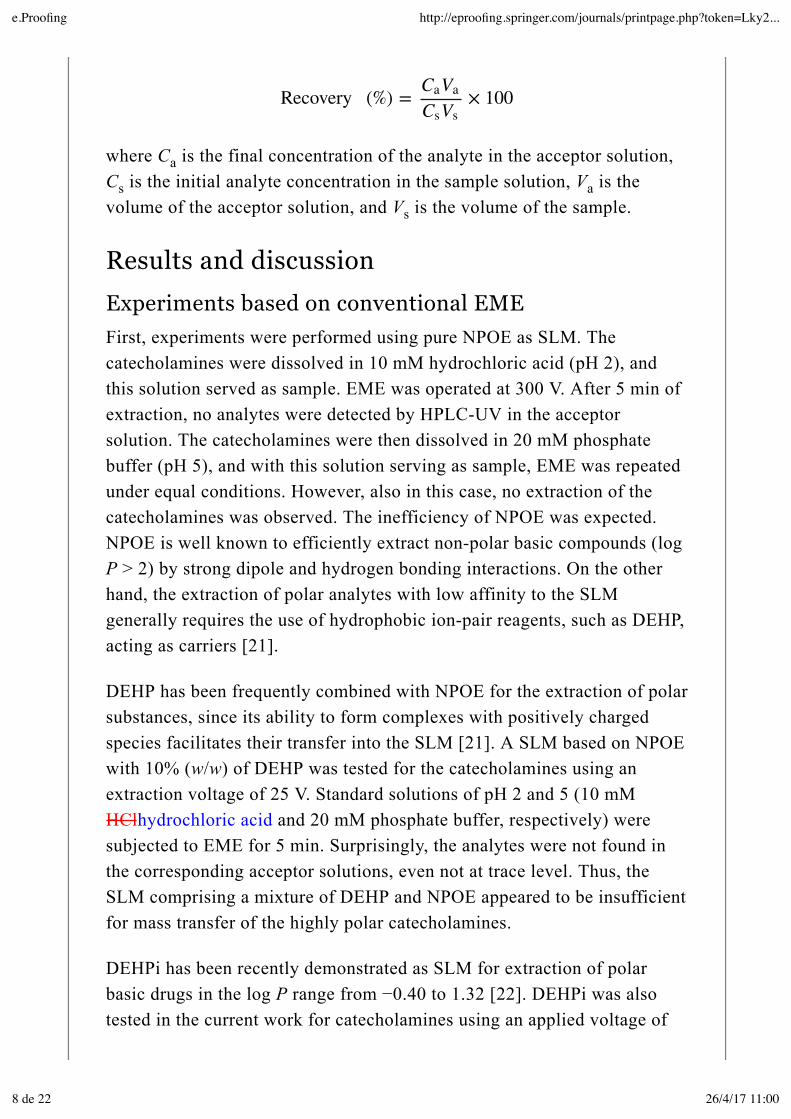
Experiments based on complexation-mediated EMEThe molecular structures of the catecholamines include two phenolicgroups in ortho position as a common feature. PBA and derivatives possessa high affinity to complex these phenols, forming boronate esters byreversible covalent binding (Fig. 1). Based on this type of complexation,previous publications [24, 25, 26] have reported the ability of PBAderivatives to facilitate transport of diol containing species (e.g., DA,glucoside, fructose) through SLMs under passive diffusion conditions. Thisconcept was transferred to EME in the present work, and tested underelectrokinetic migration conditions. The idea was to enhance the masstransfer of catecholamines due to selective complexation, whilesuppressing the general mass transfer of cationic matrix components.
Fig. 1
PBA complexation of diol groups
In a first experiment, PBA was dissolved in standard solution of pH 5 at aconcentration of 5 mM. EME was performed for 10 min at 50 V using
e.Proofing http://eproofing.springer.com/journals/printpage.php?token=Lky2...
9 de 22 26/4/17 11:00
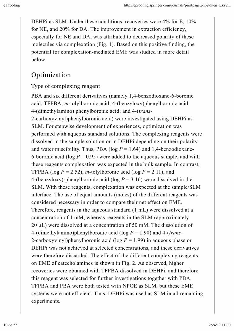
DEHPi as SLM. Under these conditions, recoveries were 4% for E, 10%for NE, and 20% for DA. The improvement in extraction efficiency,especially for NE and DA, was attributed to decreased polarity of thesemolecules via complexation (Fig. 1). Based on this positive finding, thepotential for complexation-mediated EME was studied in more detailbelow.
OptimizationType of complexing reagent
PBA and six different derivatives (namely 1,4-benzodioxane-6-boronicacid; TFPBA; m-tolylboronic acid; 4-(benzyloxy)phenylboronic acid;4-(dimethylamino) phenylboronic acid; and 4-(trans-2-carboxyvinyl)phenylboronic acid) were investigated using DEHPi asSLM. For stepwise development of experiences, optimization wasperformed with aqueous standard solutions. The complexing reagents weredissolved in the sample solution or in DEHPi depending on their polarityand water miscibility. Thus, PBA (log P = 1.64) and 1,4-benzodioxane-6-boronic acid (log P = 0.95) were added to the aqueous sample, and withthese reagents complexation was expected in the bulk sample. In contrast,TFPBA (log P = 2.52), m-tolylboronic acid (log P = 2.11), and4-(benzyloxy)-phenylboronic acid (log P = 3.16) were dissolved in theSLM. With these reagents, complexation was expected at the sample/SLMinterface. The use of equal amounts (moles) of the different reagents wasconsidered necessary in order to compare their net effect on EME.Therefore, reagents in the aqueous standard (1 mL) were dissolved at aconcentration of 1 mM, whereas reagents in the SLM (approximately20 µL) were dissolved at a concentration of 50 mM. The dissolution of4-(dimethylamino)phenylboronic acid (log P = 1.90) and 4-(trans-2-carboxyvinyl)phenylboronic acid (log P = 1.99) in aqueous phase orDEHPi was not achieved at selected concentrations, and these derivativeswere therefore discarded. The effect of the different complexing reagentson EME of catecholamines is shown in Fig. 2. As observed, higherrecoveries were obtained with TFPBA dissolved in DEHPi, and thereforethis reagent was selected for further investigations together with PBA.TFPBA and PBA were both tested with NPOE as SLM, but these EMEsystems were not efficient. Thus, DEHPi was used as SLM in all remainingexperiments.
e.Proofing http://eproofing.springer.com/journals/printpage.php?token=Lky2...
10 de 22 26/4/17 11:00
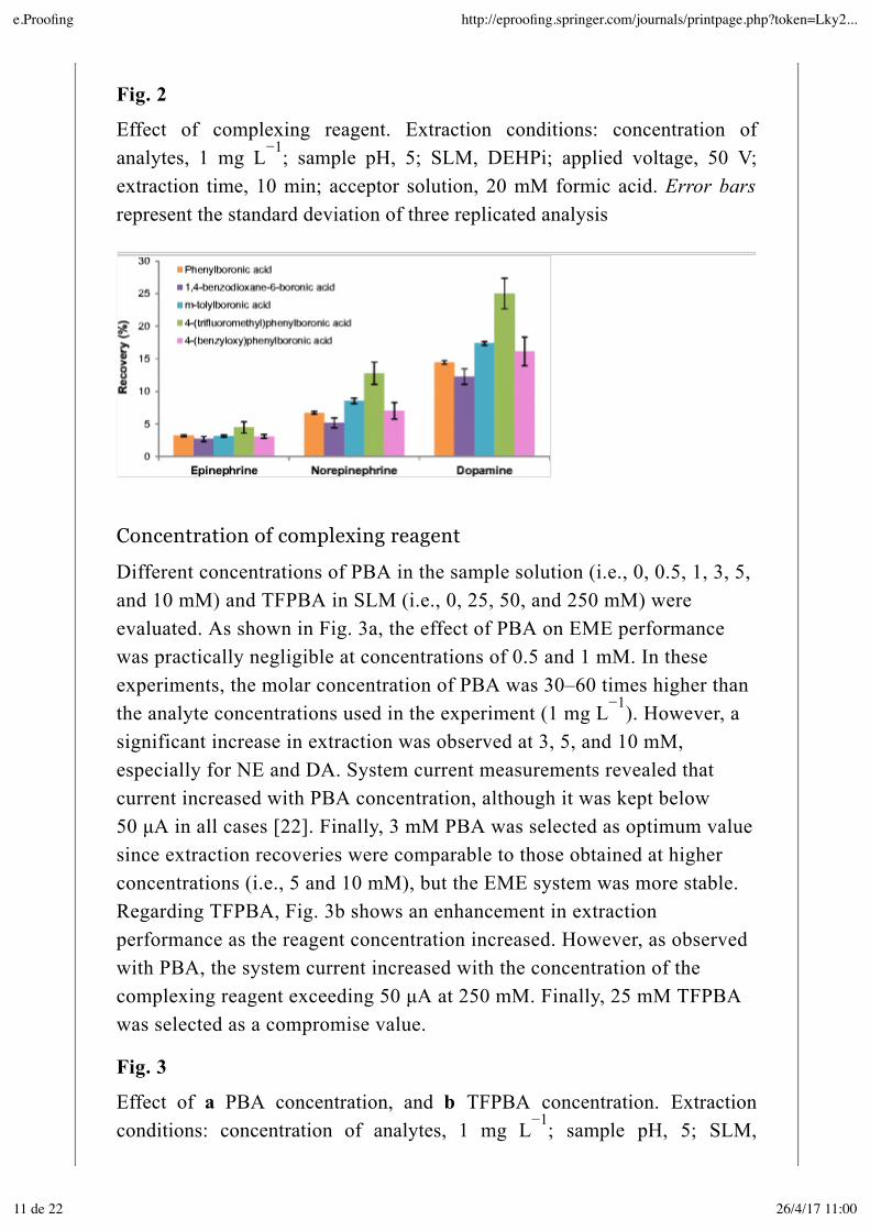
Fig. 2
Effect of complexing reagent. Extraction conditions: concentration ofanalytes, 1 mg L ; sample pH, 5; SLM, DEHPi; applied voltage, 50 V;extraction time, 10 min; acceptor solution, 20 mM formic acid. Error barsrepresent the standard deviation of three replicated analysis
Concentration of complexing reagent
Different concentrations of PBA in the sample solution (i.e., 0, 0.5, 1, 3, 5,and 10 mM) and TFPBA in SLM (i.e., 0, 25, 50, and 250 mM) wereevaluated. As shown in Fig. 3a, the effect of PBA on EME performancewas practically negligible at concentrations of 0.5 and 1 mM. In theseexperiments, the molar concentration of PBA was 30–60 times higher thanthe analyte concentrations used in the experiment (1 mg L ). However, asignificant increase in extraction was observed at 3, 5, and 10 mM,especially for NE and DA. System current measurements revealed thatcurrent increased with PBA concentration, although it was kept below50 µA in all cases [22]. Finally, 3 mM PBA was selected as optimum valuesince extraction recoveries were comparable to those obtained at higherconcentrations (i.e., 5 and 10 mM), but the EME system was more stable.Regarding TFPBA, Fig. 3b shows an enhancement in extractionperformance as the reagent concentration increased. However, as observedwith PBA, the system current increased with the concentration of thecomplexing reagent exceeding 50 µA at 250 mM. Finally, 25 mM TFPBAwas selected as a compromise value.
Fig. 3
Effect of a PBA concentration, and b TFPBA concentration. Extractionconditions: concentration of analytes, 1 mg L ; sample pH, 5; SLM,
−1
−1
−1
e.Proofing http://eproofing.springer.com/journals/printpage.php?token=Lky2...
11 de 22 26/4/17 11:00
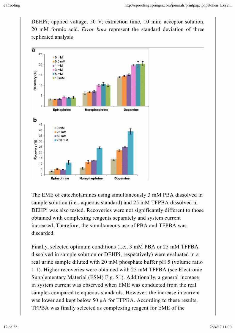
DEHPi; applied voltage, 50 V; extraction time, 10 min; acceptor solution,20 mM formic acid. Error bars represent the standard deviation of threereplicated analysis
The EME of catecholamines using simultaneously 3 mM PBA dissolved insample solution (i.e., aqueous standard) and 25 mM TFPBA dissolved inDEHPi was also tested. Recoveries were not significantly different to thoseobtained with complexing reagents separately and system currentincreased. Therefore, the simultaneous use of PBA and TFPBA wasdiscarded.
Finally, selected optimum conditions (i.e., 3 mM PBA or 25 mM TFPBAdissolved in sample solution or DEHPi, respectively) were evaluated in areal urine sample diluted with 20 mM phosphate buffer pH 5 (volume ratio1:1). Higher recoveries were obtained with 25 mM TFPBA (see ElectronicSupplementary Material (ESM) Fig. S1). Additionally, a general increasein system current was observed when EME was conducted from the realsamples compared to aqueous standards. However, the increase in currentwas lower and kept below 50 µA for TFPBA. According to these results,TFPBA was finally selected as complexing reagent for EME of the
e.Proofing http://eproofing.springer.com/journals/printpage.php?token=Lky2...
12 de 22 26/4/17 11:00
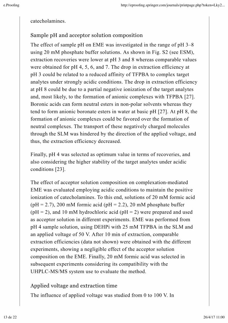
catecholamines.
Sample pH and acceptor solution composition
The effect of sample pH on EME was investigated in the range of pH 3–8using 20 mM phosphate buffer solutions. As shown in Fig. S2 (see ESM),extraction recoveries were lower at pH 3 and 8 whereas comparable valueswere obtained for pH 4, 5, 6, and 7. The drop in extraction efficiency atpH 3 could be related to a reduced affinity of TFPBA to complex targetanalytes under strongly acidic conditions. The drop in extraction efficiencyat pH 8 could be due to a partial negative ionization of the target analytesand, most likely, to the formation of anionic complexes with TFPBA [27].Boronic acids can form neutral esters in non-polar solvents whereas theytend to form anionic boronate esters in water at basic pH [27]. At pH 8, theformation of anionic complexes could be favored over the formation ofneutral complexes. The transport of these negatively charged moleculesthrough the SLM was hindered by the direction of the applied voltage, andthus, the extraction efficiency decreased.
Finally, pH 4 was selected as optimum value in terms of recoveries, andalso considering the higher stability of the target analytes under acidicconditions [23].
The effect of acceptor solution composition on complexation-mediatedEME was evaluated employing acidic conditions to maintain the positiveionization of catecholamines. To this end, solutions of 20 mM formic acid(pH = 2.7), 200 mM formic acid (pH = 2.2), 20 mM phosphate buffer(pH = 2), and 10 mM hydrochloric acid (pH = 2) were prepared and usedas acceptor solution in different experiments. EME was performed frompH 4 sample solution, using DEHPi with 25 mM TFPBA in the SLM andan applied voltage of 50 V. After 10 min of extraction, comparableextraction efficiencies (data not shown) were obtained with the differentexperiments, showing a negligible effect of the acceptor solutioncomposition on the EME. Finally, 20 mM formic acid was selected insubsequent experiments considering its compatibility with theUHPLC-MS/MS system use to evaluate the method.
Applied voltage and extraction time
The influence of applied voltage was studied from 0 to 100 V. In
e.Proofing http://eproofing.springer.com/journals/printpage.php?token=Lky2...
13 de 22 26/4/17 11:00
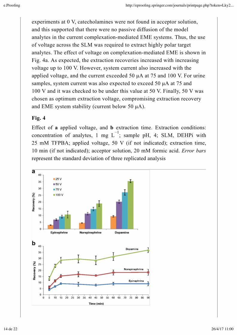
experiments at 0 V, catecholamines were not found in acceptor solution,and this supported that there were no passive diffusion of the modelanalytes in the current complexation-mediated EME systems. Thus, the useof voltage across the SLM was required to extract highly polar targetanalytes. The effect of voltage on complexation-mediated EME is shown inFig. 4a. As expected, the extraction recoveries increased with increasingvoltage up to 100 V. However, system current also increased with theapplied voltage, and the current exceeded 50 µA at 75 and 100 V. For urinesamples, system current was also expected to exceed 50 µA at 75 and100 V and it was checked to be under this value at 50 V. Finally, 50 V waschosen as optimum extraction voltage, compromising extraction recoveryand EME system stability (current below 50 µA).
Fig. 4
Effect of a applied voltage, and b extraction time. Extraction conditions:concentration of analytes, 1 mg L ; sample pH, 4; SLM, DEHPi with25 mM TFPBA; applied voltage, 50 V (if not indicated); extraction time,10 min (if not indicated); acceptor solution, 20 mM formic acid. Error barsrepresent the standard deviation of three replicated analysis
−1
e.Proofing http://eproofing.springer.com/journals/printpage.php?token=Lky2...
14 de 22 26/4/17 11:00
Finally, extraction time was investigated and the results are shown in Fig.4b. Recoveries increased as a function of time during the first 15 min ofextraction, as expected. Longer extraction times did not improve extractionrecoveries and, according to previous publications, this effect could beattributed to pH changes in the acceptor solution due to electrolysis [22,28]. The extraction time effect was also evaluated in a real urine samplediluted with 20 mM phosphate buffer pH 4 (volume ratio 1:1). As with theaqueous samples, no improvement in recoveries was observed after 15 minof extraction (data not shown). Therefore, 15 min was finally selected asoptimum time for complexation-mediated EME of the catecholamines.
Extraction performance in urine samples underoptimized conditionsThe final EME system was based on the following optimized conditions:SLM, DEHPi with 25 mM TFPBA; sample pH, 4; acceptor solution,20 mM formic acid; applied voltage, 50 V; and extraction time, 15 min.Under these conditions, recoveries were 10% for E, 15% for NE, and 29%for DA when EME was performed from aqueous standards. However,when analyzing urine samples, extraction recoveries decreasedsignificantly as discussed in “Evaluation” section.
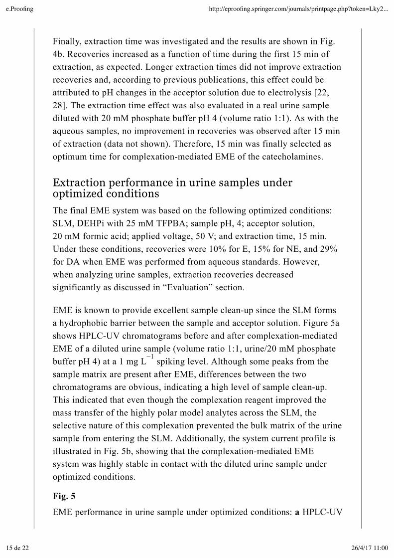
EME is known to provide excellent sample clean-up since the SLM formsa hydrophobic barrier between the sample and acceptor solution. Figure 5ashows HPLC-UV chromatograms before and after complexation-mediatedEME of a diluted urine sample (volume ratio 1:1, urine/20 mM phosphatebuffer pH 4) at a 1 mg L spiking level. Although some peaks from thesample matrix are present after EME, differences between the twochromatograms are obvious, indicating a high level of sample clean-up.This indicated that even though the complexation reagent improved themass transfer of the highly polar model analytes across the SLM, theselective nature of this complexation prevented the bulk matrix of the urinesample from entering the SLM. Additionally, the system current profile isillustrated in Fig. 5b, showing that the complexation-mediated EMEsystem was highly stable in contact with the diluted urine sample underoptimized conditions.
Fig. 5
EME performance in urine sample under optimized conditions: a HPLC-UV
−1
e.Proofing http://eproofing.springer.com/journals/printpage.php?token=Lky2...
15 de 22 26/4/17 11:00
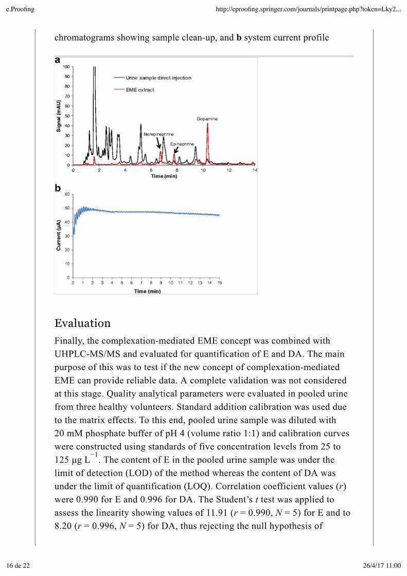
chromatograms showing sample clean-up, and b system current profile
EvaluationFinally, the complexation-mediated EME concept was combined withUHPLC-MS/MS and evaluated for quantification of E and DA. The mainpurpose of this was to test if the new concept of complexation-mediatedEME can provide reliable data. A complete validation was not consideredat this stage. Quality analytical parameters were evaluated in pooled urinefrom three healthy volunteers. Standard addition calibration was used dueto the matrix effects. To this end, pooled urine sample was diluted with20 mM phosphate buffer of pH 4 (volume ratio 1:1) and calibration curveswere constructed using standards of five concentration levels from 25 to125 µg L . The content of E in the pooled urine sample was under thelimit of detection (LOD) of the method whereas the content of DA wasunder the limit of quantification (LOQ). Correlation coefficient values (r)were 0.990 for E and 0.996 for DA. The Student’s t test was applied toassess the linearity showing values of 11.91 (r = 0.990, N = 5) for E and to8.20 (r = 0.996, N = 5) for DA, thus rejecting the null hypothesis of
−1
e.Proofing http://eproofing.springer.com/journals/printpage.php?token=Lky2...
16 de 22 26/4/17 11:00
non-linear correlation for a 5% significance level and 3 degrees of freedom(t = 3.18) [29]. The repeatability of the method, expressed ascoefficient of variation (CV), was determined by five consecutiveextractions from diluted pooled urine sample spiked at a concentrationlevel of 50 µg L . CV was 13% for both E and DA.
Extraction recoveries of the proposed procedure were found by thefollowing strategy. First, diluted pooled urine sample was spiked at50 µg L with E and DA and subjected to EME. Then, EME wasconducted from non-spiked diluted pooled urine and the final extract wasspiked at 50 µg L . Signals obtained in both experiments were comparedand, considering acceptor and sample solution volumes (i.e., 25 µL and1 mL, respectively), extraction recoveries were calculated to (5.5 ± 0.9)%for E and (15 ± 2)% for DA (n = 5). At this recovery level, LODs (S/N = 3)were 5.0 and 1.8 µg L , and LOQs (S/N = 10) were 16.5 and 6.0 µg Lfor E and DA, respectively. Enrichment factors were 2.2 for E and 6.0 forDA. Although low enrichment factor were obtained, they could be furtherimproved increasing sample and acceptor phases volume ratios.
Finally, diluted pooled urine sample was spiked at a known concentrationlevel (i.e., 50 µg L ) and analyzed by standard addition calibration usingstandards of five concentration levels from 25 to 125 µg L . Relativerecoveries were calculated as ratio between found and spikedconcentrations being (91 ± 26)% for E and (117 ± 20)% for DA, wherestandard deviation values were calculated using the s (i.e., standarddeviation of x-value estimated using regression line [29]).
ConclusionsIn this work, complexation-mediated EME of highly polar basic drugsubstances was demonstrated for the first time using selectedcatecholamines as model analytes. Complexation in the bulk sample withwater-soluble PBA derivatives added to the sample, and complexation atthe sample/SLM interface with water-insoluble PBA derivatives added tothe SLM were tested, and the latter concept appeared to be most efficient.Thus, complexation of the catecholamines with TFPBA at the sample/SLMinterface was found to enhance the mass transfer across the SLM. Becausethe complexation reaction involved substances with two phenolic groups inortho position only, the reaction was selective and therefore complexation-
0.05,3
−1
−1
−1
−1 −1
−1
−1
XE
e.Proofing http://eproofing.springer.com/journals/printpage.php?token=Lky2...
17 de 22 26/4/17 11:00
mediated EME appeared to be selective even from biological fluids. Thus,although the SLM permitted mass transfer of target analytes with −1 > logP > −2, most bulk matrix components in human urine was unable to passthe SLM, and acceptable sample clean-up was achieved. Additionally, thecurrent in the complexation-mediated EME system was easily controlledand kept below 50 µA, and therefore the system provided acceptablestability. The complexation-mediated EME concept was combined withUHPLC-MS/MS to develop a model application. Although the workpresented in this paper is preliminary in nature, complexation-mediatedEME showed potential and extraction of basic drugs with log P in therange −1 to −2 was demonstrated for the first time. Complexation-mediatedEME should be investigated in more detail in the future. With this concept,analyte detection may be performed with instruments much more simplethan mass spectrometry (as used in this initial work), and this may opennew and very interesting future possibilities.
AcknowledgementsThe authors would like to thank the Spanish Ministry of Science andInnovation (project n. CTQ2011-23968) and Generalitat Valenciana(Spain) (projects n. GVA/2014/096 and PROMETEO/2013/038) for thefinancial support. E. Fernández thanks Spanish Ministry of Education forher FPU grant (FPU13/03125) and mobility grant (EST15/00074).
Compliance with ethical standards
Informed consent was obtained from all individual participants included inthe study. Urine samples were collected from healthy volunteers andrandomized. Collection was performed in accordance with ethicalstandards and approved by the Director of School of Pharmacy (Universityof Oslo, Norway).
Conflict of interest The authors declare that they have no conflicts ofinterest.
Electronic supplementary material
ESM 1
(PDF 171 kb)
e.Proofing http://eproofing.springer.com/journals/printpage.php?token=Lky2...
18 de 22 26/4/17 11:00
References
1. Ghambarian M, Yamini Y, Esrafili A. Developments in hollow fiberbased liquid-phase microextraction: principles and applications.Microchim Acta. 2012;177:271–94.
2. Huang C, Jensen H, Seip KF, Gjelstad A, Pedersen-Bjergaard S.Mass transfer in electromembrane extraction—the link between theoryand experiments. J Sep Sci. 2016;39:188–97.
3. Fernández E, Vidal L. Liquid-phase microextraction techniques. In:Pena-Pereira F, editor. Miniaturization sample preparation. Warsaw: DeGruyter Open; 2014. p. 191–252.
4. Gjelstad A, Pedersen-Bjergaard S. Recent developments inelectromembrane extraction. Anal Methods. 2013;5:4549–57.
5. Marothu VK, Gorrepati M, Vusa R. Electromembrane extraction—anovel extraction technique for pharmaceutical, chemical, clinical andenvironmental analysis. J Chromatogr Sci. 2013;51:619–31.
6. Gjelstad A, Rasmussen KE, Pedersen-Bjergaard S. Electrokineticmigration across artificial liquid membranes tuning the membranechemistry to different types of drug substances. J Chromatogr A.2006;1124:29–34.
7. Nojavan S, Fakhari AR. Electro membrane extraction combined withcapillary electrophoresis for the determination of amlodipineenantiomers in biological samples. J Sep Sci. 2010;33:3231–8.
8. Fakhari AR, Tabani H, Nojavan S, Abedi H. Electromembraneextraction combined with cyclodextrin-modified capillaryelectrophoresis for the quantification of trimipramine enantiomers.Electrophoresis. 2012;33:506–15.
9. Davarani SSH, Najarian AM, Nojavan S, Tabatabaei M-A.Electromembrane extraction combined with gas chromatography forquantification of tricyclic antidepressants in human body fluids. AnalChim Acta. 2012;725:51–6.
e.Proofing http://eproofing.springer.com/journals/printpage.php?token=Lky2...
19 de 22 26/4/17 11:00
10. Hasheminasab KS, Fakhari AR. Development and application ofcarbon nanotubes assisted electromembrane extraction (CNTs/EME) forthe determination of buprenorphine as a model of basic drugs fromurine samples. Anal Chim Acta. 2013;767:75–80.
11. Ahmar H, Fakhari AR, Tabani H, Shahsavani A. Optimization ofelectromembrane extraction combined with differential pulsevoltammetry using modified screen-printed electrode for thedetermination of sufentanil. Electrochim Acta. 2013;96:117–23.
12. Huang C, Eibak LEE, Gjelstad A, Shen X, Trones R, Jensen H,Pedersen-Bjergaard S. Development of a flat membrane based devicefor electromembrane extraction: a new approach for exhaustiveextraction of basic drugs from human plasma. J Chromatogr A.2014;1326:7–12.
13. Asl YA, Yamini Y, Seidi S, Amanzadeh H. Dynamicelectromembrane extraction: automated movement of donor andacceptor phases to improve extraction efficiency. J Chromatogr A.2015;1419:10–8.
14. Rouhollahi A, Kouchaki M, Seidi S. Electrically stimulated liquidphase microextraction combined with differential pulse voltammetry: anew and efficient design for in situ determination of clozapine fromcomplicated matrices. RSC Adv. 2016;6:12943–52.
15. Gjelstad A, Rasmussen KE, Pedersen-Bjergaard S.Electromembrane extraction of basic drugs from untreated humanplasma and whole blood under physiological pH conditions. AnalBioanal Chem. 2009;393:921–8.
16. Eibak LEE, Gjelstad A, Rasmussen KE, Pedersen-Bjergaard S.Kinetic electro membrane extraction under stagnant conditions—fastisolation of drugs from untreated human plasma. J Chromatogr A.2010;1217:5050–6.
17. Jamt REG, Gjelstad A, Eibak LEE, Øiestad EL, Christophersen AS,Rasmussen KE, Pedersen-Bjergaard S. Electromembrane extraction ofstimulating drugs from undiluted whole blood. J Chromatogr A.
e.Proofing http://eproofing.springer.com/journals/printpage.php?token=Lky2...
20 de 22 26/4/17 11:00
2012;1232:27–36.
18. Šlampová A, Kubáň P, Boček P. Electromembrane extraction usingstabilized constant d.c. electric current—a simple tool for improvementof extraction performance. J Chromatogr A. 2012;1234:32–7.
19. Kjelsen IJØ, Gjelstad A, Rasmussen KE, Pedersen-Bjergaard S.Low-voltage electromembrane extraction of basic drugs from biologicalsamples. J Chromatogr A. 2008;1180:1–9.
20. Seip KF, Faizi M, Vergel C, Gjelstad A, Pedersen-Bjergaard S.Stability and efficiency of supported liquid membranes inelectromembrane extraction-a link to solvent properties. Anal BioanalChem. 2014;406:2151–61.
21. Huang C, Seip KF, Gjelstad A, Pedersen-Bjergaard S.Electromembrane extraction for pharmaceutical and biomedicalanalysis—quo vadis. J Pharm Biomed Anal. 2015;113:97–107.
22. Huang C, Seip KF, Gjelstad A, Pedersen-Bjergaard S.Electromembrane extraction of polar basic drugs from plasma with purebis(2-ethylhexyl) phosphite as supported liquid membrane. Anal ChimActa. 2016;934:80–7.
23. Bicker J, Fortuna A, Alves G, Falcão A. Liquid chromatographicmethods for the quantification of catecholamines and their metabolitesin several biological samples—a review. Anal Chim Acta.2013;768:12–34.
24. Paugam M-F, Valencia LS, Boggess B, Smith BD. Selectivedopamine transport using a crown boronic acid. J Am Chem Soc.1994;116:11203–4.
25. Takeuchi M, Koumoto K, Goto M, Shinkai S. Efficient glucosideextraction mediated by a boronic acid with an intramolecular quaternaryammonium ion. Tetrahedron. 1996;52:12931–40.
26. Di Luccio M, Smith BD, Kida T, Borges CP, Alves TLM.Separation of fructose from a mixture of sugars using supported liquid
e.Proofing http://eproofing.springer.com/journals/printpage.php?token=Lky2...
21 de 22 26/4/17 11:00
membranes. J Memb Sci. 2000;174:217–24.
27. Kanamori T, Funatsu T, Tsunoda M. Determination ofcatecholamines and related compounds in mouse urine using column-switching HPLC. Analyst. 2016;141:2568–73.
28. Kubán P, Boček P. The effects of electrolysis on operationalsolutions in electromembrane extraction: the role of acceptor solution. JChromatogr A. 2015;1398:11–9.
29. Miller JN, Miller JC. Statistics and chemometrics for analyticalchemistry. fifth ed. London: Pearson Prentice Hall; 2005.
e.Proofing http://eproofing.springer.com/journals/printpage.php?token=Lky2...
22 de 22 26/4/17 11:00