az élő rendszerek konzervatív...
TRANSCRIPT

Az élő rendszerek konzervatív struktúrái:
A lipidek

A lipidek hidrofób molekulák sokrétű csoportja
• A lipidek olyan különböző felépítésű és funkciójú hidrofób anyagok, melyek apoláros oldószerekben jól, vízben nem vagy alig oldódnak.
• Nem képeznek polimer makromolekulákat
• Tartalmazhatnak poláros kötéseket (pl. oxigénnel), de elsősorban szénhidrogén régiókat találunk bennük
• Csoportosíthatóak kémiai összetételük és biológiai funkcióik alapján

A lipidek csoportosítása kémiai összetétel alapján
• Egyszerű lipidek: hidrolízissel egyszerűbb alakotókra nem bonthatóak fel (nem szappanosíthatóak)
– Zsírsavak – Szteroidok – Karotinoidok – Terpének – Prosztaglandinok
• Összetett lipidek: hidrolízissel egyszerűbb alakotókra bonthatóak fel (szappanosíthatóak)
– Neutrális zsírok – Viaszok – Foszfatidok

Zsírsavak
• Hosszú szénhidrogén láncokat tartalmazó molekulák, a végükön karboxil csoporttal (páros számú szénatomból állnak)
• A zsírsavak szabadon is előfordulnak, de lehetnek az anyagcserefolyamatok köztes-termékei vagy összetett lipidek prekurzorai
• A szénhidrát lánc tartalmazhat kettős kötéseket, ez esetben telítetlen zsírsavakról beszélünk (pl. olajsav).

Zsírsavak hidrogenizálása
• A szénlánc hossza, és a kettős kötés jelenléte (mely általában cisz konformációjú), meghatározza a molekula alakját és tulajdonságait:
If the acyl groups are long enough, these molecules areinsoluble in water even though they contain three polar esterbonds. Fatty acyl groups also form the hydrophobic portionof phospholipids, which we discuss next.
Phospholipids Associate Noncovalently to Formthe Basic Bilayer Structure of BiomembranesBiomembranes are large flexible sheets that serve as theboundaries of cells and their intracellular organelles andform the outer surfaces of some viruses. Membranes liter-ally define what is a cell (the outer membrane and the con-tents within the membrane) and what is not (the extracellularspace outside the membrane). Unlike the proteins, nucleicacids, and polysaccharides, membranes are assembled by thenoncovalent association of their component building blocks.
The primary building blocks of all biomembranes are phos-pholipids, whose physical properties are responsible for theformation of the sheetlike structure of membranes.
Phospholipids consist of two long-chain, nonpolar fattyacyl groups linked (usually by an ester bond) to small, highlypolar groups, including a phosphate. In phosphoglycerides,the major class of phospholipids, fatty acyl side chains are es-terified to two of the three hydroxyl groups in glycerol. Thethird hydroxyl group is esterified to phosphate. The simplestphospholipid, phosphatidic acid, contains only these compo-nents. In most phospholipids found in membranes, the phos-phate group is esterified to a hydroxyl group on anotherhydrophilic compound. In phosphatidylcholine, for example,choline is attached to the phosphate (Figure 2-19). The neg-ative charge on the phosphate as well as the charged or polargroups esterified to it can interact strongly with water. The
44 CHAPTER 2 • Chemical Foundations
H3C
Palmitate(ionized form of palmitic acid)
H
H
C
H
H
C
H
H
C
H
H
C
H
H
C
H
H
C
H
H
C
H
H
C
H
H
C
H
H
C
H
H
C
H
H
C
H
H
C
H
H
C CO
O!
H3C
Oleate(ionized form of oleic acid)
H
H
C
H
H
C
H
H
C
H
H
C
H
H
C
H
H
C
H
H
C
H
C
H
C
H
H
C
H
H
C
H
H
C
H
H
C
H
H
C
H
H
C
H
H
C CO
O!
▲ FIGURE 2-18 The effect of a double bond on the shapeof fatty acids. Shown are space-filling mode ls and chem icalstructures of the ionized form of palm itic acid, a saturated fattyacid w ith 16 C atoms, and ole ic acid, an unsaturated one w ith
18 C atoms. In saturated fatty acids, the hydrocarbon chain is often linear; the cis double bond in oleate creates a rigid kink inthe hydrocarbon chain. [After L. Stryer, 1994, B ioch e m istry, 4th ed., W. H . Freeman and Company, p. 265.]
CholinePHOSPHATIDYLCHOLINE
Hydrophobic tail
Hydrophilic headPhosphate
Glycerol
Fatty acid chains
CH3
C
H2
H2
CN+
CH3CH3
O
CO
O
CO
OP
O
OO−
H2C
CH
CH2
▲ FIGURE 2-19 Phosphatidylcholine, a typical phospho-glyceride. A ll phosphoglycerides are amphipathic, having a hydrophobic tail (ye llow) and a hydrophilic head (blue) in whichglycerol is linked via a phosphate group to an alcohol. E ither of
or both the fatty acyl side chains in a phosphoglyceride may besaturated or unsaturated. In phosphatidic acid (red), the simplestphospholipid, the phosphate is not linked to an alcohol.
Palmitát (olvadáspont: 63,1°C)
Oleát (olvadáspont: 13,4°C)

Zsírsavak
• Példák zsírsavakra:

Esszenciális zsírsavak
• Az emberi test képes szintetizálni telített, vagy omega-9 egyszeresen telítetlen zsírsavakat, de nem képes kettős kötéseket vinni az omega-3 illetve az omega-6 helyekre, ezért az ilyen zsírsavakat kívülről kell pótolni.
Omega-3
Omega-6
•Esszenciális és nem-esszenciális zsírsavak

Omega-6 és Omega-3 zsírsavak

Szteroidok
• A Szteroidok szterán vázzal rendelkező lipidek.
Főbb csoportjaik: • Szterolok • Epesavak • Hormonok • D-vitamin

Koleszterin
• Koleszterin elsősorban a biológiai membánokban előforduló amfipatikus molekula. A membránalkotók 50-70%-át is kiteheti. A membránok fluiditását csökkenti, azokat merevíti.
Koleszterol
•C3 atomon –OH csoport•C10 atomon –CH3 csoport•C13 atomon –CH3 csoport•C17 atomon 8 C atomból állószénhidrogén lánc
•Amfipatikus molekula•-OH csoport – poláris fejcsoport•Szterán váz + CH lánc – apoláris rész
poláros feji rész
apoláros Szénhidrogén lánc
Merev planáris gyűrűsruktúra
A koleszterint a legtöbb szerv szintetizálja (elsősorban a máj), másrészt a táplálékkal kerül a szervezetbe. A koleszterin előanyaga az epesavaknak, szteroidhormonoknak és a D-vitaminnak.

Koleszterin
• A plazmamembránon kívül a szérum lipoproteinek (LDL, HDL, VLDL) tartalmaznak koleszterint. Ezek a lipoproteinek nem- kovalens kötéssel összekapcsolt lipidek és fehérjék (apolipoproteinek) változó arányú keverékének komplexei.
HDL

Epesavak
• Az epesavak a májban koleszterinből képződnek. Szintézisük jelenti a koleszterin lebontásának és eltávolításának fő útját.
• Az epesavas sók természetes detergensek, poláros és apoláros részeik vannak. Vizes oldatban micellákat képeznek.
• Epesavas sók jelenlétében a lipidekből egyszerű rázással emulzió képződik, amit a diszpergált részecskék felületének negatív töltése stabilizál
phosphate and its associated esterified group, the “head”group of a phospholipid, is hydrophilic, whereas the fattyacyl chains, the “tails,” are hydrophobic.
The amphipathic nature of phospholipids, which governstheir interactions, is critical to the structure of biomem-branes. When a suspension of phospholipids is mechanicallydispersed in aqueous solution, the phospholipids aggregateinto one of three forms: sphericalmicelles and liposomes andsheetlike, two-molecule-thick phospholipid bilayers (Figure2-20). The type of structure formed by a pure phospholipidor a mixture of phospholipids depends on several factors, in-cluding the length of the fatty acyl chains, their degree ofsaturation, and temperature. In all three structures, the hy-drophobic effect causes the fatty acyl chains to aggregate andexclude water molecules from the “core.” Micelles are rarelyformed from natural phosphoglycerides, whose fatty acylchains generally are too bulky to fit into the interior of a micelle. If one of the two fatty acyl chains is removed by hydrolysis, forming a lysophospholipid, the predominanttype of aggregate that forms is the micelle. Common deter-gents and soaps form micelles in aqueous solution that be-have as tiny ball bearings, thus giving soap solutions theirslippery feel and lubricating properties.
Under suitable conditions, phospholipids of the compo-sition present in cells spontaneously form symmetric phos-pholipid bilayers. Each phospholipid layer in this lamellar
structure is called a leaflet. The fatty acyl chains in eachleaflet minimize contact with water by aligning themselvestightly together in the center of the bilayer, forming a hydrophobic core that is about 3 nm thick (see Figure 2-20).The close packing of these nonpolar tails is stabilized by thehydrophobic effect and van der Waals interactions betweenthem. Ionic and hydrogen bonds stabilize the interaction ofthe phospholipid polar head groups with one another andwith water.
A phospholipid bilayer can be of almost unlimited size—from micrometers (!m) to millimeters (mm) in length orwidth—and can contain tens of millions of phospholipidmolecules. Because of their hydrophobic core, bilayers arevirtually impermeable to salts, sugars, and most other smallhydrophilic molecules. The phospholipid bilayer is the basicstructural unit of nearly all biological membranes; thus, al-though they contain other molecules (e.g., cholesterol, gly-colipids, proteins), biomembranes have a hydrophobic corethat separates two aqueous solutions and acts as a perme-ability barrier. The structural organization of biomembranesand the general properties of membrane proteins are de-scribed in Chapter 5.
KEY CONCEPTS OF SECTION 2.2
Chemical Building Blocks of Cells■ Three major biopolymers are present in cells: proteins,composed of amino acids linked by peptide bonds; nucleicacids, composed of nucleotides linked by phosphodiesterbonds; and polysaccharides, composed of monosaccharides(sugars) linked by glycosidic bonds (see Figure 2-11).
■ Many molecules in cells contain at least one asymmet-ric carbon atom, which is bonded to four dissimilar atoms.Such molecules can exist as optical isomers (mirror im-ages), designated D and L, which have different biologicalactivities. In biological systems, nearly all sugars are D iso-mers, while nearly all amino acids are L isomers.
■ Differences in the size, shape, charge, hydrophobicity,and reactivity of the side chains of amino acids determinethe chemical and structural properties of proteins (see Fig-ure 2-13).
■ Amino acids with hydrophobic side chains tend to clus-ter in the interior of proteins away from the surroundingaqueous environment; those with hydrophilic side chainsusually are toward the surface.
■ The bases in the nucleotides composing DNA and RNAare heterocyclic rings attached to a pentose sugar. Theyform two groups: the purines—adenine (A) and guanine(G)—and the pyrimidines—cytosine (C), thymine (T), anduracil (U) (see Figure 2-15). A, G, T, and C are in DNA,and A, G, U, and C are in RNA.
■ Glucose and other hexoses can exist in three forms: anopen-chain linear structure, a six-member (pyranose) ring, and
2.2 • Chemical Building Blocks of Cells 45
Phospholipid bilayer
Liposome
M icelle
▲ FIGURE 2-20 Cross-sectional views of the three struc-tures formed by phospholipids in aqueous solutions. Thewhite spheres depict the hydrophilic heads of the phospholipids,and the squiggly black lines (in the ye llow regions) represent the hydrophobic tails. Shown are a spherical m ice lle w ith a hydrophobic interior composed entire ly of fatty acyl chains; aspherical liposome , which has t wo phospholipid layers and anaqueous center; and a t wo-molecule-thick sheet of phospholipids,or bilayer, the basic structural unit of biomembranes.

Szteroid hormonok
• Kortikoszteroidok (mellékvese kéreg hormonok)
• glikokortikoidok: kortizol (Stressz hormon - fokozza a glukoneogenezist, segíti a glükóz felhasználást, és jelentős gyulladáscsökkentő hatása is van)
• mineralokortikoidok: aldoszteron (só- és vízháztartását szabályozzák)
• Nemi hormonok
• Női nemi hormonok: ösztrogének és progesztinek (ösztradiol, progeszteron)
• Hím nemi hormonok: tesztoszteron és adrogének

D-vitamin
• Szintézise koleszterolból történik, a szteránváz felnyílásával
• Hatása: a Ca2+ és a foszfát szintjét szabályozza: csontok képződése, növekedése, ásványok beépülése
D-vitamin szintézis

Terpének: Karotinoidok
• 5 szénatomos, izoprén egységekből felépülő apoláros vegyületek (pl. β-karotin)
A β-karotin, az A-vitamin és a retinálprekurzora
Konjugált kettős kötés rendszert tartalmaznak, ezért színesek.

Prosztaglandinok
• Arachidonsavból szintetizálódnak
Biológiai hatás: simaizom kontrakció, vérnyomás csökkentés, véralvadás, anyagcserefolyamatok szabályozása. A ciklooxigenáz enzim blokkolása gyulladáscsökkentő (pl. acetilszalicilsav).
Prosztaglandinok

Ciklooxigenáz-gátló gyulladáscsökkentő hatóanyagok

A lipidek csoportosítása kémiai összetétel alapján
• Egyszerű lipidek: hidrolízissel egyszerűbb alakotókra nem bonthatóak fel (nem szappanosíthatóak)
– Zsírsavak – Szteroidok – Karotinoidok – Terpének – Prosztaglandinok
• Összetett lipidek: hidrolízissel egyszerűbb alakotókra bonthatóak fel (szappanosíthatóak)
– Neutrális zsírok – Viaszok – Foszfatidok

Neutrális zsírok
• A neutrális zsírok glicerinre és zsírsavakra bonthatóak.
• A glicerin mindhárom hidroxil csoportja észteresítve van zsírsavakkal (triglicerid)
• Általában különböznek a savkomponensek.

Zsírsav (palmitinsav)
Glicerol (a) Dehirációs reakció: észterkötés kialakulása
Észter kötés
(b) Zsír molekula (triacilglicerol)

(a) Telített zsírok
Telített zsír molekula
Sztearinsav, telített zsírsav

(b) Telítetlen zsír
Telítetlen zsír molekula
Olajsav, telítetlen zsírsav
cisz kettős kötés

Telítetlen kötések hidrogénezése
A telítetlen kötése hidrogénezése általában nem teljes. (szobahőmérsékleten félkemény állapot)

• A telített zsírok szobahőmérsékleten szilárdak (állati zsírok)
• A telítetlen zsírok vagy olajok szobahőmérsékleten folyékonyak
• A telített és transz-zsírsavakat tartalmazó zsírok közrejátszhatnak az érelmeszesedés kialakulásában
• A zsírok fő funkciója az energiaraktározás, a hőszigetelés és a mechanikai védelem.

Foszfolipidek
Az amino-alkohol csoport lehet:
A glicerofoszfolipidek esetében a glicerinhez két zsírsav és egy foszfát csoport kapcsolódik

(b) Tér-kitöltő modell(a) (c) Szerkezeti rajz Foszfolipid szimbólum
Zsírsavak
Hidrofil fej
Hidrofób farok
Kolin
Foszfát
Glicerol
Hid
rofó
b fa
rok
Hid
rofil
fej
A két zsírsav oldallánc hidrofób karakterű farki, a foszfát csoport és az ahhoz kapcsolódó részek hidrofil, feji véget képeznek. Így a molekula amfipatikus tulajdonságú lesz.

Foszfolipidek II.
• A szfingolipidek esetében a szfingozinhoz egy zsírsav és egy foszfát csoport kapcsolódik

Foszfolipidek hiányában IRDS alakulhat ki
Foszfolipidek (pl. lecitin és szfingomielin) vesznek részt a tüdő surfactant ayagának képzésében. Koraszülöttek esetén (28. előtt) a surfactant kevés lehet, és infant respiratory distress szindróma (IRDS) alakulhat ki.
A rizikó felméréhez a magzatvízből meghatározzák a lecitin/szfingomilein arányt. (<1,5 magas rizikót jelent)

65
Az amfipatikus molekula alakjától függően jellemzően kétféle végeredmény születhet (lásd 2.28. ábra). Ha az apoláros rész kisebb átmérőjű, mint a poláros (feji résznek nevezett) rész, akkor a molekula ék alakú lesz. Ebben az esetben az apoláros részek úgy temetődhetnek el, ha apoláros belsejű, poláros külsejű gömböket, micellákat képeznek. A micellák sugara megegyezik az amfipatikus molekulák hosszával. Ha az apoláros rész átmérője nagyjából megegyezik a poláros (feji) rész átmérőjével, akkor nem gömbök képződnek, hanem kettős membránok. Ezek olyan síkok, amelyeket két, amfipatikus molekulákból álló réteg alkot. Az apoláros molekularészek ebben a kettősrétegben egymás felé, míg a poláros részek kifelé, a víz felé fordulnak. A kettősmembránok maguk is gömböket képeznek, így nem alakulnak ki a kettősmembrán sík szélén olyan területek, ahol az apoláros részek vízzel érintkeznek. A hidrofób hatásnak (a szakirodalomban: hidrofób effektus) óriási jelentősége van a biológiai rendszerekben. A globuláris fehérjék natív szerkezetének kialakulásában például döntő szerepe van a makromolekula belsejébe temetődő, zömmel apoláros oldalláncokat tartalmazó úgynevezett hidrofób magnak. A globuláris fehérjék felszínén zömmel poláros csoportok helyezkednek el, tehát natív térszerkezete a micellákra emlékeztet (bár a részletek tekintetében természetesen sokkal összetettebb).
2.28. ábra: Az amfipatikus molekulák vízben szerkezetüktől függően micellákat, vagy membrán kettősréteget képeznek
A biológiai membránok, például a sejtmembrán, vagy az eukarióták számos organellumának a membránjai döntően foszfolipidekből állnak. A foszfolipidek olyan amfipatikus molekulák, amelyek apoláros részének

• A foszfolipidek vízben kétrétegű struktúrákat alkotnak, fő komponensei a biológiai membránoknak:
A viaszok
20
z Zsiradékok fĘ kémiai tulajdonságai (folytatás)z romlás ~ fĘként kémiai okok
z savasodás:gliceridek hidrolízise Ź szabad zsír-savak (hĘmérséklet, oxigén, fény, nedvesség, fémnyom katalizálhat)
z faggyúsodás:hidroxilsavak képzĘdése + polimeri-záció
z aldehid-avasság (leggyakoribb):gyökös folyamatok Ź a keletkezĘaldehidek adják a jellegzetes avas szagot (továbbá gyakran elszínezĘ-dött zsír)� védekezés: antioxidánsok használata
(élelmiszerekben pl.- mesterséges: BHA, BHT;- természetes: Į-tokoferolok)
EgyszerĦ lipidek – fizika vs. kémia III.R1 CH2 R2
CHR1 R2
CH R2
OO
R1
CH R2R1
OOH
CH R2R1
O
R1 CHO
aldehid-avasság
H
O2
R3 CH2 R4
CH R4R3
OH
R2
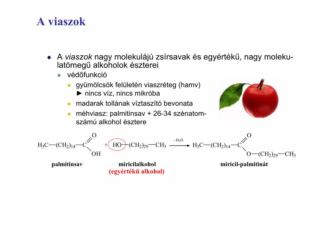
z A viaszok nagy molekulájú zsírsavak és egyértékĦ, nagy moleku-latömegĦ alkoholok észtereiz védĘfunkció
z gyümölcsök felületén viaszréteg (hamv)Ź nincs víz, nincs mikróba
z madarak tollának víztaszító bevonataz méhviasz: palmitinsav + 26-34 szénatom-
számú alkohol észtere
EgyszerĦ lipidek – viaszok definíciója
H3C (CH2)14
CH3(CH2)29O
OC
palmitinsav miricilalkohol miricil-palmitinát
H3C (CH2)14
OC
OHHO (CH2)29 CH3+
- H2O
(egyértékĦ alkohol)

Az élő rendszerek konzervatív struktúrái:
A nukleinsavak

A nuklein savak tárolják és továbbítják az öröklődés információit
• A polipeptidek aminosav szekvenciáját a öröklődés egységei, vagyis a gének szabják meg
• A géneket DNS, egy nukleinsav szekvenciája határozza meg

A nuklein savak szerepei
• Kétféle nukleinsavat különítünk el:
– Dezoxiribonuklein sav (DNS)
– Ribonuklein sav (RNS)
• A DNS irányítja a saját megkettőződését
• A DNS irányítja a hírvivő (messenger) RNS szintézisét és rajtuk keresztül a fehérje színtézist
• A fehérjék szintézise a riboszómákon történik

Riboszóma
Amino savakPolipeptid
Fehérje szintézis
3
mRNSAz mRNS a citoplazmába kerül
2
mRNS
mRNS szintézise a sejtmagban
DNS
SEJTMAGCITOPLAZMA
1

A nuklein savak szerkezete
• A nukleinsavak polimerek, polinukleotidok
• A monomer egységek a nukleotidok
• Minden nukleotid szerves bázisból, pentózból és foszfát csoportból áll
• A foszfát csoport mentes nukleotid részletet nukleozidnak nevezzük

Nukleotid monomerek
• Nucleozid = nitrogén tartalmú szerves bázis + cukor
• A N-tartalmú szerves bázisoknak két csoporja:
– Pirimidinek (citozin, timin, uracil) egyszerű hattagú gyűrűből épülnek fel
– Purinok (adenin és guanin) hat és öttagú gyűrű kombinációjából állnak
• A DNS-ben a cukor dezoxiribóz; az RNS-ben a cukor ribóz
• nukleotid = nukleozid+ foszfát csoport

5’ vég
Nukleozid
Nitrogén tartalmú bázis
Foszfát csoport Cukor
(pentóz)
(b) Nukleotid
(a) Polinukleotid vagy nukleinsav
3’ vég
3’C
3’C
5’C
5’C
Nitrogén tartalmú szerves bázisok Pirimidinek
Citozin (C) Timin (T, DNS-ben) Uracil (U,RNS-ben)
Purinok
Adenin (A) Guanin (G)
Cukrok
Dezoxiribóz (DNS-ben) Ribóz (RNS-ben)
(c) Nukleozid komponensek

Nukleotid polimerek
• A nukleotid polimerek összekapcsolódnak és polinukleotidokat hoznak létre
• A szomszédos nukleotidok kovalens kötéssel kapcsolódnak össze, mely a nukleotid 3ʹ szénatomjának –OH csoportja és a következő nukleotid 5ʹ szénatomjának foszfát csoportja között jön létre (foszfodiészter kötés)
• Így egy cukor-foszfát gerinc alakul ki • a bázisok sorrendje a DNS-ben vagy az mRNS-
ben minden génre egyedi

A DNS kettős hélixe
• A DNS molekulának két polinukleotid szála van, mely egy képzeletbeli tengely körül kettős hélixet képez.
• A DNS kettős hélixben a két gerinc egymásnak ellentétesen fut 5ʹ → 3ʹ irányba, vagyis antiparalell elrendeződésű
• Egy DNS molekula számos gén hordozhat
• A nitrogén tartalmú bázisok az antiparalell szálakban párba állnak és hidrogén kötésekkel kapcsolódnak: adenin (A) - timin (T),és guanin (G) - citozin (C)

A DNS kettős hélixe
A nitrogén tartalmú bázisok az antiparalell szálakban párba állnak és hidrogén kötésekkel kapcsolódnak: adenin (A) - timin (T) és guanin (G) - citozin (C)
of the double helix. Hydrophobic and van der Waals inter-actions between the stacked adjacent base pairs further sta-bilize the double-helical structure.
In natural DNA, A always hydrogen bonds with T andG with C, forming A·T and G·C base pairs as shown in Fig-ure 4-3b. These associations between a larger purine andsmaller pyrimidine are often called Watson-Crick base pairs.Two polynucleotide strands, or regions thereof, in which allthe nucleotides form such base pairs are said to be comple-mentary. However, in theory and in synthetic DNAs otherbase pairs can form. For example, a guanine (a purine) couldtheoretically form hydrogen bonds with a thymine (a pyrim-idine), causing only a minor distortion in the helix. The spaceavailable in the helix also would allow pairing between thetwo pyrimidines cytosine and thymine. Although the non-standard G·T and C·T base pairs are normally not found inDNA, G·U base pairs are quite common in double-helicalregions that form within otherwise single-stranded RNA.
Most DNA in cells is a right-handed helix. The x-ray dif-fraction pattern of DNA indicates that the stacked bases areregularly spaced 0.36 nm apart along the helix axis. The
helix makes a complete turn every 3.6 nm; thus there areabout 10.5 pairs per turn. This is referred to as the B formof DNA, the normal form present in most DNA stretches incells. On the outside of B-form DNA, the spaces between theintertwined strands form two helical grooves of differentwidths described as the major groove and the minor groove(see Figure 4-3a). As a consequence, the atoms on the edgesof each base within these grooves are accessible from out-side the helix, forming two types of binding surfaces. DNA-binding proteins can “read” the sequence of bases in duplexDNA by contacting atoms in either the major or the minorgrooves.
In addition to the major B form, three additional DNAstructures have been described. Two of these are comparedto B DNA in Figure 4-4. In very low humidity, the crystallo-graphic structure of B DNA changes to the A form; RNA-DNA and RNA-RNA helices exist in this form in cells and invitro. Short DNA molecules composed of alternating purine-pyrimidine nucleotides (especially Gs and Cs) adopt an al-ternative left-handed configuration instead of the normalright-handed helix. This structure is called Z DNA because
104 CHAPTER 4 • Basic Molecular Genetic Mechanisms
N H
N HH
O
O
N HH
H
H N
O
O
H N
H N
O
O
C H 2
H
H
H
(a)
M a j o rg r o o v e
M i n o rg r o o v e
5!
3!
3!
5!
OO
O
O
OO
OO
O
O
OO
OO
O
OO
OO
O
O
P
3!
(b)
C H 2
P
C H 2
P
C H 2
P
C H 25!
5!
5!5! C H 2
O
OO
OO
O
OO
OO
O
O
OO
O
P
P
P
C H 2
C H 2
O
OO
OP
3!
C H 3
T A
GC
AT
C G
N
N H N
N H N
N H N
N H N
▲ FIGURE 4-3 The DNA double helix. (a) Space-filling mode lof B DNA , the most common form of DNA in ce lls. The bases(light shades) project inward from the sugar-phosphate backbones(dark red and blue) of each strand, but the ir edges are accessiblethrough major and m inor grooves. Arrows indicate the 5’n3’ direction of each strand. Hydrogen bonds bet ween the bases arein the center of the structure . The major and m inor grooves
are lined by potential hydrogen bond donors and acceptors (highlighted in ye llow). (b) Chem ical structure of DNA double he lix. This extended schematic shows the t wo sugar-phosphatebackbones and hydrogen bonding bet ween the Watson-Crickbase pairs, A"T and G"C . [Part (a) from R. W ing et al., 1980, Nature287:755; part (b) from R. E . D ickerson, 1983, Sci. Am . 249:94.]

A DNS kettős hélixe
8
Nukleotidok és nukleinsavak
A DNS egyéb konformációi
z B-DNSz élĘlényekben, vizes
oldatban ez a leg-gyakoribb, a bázi-sok síkja majdnem merĘleges a cukor-foszfát gerincre
z A-DNSz dehidrált körülmé-
nyek között egy tö-mörebb forma jön létre, a bázisok síkja megdĘl
z Z-DNSz hosszú GCGCGC...
ismétlĘdések ezt a formát vehetik fel, amely balmenetes, zeg-zugos lefutású és megnyúlt
A-DNS B-DNS Z-DNS
Nukleotidok és nukleinsavak
Többedleges szerkezetek(5’ vég)
(5’ vég) (3’ vég)
(3’ vég)
(bázisok)
(cukor)
(hidrogén kötés)(foszfodiészter kötés)
(a DNS kettĘs spirálegy rövid szakasza)
(a kromatin* „gyöngy-fĦzér” formája)* kromatin = DNS + hisz-
ton fehérjék
(az összecsomagoltnukleoszóma 30 nm-eskromatin fonala)
(a kromoszóma egyrésze kinyújtott for-mában)
(metafázisú kromo-szóma tömörítettrészlete)
(teljes, metafázisú kromoszóma)
a DNS két ellentétes polaritású, antiparallel
szála, ahol az irányultságot a cukor
5’ ĺ 3’ iránya adja Ź
bakteriális genom: általában cirkuláris Źeukarióta genom: kromoszómákba rendezĘdik Ź
2 m DNS 6 µm-ben (~ 40 km madzag egy teniszlabdában) Ź

Cukor-foszfát “gerinc”
3' vég
3' vég
3' vég
3' end
5' vég
5' vég
5' vég
5' vég
Bázispár ( hidrogén kötésekkel összekötve)
Régi szálak
Új szálak
Az új szálhoz hozzáadódó nukleotidok

Ribonukleinsavak
9
z HírvivĘ (messenger) RNS (mRNS)z genetikai kód közvetítése a DNS-rĘl a fehérjeszintézis helyére
z Riboszómális RNS (rRNS)z fehérjékkel együtt alkotja a riboszó-
mát, melyek a fehérjeszintézis helyei
z Szállító (transfer) RNS (tRNS)z megfelelĘ aminosavak biztosítása
a fehérjeszintézishezz 80 nukleotidból áll; 1 szálas,
de a szálon belül H-kötések,így „lóhere alakú” Ź
z Kis magi RNS (snRNS)z átírás utáni módosítás
z Kis RNS-ekz kromatinszerkezet módosítása, szabályozás, védelem vírusok ellen
(újabb felfedezések, funkciók)
Nukleotidok és nukleinsavakRNS-ek csoportosítása
Nukleotidok és nukleinsavakA cél… (centrális dogma)
Ź
z A DNS-ben információ a fehérjék szintéziséhez, mely az RNS-ek segítségével valósul meg (DNS ĺ RNS ĺ fehérjék)
(foszfát)(cukor)
(bázis)
(cukorfoszfát)
(nukleotid)
(DNS szál)
(hidrogén kötéssel kapcsolódó bázispárok)
(cukorfoszfátgerinc)
(DNS építĘkövei) (kétszálú DNS) (DNS kettĘs spirál)
(DNS szintézis(replikáció, „másolatkészítés”))
(DNS)
(RNS szintézis(transzkripció,
„átírás”)) (RNS)
(fehérje szintézis(transzláció,„átfordítás”)) (fehérje)
(aminosavak)

A nukleitod származékok a sejt energetikai folyamataiban, redox reakciókban vesznek részt
giving a !G of "887 cal/mole. Under these conditions, the reaction will proceed in the direction of formation ofG3P.
The !G for a reaction is independent of the reaction rate.Indeed, under usual physiological conditions, few, if any, ofthe biochemical reactions needed to sustain life would occurwithout some mechanism for increasing reaction rates. As wedescribe in Chapter 3, the rates of reactions in biological sys-tems are usually determined by the activity of enzymes, theprotein catalysts that accelerate the formation of productsfrom reactants without altering the value of !G.
The !G º! of a Reaction Can Be Calculated from Its Keq
A chemical mixture at equilibrium is already in a state ofminimal free energy; that is, no free energy is being generatedor released. Thus, for a system at equilibrium (!G # 0, Q # Keq), we can write
!Gº$ # %2.3RT log Keq # %1362 log Keq (2-8)
under standard conditions (note the change to base 10 loga-rithms). Thus, if the concentrations of reactants and prod-ucts at equilibrium (i.e., the Keq) are determined, the value of!Gº$ can be calculated. For example, Keq for the intercon-version of glyceraldehyde 3-phosphate to dihydroxyacetonephosphate (G3P DHAP) is 22.2 under standard condi-tions. Substituting this value into Equation 2-8, we can eas-ily calculate the !Gº$ for this reaction as %1840 cal/mol.
By rearranging Equation 2-8 and taking the antiloga-rithm, we obtain
Keq # 10%(!Gº$/2.3RT) (2-9)
From this expression, it is clear that if !Gº$ is negative, theexponent will be positive and hence Keq will be greater than1. Therefore at equilibrium there will be more products thanreactants; in other words, the formation of products from re-actants is favored. Conversely, if !Gº$ is positive, the expo-nent will be negative and Keq will be less than 1.
An Unfavorable Chemical Reaction Can Proceed If It Is Coupled with an Energetically Favorable ReactionMany processes in cells are energetically unfavorable (!G & 0)and will not proceed spontaneously. Examples include thesynthesis of DNA from nucleotides and transport of a sub-stance across the plasma membrane from a lower to a higherconcentration. Cells can carry out an energy-requiring reac-tion (!G1 & 0) by coupling it to an energy-releasing reac-tion (!G2 ' 0) if the sum of the two reactions has a netnegative !G.
Suppose, for example, that the reaction A B " X hasa !G of "5 kcal/mol and that the reaction X Y " Z hasa !G of %10 kcal/mol.
(1) A B " X !G # "5 kcal/mol
(2) X Y" Z !G # %10 kcal/mol
Sum: A B " Y " Z !Gº' # %5 kcal/mol
In the absence of the second reaction, there would be muchmore A than B at equilibrium. However, because the conver-sion of X to Y " Z is such a favorable reaction, it will pullthe first process toward the formation of B and the con-sumption of A. Energetically unfavorable reactions in cellsoften are coupled to the hydrolysis of ATP, as we discussnext.
Hydrolysis of ATP Releases Substantial FreeEnergy and Drives Many Cellular ProcessesIn almost all organisms, adenosine triphosphate, or ATP, isthe most important molecule for capturing, transiently stor-ing, and subsequently transferring energy to perform work(e.g., biosynthesis, mechanical motion). The useful energyin an ATP molecule is contained in phosphoanhydride bonds,which are covalent bonds formed from the condensation oftwo molecules of phosphate by the loss of water:
An ATP molecule has two key phosphoanhydride bonds(Figure 2-24). Hydrolysis of a phosphoanhydride bond (~) ineach of the following reactions has a highly negative !Gº$of about %7.3 kcal/mol:
O"
O"
P
O
OO" O"
O"
P
O
O"
P
O
O"
O"
P
O
H2O#
O H H O
52 CHAPTER 2 • Chemical Foundations
CH2
N H2
O
H
H O
HH
O H
H
N
N
N
N
CC
CHCCH
Adenosine triphosphate(ATP)
O
O
O%
P
O
O%
P O
O
Phosphoanhydride bonds
O
O%
P%O
▲ FIGURE 2-24 Adenosine triphosphate (ATP). The t wophosphoanhydride bonds (red) in ATP, which link the three phosphate groups, each has a !G $̊ of %7.3 kcal/mol for hydroly-sis. Hydrolysis of these bonds, especially the term inal one , drivesmany energy-requiring reactions in biological systems.
A foszfátcsoportok között foszfoanhidrid kötések alakulnak ki Ap~p~p ! H2O On Ap~p ! Pi ! H!
(ATP) (ADP)
Ap~p~p ! H2O On Ap ! PPi ! H!
(ATP) (AMP)
Ap~p ! H2O On Ap ! Pi ! H!
(ADP) (AMP)
In these reactions, Pi stands for inorganic phosphate (PO43")
and PPi for inorganic pyrophosphate, two phosphate groupslinked by a phosphodiester bond. As the top two reactionsshow, the removal of a phosphate or a pyrophosphate groupfrom ATP leaves adenosine diphosphate (ADP) or adenosinemonophosphate (AMP), respectively.
A phosphoanhydride bond or other high-energy bond(commonly denoted by ~) is not intrinsically different fromother covalent bonds. High-energy bonds simply release es-pecially large amounts of energy when broken by addition ofwater (hydrolyzed). For instance, the #Gº$ for hydrolysis ofa phosphoanhydride bond in ATP ("7.3 kcal/mol) is morethan three times the #Gº$ for hydrolysis of the phosphoesterbond (red) in glycerol 3-phosphate ("2.2 kcal/mol):
A principal reason for this difference is that ATP and its hy-drolysis products ADP and Pi are highly charged at neutralpH. During synthesis of ATP, a large input of energy is re-quired to force the negative charges in ADP and Pi together.Conversely, much energy is released when ATP is hydrolyzedto ADP and Pi. In comparison, formation of the phospho-ester bond between an uncharged hydroxyl in glycerol and Pirequires less energy, and less energy is released when thisbond is hydrolyzed.
Cells have evolved protein-mediated mechanisms fortransferring the free energy released by hydrolysis of phos-phoanhydride bonds to other molecules, thereby driving re-actions that would otherwise be energetically unfavorable.For example, if the #G for the reaction B ! C On D is pos-itive but less than the #G for hydrolysis of ATP, the reactioncan be driven to the right by coupling it to hydrolysis of theterminal phosphoanhydride bond in ATP. In one commonmechanism of such energy coupling, some of the energystored in this phosphoanhydride bond is transferred to theone of the reactants by breaking the bond in ATP and form-ing a covalent bond between the released phosphate groupand one of the reactants. The phosphorylated intermediategenerated in this fashion can then react with C to form D !Pi in a reaction that has a negative #G :
B ! Ap~p~p On B~p ! Ap~p
B~p ! C On D ! Pi
H O P CH2O HCH2O
O
O!
O H
CH
Glycerol 3-phosphate
The overall reaction
B ! C ! ATP D ! ADP ! Pi
is energetically favorable (#G ! 0).An alternative mechanism of energy coupling is to use the
energy released by ATP hydrolysis to change the conforma-tion of the molecule to an “energy-rich” stressed state. Inturn, the energy stored as conformational stress can be re-leased as the molecule “relaxes” back into its unstressed con-formation. If this relaxation process can be mechanisticallycoupled to another reaction, the released energy can be har-nessed to drive important cellular processes.
As with many biosynthetic reactions, transport of mole-cules into or out of the cell often has a positive #G and thusrequires an input of energy to proceed. Such simple transportreactions do not directly involve the making or breaking ofcovalent bonds; thus the #Gº$ is 0. In the case of a substancemoving into a cell, Equation 2-7 becomes
#G (2-10)
where [Cin] is the initial concentration of the substance in-side the cell and [Cout] is its concentration outside the cell. Wecan see from Equation 2-10 that #G is positive for transportof a substance into a cell against its concentration gradient(when [Cin] % [Cout]); the energy to drive such “uphill” trans-port often is supplied by the hydrolysis of ATP. Conversely,when a substance moves down its concentration gradient([Cout] % [Cin]), #G is negative. Such “downhill” transportreleases energy that can be coupled to an energy-requiring re-action, say, the movement of another substance uphill acrossa membrane or the synthesis of ATP itself (see Chapter 7).
ATP Is Generated During Photosynthesis and RespirationClearly, to continue functioning cells must constantly re-plenish their ATP supply. The initial energy source whose en-ergy is ultimately transformed into the phosphoanhydridebonds of ATP and bonds in other compounds in nearly allcells is sunlight. In photosynthesis, plants and certain mi-croorganisms can trap the energy in light and use it to syn-thesize ATP from ADP and Pi. Much of the ATP producedin photosynthesis is hydrolyzed to provide energy for theconversion of carbon dioxide to six-carbon sugars, a processcalled carbon fixation:
In animals, the free energy in sugars and other molecules de-rived from food is released in the process of respiration. Allsynthesis of ATP in animal cells and in nonphotosyntheticmicroorganisms results from the chemical transformation ofenergy-rich compounds in the diet (e.g., glucose, starch). Wediscuss the mechanisms of photosynthesis and cellular respi-ration in Chapter 8.
6 C O2 6 H2O
ATP Pi"A DP
C6H12O6 6 O2""
& RT ln[Cin]
[Cout]
2.4 • Biochemical Energetics 53
• Az adenozin trifoszfát (ATP) molekula a sejt fő energia raktározó /szállító molekulája
foszfoanhidrid kötések felbomlása 7,3 kcal/mol energiát szabadít fel.

A nukleitod származékok a sejt energetikai folyamataiban, redox reakciókban vesznek részt
• A nikotinamid adenin dinukleotid (NAD) és a flavin adenin dinukleotid (FAD) a számos biológiai redox folyamatban vesz részt
The complete oxidation of glucose to yield carbon dioxide
C6H12O6 ! 6 O2 On 6 CO2 ! 6 H2O
has a "Gº# of $686 kcal/mol and is the reverse of photo-synthetic carbon fixation. Cells employ an elaborate set of enzyme-catalyzed reactions to couple the metabolism of 1 molecule of glucose to the synthesis of as many as 30 molecules of ATP from 30 molecules of ADP. This oxygen-dependent (aerobic) degradation (catabolism) of glucose is the major pathway for generating ATP in all an-imal cells, nonphotosynthetic plant cells, and many bac-terial cells.
Light energy captured in photosynthesis is not the onlysource of chemical energy for all cells. Certain microorgan-isms that live in deep ocean vents, where sunlight is com-pletely absent, derive the energy for converting ADP and Piinto ATP from the oxidation of reduced inorganic com-pounds. These reduced compounds originate in the centerof the earth and are released at the vents.
NAD! and FAD Couple Many BiologicalOxidation and Reduction ReactionsIn many chemical reactions, electrons are transferred fromone atom or molecule to another; this transfer may or maynot accompany the formation of new chemical bonds. Theloss of electrons from an atom or a molecule is called oxida-tion, and the gain of electrons by an atom or a molecule iscalled reduction. Because electrons are neither created nordestroyed in a chemical reaction, if one atom or molecule isoxidized, another must be reduced. For example, oxygendraws electrons from Fe2! (ferrous) ions to form Fe3! (fer-
ric) ions, a reaction that occurs as part of the process bywhich carbohydrates are degraded in mitochondria. Eachoxygen atom receives two electrons, one from each of twoFe2! ions:
2 Fe2! ! 1⁄2 O2 On 2 Fe3! ! O2$
Thus Fe2! is oxidized, and O2 is reduced. Such reactions inwhich one molecule is reduced and another oxidized oftenare referred to as redox reactions. Oxygen is an electron ac-ceptor in many redox reactions in aerobic cells.
Many biologically important oxidation and reduction re-actions involve the removal or the addition of hydrogenatoms (protons plus electrons) rather than the transfer of iso-lated electrons on their own. The oxidation of succinate tofumarate, which also occurs in mitochondria, is an example(Figure 2-25). Protons are soluble in aqueous solutions (asH3O!), but electrons are not and must be transferred di-
54 CHAPTER 2 • Chemical Foundations
Succinate
O
O
C O$
2 e$ 2 H!
C O$
C HH
C HH
Fumarate
O
O
C O$
C O$
C H
C H
▲ FIGURE 2-25 Conversion of succinate to fumarate. In thisoxidation reaction, which occurs in m itochondria as part of thecitric acid cycle , succinate loses t wo e lectrons and t wo protons.These are transferred to FAD, reducing it to FADH2.
Oxidized: NAD!
Nicotinamide
N A D! ! H! ! 2 e" N A DH
Flavin
! H!
! 2 e"
! 2 H!
! 2 e"
Ribitol
A denosine
2P
Ribitol
A denosine
2P
FA D ! 2 H! ! 2 e" FA DH2
(a)
Reduced: FADH2Oxidized: FAD
(b)
H3C
H3C
H
H
H H
O O
OO
ON
N
NN H3C
H3C
H H
H H
HN
N N
N
C N H2
N+
Reduced: NADH
H HO
C N H2
N• •
Ribose
A denosine
2P
Ribose
A denosine
2P
▲ FIGURE 2-26 The electron-carrying coenzymes NAD! andFAD. (a) NAD! (nicotinam ide adenine dinucleotide) is reduced toNADH by addition of two e lectrons and one proton simultaneously.In many biological redox reactions (e .g., succinate n fumarate), a pair of hydrogen atoms (two protons and two e lectrons) are removed from a molecule . One of the protons and both e lectrons
are transferred to NAD!; the other proton is re leased into solution.(b) FAD (flavin adenine dinucleotide) is reduced to FADH2 by addition of two electrons and two protons. In this two-step reaction,addition of one electron together w ith one proton first generates a short-lived semiquinone intermediate (not shown), which then accepts a second electron and proton.