ce determination of the cytokine tnf-α in perfusion samples from cancer patients
TRANSCRIPT
CE Determination of the Cytokine TNF-a in Perfusion Samples from Cancer Patients G. De Boeck and K. Van Cauwenberghe" Department of Chemistry, University of Antwerp (UIA), B-2610 Antwerp, Belgium
A.M.M. Eggermont Rotterdam Cancer Center, Rotterdam, The Netherlands
A.T. Van Oosterom and E.A. de Bruijn Lab of Experimental Oncology, Div. Clin. Pharmacol & Bioanalysis, University of Leuven (KUL), 8-3000 Leuven, Belgium
Key Words: Capillary electrophoresis
Cytokines TNF-W.
1 Introduction
Limb tumors are currently treated effectively by isolated perfu- sion with the classical alkylating agent phenylalanine mustard (Melphalan) and concomitant protein tumor necrosis factor-a (TNF-a). TNF-a is a cytokine with a molecular weight of ap- proximately 17000 Daltons and an isoelectric point of 5.3 [l]. Isolated limb perfusion is preferred, as systemic administration can lead to unacceptable adverse effects.
Cytokine concentrations in biological fluids are usually deter- mined by the immunological ELISA technique, using specific antibodies labeled with the enzyme alkaline-phosphatase which are directed against different cytokines. However problems with the use of the ELISA technique have been highlighted recently in the literature [2].For example preliminary isolation of individ- ual cytokines is necessary before compounds can be identified by ELISA, due to the appearance of cross-reactions between different cytokines and labeled antibodies. These cross-reactions make the quantification of mixtures of cytokines by ELISA difficult, and usually produce inaccurate and exaggerated quan- titative results. Capillary electrophoresis can provide a good separation of individual cytokines [2] and presents an alternative for the quantitative determination of proteins in biological fluids. This communication contains the first data for the determination of TNF-a in perfusion blood samples, based on capillary elec- trophoresis with classical UV detection of the peptide bond at 214 nm.
The two problems to overcome were the presence of an excess of albumin in both the TNF-a formulation and plasma samples, and the small difference in mobility between TNF-a and albumin, which makes their separation difficult.
A large amount of the albumin in perfusion plasma samples can be readily eliminated by ultra-filtration over a hydrophilic mem- brane with a molecular weight cut-off of 50000 prior to the CE analysis. The injection of filtered perfusion samples was per- formed both in pressure and electromigration modes. Three dif- ferent types of capillaries were explored for the separation of TNF-a from albumin.
The results presented in this paper are only preliminary and the detection sensitivity needs to be improved for quantitative stud- ies. However, the data presented demonstrate the possible appli- cability of CE in cancer research.
2 Experimental
A QUANTA 4000 CE instrument from Waters Chromatography Division, with a UV absorption detector at 214 nm was used in the experiments.
TNF-a was obtained from Boehringer Ingelheim, Germany in formulations containing an excess of serum albumin in a 150 weight ratio for stabilization.
The hydrophilic membrane filters were supplied by Amicon, USA and had a molecular weight cut-off of relative molecular mass 50000.
Three different types of capillaries were explored in our experi- ments.
The first capillary was a Supelco CElect P150 coated with a hydrophilic coating of unknown structure, internal diameter 50 pm, total length 72.5 cm, and a length of 65 cm to the detector.
The second capillary, supplied by Applied Biosystems, had an internal diameter of 50 pm, total length of 57.5 cm, and a length to the detector of 50 cm. It was coated with the Micro Coat Solution (Applied Biosystems) to reverse the wall charge from negative to positive. The separations of TNF-a and albumin as cationic species on commercially available capillaries with a hydrophilic wall coating (CElect P150, Supelco) and with re- versed wall charge (Micro Coat, Applied Biosystems) were in- complete.
The final capillary was the Applied Biosystems capillary, used under the conditions of micellar electrokinetic chromatography, adding 50 or 100 mM sodium dodecyl sulfate (SDS) to the electrolyte. The addition of the sodium dodecyl sulfate gave good qualitative results on several samples of a leg perfusate, as shown by comparing the electropherograms of the original filtered per- fusion samples with those of samples spiked with TNF-a.
J. High Resol. Chromatogr. VOL. 20, MAY 1997 295
Short Communications
3 Results
3.1 Capillary with Hydrophilic Coating
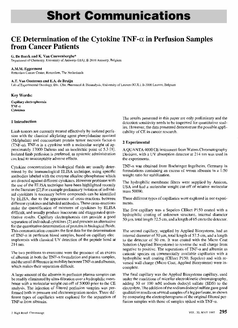
The Supelco CElect P150 capillary of Supelco has a coating of unknown hydrophilic structure. Before the analyses, the capillary was conditioned by rinsing consecutively with a phosphate buffer pH7,O.l N NaOH and phosphate buffer pH 7 again, each rinse lasting for 15 min. Analyses were performed with a positive voltage of 23 kV.
Measurements using electrolytes with different pH values showed that pH7 gave the best separation of acidic proteins.
Figure 1 illustrates the electropherogram of the separation of human albumin and TNF-a directly in the TNF-a formulation, which contains a mixture of TNF-a and albumin in a 150 weight ratio. The separation is not complete and an excess of albumin dominates the electropherogram. Both by pressure and electrok- inetic injection the excess of albumin is important in the picture obtained. However the electrokinetic injection will introduce a higher amount of albumin, the compound with higher mobility. Therefore pressure injection is to be preferred for quantitative measurements.
1
, $ 1 I
0 2 4 6 8 10 12 14 16 18 20 min
Figure 1. Electropherogram of the TNF-a formulation using a capillary with hydrophilic coating (Supelco CElect P150). Electrokinetic injection during 15 s at 5 kV - high voltage: positive, 23 kV -buffer pH 7. 1 = HSA (Human Serum Albumin), 2 = TNF-a.
3.2 Capillary with Reversed Wall Charge
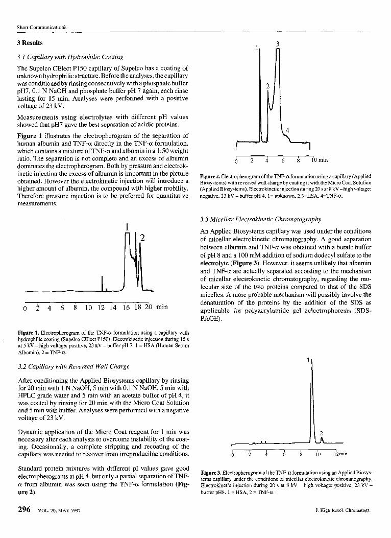
After conditioning the Applied Biosystems capillary by rinsing for 20 min with 1 N NaOH, 5 min with 0.1 N NaOH, 5 min with HPLC grade water and 5 min with an acetate buffer of pH 4, it was coated by rinsing for 20 min with the Micro Coat Solution and 5 min with buffer. Analyses were performed with a negative voltage of 23 kV.
Dynamic application of the Micro Coat reagent for 1 min was necessary after each analysis to overcome instability of the coat- ing. Occasionally, a complete stripping and recoating of the capillary was needed to recover from irreproducible conditions.
Standard protein mixtures with different PI values gave good electropherograms at pH 4, but only a partial separation of TNF- a from albumin was seen using the TNF-a formulation (Fig- ure 2).
Figure 2. Electropherogram of the TNF-a formulation using a capillary (Applied Biosystems) with reversed wall charge by coating it with the Micro Coat Solution (Applied Biosystems). Electrokinetic injection during 20 s at 8 kV -high voltage: negative, 23 kV - buffer pH 4. 1= unknown, 2,3=HSA, 4=TNF-a.
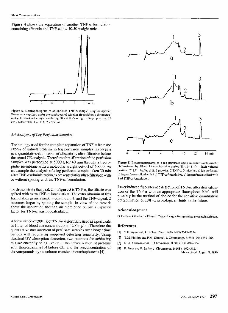
3.3 Micellar Electrokinetic Chromatography
An Applied Biosystems capillary was used under the conditions of micellar electrokinetic chromatography. A good separation between albumin and TNF-a was obtained with a borate buffer of pH 8 and a 100 mM addition of sodium dodecyl sulfate to the electrolyte (Figure 3). However, it seems unlikely that albumin and TNF-a are actually separated according to the mechanism of micellar electrokinetic chromatography, regarding the mo- lecular size of the two proteins compared to that of the SDS micelles. A more probable mechanism will possibly involve the denaturation of the proteins by the addition of the SDS as applicable for polyacrylamide gel eclectrophoresis (SDS- PAGE).
' I
2
Figure 3. Electropherogram of the TNF-a formulation using an Applied Biosys- terns capillary under the conditions of micellar electrokinetic chromatography. Electrokinetic injection during 20 s at 8 kV - high voltage: positive, 23 kV - buffer pH8. 1 = HSA, 2 = TNF-a.
296 VOL. 20, MAY 1997 I . High Resol. Chromatogr.
Short Communications
Figure 4 shows the separation of another TNF-a formulation containing albumin and TNF-a in a 5050 weight ratio.
0 2 4 6 8 lbmin
Figure 4. Electropherogram of an enriched TNF-a sample using an Applied Biosystems capillary under the conditions of micellar electrokinetic chromatog- raphy. Electrokinetic injection during 20 s at 8 kV -high voltage: positive, 23 kV - buffer pH8. 1 = HSA, 2 = TNF-a.
3.4 Analyses of Leg Pelfusion Samples
The strategy used for the complete separation of TNF-a from the excess of natural proteins in leg perfusion samples involves a near quantitative elimination of albumin by ultra-filtration before the actual CE analysis. Therefore ultra-filtration of the perfusion samples was performed at 5000 g for 40 min through a hydro- philic membrane with a molecular weight cut-off of 50000. As an example the analysis of a leg perfusate sample, taken 30 min after TNF-a administration, is presented after ultra-filtration with or without spiking with the TNF-a formulation.
To demonstrate that peak 2 in Figure 5 is TNF-a, the filtrate was spiked with extra TNF-a formulation. The extra albumin of this formulation gives a peak in continuum 1, and the TNF-a peak 2 becomes larger by spiking the sample. In view of the remark about the separation mechanism mentioned before a capacity factor for TNF-a was not calculated.
A formulation of 200 pg of TNF-a is normally used as a perfusate in 1 liter of blood at a concentration of 200 ng/ml. Therefore the quantitative measurement of perfusate samples over longer time periods will require an improved detection sensitivity. Using classical UV absorption detection, two methods for achieving this are currently being explored: the derivatization of proteins with fluorescamine [3] before CE, and the preconcentration of the compounds by on column transient isotachophoresis [4].
1 3
3 .
Figure 5. Electropherograrns of a leg perfusate using micellar electrokinetic chromatography. Electrokinetic injection during 20 s by 8 kV - high voltage: positive, 23 kV -buffer pH8. 1 proteins, 2 TNF-a, 3 micelles. a) leg perfusate, b) leg perfusate spiked with 1 p1TNF-a formulation, c) leg perfusate spiked with 2 a1 TNF-a formulation.
Laser induced fluorescence detection of TNF-a, after derivatiza- tion of the TNF-a with an appropriate fluorophore label, will possibly be the method of choice for the sensitive quantitative determination of TNF-a in biological fluids in the future.
Acknowledgment G. De Boeck thanks the Flemish Cancer League for a grant as a research assistant.
References [l] B.B. Aggarwal, J. Biolog. Chem. 260 (1985) 2345-2354.
[2] T.M. Phillips and P.M. Kimmel, J. Chromatogr. B 656(1994) 259-266.
[3] N. A. Guzman etal., J. Chromatogr. B 608 (1992)197-204.
[4] F. Foret and E. Szoko, J. Chromatogr. B 608 (1992) 312. Ms received: August 8, 1996
J. High Resoi. Chromatogr. VOL. 20, MAY 1997 297