copy right- hongqi zhang-department of anatomy-fudan...
TRANSCRIPT

Copy Right- Hongqi ZHANG-Department of Anatomy-Fudan University
Copy Right- Hongqi ZHANG-Department of Anatomy-Fudan University1
Dr.Hongqi Zhang (张红旗)Email: [email protected]
Systematic Anatomy
Locomotor system - Part 5 Introduction of myology
The muscles of head, neck and trunk

Copy Right- Hongqi ZHANG-Department of Anatomy-Fudan University
Copy Right- Hongqi ZHANG-Department of Anatomy-Fudan University
Myology
Copy Right- Hongqi ZHANG-Department of Anatomy-Fudan University
Copy Right- Hongqi ZHANG-Department of Anatomy-Fudan University
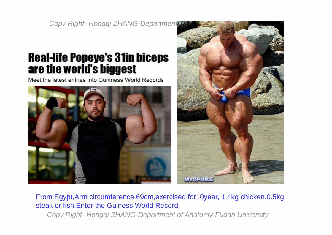
From Egypt,Arm circumference 69cm,exercised for10year, 1.4kg chicken,0.5kg steak or fish,Enter the Guiness World Record.
Copy Right- Hongqi ZHANG-Department of Anatomy-Fudan University
Copy Right- Hongqi ZHANG-Department of Anatomy-Fudan University
Copy Right- Hongqi ZHANG-Department of Anatomy-Fudan University
Copy Right- Hongqi ZHANG-Department of Anatomy-Fudan University
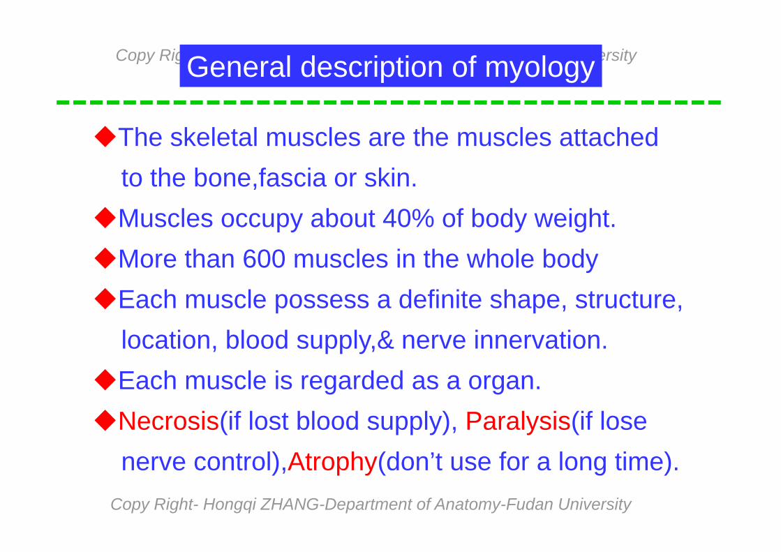
The skeletal muscles are the muscles attachedto the bone,fascia or skin.
Muscles occupy about 40% of body weight.More than 600 muscles in the whole bodyEach muscle possess a definite shape, structure,
location, blood supply,& nerve innervation.Each muscle is regarded as a organ.Necrosis(if lost blood supply), Paralysis(if lose
nerve control),Atrophy(don’t use for a long time).
General description of myology

Copy Right- Hongqi ZHANG-Department of Anatomy-Fudan University
Copy Right- Hongqi ZHANG-Department of Anatomy-Fudan University
Cardiac muscle (in heart)
Skeletal muscle(attach to bones) voluntary muscle
Classification of the muscle according to structure
Smooth muscle(in wall of vessel & viscera)
striated muscle
involuntary muscle
(Alternating light & dark pattern)
Copy Right- Hongqi ZHANG-Department of Anatomy-Fudan University
Copy Right- Hongqi ZHANG-Department of Anatomy-Fudan University
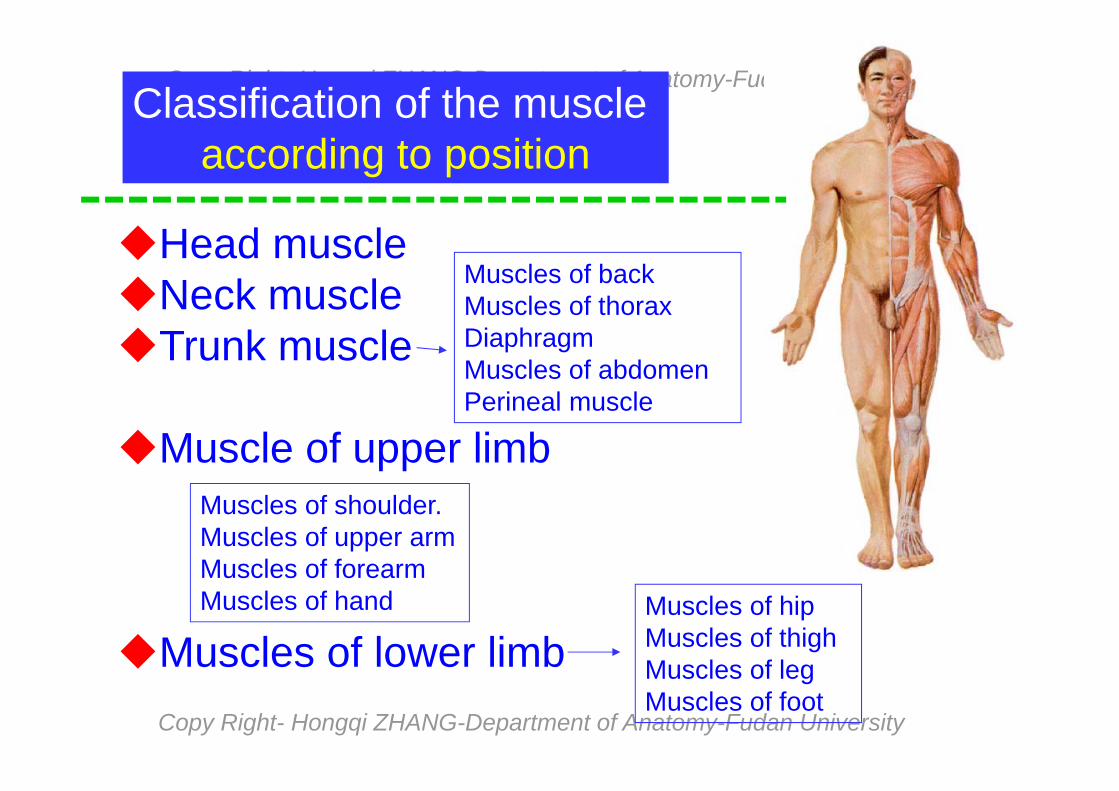
Classification of the muscle according to position
Head muscleNeck muscleTrunk muscle
Muscle of upper limb
Muscles of lower limb
Muscles of backMuscles of thoraxDiaphragm Muscles of abdomenPerineal muscle
Muscles of shoulder. Muscles of upper armMuscles of forearmMuscles of hand Muscles of hip
Muscles of thighMuscles of legMuscles of foot
Copy Right- Hongqi ZHANG-Department of Anatomy-Fudan University
Copy Right- Hongqi ZHANG-Department of Anatomy-Fudan University
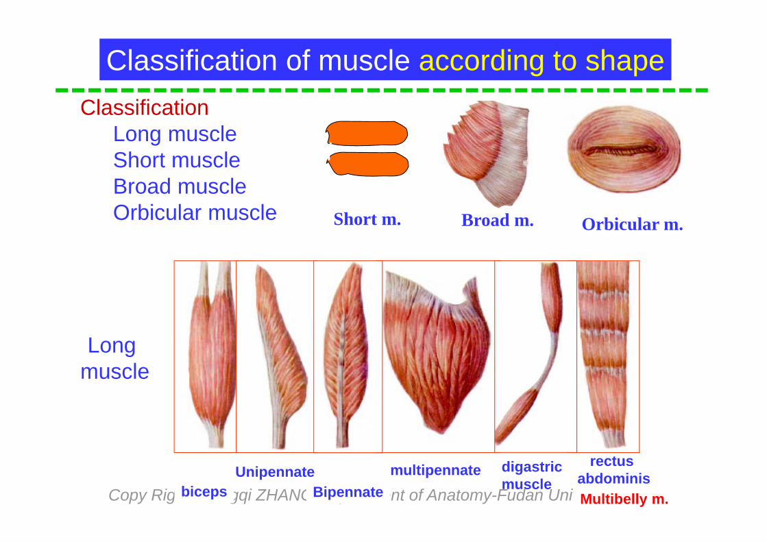
Short m. Broad m. Orbicular m.
ClassificationLong muscleShort muscleBroad muscleOrbicular muscle
Long muscle
bicepsmultipennate digastric
muscleUnipennate
Bipennate
Classification of muscle according to shape
rectus abdominisMultibelly m.

Copy Right- Hongqi ZHANG-Department of Anatomy-Fudan University
Copy Right- Hongqi ZHANG-Department of Anatomy-Fudan University
Structure of skeleton muscle
Belly
Tendon
Aponeurosis
Tendinous intersection
Belly-contractiveTendon-don’t contract
Muscularpart
Muscularpart
Muscular part & tendinous part
Tendon

Copy Right- Hongqi ZHANG-Department of Anatomy-Fudan University
Copy Right- Hongqi ZHANG-Department of Anatomy-Fudan University
Muscle attachment
1-Origin 2-Belly3-Tendon4-Insertion
1
234
Usually muscle distribute pairedMuscle works as functional groupsExtensor & flexorPronator & supinatorAbductor & adductormedial rotator. & lateral rotator
Copy Right- Hongqi ZHANG-Department of Anatomy-Fudan University
Copy Right- Hongqi ZHANG-Department of Anatomy-Fudan University
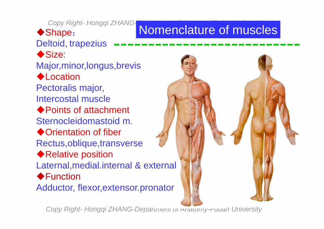
Shape:Deltoid, trapeziusSize:Major,minor,longus,brevisLocationPectoralis major, Intercostal musclePoints of attachmentSternocleidomastoid m.Orientation of fiberRectus,oblique,transverseRelative positionLaternal,medial.internal & externalFunctionAdductor, flexor,extensor.pronator
Nomenclature of muscles

Copy Right- Hongqi ZHANG-Department of Anatomy-Fudan University
Copy Right- Hongqi ZHANG-Department of Anatomy-Fudan University
Movement of muscleHow to analyze the motion of the muscle
According to attachment of the musclePass the number of joints.Location of the muscle in joints
Copy Right- Hongqi ZHANG-Department of Anatomy-Fudan University
Copy Right- Hongqi ZHANG-Department of Anatomy-Fudan University
Supplementary structures of muscle
FasciaSynovial bursaTendinous sheathSesamoid bone
Copy Right- Hongqi ZHANG-Department of Anatomy-Fudan University
Copy Right- Hongqi ZHANG-Department of Anatomy-Fudan University
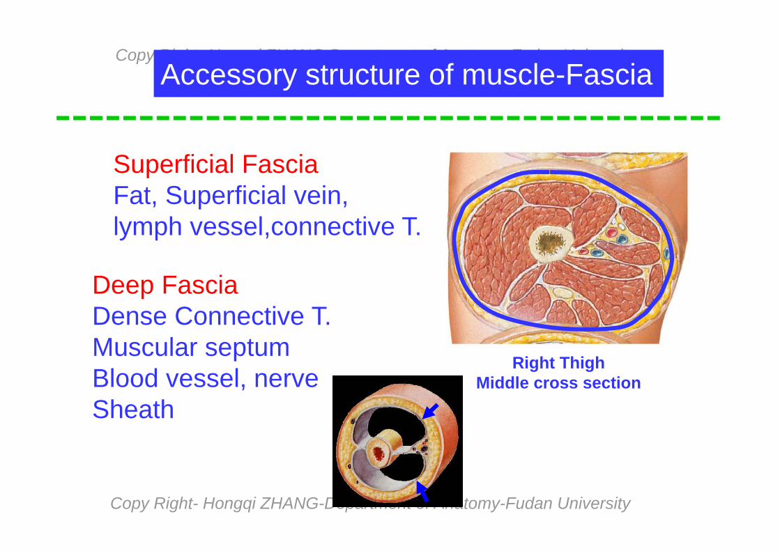
Right ThighMiddle cross section
Superficial FasciaFat, Superficial vein,lymph vessel,connective T.
Deep FasciaDense Connective T.Muscular septumBlood vessel, nerve Sheath
Accessory structure of muscle-Fascia
Copy Right- Hongqi ZHANG-Department of Anatomy-Fudan University
Copy Right- Hongqi ZHANG-Department of Anatomy-Fudan University
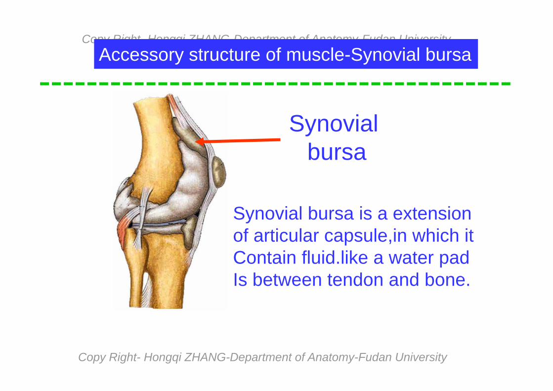
Accessory structure of muscle-Synovial bursa
Synovial bursa
Synovial bursa is a extension of articular capsule,in which itContain fluid.like a water pad Is between tendon and bone.

Copy Right- Hongqi ZHANG-Department of Anatomy-Fudan University
Copy Right- Hongqi ZHANG-Department of Anatomy-Fudan University
Accessory structure of m.- Sesamoid bone
Sesamoidbone
Sesamoid bone generally is locatedIn the tendon, which functions as a pad and Make tendon bearing rub
Copy Right- Hongqi ZHANG-Department of Anatomy-Fudan University
Copy Right- Hongqi ZHANG-Department of Anatomy-Fudan University
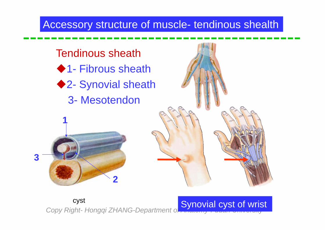
Accessory structure of muscle- tendinous shealth
Tendinous sheath1- Fibrous sheath2- Synovial sheath
3- Mesotendon
Synovial cyst of wrist
1
2
3
cyst

Copy Right- Hongqi ZHANG-Department of Anatomy-Fudan University
Copy Right- Hongqi ZHANG-Department of Anatomy-Fudan University
Function of the skeletal muscle
Movement
Heat production
Body support
maintenance of posture
Copy Right- Hongqi ZHANG-Department of Anatomy-Fudan University
Copy Right- Hongqi ZHANG-Department of Anatomy-Fudan University
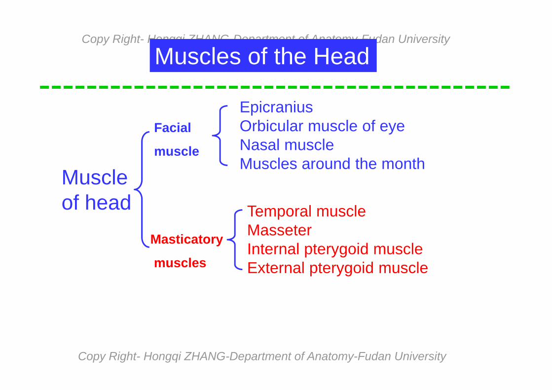
Muscles of the Head
Muscle of head
Facial
muscle
Masticatory
muscles
Epicranius Orbicular muscle of eyeNasal muscleMuscles around the month
Temporal muscle Masseter Internal pterygoid muscleExternal pterygoid muscle
Copy Right- Hongqi ZHANG-Department of Anatomy-Fudan University
Copy Right- Hongqi ZHANG-Department of Anatomy-Fudan University
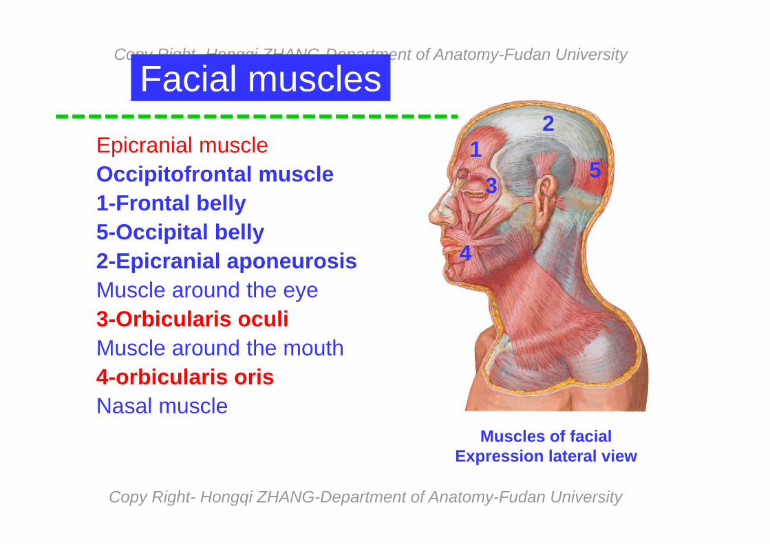
Muscles of facial Expression lateral view
Facial muscles
Epicranial muscleOccipitofrontal muscle1-Frontal belly5-Occipital belly2-Epicranial aponeurosisMuscle around the eye3-Orbicularis oculiMuscle around the mouth4-orbicularis orisNasal muscle
15
2
3
4
Copy Right- Hongqi ZHANG-Department of Anatomy-Fudan University
Copy Right- Hongqi ZHANG-Department of Anatomy-Fudan University
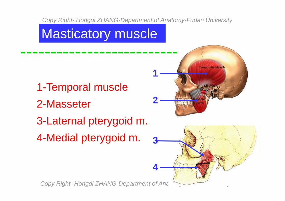
Masticatory muscle
1-Temporal muscle 2-Masseter 3-Laternal pterygoid m.4-Medial pterygoid m.
1
2
3
4
Copy Right- Hongqi ZHANG-Department of Anatomy-Fudan University
Copy Right- Hongqi ZHANG-Department of Anatomy-Fudan University
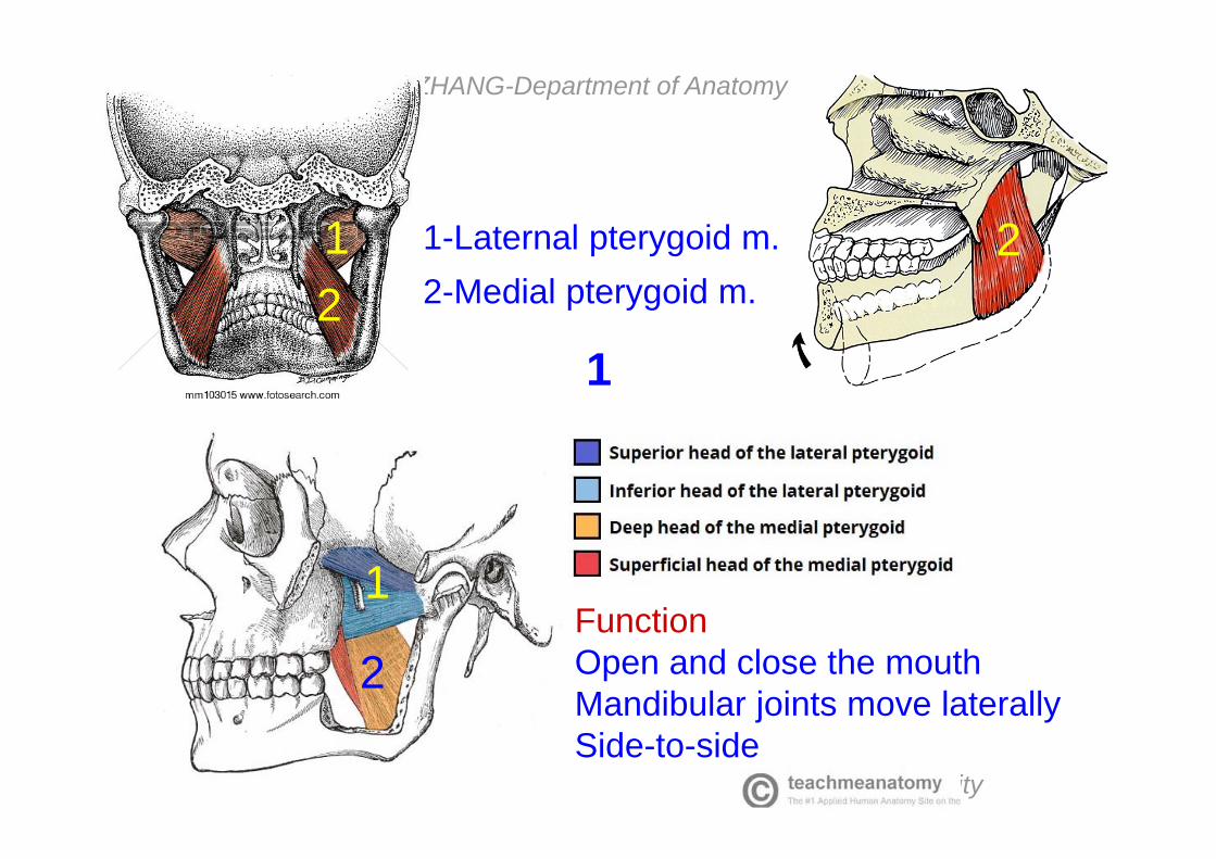
Function Open and close the mouthMandibular joints move laterallySide-to-side
1-Laternal pterygoid m.2-Medial pterygoid m.
1
12
2
1
2

Copy Right- Hongqi ZHANG-Department of Anatomy-Fudan University
Copy Right- Hongqi ZHANG-Department of Anatomy-Fudan University
Muscles of the neck
Muscles of the neck
Superficial
Anterior cervical m
Deep
Platysma m.
Sternocleidomastoid m.Suprahyoid mm.
Infrahyoid mm.
Lateral group
Medial group
Copy Right- Hongqi ZHANG-Department of Anatomy-Fudan University
Copy Right- Hongqi ZHANG-Department of Anatomy-Fudan University
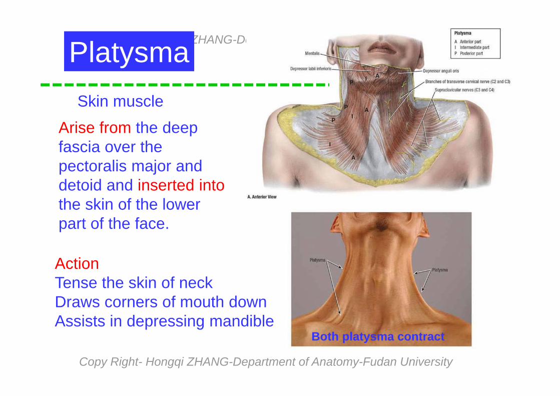
PlatysmaSkin muscle
Arise from the deep fascia over the pectoralis major and detoid and inserted intothe skin of the lower part of the face.
ActionTense the skin of neckDraws corners of mouth downAssists in depressing mandible
Both platysma contract

Copy Right- Hongqi ZHANG-Department of Anatomy-Fudan University
Copy Right- Hongqi ZHANG-Department of Anatomy-Fudan University
Muscle of neck lateral view
Sternocleidomastoid m.Superficial muscles of the neck
Name from origin and insertionA prominent visible landmarkAction- Acting alone: The head is inclined laterally & the face rotateto the opposite sideActing together- they draw thehead backward or raise the head
Both mm contract One m contracts

Copy Right- Hongqi ZHANG-Department of Anatomy-Fudan University
Copy Right- Hongqi ZHANG-Department of Anatomy-Fudan University
1-Digastric m.2-Stylohyoid m.3-Mylohyoid m.4-Geniohyoid m.
Suprahyoid muscle - 4
3 4
Pull the hyoid upward,backward & help to Depress the Mandible when the hyoid is fixed
Post.view
1
2
3
1
2
3

Copy Right- Hongqi ZHANG-Department of Anatomy-Fudan University
Copy Right- Hongqi ZHANG-Department of Anatomy-Fudan University
Infrahyoid muscleSuperficial layer
1-Sternohyoid m.2-Omohyoid m.Deep layer
3-Sternothyroid m.4-Thyrohyoid m. 1
2
32
4
Action1-Depresses the hyoid bone as it contracts2-Larynx is pulled downward3-Elevates the larynx and lowers the hyoid bone4-it acts to depress the hyoid bone
Length of muscle 1= m 3 + m 42
2
Copy Right- Hongqi ZHANG-Department of Anatomy-Fudan University
Copy Right- Hongqi ZHANG-Department of Anatomy-Fudan University
12
3 1 3
2
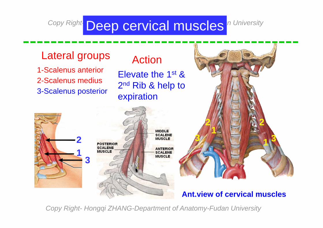
Lateral groups
Deep cervical muscles
1-Scalenus anterior2-Scalenus medius3-Scalenus posterior
Action Elevate the 1st &2nd Rib & help toexpiration
Ant.view of cervical muscles
13
2

Copy Right- Hongqi ZHANG-Department of Anatomy-Fudan University
Copy Right- Hongqi ZHANG-Department of Anatomy-Fudan University
Scalene fissureBetween scalenus ant. & mid. subclavian a. & brachial plexus pass from the fissure
Scalenus ant.
Scalenus mid.
Brachial n.
Subclavicular a.
Copy Right- Hongqi ZHANG-Department of Anatomy-Fudan University
Copy Right- Hongqi ZHANG-Department of Anatomy-Fudan University
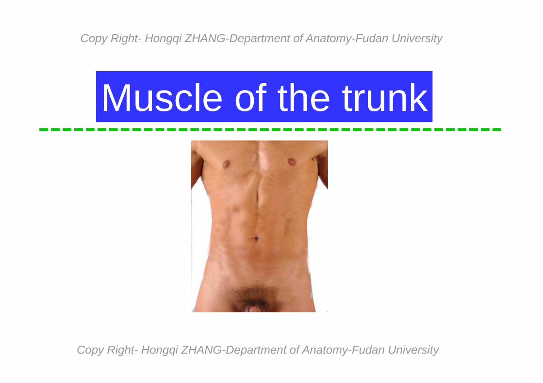
Muscle of the trunk
Copy Right- Hongqi ZHANG-Department of Anatomy-Fudan University
Copy Right- Hongqi ZHANG-Department of Anatomy-Fudan University
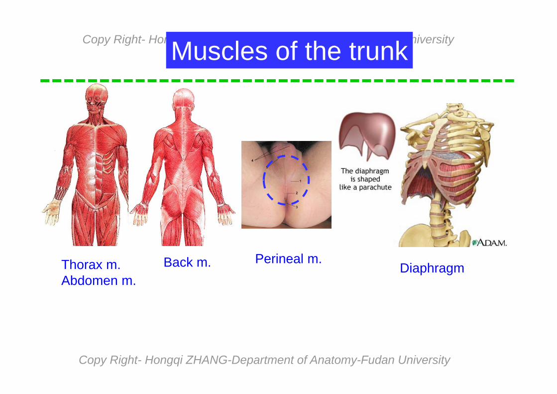
Muscles of the trunk
Back m.Thorax m.Abdomen m.
DiaphragmPerineal m.
Copy Right- Hongqi ZHANG-Department of Anatomy-Fudan University
Copy Right- Hongqi ZHANG-Department of Anatomy-Fudan University
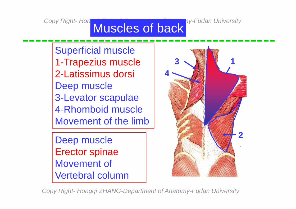
Superficial muscle1-Trapezius muscle2-Latissimus dorsiDeep muscle3-Levator scapulae4-Rhomboid muscleMovement of the limb
Deep muscleErector spinaeMovement of Vertebral column
Muscles of back
1
2
34

Copy Right- Hongqi ZHANG-Department of Anatomy-Fudan University
Copy Right- Hongqi ZHANG-Department of Anatomy-Fudan University
Trapezius
Latissimus dorsi
Superficial muscle of backLarge triangular in neck and backOrigin:ext. occipital protuberanceSpine of seventh cervical vertebraSpine all thoracic vertebraeInsertionThe lat. one third of the clavicleThe acromion and spine of scapulaAction:raises, descends, retracts & rotates The scapula & extends the head
Large wide & triangular flat m.in the backOrigin:Spinous process of lower six thoracic C.Thoracolumbar fascia, spinous process ofLumbar vertebrae, iliac crestInsertion Crest of lesser tuberosity of the humerusAction: extends, adducts and medially rotates humerus at shoulder joint
Copy Right- Hongqi ZHANG-Department of Anatomy-Fudan University
Copy Right- Hongqi ZHANG-Department of Anatomy-Fudan University
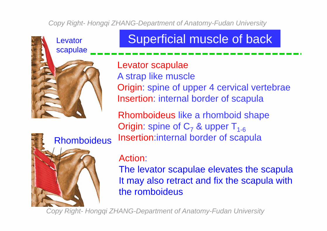
Superficial muscle of backLevatorscapulae
Rhomboideus
Levator scapulaeA strap like muscleOrigin: spine of upper 4 cervical vertebraeInsertion: internal border of scapula
Rhomboideus like a rhomboid shapeOrigin: spine of C7 & upper T1-6Insertion:internal border of scapula
Action:The levator scapulae elevates the scapulaIt may also retract and fix the scapula withthe romboideus

Copy Right- Hongqi ZHANG-Department of Anatomy-Fudan University
Copy Right- Hongqi ZHANG-Department of Anatomy-Fudan University
Deep muscle of backErector
spinae
It is a collecting name (groups)Origin: Sacrum, ilium & related ligs.Insertion: Ribs and vertebraeAction:When acting on one side it Bends and rotates the spinal Column toward the opposite side.when acting on Both sides It extends the spinal column.
Erector spinae

Copy Right- Hongqi ZHANG-Department of Anatomy-Fudan University
Copy Right- Hongqi ZHANG-Department of Anatomy-Fudan University
Extrinsic m.(origin in thoracic wall,
Insertion in upper limb bone)
Pectoralis major
Pectoralis minor
Serratus anterior
Intrinsic m.(both origin and insertion are in thoracic wall)
Ext.Intercostal m. int.Intercostal m.Transversus thoracis
Intercostates intimi
Muscles of thorax
External intercostal membraneInternal intercostal membrane
Copy Right- Hongqi ZHANG-Department of Anatomy-Fudan University
Copy Right- Hongqi ZHANG-Department of Anatomy-Fudan University
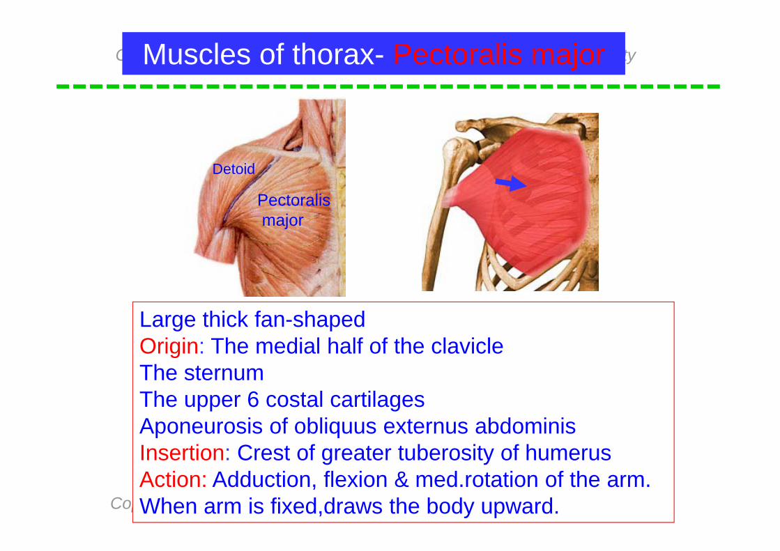
Large thick fan-shapedOrigin: The medial half of the clavicleThe sternumThe upper 6 costal cartilagesAponeurosis of obliquus externus abdominisInsertion: Crest of greater tuberosity of humerusAction: Adduction, flexion & med.rotation of the arm.When arm is fixed,draws the body upward.
Muscles of thorax- Pectoralis major
Detoid
Pectoralismajor
Copy Right- Hongqi ZHANG-Department of Anatomy-Fudan University
Copy Right- Hongqi ZHANG-Department of Anatomy-Fudan University
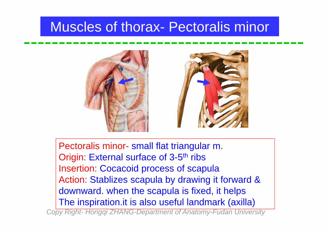
Pectoralis minor- small flat triangular m.Origin: External surface of 3-5th ribsInsertion: Cocacoid process of scapulaAction: Stablizes scapula by drawing it forward &downward. when the scapula is fixed, it helps The inspiration.it is also useful landmark (axilla)
Muscles of thorax- Pectoralis minor

Copy Right- Hongqi ZHANG-Department of Anatomy-Fudan University
Copy Right- Hongqi ZHANG-Department of Anatomy-Fudan University
Muscles of thorax- Serratus anterior
A large thin powerful in lateral part of thoraxOrigin: external surface of upper 8 or 9 ribsInsertion: internal border of the scapulaAction: Holds the scapula against the chest wallPulls the scapula forwards in throwing and pushing by fixing the scapula it helps the inspiration

Copy Right- Hongqi ZHANG-Department of Anatomy-Fudan University
Copy Right- Hongqi ZHANG-Department of Anatomy-Fudan University
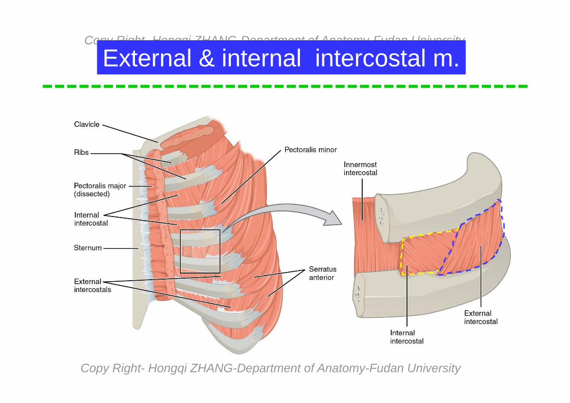
External & internal intercostal m.
1-Ext.intercostal m.2-Ext.intercontstal membrane3-Int.intercostal m.4-Int.intercostal membrane
External intercostal m.Help to inspirationInternal intercostal m.Help to expiration
Puncture of thoracic wall
12
3
4
Copy Right- Hongqi ZHANG-Department of Anatomy-Fudan University
Copy Right- Hongqi ZHANG-Department of Anatomy-Fudan University
External & internal intercostal m.
Copy Right- Hongqi ZHANG-Department of Anatomy-Fudan University
Copy Right- Hongqi ZHANG-Department of Anatomy-Fudan University
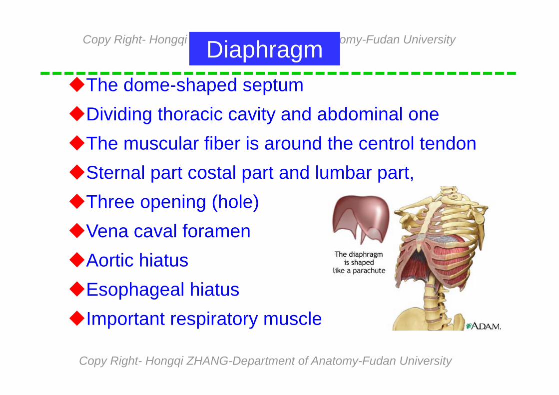
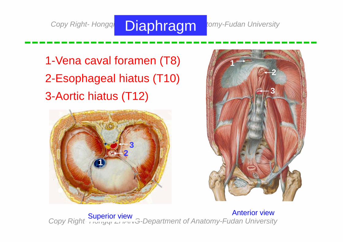
DiaphragmThe dome-shaped septumDividing thoracic cavity and abdominal oneThe muscular fiber is around the centrol tendonSternal part costal part and lumbar part,Three opening (hole)Vena caval foramenAortic hiatusEsophageal hiatus Important respiratory muscle
Copy Right- Hongqi ZHANG-Department of Anatomy-Fudan University
Copy Right- Hongqi ZHANG-Department of Anatomy-Fudan University
1-Vena caval foramen (T8)2-Esophageal hiatus (T10)3-Aortic hiatus (T12)
Diaphragm
1
1
2
2
3
3
Superior view Anterior view

Copy Right- Hongqi ZHANG-Department of Anatomy-Fudan University
Copy Right- Hongqi ZHANG-Department of Anatomy-Fudan University
Aorta
Phrenic nerve Inf.venavava Esophagus

Copy Right- Hongqi ZHANG-Department of Anatomy-Fudan University
Copy Right- Hongqi ZHANG-Department of Anatomy-Fudan University
Master the shape, classification and nomenclature of muscles.
Master the accessory structures of muscleMaster name, location and function of the
masticatory muscle.Master the insertion, origin and function of
the stenocledomastoid & infrahyoid musclesMaster the distribution & function of
thoracic m. and back muscles.
The important contents today
Copy Right- Hongqi ZHANG-Department of Anatomy-Fudan University
Copy Right- Hongqi ZHANG-Department of Anatomy-Fudan University
See you next time!