occult ca oral cavity
TRANSCRIPT

Journal club
Investigations for occult carcinoma of Oral Cavity
ByDr Madhu Kumar
Under Guidance ofDr DSVL Narasimham MSDr R Hemanthi MSDr PS Sitaram MS
Oral Cavity
bull Lips
bull Oral tongue
bull Floor of mouth
bull Alveolus
bull Buccal mucosa
bull Palate
bull Oropharynx
Risk factors
bull Tobacco
bull Alcohol
bull Areca nutpan masala
bull Human papillomavirus
bull EpsteinndashBarr virus
bull PlummerndashVinson syndrome
bull Poor nutrition
Pathology
bull Squamous cell carcinoma commonest
bull Lymphoma
Premalignant conditions
bull High-risk lesionsndash Erythroplakiandash Speckled erythroplakiandash Chronic hyperplastic candidiasis
bull Medium-risk lesionsndash Oral submucous fibrosisndash Syphilitic glossitisndash Sideropenic dysphagia (PatersonndashKelly syndrome)
bull Low-riskequivocal-risk lesionsndash Oral lichen planusndash Discoid lupus erythematosusndash Discoid keratosis congenita
CLINICAL FEATURES
bull Persistent oral swelling for gt 4 weeks
bull Mouth ulceration for gt 4 weeks
bull Sore tongue
bull Difficulty swallowing
bull Jaw or facial swelling
bull Painless neck lump
bull Unexplained tooth mobility
bull Trismus
Occult oral cavity malignancy
bull Presentation
bull Cervical lymphadenopathy
ndash Primary eg lymphoma
ndash Secondary eg squamous cell carcinoma
ndash Known primary
ndash Occult primary
Lympahatic spread
Investigations
bull Radiography
bull Magnetic resonance imaging
bull Computerised tomography
bull Radionucleotide studies
bull Fine-needle aspiration cytology
bull Ultrasound
bull FG PET CT
bull Panendoscopy
Radiography
bull Plain radiography of the jaw
bull for dental assessment
bull Orthopantomogram of the jaws is helpful to assess bony invasion particularly from tumours arising on the alveolus and maxillary antrum
Magnetic resonance imaging
bull MRI is the investigation of choice for cancer of the oral cavity and oropharynx
bull Visualisation of soft-tissue infiltration of the tumour
bull Its specificity and sensitivity in diagnosing cervical node metastasis is similar to that of CT
Computerised tomography
bull CT is much more widely available
bull Useful when bony invasion is suspected
bull CT of the thorax and abdomen is also indicated for patients who have proven cervical lymph node metastasis or who have large-volume disease in which the risk of distant metastasis is high
Radionucleotide studies
bull Radioisotope bone scan of the facial skeleton
bull The scan is not specific and tends to show increased uptake wherever there is increased metabolic activity in bone
bull A false-positive diagnosis is common and lsquoover-stagingrsquo of the disease frequent
Fine-needle aspiration cytology
bull for the assessment and pathological diagnosis of enlarged cervical lymph nodes
bull Involves the use of a fine-needle puncture into the mass and immediate aspiration for cytological examination
bull Equipment a 21G or 23G needle and a 10 ml syringe
bull Aspiration should be carried out only when the needle enters the swelling
bull The positive yield from FNAC is dependent not only on the quality of the aspirate but also on the skill of the cytologist
Ultrasound
bull Useful as an adjunct in FNAC
bull It also has a place in abdominal assessment particularly when metastases of the liver are suspected
FG PET CTbull 18F-fluoro-2-deoxyglucose positron emission
tomography (FDG PET)computed tomography (CT)
bull DiagnosisStagingndash M and bilateral nodal staging of advanced HNSCC
displaying equivocal conventional imaging
ndash Identification of unknown primary site in addition to conventional imaging and diagnostic panendoscopy
ndash Staging of nasopharyngeal carcinoma without evidence of distant disease
bull RecurrenceRestagingndash Restaging patients who are being considered for major
salvage treatment (surgery or other)
Panendoscopy
bull Rhinoscopy
bull Nasopharyngoscopy
bull Direct laryngoscopy
bull Hypopharyngoscopy
bull Esophagoscopy
bull Bronchoscopy
bull Indications for Panendoscopy
ndash To biopsy a tumor
ndash Tumor mapping to identify extent of tumorthrough inspection palpation and sampling biopsies
ndash Rule out associated malignancy
bull Panendoscopy is ideally done prior to definitive treatment planning
Journals
bull Incidental detection of an occult oral malignancy with autofluorescence imaging a case report
bull Nadarajah Vigneswaran et al
bull Head Neck Oncol 2009
bull Autofluorescence imaging is used widely for diagnostic evaluation of various epithelial malignancies Cancerous lesions display loss of autofluorescence due to malignant changes in epithelium and subepithelial stroma
bull Carcinoma of unknown primary site presents with lymph node or distant metastasis for which the site of primary tumour is not detectable
bull Use of autofluorescence imaging for detecting a clinically innocuous appearing occult malignancy of the palate which upon pathological examination was consistent with a metastatic squamous cell carcinoma
bull Down regulation of E-Cadherin (ECAD) - a predictor for occult metastatic disease in sentinel node biopsy of early squamous cell carcinomas of the oral cavity and oropharynx
bull Gerhard F Huber et al
bull BMC Cancer 2011
bull Background
ndash Prognostic factors in predicting occult lymph node metastasis in patients with head and neck squamous-cell carcinoma (HNSCC) are necessary to improve the results of the sentinel lymph node procedure in this tumour type
ndash The E-Cadherin glycoprotein is an intercellular adhesion molecule in epithelial cells which plays an important role in establishing and maintaining intercellular connections
bull Objectives
ndash To determine the value of the molecular marker E-Cadherin in predicting regional metastatic disease
bull Methods
bull E-Cadherin expression in tumour tissue of 120 patients with HNSCC of the oral cavity and oropharynx were evaluated using the tissue microarray technique
bull 110 tumours were located in the oral cavity (917 mostly tongue) 10 tumours in the oropharynx (83)
bull Intensity of E-Cadherin expression was quantified by the Intensity Reactivity Score (IRS)
bull These results were correlated with the lymph node status of biopsied sentinel lymph nodes
bull Univariate and multivariate analysis was used to determine statistical significance
bull Resultsndash pT-stage gender tumour side and location did not
correlate with lymph node metastasis
ndash Differentiation grade (p = 0018) and down regulation of E-Cadherin expression significantly correlate with positive lymph node status (p = 0005) in univariateand multivariate analysis
bull Conclusionndash These data suggest that loss of E-cadherin expression
is associated with increased lymhogeneous metastasis of HNSCC E-cadherin immunohistochemistry may be used as a predictor for lymph node metastasis in squamous cell carcinoma of the oral cavity and oropharynx
bull 18F-FDG PET and CTMRI in Oral Cavity Squamous Cell Carcinoma A Prospective Study of 124 Patients with Histologic Correlation
bull By Shu-Hang Nget albull Journal of nuclear medicine 2005bull Accurate evaluation of primary tumors and cervical
lymph node status of squamous cell carcinoma (SCC) of the oral cavity is important to treatment planning and prognosis prediction
bull In this prospective study we evaluated the use of 18F-FDG PET CTMRI and their visual correlation for the identification of primary tumors and cervical nodal metastases of SCC of the oral cavity with histologiccorrelation
bull Methodsbull One hundred twenty-four patients with pathologically
proven diagnoses of oral cavity SCC underwent 18F-FDG PET and CTMRI within 2 wk before surgery
bull We interpreted 18F-FDG PET CTMRI and visually correlated 18F-FDG PET and CTMRI separately to assess the primary tumors and their regional lymph node status
bull We recorded lymph node metastases according to the neck level system of imaging-based nodal classification
bull Histopathologic analysis was used as the gold standard for assessment of the primary tumors and lymph node involvement
bull We analyzed differences in sensitivity and specificity among the imaging modalities using the McNemar test
bull The receiver-operating-characteristic (ROC) curve and calculation of the area under the curve were used to evaluate their discriminative power
bull Resultsbull The accuracy of 18F-FDG PET CTMRI and their visual
correlation for the identification of primary tumors was 984 871 and 992 respectively
bull The sensitivity of 18F-FDG PET for the identification of nodal metastases on a level-by-level basis was 221 higher than that of CTMRI (747 vs 526 P lt 0001) whereas the specificity of 18F-FDG PET was 15 lower than that of CTMRI (930 vs 945 P = 0345)
bull The sensitivity and specificity of the visual correlation of 18F-FDG PET and CTMRI were 32 and 15 higher than those of 18F-FDG PET alone (779 vs 747 P = 025 945 vs 930 P = 018 respectively)
bull The area under the curve obtained from the ROC curve showed that 18F-FDG PET was significantly superior to CTMRI for total nodal detection (0896 vs 0801 P = 0002) whereas the visual correlation of 18F-FDG PET and CTMRI was modestly superior to 18F-FDG PET alone (0913 vs 0896 P = 028)
bull Conclusion
bull 18F-FDG PET is superior to CTMRI in the detection of cervical status of oral cavity SCC
bull The sensitivity of 18F-FDG PET for the detection of cervical nodal metastasis on a level-by-level basis was significantly higher than that of CTMRI whereas their specificities appeared to be similar
bull Visual correlation of 18F-FDG PET and CTMRI showed a trend of increased diagnostic accuracy over 18F-FDG PET alone but without a statistically significant difference and its sensitivity was still not high enough to replace pathologic lymph node staging based on neck dissection
bull AN IMMUNOLOGIC BASIS FOR DETECTION OFOCCULT PRIMARY MALIGNANCIES OF THE HEAD AND NECK
bull HARVEYL COATESM
bull Cancer 41912-918 1978
bull After extensive evaluation of patients with metastatic neck disease and clinically undetectable primary cancer of the head and neck the clinician is often faced with the difficult question of subsequent management
bull In this study sera from 11 patients with clinically occult carcinoma and metastatic lymphadenopathy were studied for Epstein-Barr virus-associated antigens
bull These were compared with 35 sera from patients with known nasopharyngeal carcinoma at all stages of disease and treatment and with 212 sera from control patients with other head and neck tumors patients with lymphoma and normal controls
bull There was a significant correlation between high antibody titers to Epstein-Barr virus especially in the serum IgAfraction and the presence of nasopharyngeal carcinoma
bull Thus identification of occult nasopharyngeal carcinoma by immunologic means may have important application in the selective management of the patient with an unknown head and neck primary malignancy
bull Occult Primary Head and Neck Carcinoma
bull Cecelia E Schmalbach
bull Current Oncology Reports 2007
bull Unknown primary carcinoma presenting as cervical lymph node metastasis accounts for approximately 5 of all head and neck malignancies
bull Over 90 of these malignancies represent squamous cell carcinoma originating within Waldeyerrsquos ring
bull Adenocarcinoma
bull Melanoma
bull other rare histologic variants
bull PET scanning and PET-CT fusion
bull Immunohistochemical analysis
bull Panendoscopy
bull Conclusionsndash Carcinoma with an unknown primary presenting as
cervical lymph node metastasis is estimated to represent 3 to 5 of all head and neck malignant neoplasms
ndash At a minimum all patients presenting with an unknown primary carcinoma of the cervical region require a thorough head and neck history
ndash physical examinationndash FNA of the neck mass radio-ndash graphic imagingndash panendoscopy with directed biopsies ndash bilateral tonsillectomyndash Improvement in the ability to identify occult primary
carcinomas responsible for cervical lymph node metastasis ultimately hinges upon promising research in the areas of biomolecular testing PET and PET-CT fusion
bull Detection of unknown primary tumours and distant metastases in patients with cervical metastases value of FDG-PET versus conventional modalities
bull Gerreke Regelink
bull European Journal of Nuclear Medicine and Molecular Imaging
bull In 1ndash2 of head and neck oncology patients the only symptom of a malignancy is a positive cervical node
bull The aim of this study was to compare the value of positron emission tomography using fluorine-18 fluoro-2-deoxy-D-glucose (FDG-PET) and conventional diagnostic modalities (CT andor MRI panendoscopy) in detecting unknown primary tumours and distant metastases in patients suffering from such a cervical metastasis
bull Fifty patients (37 men and 13 women) with cervical metastases of an unknown primary tumour were included
bull All patients underwent FDG-PET In addition CT andor MRI was obtained and panendoscopy was performed
bull All clinically known metastases were detected by FDG-PET
bull The primary tumour could be diagnosed in 16 patients (four primary tumours were detected exclusively by FDG-PET)
bull Seven patients had multiple distant metastases that in six cases were detected exclusively by FDG-PET
bull The sensitivity and specificity of FDG-PET for detection of unknown primary tumours were 100 and 94 respectively
bull For the conventional diagnostic modalities these values were 92 and 76 FDG-PET had an exclusive effect on the applied therapy in 20 of the patients referred for diagnosis of an unknown primary tumour
bull The data obtained in this study strongly support the diagnostic strategy of performing FDG-PET in patients suffering from cervical metastases of an unknown primary tumour before any other diagnostic technique
bull The role of FDG-PET and PETCT in the diagnosis and staging of head and neck cancer
bull Adrian mattews
bull UWOMJ 2011
bull CARCINOMA OF UNKNOWN PRIMARYbull In 2-9 of patients with newly diagnosed HNSCC cervical node
metastases are clinically evident at biopsy but the primary tumour cannot be identified by conventional workup which includes physical examination CT MRI and endoscopic-guided biopsy
bull 15 PETCT has proven to be significantly more sensitive than CT (940 versus 716 respectively P lt 0001) at detecting carcinomas of unknown primary
bull Rusthoven et al reviewed 16 studies published between 1994 and 2003 and found that among 302 patients with a negative conventional workup FDG-PET detected the primary tumour in 74 patients (245)
bull In a more recent review Al-Ibraheem et al performed a meta-analysis of 8 studies published between 2000 and 20095 FDG-PET or PETCT were able to detect the unknown primary in 51 of 180 patients with an otherwise inconclusive workup
bull Delineation of a primary tumour is essential for delivering targeted therapy minimizing therapeutic morbidity caused by wide-field irradiation and improving prognosis
bull A recent report noted that findings made by FDG-PET changed therapeutic management in 25 of patients
bull In light of this evidence PETCT may have an important role in the diagnostic assessment of carcinoma of unknown primary
bull Diagnosis and Staging of Head and Neck CancerA Comparison of Modern Imaging Modalities (Positron Emission Tomography Computed Tomography Color-Coded Duplex Sonography) With Panendoscopic and Histopathologic Findings
bull Ercole Di Martino MD etal
bull Arch Otolaryngol Head Neck Surg 2000
bull Objective To compare the clinical value of positron emission tomography (PET) using fludeoxyglucose F 18 computed tomography (CT) color-coded duplex sonography (CCDS) and panendoscopy in the detection and staging of head and neck cancer
bull Design Prospective nonrandomized controlled study
bull Setting Medical school
bull Patients Convenience sample of 50 patients with suspected primary or recurrent head and neck cancer
bull Intervention Biopsy tumor surgery
bull Main Outcome Measures Information of diagnostic procedures compared with histopathologic features
bull Resultsndash Both PET and panendoscopy had a sensitivity of 95 and
100 for detection of primary tumor or recurrent carcinomas respectively
ndash Specificity for PET and panendoscopy was 92 and 85 in primary tumors and 100 and 80 in recurrent carcinoma respectively
ndash Sensitivity of CCDS and CT was 74 and 68 in primary tumors and 67 and 63 in recurrent carcinomas respectively
ndash Specificity was 75 and 69 in primary tumors and 100 and 80 in recurrent neoplasms
ndash When assessing neck nodes all imaging procedures exhibited identical sensitivity (84)
ndash Specificity was 90 96 and 88 in PET CT and CCDS respectively
ndash In recurrent lymph node metastases sensitivity was 100 67 and 67 and specificity was 87 91 and 87 for PET CT and CCDS respective
bull Conclusionsndash Positron emission tomography was the most reliable
imaging procedure in the detection of primary tumorand recurrent carcinomas localized in the head and neck region
ndash Owing to its limited anatomical depiction it cannot as yet replace other diagnostic procedures in preoperative planning but does contribute valuable complementary diagnostic information
ndash Computed tomograpy may have difficulties in identifying recurrent carcinomas
ndash For routine diagnosis of nodal spread in the neck CCDS is recommended
ndash Panendoscopy is a valuable diagnostic procedure that can provide key information in cases of superficial mucosal tumor involvement
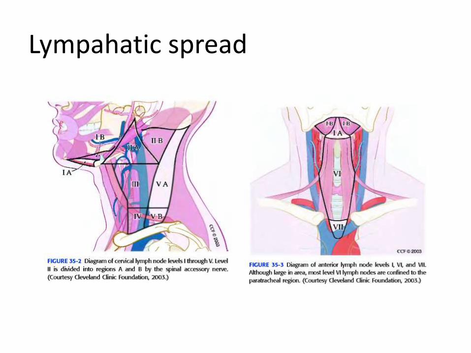
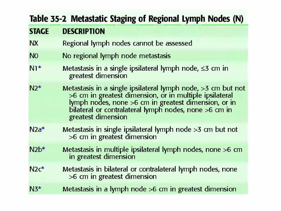
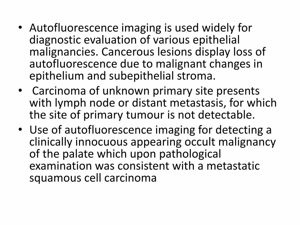
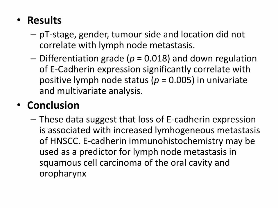
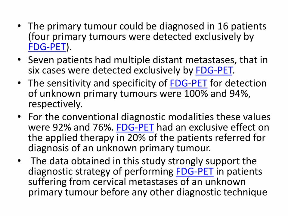
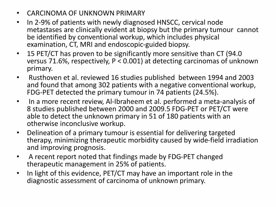
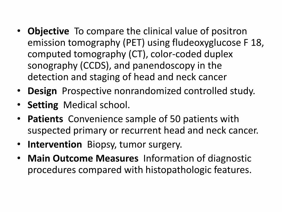
