北海道教育大学紀要. 第二部. a, 数学・物理学・化学・工学編,...
TRANSCRIPT

Hokkaido University of Education
�
�
Title イカ筋肉のアデノシン脱アミノ酵素
Author(s) 坂内, 緑; 吉田, 邦子; 奈良, 盛
Citation北海道教育大学紀要. 第二部. A, 数学・物理学・化学・工学編, 19(1)
: 55-66
Issue Date 1968-09
URL http://s-ir.sap.hokkyodai.ac.jp/dspace/handle/123456789/5895
Rights

Vol. 19, No, 1 Journal of Hokkaido University of Education (Section II A) September, 1968
Purification and Properties of Adenosine Deaminase
from the IVtuscle of a Squid(^Ommastrephes sloani pacificusy*
Midori SAKAITOHI, Kuniko YOSHIDA and Sakari NARA*'"
Chemical Laboratory, Hokkaido University of Education, Hakodate
S?. isW?-. zisX^ : -< A%l^®77:'^ ^yfi7 ^@N?
Summary
Adenosine deaminase (adenosine aminohydrolase, EC 3. 5,4. 4) has been purified about
200-fold from the squid muscle by the following techiques : aqueous extraction, heating to
50°, precipitation with ammonium sulfate, and column chromatography on DEAE-cellulose.
The purified enzyme was chromatographically homogeneous and a bulk of the proteinmigrated paper-ionophoretically was highly active. The crude enzyme resisted to heat, but
the activity of the purified enzyme was quitely lost in half an hour at 50 °C. The optimalpH was observed at 7. 5 with phosphate buffer and Mcllvaine buffer.
The apparent activation energy was found to be approximately 12,OOOcaVmol. The
Michaelis-Menten constant was calculated to be 1. 39 x 10-5M. Finally, the effect of various
additions on the enzymatic activity was described. Especially Hg2+ was a potent inhibitor
whose Ki (dissociation constant of enzyme-inhibitor complex) was calculated to be 6. 5 X
10-6M in its competitive inhibition.
It is well known that adenosine deaminase (adenosine aminohydrolase, EC 3. 5. 4. 4) is
distributed in varying concentration not only in the organs of animals, but also in bacteria.
Since this enzyme was shown to be present in the muscle of a rabbit by SchmidtD in 1928,it has been extensively studied enzymo-chemically and physiologically by many authors.
Conway et al.W found its presence with 5/-adenylic acid deaminase (AMP aminphydro-
lase, EC 3.5.4, 6) in the blood and tissues of rabbit and suggested that it might be importantin detoxifying the vasodepressant adenosine into relatively inactive inosine for the reason
of its high concentration in the intestine. Kornberg et fl/.3) reported on the extract of calf
intestinal mucosa that this enzyme deaminated slowly synthetic 2,6-diaminopurine riboside
(2-aminoadenosine).
In regard to the detailed purification procedures of this enzyme from the calf intestinalmucosa was given by Brady and O'Cnnnell.4) They obtained first the homogeneous preparation
judged by the method of ultracentrifugal analysis.
* A part of this work was presented at the 40th Annual Meeting of the Japanese Biochemical Society,
Osaka (1967).** To whom requests for reprints should be submitted.
( 55)

Adenosine Deaminase of a Squid Muscle
And moreover, one of them, BradyS) reported in detail with his co-worker that the
deamination of adenosine and deoxyadenosine was different a little from the organs of six
mammalian animals in the view of a comparative biochemistry. Fischer et alfo) suggested
that the activity of this enzyme in the chicken duodenum could be increased 40-fold byan injection of adenosine. This result was not consistent with the observation performed
by Gordon et al. who has found the similar finding rather in its embryos.7)
Recently, Hoagland and Fischei-8) have partially purified adenosine deaminase from the
chicken duodenum and observed some properties with particular emphasis on its inhibition
by mercurials. Aikawa9) has reviewed the physicochemical properties of this enzyme from
the clam mid-gut gland.
Concerning to the non-specific adenosine deaminase, Nakanishi et al.W-W have
examined its specificity of substrates and inhibitors. On the other hand, the activity ofthis enzyme in plants was found in the dried barley roots by Fiers et al.W and the
discussions relating to its presence in plant seeds has been arise in the recent paper, is)
However, little has known on this enzyme from an invertebrate muscle.
The present paper described the purification procedures and some enzymatic properties
on this enzyme from the squid muscle.
MATERIAL AND METHODS
Materials — Fresh squids (Ommastrephes sloani pacificns') were caught on the
southern coast of Hokkaido from August to November, 1967.
All chemicals were of analytical grade and obtained from Wako Pure ChemicalIndustries Co. DEAE-cellulose (BROWN) was purchased from Seikagaku Kogyo Co.Filter paper (No. 5, 2X 30cm) for an ionophoresis was obtained from Toyo Roshi Co.
Methods — Adenosine deaminase activity was measured by reading the decrease in
the optical density at 265m,u according to the method of Kalcker.16) The standard assay
system consists of 0.05 ml of 1.87xlO-3M adenosine solution in 0.1M phosphate buffer,
pH 7. 0, 0.1 ml of the enzyme solution and the similar buffer solution to make 3 ml of the
resulting solution in a quartz cell having a lcm light path.
The reaction was initiated by adding the enzyme solution and read at 265m/< for each30-second intervals using a Shimadzu Model DU spectrophotometer. One unit of the enzyme
is defined at the amount which causes an initial rate of change in optical density (^fE^)ot 0.01 per minute under the condition mentioned above. Also ammonia liberated after the
deamination was determined colorimetrically by a Nessler reagent for some experiments.
Protein was determined by a biuret reactionl?) with using casein as a standard and also
by a micro-Kjeldahl method. Specific activity was expressed as units per mg of protein.
The copper was determined colorimetrically by the method of Peterson and BollierlS) basedon producing a blue complex between copper (cupric) and cupferazone.
The migration of protein on the paper with veronal buffer (/<=0.1, pH 8.6) was
performed under the following conditions ; 420 volts, 0. 3 mA/cm, and 3 hours by using a
Toyo Roshi Type C apparatus.
( 56 )

M. SAKAUCHI, K. YOSHIDA and S. NARA
RESULTS
Purification of Adenosine DeaminaseCrude extraction from Muscle — Flesh muscles taken off from an alive squid were
minced and homogenized carefully in a homogenizer (Nihon Seiki Kogyo Co.) for 15 seconds
with chilled water (1: 5 W/V). The resulting homogenates were centrifuged at 5,000 r.p.m.for 15 minutes. The precipitates were repeated by a similar procedure and the combined
upper layer was called as Fraction 1.
Heat Treatment — Each 300ml of Fraction 1, efficiently stirred, were heated to 50°±1°
in a 500 ml stainless steel container by running warm water (55°±1°C). The suspension
was kept at 50°±1°C for 3 minutes accurately and then chilled in a -10°C ice bach until
the temperature dropped to 5°C.
The denatured protein was centrifuged at 5,000 r.p.m. for 10 minutes and discarded.
The slight opalescent supernatant was called as Fraction 2.
Ammonium Sulfate Precipitation and Dialysis-Solid ammonium sulfates were carefully
added into the Fraction 2 to bring about 65% saturation. Much cloudy suspension wasfiltered through a Buchner funnel prepared by a large amounts of Hyflo Super-Cel, Deep
blue precipitates on the layer were collected immediately with a spatula and dissolved ina small amount of water. The resulting fairly bluish solution put into a cellophane tubing
was dialysed against chilled water (1: 20 V/V) for 20 hours with changing 3 times of theouter water. Many white precipitates appeared in the tubing during the dialysis.
Eliminating the precipitates by centrifuge, adenosine deaminase with a high activitywas found in a pale blue supernatant liquid which was called as Fraction 3.
i-0GO£! 4
<dQ.0
O.OIM P.B. 0.05M RB. 0.1 MRB.
(pH6.5) (pHT.O) IpHT.OI
50 75
TUBE NUMBER
O.IMf?B.+2%NaCI
(pH 7.0)
i400I-
>
125
Fig. 1. Column chromatography of the crude enzyme on DEAE-cellulose,DEAE-cellulose column, 2 x 50 cm, equilibratfid with 0.1M phosphate buffer (pH
6. 5) and then eluted with buffer as shown in this figure. Each tube, 6ml. Approx.500 mg protein was loaded.
: Protein measured by the change of optical density at 280m/<.: Enzymatic activity assayed by the change of optical density at 265m/<.
(57)

Adenosine Deaminase of a Squid Muscle
DEAE-Cellulose Chromatography — Each 30 ml of the Fraction 3 was loaded onto a
DEAE-cellulose column (2x50 cm) which was pre-equilibrated with 0.01M phosphate buffersolution, pH 7. 0. The elution of protein was subjected to a stepwise elution method and
the buffer solution system at pH 7. 0 consisted of 0. OlM, 0. 05M, 0. 1M and 0.1M containing
sodium chloride (final concentration to 2%). A 6ml of the eluates was collected by a fractioncollector at the rate of one ml per minute. A coloriess highly active enzyme fraction which
was called as Fraction 4, was obtained from the last buffer solution as shown in Fig. 1.
A blue fraction eluted with 0.1M buffer solution contained much protein but no enzymatic
activity was observed.
Rechromatography on DEAE-Cellulose — Fraction 4 was concentrated by saturated
ammonium sutfate solution (adjusted at pH 7. 0) and then dialysed against water till thedesalting of dialysates entirely ceased. A 10 ml of this dialysates was rechromatographiedon DEAE-cellulose (2x30 cm) which was prepared with 0.01M phoshate buffer solution,
1500
luoo
50 100 150 200TUBE NUMBER
250
Fig. 2. Rechromatography of Fraction 4 on DEAE-cellulose.The elution condition was almost the same as shown in Fig. 1
except the height of column (2 x 30 cm) and the ionic strength of thebuffer. Each tube, 6 ml. Approx. 20 mg was loaded.- : Protein,
1.
2.
3.
4.
5.
Table I. Ptirification of
Fraction
crude extract
Heat-treatment
(NH4)siSO., 0.65-saturation and dialysis
DEAE-cellulosechromatography
Rechromatographyon DEAE-cellulose
Volume(ml)
950
830
60
16
5
adenosme deamhiase from
Activity(unit)
57,000
46,480
35,760
9,312
7,270
Protein(mg)
8,740
1,992
510
19.
4.
the squid musrle
Specific activity(units/rng)
6.5
23.3
70.2
2 485.0
8 1,381.3
(200.S- muscle')
^ds Purification
100
81
62
16
12
1
3.6
10.8
74.6
212,5
(58)

M. SACAUCHI. K. YOSHIDA and S. NARA
pH 7.0. The buffer solution system used here was almost similer to the previous step
except using 0. 3M buffer instead of 0.1M phosphafce buffer containing sodium chloride.
The highest active enzyme preparation, named as Fraction 5, was observed in the
eluates with 0.1M buffer solution as shown in Fig. 2. Overall results of the purification
of the enzyme were summarized in Table I. Ultimately the enzyme preparation was
purified more than 200 times as a specific activity.
Properties of Purified Adenosine DeaminasePurity — A homogeneity of the enzyme preparation has not been rigorously established
yet, but it has been examined by paper-ionophoresis as given in Fig. 3. A bulk of protein
migrated for anode was highly active. Also, a pattern observed by the column chromato-
graphy on DEAE-cellulose indicated a partial homogeneity.
tFig. 3, Paper-ionophoregram of native enzyme.
Filter-paper was prepared with veronal buffer (/<==0,1, pH8.6). Condition of migration ; 420 volt, O.SmA/cm, 15 °, and 3hours. Arrow indicates slot for addition of the sample. Thedistance of migration was about 8 cm for anode from arrowunder the condition mentioned above,
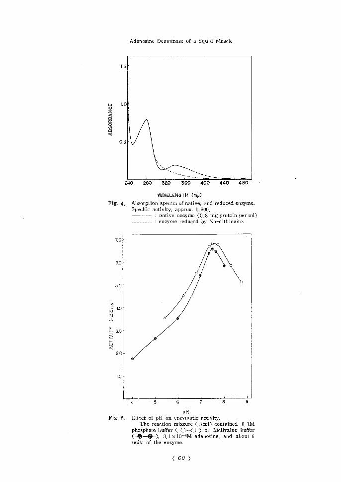
Absorption Spectrum — A spectrum of the native enzyme preparation is shown in Fig.
4. Besides a major peak of an absorption at 280m/.<, a minor one around 340nv< was found
in this figure. This minor component disappared immeadiately by adding a small amount
of sodium dithionite, but recovered partially by an airation.
Opiimal-PH'—Wect of pH on the enzymatic activity was carried out by using phosphate
buffer (pH 4.0-9.0) and Mcllvaine buffer (pH 5.5-8.0). Each buffer solution was used in
the reaction mixture instead of that in a standard assay system for a determination of the
activity.
A sharp peak appeared at pH 7.5. The results obtained are shown in Fig. 5.
Stability — The enzyme preparation kept at 4°C was entirely inactive in one month,
but its activity remained about S0% in deep-freezing. No loss showed in freezing even
over one month, if ammonium sulfate was added to the preparation to make a final
concentration of 10%, A pH-stability was shown in Fig. 6. The enzyme preparation
was diluted (1 : 10 V/V) with phosphate buffer solutions having various pHs (from 4 to9) and was kept in a water bath at 35 °C. The enzyme showed the highest stability atpH 7.0 and retained its activity of 90% even after 6 hours.
(55)
Adenosine Deaminase of a Squid Muscle
1.5
uuz<00a:00)m<
0.51-
320 360 400 440 480
WAVELENGTH (mp)Fig. 4. Absorption spectra of native, and reduced enzyme.
Specific activity, approx. 1,300.: native enzyme (0. 8 mg protein per ml)
.- : enzyme reduced by Na-dithionite.
PHFig. 6. Effect of pH on enzymatic activity.
The reaction mixture ( 3 ml) contained 0.1Mphosphate buffer (-0—0-) or Mcllvaine buffer(-®—®-), 3.1xlO-3M adenosine, and about 6units of the enzyme.
(60 )

M. SAKAUOHT. K. YOSHIDA and S. NARA
100
TIME (hours)Fig. G. PH-stability of adenosine deaminase.
The enzyme was 10-fold diluted with 0.1M phosphate buffer (pH4.0-9.0) and kept in a water bath at 35 °C. The activity was measured bya standard method at various time intervals.
Also the stability was comparatively high at pH 6 or 8, but it dropped half in onehour at pH 5 or 9. At pH 4, no enzymatic activity showed after one hour. That is, a
stability of the enzyme was higher in a neutral state, but decreased remarkably in bothacidic and alkaline sides. A thermostability was determined in the following procedure.
Aliquots of the enzyme preparation were kept at pH 7.0 in a water bath adjusted with a
100
8C
°s
t GOI—u<I 40S£
ILL]tK 20
?^;=
s,50°
•o•a-
35°-o——o-••- .s
40°
45-
10 30 60 90 1?0
TIME (minutes)Fig. 7. Thermostability of adensoine deaminase.
The enzyme was kept at pH 7.0 in a water bath adjusted with arange of temperature from 35 °C to 50 °C. The activity was measured bya standard method at various time intervals as shown in this figure.
(62)

Adenosine Deaminase of a Squid Muscle
temperature ranging from 35°C to 50°C. The remaining activity was determined by a
usual method during constant time intervals.
The results obtained are given in Fig. 7. The highest thermostability was found at35°C and 40°C, and the enzymatic activity of about 90% remained even after two hours.
The activity decreased by half in 2 hours at 45°C and perfectly in half an hours at 50°C.Effect of Temperature on the Reaction Velocity — An apparent activation energy of
the enzyme reaction was approximately 12,000 caVmol as plotted after Arrhenius' equation.
In this experiment, the reaction started by adding one ml of the enzyme preparation to
1.25xlO-3M adenosine (final concentration) mixed with 2ml of 0.1M phosphate buffer
solution, pH 7.0. A determination of the activity was carried out colorimetrically at 450m/-<
by combing ammonia liberated with a Nessler's reagent at various temperatures for 10
minutes. The results obtained were shown in Fig. 8. It appears that a little denaturation
of the enzyme occured at higher than 35 °C.
34 35 36
1/TXIO"
Fig. 8. Arrhenius' plot of the temperaturedependence of deaminase activity.
The reaction started by adding 1 ml of theenzyme to 1.25xlO-3M adenosine mixed with 2mlof 0.1M phosphate buffer (pH 7.0) and kept for 10minutes at various temperatures. The activity wasdetermined colorimetrically at 450m^.
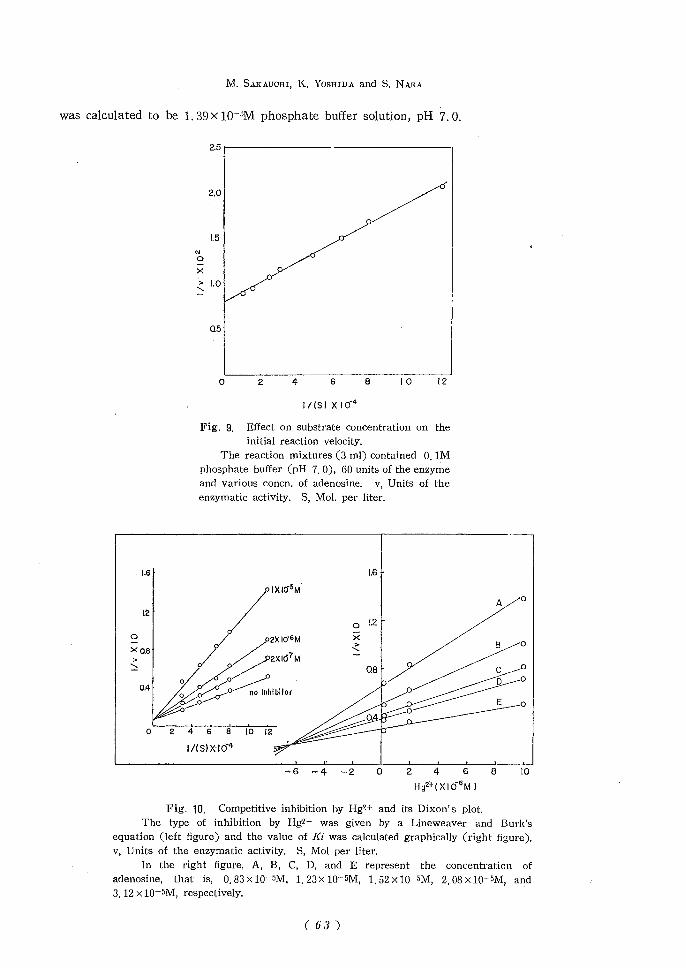
Effect of Sttbsirate Concentration on the Reaction Velocity — Several concentrations of
adenosine were prepared within the range from 8. 30 x 10-eM to 8. 30 x 10-5M. The enzymatic
activity was measured by a standard assay system and the results obtained are shown in
Fig. 9 according to a Lineweaver and Burk's plotl9)' and a Michaelis-Menten constant
( 62 )
M. SAEAUOHI. K. YOSHIDA and S. NARA
was calculated to be 1.39xlO-5M phosphate buffer solution, pH 7.0.
2.5
1.6
12
Fig. 9. Effect on substrate concentration on theinitial reaction velocity.
The reaction mixtures (3 ml) contained 0.1Mphosphate buffer (pH 7. 0), 60 units of the enzymeand various concn. of adenosine. v. Units of theenzymatic activity, S, Mol. per liter.
1.6
'IXIO-5M
24 6 8 10 12
1/(S)XIO'4
-6 -4 -2 2468Hg21-(XIOeM)
Fig'. 10. Competitive inhibition by Hg?.+ and its Dixon's plot.The type of inhibition by Hg2+ was given by a Lineweaver and Burk's
equation (left figure) and the value of Kt was calculated graphically (right figure).v, Units of the enzymatic activity. S, Mol per liter.
In the right figure, A, B, C, D, and E represent the concentration ofadenosine, that is, 0.83xlO-5M, 1.23X10⁻⁵M, 1.52xlO-5M, 2.08X10⁻⁵M, and3.12 x 10-5M, respectively.
( 63)

Adenosine Deaminase of a Squid Muscle
Table II. Effect of various additions on the activity of adenosine deamhiase
from the squid muscle
The reaction mixture except adenosine was preincubated for 15 minutes at room temperature
and then the reaction was started by mixing the substrate.
Additions
Na+
K+Ag+Mg2+
Ca2+
Co2+
Ba2+
Zn2+
Hg2+,D
Pb2+
Cu2+
F-
Final concentration(M)
1X10⁻²
1X10⁻²
5xl0-6
1X10⁻²
1X10⁻²
1X10⁻³
1X10⁻³
1X10⁻³
3x10⁻⁸
3 x 10-''
1X10⁻³
6x10⁻²
Inhibitionm( 5.0)(10.2)
50,5
( 2.5)( 3.2)
5.0
4.3
062.6
45.7
42.2
57.5
Additions Final
NOs-
so.^-
Pyrophosphate
EDTA^-CMB"Monoidoacetic acid
Fericyanide
Oxalic acid
Cysteine
Ascorbic acid
Hydroquinone
H;A
concentration(M)
1X10⁻³
1X10⁻³
1X10⁻³
1 X 10⁻³
1 X 10⁻⁵
1 X 10-3
T x 10-a
1X10⁻³
1 X 10⁻²
1 x 10-*
1 X 10⁻³
1 X 10⁻²
Inhibition(%)( 2.5)( 8.4)( 8.0)
12.6
48.3
4.0
(15.2)( 7.6)( 4.3)
0( 2.3)
0
1) The preincubation time was used for 6 minutes.Figures in parlentheses represent acceleration, also in per cent,
Effect of Various Additions of ihe Reaction Velocity — Some additions including
cathions, anions, oxidants, reducing agents, metal chelating agents and sulfhydryl binding
agents were tested how they did affect on the reaction velocity. The concentration of
additions was applied temporally in the range from 3xlO-6M to lx!0-ZM. The reaction
mixture except adenosine was preincubated for 15 minutes at room temperature and then the
reaction started by mixing the substrate. Hgz+ and p-CMB in mercurials were preincubated
for 6 minutes, because a longer period in time intervals resulted in a decrease of its activity.
Results obtained are listed in Table II. Heavy metals were generally strongly inhibitoryand especially, Hgz+, Ag+ and p-CMB had a potent inhibitory effect. The inhibition byHg2+ is shown in Fig. 10, and also this figure20) indicates that Hg2+ was a competitive
inhibitor for adenosine. It was much striking that the enzymatic activity in a low
concentration of p-CMB was conversely accelerated as suggested by Fisher et al.s) That
is, the inhibition rate by p-CMB varied with time intervals of the preincubation. Concerningthe inhibition by mercurials, the detailed experiments are in progress.
DISCUSSION
As far as the purification of adenosine deaminase is concerned, an acetone fractionation
adopted in the bovine intestinal mucosa'021), and an isoelectric point precipitation in the
mid-gut gland of a clam9) were neither successful in this case because much denaturation
occurred. While a calcium phosphate gel absorption in the beef livei-22) and Tenerds Cr2D
seemed to be better, they were not applied here by reason of having a low yield. To obtain
Fraction 3, the time interval of dialysis was taken for 20 hours under the condition
described previously and this fact appeared to be much important to elevate the yields in
(fi4)

M, SAKAUOHI, K. YOSHIDA and S. NARA
the enzyme purification. Dialysing against water for more than 20 hours, many precipitates
examplified as myosin or actomyosin in the squid muscle occurred gradually, but the time
intervals resulted simultaneously in some inactivation of the enzyme.
A column chromatography on DEAE-cellulose may be suitable for a separation of
this enzyme from the other fractions, especially an unknown blue material containing
approximately 6.5 w copper mg of protein at the highest. The purified enzyme gave one
major band paper-ionophoretically and also indicated comparatively symmetrical curves in
both the enzymatic activity and the protein as shown in Fig. 2, but no other check for the
homogeneity has been performed. Further detailed studies should be done and are still inprogress. By a spectral analysis, the enzyme preparation has no absorption around 600m/-<
based on a blue color, but a different spectrum affected by sodium dithionite was observedat 340 rn/.i. It is not sure whether this spectrum is due to the enzyme itself or any
contaminants.
The pH activity curves of this enzyme preparation are given in Fig. 5. It has an
optimum at pH 7, 5 which declined sharply on both acidic and alkaline sides. This resultwas something different from that of the rabbit (6.6), of the dog (6.8), of the clamand of the calf (7.0). In regard to thermostability, a remaining activity of the enzymepreparation showed about 90% even when maintained at 40 °C for 2 hours.
This fact was in contrast to the behaviour of adenosine deaminase from a clam9)
which was quite inactivated at 40 °C in 5 minutes.
It was evident that a Lineweaver and Burk's plot of the reciprocal of the initialvelocity against the reciprocal of adenosine concentration gave a straight line as shown in
Fig. 9. The value of Km for adenosine was calculated graphically as 1.39xlO-5M.
Comparing this value with that of the other sources, it was almost the same order and
especially it was close to that of the chicken duodenum deaminase (1.30xlO~5M).
It is possible that this enzyme is classified as a sulfhydryl-enzyme, because it was
strongly inhibited by Hg2+, and p-CMB indicating the sulfhydryl nature of the enzyme andalso activated by some reducing agents23\ Hg+z was competitively inhibitor/ at some
range of its concentration for adenosine. However, it is noticeable that the enzymatic
activity was observed to increase conversely, if less than 10-5M of p-CMB were incubated
with the reaction mixtures.
That the value of Ki for Hg2+ was smaller than Km for adenosine, might mean that
the affinity of Hgz+ for the enzyme is larger than that of adenosine.
According to Brady et al.,5) the activity of adenosine deaminase is generally low in a
muscle of all species which are at least classified as vertebrates, but it appears to be high
in the squid muscle. It is much interesting to ascertain whether or not some specificities
may exist in the two classifications.
REFERENCES
1) G. Schmidt, Z. Physiol. Cliem., 179, 243 (1928)2) D. J. Conway and R. Cooke, Biochein. /., 33, 479 (1939)3) A. Kornberg and W. E. Pricer, Jr., /. Biol. Chem., 193, 481 (1951)
( 65 )

Adenosine Deaminase of a Squid Muscle
4) T. G. Brady and W. O'Connell, Biochim. Biophys. Acta, 62, 216 (1962)5) T. G. Brady and C. I. O'Donovan, Comp. Biochem. Physiol., 14, 101 (1965)6) J. R. Fisher, 0. P. Chilson and S. K. Chan, Biochim. Biophys. Ada, 58, 371 (1962)7) M. W. Gordon and M. Roder, /. Biol. Chem., 200, 859 (1953)8) V, D. Hoagland, Jr. and J. R. Fisher, /. Biol. Chem., 242, 4341 (1967)
9) T, Aikawa, Comp. Biochem. Physiol., 17, 271 (1966)10) S. Minato, T. Tagawa and K. Nakanishi, /. Biochem., 58, 519 (1965)11) S. Minato, T. Tagawa, M. Miyalu, B. Shimizu and K. Nakanishi, /. Biochem., 59, 265, (1966)12) S. Minato, T. Tagawa and K. Nakanishi, /. Biochem., 60, 352 (1966)13) S. Minato and K. Nakanishi, /. Biochem., 62, 21 (1967)14) W. Piers and L. Vandendriessche, Arch. inter. Physiol. et Biochem., 68, 203 (1960)
15) T. G. Brady and V. J. Hegarty, Nature, 209, 1027 (1966)16) H. M. Kalckar, /. Biol. Chein., 167, 461 (1947)17) A. G. Gornall, C. J. Bardawill and M. M. David, /. Biol. Cliem., 177, 751 (1949)18) P. E. Peterson and M. E. Bollier, And. Chem., 27, 1195 (1955)19) H. Lineweaver and D. Burk, /. Am. Chem. Soc., 66, 658 (1934)
20) M. Dixon, Biochem. J., 55, 170 (1953)21) W. Klein, Z. Physwl., Chein., 324, 163 (1961)22) K. Hagiwara, K. Tsujimoto, M. Sakamoto and T. Hiroo, /. Female College of Shitennoji (in Japanese),
p. 102 (1966)23) P. D. Boyer, "The Enzymes", ed. by P. D. Boyer, H. Lardy and K. Myrback, Academic Press Inc.,
New York, Vol. I. p. 511 (1959)
( 66 )