technique of chest x-ray reading · technique of chest x-ray reading a. check the request form and...
TRANSCRIPT

Basic Technique of
Chest X-Ray Reading
馬偕醫院 胸腔內科
郭許達 醫師
2017/12/26
1

Technique of Chest X-Ray Reading
A. Check the request form and data on the film
B. Check the the technical quality of chest x-ray
Patient’s position --PA, sternal end
Depth of breathing -- diaphragm level
Exposure of film
C. Find abnormal picture
D. Interpretation
Hsu-Tah Kuo,MD 2

The Effect of Standing and Supine Position
3

Effects of Body Position on CXR
Standing Sitting Supine
4

同一病人在相同時間呼氣和吸氣之影像
5

Checkpoints to Evaluate Technical Quality
1.Medial tips of clavicle equidis-
tant from spinous process
2. Diaphragm at least as low as
the 10th posterior rib
3. Intervertebral spaces visible
through mediastinal shadow
4. Symmetrical radiodensity of
shoulder and soft tissues
5. Clavicles overlying 3-4th
posterior interspace
1 2 3 4 5 6
7
8
9
10
11
Hsu-Tah Kuo,MD
Mackay Memorial Hospital
6

Technique of Chest X-Ray Reading
Read the film systemically
Soft tissue and bony structure
A. Airway -- trachea
B. Blood vessels -- aorta, hilar
vessels
C. Cardiac & mediastinum
D. Diaphragm -- level, shape
E. Effusion -- CP angle
F. Field --lung fields symmetric
bilaterally.
G. Gas shadow -- stomach gas
A
B
C
D E
F
G
Soft tissue and bone
Bone
Soft
tissue
Hsu-Tah Kuo,MD
Mackay Memorial Hospital
7

Anatomical Landmarks on Chest X-Ray
Trachea
Central, slightly to r’t in lower 3rd
Hilar
Level: R’t opposite 6th rib in ax,
L’t 1.5 cm higher
Size: R’t basal artery 9-16 mm
Lung Vessels
Larger in lower, roughly
symmetrical bilaterally
Horizontal Fissure
Level: 6th rib in axilla
Tr.
Hilar
Fissure Lung vessels
6th
Hsu-Tah Kuo,MD
Mackay Memorial Hospital
8

Contours of the Mediastinum
L subclavian
Aortic knob
Pulm a.
Cardiac
SVC Tr.
Bi
lmb rmb
changes in the contours of mediastinal pleural at its interface with adjacent lung.
–Concave contours to straight or convex
–Loss of sharp margins about such anatomic structures
–Widening of spaces between mediastinal boundaries and mediastinal contents
As aorta
9

Cardiac silhouette and mediastinum
10

Hilar and Blood Vessels
11

Mediastinal
window on chest
CT
contrast media
12

Airway and Blood Vessels
A
B
13

Hilar and Blood Vessels
14

Anatomical Landmarks on Chest X-Ray
Heart
Size: TD < 16 cm, change < 2 cm
Shape: Classical
Position: Central, 2/3 to the left
of mid-line
Diaphragm
Level: Ant. 5-6.5 rib,
R’t 0.5-2.5 cm higher than L’t
Curve: Vertical line > 1.5 cm
Outline: shape, CP angle acute
Background Blacking
Same at equivalent level
1 2
3
4
5
6
7
Hsu-Tah Kuo,MD
Mackay Memorial Hospital
15

Anatomical position -- PA & Lat view
c.Anatomical division
red= upper lobes
yellow= middle lobe
blue= lower lobe
a. division of the lungs into
1= hilar portion
2= central portion
3= peripheral portion
b.division of the lung zones
1= apex
2= upper lung field
3= middle lung field
4= lower lung field
16

Lateral View
2 Clear Spaces
Retrosternal
Retrocardiac
17

Lateral Chest X-ray Image
1. Judge the size and shape of the lungs and position and
shape of the diaphragms.
Flattening of the diaphragms (height of < 2.7cm)
2. Follow the airway from neck to the hilum
Tracheal position,
Center of hilar structures - left main bronchus
anterior – RPA; Posterior - LPA.
3. Fissure lines
4. Retrosternal, retrocardiac darkening
5. Trace around the periphery of the image.
Bowel gases , pneumoperitoneum,
18

Lordotic view
19

利用正面和側位判斷病灶位置
20
15% of the lung can be hidden by
cardiovascular structures and the diaphragm.
The lateral image can be helpful in looking
for these obscure lesions on PA films.

Identify the lesion-- PA & Lat
Hsu-Tah Kuo,MD
Mackay Memorial Hospital
21

22

23

Apical lesion -Lordotic View
PA
Lordotic
CT 24

Adjust Digital Imaging
1. Adjust contrast and density to maximize
visualization of all structures
2. Measure the size of the lesion
3. Compare with the prior films
25

Terms use in chest x-ray interpretation
1. Atelectasis, collapse
2. Bulla
3. Circular, ovoid shadow
– multiple fine, nodular, coarse mottling
– small large circular shadow 2cm
4. Consolidation
5. Density- low, fairly high, very high
6. Disseminated, diffused nodular shadow
Hsu-Tah Kuo,MD 26

Terms use in chest x-ray interpretation
7. Effusion
8. Honeycomb shadow
9. Ill-defined opacity
10. Linear, band-like shadow
11. Patchy clouding
12. Reticulation
13. Cavity
14. Septal line
15. Thickened pleura
16. Tubular shadow Hsu-Tah Kuo,MD 27

Terms use in chest x-ray interpretation
28

Terms use in chest x-ray interpretation
29

Blind Area at Chest PA Film
1. Behind the inner end of the clavicle or anterior end of 1st rib
2. Apex of lower lobe on either side
3. Left lower lobe
4. Posterior costophrenic recess
5. Central part of the mediastinum
30
15% of the lung can be hidden by
cardiovascular structures and the diaphragm.
The lateral image can be helpful in looking
for these obscure lesions on PA films.

31
Silhouette

Adjacent Structures whose interfaces create a
Radiographic Silhouette PA view
1.Diaphragm & lower lobe
2.R’t cardiac border & RML
3.L’t cardiac border & Lingual segment
4.Ascending aorta & anterior segment RUL
5.Aortic knob & apical-post. Segment LUL
6.Descending aorta & superior posterior segment LLL
7.Left ventricle & LLL
Lateral view
1.Diaphragm & lower lobe
2.Inferior vena cava & medial basal segment RLL
3.Left ventrilce & LLL
Hsu-Tah Kuo,MD
1
2 3
4 5
6
7
32

Silhouette Sign
Hsu-Tah Kuo,MD 33

Silhouette Sign
34

Pattern of Chest Lesion
1. Chest wall lesion
2. Pleural lesion
3. Diaphragmatic lesion
4. Mediastinal lesion
5. Hilar enlargement
6. Atelectasis
7. Segment and lobar opacity
8. Diffuse air-space opacity
9. Multifocal ill-defined opacity
10. Diffuse fine nodular opacity
11. Fine reticular opacity
12. Coarse reticular opacity
13. Solitary nodule
14. Multiple nodules
15. Hyperlucent abnormality
35

Steps of CXR reading

This is a plain, PA chest radiograph of Mr. Clause taken on the 25
December at the North Pole General Hospital.
The most obvious abnormality is the appearance of cardiomegaly.
I will focus on this after I have first studied the radiograph
systematically. The radiograph is correctly oriented and the exposure is
satisfactory. The patient is not rotated, There are no obvious
abnormalities within the bones. The trachea is not deviated. The
mediastinum has clearly defined borders and there is no enlargement or
change in density of it or the hilar regions. There is no CXR pathology in
the lung and they are expanded normally. The diaphragms are clearly
seen although there is blunting at the right costophrenic angle with a
meniscus suggesting a small effusion. There are no abnormalities below
the diaphragm or within the soft tissues. The cardiothoracic ratio exceeds
1:2. As this is a PA film, this confirms the heart is enlarge.
With regard to the cardiomegaly, four causes are IHD, VHD, pericardial
effusion, and cardiomyopathy.

這是王大川先生今年12月5日在本院拍攝PA view 的胸部X光片
最明顯異常的地方是在左肺上肺野可以看到一個腫塊。 先全部系統性讀完整個片子以後再來討論這個腫塊部分。
拍攝時病人的位置正常, 沒有歪斜,且在吸飽氣下拍攝,肺組織的影像清晰。
骨骼部份在鎖骨,肋骨部分沒有看見異常。
氣管在中央沒有偏移。
Aortic arch, ascending aorta, SVC, 和肺門血管正常大小。
心臟影像正常, mediastinum 沒有變寬。
兩邊橫膈高度正常。
肋膜腳尖銳,沒有看見鈍化積水現象。
肺野沒有異常病灶出現。
橫膈下肺部沒有異常陰影。

Chest X-Ray Reading
1. Give a Factual Report
– What you sees or think
– Position, size, shape, character, effect on surrounding
2. Diagrammatic Drawing
3. Interpretation
– Anatomical site, underlying pathological process
– Correlate any tentative conclusions with clinical
picture
Hsu-Tah Kuo,MD 39

Interpretation of Chest Lung Abnormality James C. Reed Chest Radiology
40
1. Find a true abnormality
2. Localize the abnormality soft tissue, chest wall, pleural, diaphragm, mediastinum,
hilum, peripheral vessels, or the lung parenchyma
3. Classified or describe the pattern patterns of parenchymal lung: nodule, mass,
diffuse opacity, cavity, calcification, atelectasis
4. Assess the distribution localized or diffuse, peripheral or central,
in the upper vs. lower lobe, or alveolar vs. interstitial

Algorithmic Application of Chest Patterns
James C. Reed Chest Radiology
41
1. Pattern identification
2. Differential diagnosis (all the categories of disease that might lead to the identified pattern)
3.Narrow the differential diagnosis by 1.Analysis of the film for additional radiologic findings
2.Evolving patterns of the disease by review of serial examinations
3.Correlation of the patterns with clinical and laboratory data

Hsu-Tah Kuo,MD
Mackay Memorial Hospital
Large Air way Obstruction
42

Pneumothorax Inspiration, Expiration
Inspiration Expiration
Mackay Memorial Hospital
氣胸呈現在胸腔的上方及外緣,看到如髮絲,不透亮的臟膜
少量的氣胸時,吐氣胸部攝影
Mackay Memorial Hospital. HT Kuo,MD
Mackay Memorial Hospital
43

Thoracic Cage & Soft Tissue
Mammoplasty Kyphosis
44

Breast Shadow
45

Thoracic Cage & Soft Tissue
Hepatoma and rib metastasis
Mackay Memorial Hospital. HT Kuo,MD
Multiple myeloma
46

47

Thoracic Cage & Soft Tissue
Mackay Memorial Hospital. HT Kuo,MD 48

Lung
Collapse
Mackay Memorial Hospital. HT Kuo,MD 49

Lung Collapse
Mackay Memorial Hospital. HT Kuo,MD 50

Lung Consolidation after Epistaxis
Mackay Memorial Hospital. HT Kuo,MD 51
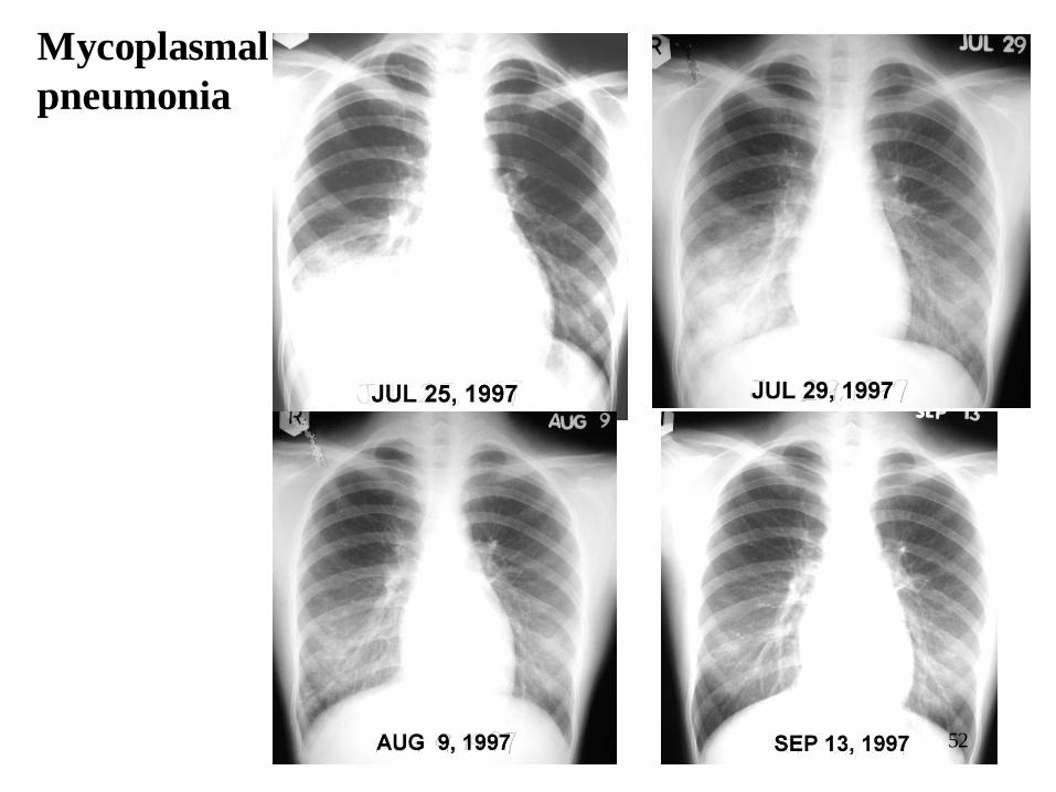
Mycoplasmal
pneumonia
52

Klebsiella
Pneumonia
訾 x 玲
2003/3/14 2003/3/18
2003/3/26 2003/3/30 53

54
Pulmonary TB
病史:
危險因素: DM, Alcoholism, immune abn., Aborigines, Family TB
好發部位: Apico-posterior seg., Superior segment of lower lobe

55
男 , 51, DM, Persistent cough for 3 months; BW loss 7
kg in 4 months; poor intake(+), Fever (-)

Pulmonary Tuberculosis
肺結核 – 兩側上肺葉浸潤 肺結核 – 兩側上肺葉浸潤
粟粒性結核 重度肺結核 56

57
Productive Fibrotic
Cavity Pleural Effusion Milinary
Calcified

Severity of Pulmonary TB
58
Far advanced degree
0 (正常), 1(輕度), 2(中度無空洞), 3(中度有空洞), 4(重度無空洞),
5(重度有空洞), 6(肋膜積水), 7(陳舊性肺結核)

59
TB treat 朱哲X
2002-12-30 2003-2-27
2003-4-18 2003-7-11

60
Follow up Chest x-Ray

Severe Acute Respiratory Syndrome (SARS)
61

MR
ML
HCVD
Radiographic Appearance of Cardiac
Pulmonary Edema LA Myxoma
62

大量胸水會把氣管、縱膈、心臟推到他側 沒有出現推移 (1)endobronchial obstruction (Foreign body,neoplasm)
(2)Pleural adhesion, (3)Fixed mediastinum
Mackay Memorial Hospital Mackay Memorial Hospital
Mackay Memorial Hospital. HT Kuo,MD
63

MR
ML
Radiographic Finding of Pleural Effusion Upright film Supine film
64

Empyema
Streptokinase inj 65

Intrathoracic Goiter Well-defined mass 把氣管推到另一側
CT可見甲狀腺自頸部延伸到縱膈甲狀腺影像密度不均勻,會有局部鈣化注射顯影劑後,甲狀腺的CT密度會增加; 放射碘核醫掃瞄
Mackay Memorial Hospital. HT Kuo,MD
Mackay Memorial Hospital Mackay Memorial Hospital
陳 X 招 88 F 66

Lymphoma 游 X 婷 18 yrs. F
Mackay Memorial Hospital
Mackay Memorial Hospital
Mackay Memorial Hospital. HT Kuo,MD
67

Lung Cancer with Mediastinal Metastasis
Mediastinal mass contiguous with a lung
tumor; Tumor conduct > 1/4 circum of
the aortic wall ; tumor conduct > 3cm
mediastinum
LN > 1 cm Dx metastasis, Accuracy 70%.
Lung Ca 30% mediastinoscopy (-) have
LN metastasis at surgery.
T 4 diagnosis
Mackay Memorial Hospital. HT Kuo,MD
Mackay Memorial Hospital
Mackay Memorial Hospital
68

Sarcoidosis
兩側對稱性淋巴結腫大. 最常發生在支氣管肺部淋巴結、右側氣管旁淋巴結, aorticopulmonary window
Mackay Memorial Hospital Mackay Memorial Hospital
Mackay Memorial Hospital. HT Kuo,MD
69

Aortic Aneurysm
Atherosclerosis
Hypertension
age>40
Mass at aortic contour
contiguous with the aorta.
Aortic dissection identify an
intimal flap
Mackay Memorial Hospital. HT Kuo,MD
Mackay Memorial Hospital
70

Achalasia 27 yrs F. Freq. choking
Mackay Memorial Hospital. HT Kuo,MD
Mackay Memorial Hospital Mackay Memorial Hospital
Bird beak appearance 71

Hiatal Hernia, Para-esophageal 陳 X 娥
Mackay Memorial Hospital. HT Kuo,MD
Mackay Memorial Hospital
Mackay Memorial Hospital
72

Emphysema
Hyperinflation
1. Increase lung height
2. Flattened diaphragm
3. Retrosternal space >2.5cm
Hyperlucency of the lungs
Rapid tapering of the vascular
markings
73

74
Portable Chest X-ray
Endotracheal tube

Malpositioned CVP catheter
75

Pulmonary Artery Catheter
Tip in the interlobar pul
artery: <2 cm lateral to hilum
Complications:
– Malposition: too proximal or too distal (24%)
– Arrhythmia, cardiac damage
– Pulmonary hemorrhage, infarction
76

Malpositioned PA catheter
77

Pneumomediastinum and subcutaneous
emphysema - The early sign of barotrauma
78

Cardiomegaly , pulmonary vessel cephalization
79

Try to read this film
• Thorax
• A
• B
• C
• D
• E
• F
• G
Thorax A
B
C
D
E
F
G
80

Try to read this film
• Thorax
• A
• B
• C
• D
• E
• F
• G
Thorax
A
B
C
D E
F
G 81

Try to read this film
• Thorax
• A
• B
• C
• D
• E
• F
• G
A
B
C
D E
F
G
Thorax
82

Try to Read this film
• Thorax
• A
• B
• C
• D
• E
• F
• G
A
B
C
D
E
F
G
Thorax
83

Try to read this film
• Thorax
• A
• B
• C
• D
• E
• F
• G
A
B
C
D E
F
G
Thorax
84

Try to read this film
• Thorax
• A
• B
• C
• D
• E
• F
• G
A
B
C
D E
F
G
Thorax
85

86
Try to read
this film
• Thorax
• A
• B
• C
• D
• E
• F
• G

87
Try to read
this film
• Thorax
• A
• B
• C
• D
• E
• F
• G

Try to read this film
• Thorax
• A
• B
• C
• D
• E
• F
• G
A
B
C
D E
F
G
Thorax
88

Try to read this film
• Thorax
• A
• B
• C
• D
• E
• F
• G
89

90

判讀胸部X 光片
1.說出攝影為PA, AP,Lat. 如果照得不好指出缺失
2.估計大約年齡或性別
3.再按ABCDEFG 讀出正常或異常點
4.按照異常 pattern說出可能的疾病診斷
5. 可能再進一步的檢查或方法會幫忙診斷
91