university of groningen histopathology, salivary flow and
TRANSCRIPT

University of Groningen
Histopathology, salivary flow and ultrasonography of the parotid glandMossel, Esther; van Ginkel, Martha S; Haacke, Erlin A; Arends, Suzanne; Liefers, Silvia C;Delli, Konstantina; van Nimwegen, Jolien F; Stel, Alja J; Spijkervet, Fred K L; Vissink, ArjanPublished in:Rheumatology
DOI:10.1093/rheumatology/keab781
IMPORTANT NOTE: You are advised to consult the publisher's version (publisher's PDF) if you wish to cite fromit. Please check the document version below.
Document VersionVersion created as part of publication process; publisher's layout; not normally made publicly available
Publication date:2021
Link to publication in University of Groningen/UMCG research database
Citation for published version (APA):Mossel, E., van Ginkel, M. S., Haacke, E. A., Arends, S., Liefers, S. C., Delli, K., van Nimwegen, J. F., Stel,A. J., Spijkervet, F. K. L., Vissink, A., van der Vegt, B., Kroese, F. G. M., & Bootsma, H. (Accepted/Inpress). Histopathology, salivary flow and ultrasonography of the parotid gland: three complementarymeasurements in primary Sjögren's syndrome. Rheumatology.https://doi.org/10.1093/rheumatology/keab781
CopyrightOther than for strictly personal use, it is not permitted to download or to forward/distribute the text or part of it without the consent of theauthor(s) and/or copyright holder(s), unless the work is under an open content license (like Creative Commons).
The publication may also be distributed here under the terms of Article 25fa of the Dutch Copyright Act, indicated by the “Taverne” license.More information can be found on the University of Groningen website: https://www.rug.nl/library/open-access/self-archiving-pure/taverne-amendment.
Take-down policyIf you believe that this document breaches copyright please contact us providing details, and we will remove access to the work immediatelyand investigate your claim.
Downloaded from the University of Groningen/UMCG research database (Pure): http://www.rug.nl/research/portal. For technical reasons thenumber of authors shown on this cover page is limited to 10 maximum.
Download date: 18-12-2021
1
Histopathology, salivary flow and ultrasonography of the parotid gland: three
complementary measurements in primary Sjögren’s syndrome
Esther Mossel*1, Martha S. van Ginkel*1, Erlin A. Haacke1,2, Suzanne Arends1, Silvia C. Liefers1,
Konstantina Delli3, Jolien F. van Nimwegen1, Alja J. Stel1, Fred K.L. Spijkervet3, Arjan Vissink3, Bert van
der Vegt2, Frans G.M. Kroese1, Hendrika Bootsma1
* Both authors contributed equally to this manuscript
Departments of 1Rheumatology and Clinical Immunology, 2Pathology and Medical Biology and 3Oral
and Maxillofacial Surgery, University of Groningen and University Medical Center Groningen,
Groningen, the Netherlands.
Corresponding author
Martha S. van Ginkel
Department of Rheumatology and Clinical Immunology, University of Groningen, University Medical
Center Groningen
HPC AA21, Hanzeplein 1, 9713 GZ Groningen, the Netherlands
Tel: +31503616604
Email: [email protected]
ORCID: 0000-0002-0867-0282
Page 2 of 31Rheumatology
123456789101112131415161718192021222324252627282930313233343536373839404142434445464748495051525354555657585960
© The Author(s) 2021. Published by Oxford University Press on behalf of the British Society for Rheumatology.
This is an Open Access article distributed under the terms of the Creative Commons Attribution Non-Commercial License
(http://creativecommons.org/licenses/by-nc/4.0/), which permits non-commercial re-use, distribution, and reproduction in any
medium, provided the original work is properly cited. For commercial re-use, please contact [email protected]
Dow
nloaded from https://academ
ic.oup.com/rheum
atology/advance-article/doi/10.1093/rheumatology/keab781/6407159 by U
niversity Library user on 03 Novem
ber 2021

2
ABSTRACT
Objective
The involvement of salivary glands in primary Sjögren’s syndrome (pSS) can be assessed in different
ways: histopathology, salivary flow and ultrasonography. To understand the relative value of these
different approaches, it is crucial to understand the relationship between them. As we routinely
perform these three modalities in the parotid gland for disease evaluation, our aim was to
investigate the construct validity between these modalities in one and the same gland.
Methods
Consecutive sicca patients underwent a multidisciplinary diagnostic work-up including parotid gland
biopsy, collection of parotid gland-specific saliva and parotid gland ultrasonography. Patients who
were classified as pSS according to the ACR-EULAR criteria were included. Construct validity was
assessed using Spearman’s correlation coefficients.
Results
The 41 included pSS patients completed a full work-up within mean time interval of 2.6 months.
Correlations between histopathological features and stimulated parotid salivary flow were fair (ρ=-
0.123 for focus score, and ρ=-0.259 for percentage of CD45+ infiltrate). Likewise, poor correlations
were observed between stimulated parotid salivary flow and parotid ultrasonography (ρ=-0.196).
Moderate to good associations were found between the histopathological items focus score and
percentage of CD45+ infiltrate, with parotid ultrasound scores (total ultrasound score: ρ=0.510 and
ρ=0.560; highest for homogeneity: ρ=0.574 and ρ=0.633).
Conclusion
Although pSS associated ultrasonographic findings did correlate with histopathological features, the
three modalities that evaluate salivary gland involvement assess different (or at best partly related)
constructs. Therefore, histopathology, salivary flow and ultrasonography are complementary
measurements and cannot directly replace each other in the work-up of pSS.
Page 3 of 31 Rheumatology
123456789101112131415161718192021222324252627282930313233343536373839404142434445464748495051525354555657585960
Dow
nloaded from https://academ
ic.oup.com/rheum
atology/advance-article/doi/10.1093/rheumatology/keab781/6407159 by U
niversity Library user on 03 Novem
ber 2021

3
KEYWORDS
Primary Sjögren’s syndrome, parotid gland, biopsy, histopathology, salivary flow, ultrasonography
KEY MESSAGES
- Parotid gland histopathology, salivary flow and ultrasonography assess different constructs in
primary SS (pSS) patients
- Parotid salivary flow is not related to parotid histopathology or parotid ultrasonography in
pSS
- The three different modalities cannot directly replace each other in the work-up of pSS
Page 4 of 31Rheumatology
123456789101112131415161718192021222324252627282930313233343536373839404142434445464748495051525354555657585960
Dow
nloaded from https://academ
ic.oup.com/rheum
atology/advance-article/doi/10.1093/rheumatology/keab781/6407159 by U
niversity Library user on 03 Novem
ber 2021
4
INTRODUCTION
Primary Sjögren’s syndrome (pSS) is a chronic, systemic auto-immune disease characterized by oral
and ocular sicca complaints [1]. Inflammation of the salivary and lacrimal glands is a hallmark of the
disease and plays a central role in the current classification criteria [2]. The involvement of salivary
glands in the disease process can be assessed in different ways. The three following modalities are
most commonly used: salivary gland biopsy, saliva collection and salivary gland ultrasonography. The
histopathological assessment of salivary glands and collection of salivary secretion are included in the
current classification criteria as separate items, and it has recently been proposed to add
ultrasonography as an item [2–4].
The inflammatory process in salivary glands of pSS patients is characterized by periductal clusters of
≥50 lymphocytes, called foci. The number of foci per 4 mm2 glandular parenchyma can be calculated
into a focus score (FS) [5,6]. For classification of pSS, in which histopathology of salivary glands is one
of the major items, biopsies with FS≥1 are considered positive [2]. Other histopathological features
are presence of lymphoepithelial lesions and germinal centers, and a relative increase of IgG and IgM
plasma cells, compared to the number of IgA plasma cells (plasma cell shift) [7–11]. Furthermore,
salivary glands of pSS patients contain higher proportions of fibrosis and acinar atrophy than controls
[12,13]. Whether fatty infiltration is specific for pSS or an age-associated condition remains to be
shown [14,15]. During the diagnostic work-up of pSS, biopsies are mostly taken from labial glands.
However, taking biopsies from parotid glands offers several advantages, such as increased specificity
for diagnosing pSS, the possibility to take multiple biopsies of the same gland and the advantage of
early detection of pSS associated lymphomas localized in the parotid gland [16,17]. For these
reasons, we routinely perform parotid gland biopsies in patients suspected of pSS.
The second tool to evaluate salivary gland involvement in pSS is the assessment of saliva secretion.
Unstimulated whole salivary flow (UWSF) is currently included in the ACR-EULAR criteria as an item
[2]. UWS is composed of contributions of all minor and major glands, albeit that UWS contains minor
amounts of saliva from the parotid glands, since the parotid glands only contribute significantly to
whole saliva after stimulation [18]. Saliva can also be collected under stimulated conditions, such as
masticatory (chewing) or gustatory (citric acid solution) stimulation. Stimulated whole salivary flow
(SWSF) is also a mixture of saliva of all minor and major salivary glands. In addition to collecting saliva
from all glands together it is possible to collect saliva exclusively from the parotid glands [18].
Herewith, parotid flow rates can be used for direct functional assessment of the parotid glands.
The third tool to assess salivary gland involvement in pSS is salivary gland ultrasonography (SGUS).
SGUS evaluates major salivary glands, i.e., parotid and submandibular glands. This upcoming tool is
non-invasive, well-tolerated, inexpensive, increasingly available in the outpatient clinic, and can be
Page 5 of 31 Rheumatology
123456789101112131415161718192021222324252627282930313233343536373839404142434445464748495051525354555657585960
Dow
nloaded from https://academ
ic.oup.com/rheum
atology/advance-article/doi/10.1093/rheumatology/keab781/6407159 by U
niversity Library user on 03 Novem
ber 2021

5
performed repeatedly [19,20]. Multiple scoring systems of SGUS are in use, of which the Hocevar
scoring system is most commonly applied [21]. We previously showed that scoring hypoechogenic
areas suffices for classification, which further increases the feasibility of SGUS [22].
To understand the relative value of these different modalities in the work-up of Sjögren’s syndrome,
it is crucial to understand the relationship between them. Although patients secreting less than 0.1
ml/min UWS were two times more likely to have a FS≥1 [23], correlations between histopathological
findings, such as FS and amount of fibrosis, and UWSF are poor [24–26]. When comparing SGUS
scores (from major salivary glands) with labial gland FS and UWSF, correlation coefficients appear to
be higher [27–29]. However, interpretation of these findings is complicated since histopathology,
collection of saliva and ultrasonography were not assessed in the same salivary gland. Furthermore,
there were differences in methods of saliva collection and SGUS scoring systems. Since we routinely
perform parotid gland biopsies, collect parotid gland-specific saliva, and perform parotid gland
ultrasonography in the diagnostic work-up of pSS, we are in the unique position to analyze the
relationship between these three modalities in one and the same gland. In the work presented here,
we aim to explore this relationship by assessing construct validity between histopathology, salivary
secretion, and ultrasonography of the same parotid gland in pSS patients.
Page 6 of 31Rheumatology
123456789101112131415161718192021222324252627282930313233343536373839404142434445464748495051525354555657585960
Dow
nloaded from https://academ
ic.oup.com/rheum
atology/advance-article/doi/10.1093/rheumatology/keab781/6407159 by U
niversity Library user on 03 Novem
ber 2021

6
MATERIALS AND METHODS
Patients
In this cross-sectional study, we included consecutive patients, aged ≥ 18 years, who were referred to
the Sjögren’s expertise center at the University Medical Center Groningen (UMCG) for suspicion of
pSS and were classified as pSS according to the ACR-EULAR criteria [2]. All patients of this inception
cohort underwent a full diagnostic work-up including a parotid gland biopsy, collection of parotid
gland specific saliva and ultrasonography. Informed consent was not required by Dutch Law for
Medical Research and by institutional guidelines. No objection against use of redundant diagnostic
material was recorded from patients in the institutional record of objection. Patient material was
handled according to the ‘Code of conduct for health research’ of the Dutch Federation of Biomedical
Scientific Societies [30].
Histochemical and immunohistochemical staining
Parotid gland biopsies were formalin fixed, paraffin embedded and sectioned at 3 µm thickness. On
consecutive slides a hematoxylin and eosin (H&E) and a Masson staining was performed.
Immunohistochemical staining for CD45, IgA/IgG and IgM was performed on an automated staining
platform, following the manufacturer’s protocols. For the Bcl-6 staining, tissue sections were
deparaffinized and antigen retrieval was performed (EDTA pH 8, 15 minutes 98°C). After blocking of
endogenous peroxidase, slides were incubated with anti-Bcl6 for 75 min. Sections were incubated
with anti-mouse Ig HRP-polymer for 40 minutes. Staining was visualized with diaminobenzidine and
slides were counterstained with hematoxylin. For further details see Supplementary Table S1,
available at Rheumatology online.
Histological analysis
FS and presence of lymphoepithelial lesions were assessed on H&E-stained sections (Figure 1A). An
arbitrary maximum FS of 12 was given to confluent foci. Lymphoepithelial lesions were determined
by the presence of lymphocytes within the ductal epithelium with concurrent epithelial hyperplasia
(Figure 1A,1B). A cluster of ≥5 Bcl6+ cells, lying next to each other within a focus was classified as
germinal center (Figure 1C) [31,32]. Presence of a plasma cell shift was assessed using IgA/IgG double
staining and IgM staining (Figure 1D). A relative decrease of ≤70% IgA+ plasma cells in relation to the
total plasma cell population (present in foci and unaffected parenchyma) was considered as
abnormal [11]. Biopsies were independently scored by two pathologists (EAH and BvdV).
Inconsistencies were solved during a consensus meeting.
Page 7 of 31 Rheumatology
123456789101112131415161718192021222324252627282930313233343536373839404142434445464748495051525354555657585960
Dow
nloaded from https://academ
ic.oup.com/rheum
atology/advance-article/doi/10.1093/rheumatology/keab781/6407159 by U
niversity Library user on 03 Novem
ber 2021

7
Relative area of CD45+ infiltrate in relation to the total area of glandular parenchyma (including
intervening fat cells) was evaluated on CD45 stained slides, and relative areas of fatty and fibrotic
tissue were evaluated on the modified Masson staining. Digital image analyses (DIA) of both slides
were performed by using the open software QuPath v0.2.3 (Figure 1E-1H).
Salivary secretion
Unstimulated whole saliva (UWS) and chewing stimulated whole saliva (SWS) were collected. UWS
secretion rates of ≤1.5mL/15min and SWS secretion rates of ≤3.5mL/5 min were considered
abnormal [33]. Unstimulated parotid salivary flow (UPF) and 2% citric acid stimulated parotid salivary
flow (SPF) were measured using Lashley cups using a standardized assessment protocol [18].
Ultrasonography
SGUS was performed using an ultrasonographic scanner (Esaote MyLabSeven, Genova, Italy),
equipped with a high-resolution probe (4-13MHz), by three well-trained ultrasonographers (AJS, KD,
JFN), with good to excellent inter- and intra-observer reliability [34]. Patients were examined in a
supine position with their head turned towards the opposite side and neck slightly extended. The
scoring system of Hocevar et al. was used in both parotid and submandibular glands [21]
(Supplementary Figure S1, available at Rheumatology online). Separate analyses were performed for
one-sided parotid gland ultrasonography (PGUS), with emphasis on the components ‘hypoechogenic
areas’ and ‘hyperechogenic reflections’ [22]. For the total Hocevar score, which includes all four
major glands, a score of ≥15 was considered positive [35].
Statistical analysis
Descriptive parameters were expressed as number (%), mean (SD) or median (IQR) as appropriate.
Construct validity was assessed using Spearman’s correlation coefficients (ρ) between
histopathological parameters and parotid salivary secretion, between histopathological parameters
and PGUS scores, and between parotid salivary flow and PGUS scores; all three modalities were
assessed in the same parotid gland. Spearman’s correlation coefficient (ρ) was interpreted as poor
agreement (0.0-0.2), fair (0.2-0.4), moderate (0.4-0.6), good (0.6-0.8) or excellent agreement (0.8-
1.0) [35]. Hypotheses for assessing construct validity were predefined, as recommended in construct
validity research [37]. Since fair correlations were found between histopathology and salivary flow in
previous studies [24–26], it was hypothesized that there would be fair to moderate correlations
between parotid histopathological parameters and parotid salivary secretion, as they assess related,
but different types of outcomes. Furthermore, it is known that intra-individual variation of parotid
Page 8 of 31Rheumatology
123456789101112131415161718192021222324252627282930313233343536373839404142434445464748495051525354555657585960
Dow
nloaded from https://academ
ic.oup.com/rheum
atology/advance-article/doi/10.1093/rheumatology/keab781/6407159 by U
niversity Library user on 03 Novem
ber 2021

8
salivary flow is around 24% [38], and that salivary flow fluctuates with circadian rhythm and age
[39,40]. The relatively high measurement variation in salivary flow rates will lower the expected level
of correlation with histopathological parameters. As parotid gland biopsies are always taken in the
same region of the gland, representing only a small part of the parotid gland that is analyzed by
ultrasound, abnormalities that are detected by ultrasound can be missed in the salivary gland biopsy.
Therefore, correlations between histopathological parameters and PGUS scores were hypothesized
to be moderate as they only partly assess the same construct. Finally, it was hypothesized that there
would be fair correlations between parotid salivary flow and PGUS, as they assess different outcomes
within the same construct, and the measurement variation in parotid salivary flow was also taken
into account.
As exploratory analyses, Kruskal Wallis tests were used to compare histopathological parameters
between various groups based on PGUS-hypo and PGUS-hyper scores. Mann Whitney U tests were
used to compare variables between groups based on presence of histopathological features and
between groups based on presence of multiple hypoechogenic areas (PGUS-hypo≥2) vs. limited or no
hypoechogenic areas (PGUS-hypo≤ 1).
Percentages of absolute agreement between the different methods were calculated, based on the
cut-off values that are used for classification of pSS: FS≥1, UWSF≤0.1 ml/min, SGUS≥15. Furthermore,
the associations between FS, UWS and SGUS were explored using Spearman’s correlation coefficients
(ρ). Statistical analyses were executed using IBM SPSS Statistics 23 (SPSS, Chicago, IL, USA). P-values
<0.05 were considered statistically significant.
Page 9 of 31 Rheumatology
123456789101112131415161718192021222324252627282930313233343536373839404142434445464748495051525354555657585960
Dow
nloaded from https://academ
ic.oup.com/rheum
atology/advance-article/doi/10.1093/rheumatology/keab781/6407159 by U
niversity Library user on 03 Novem
ber 2021

9
RESULTS
Patients and time interval between methods
Forty-six consecutive pSS patients were evaluated for eligibility. Four patients had to be excluded
because of insufficient biopsy material (surface area <4mm2) and one patient because of presence of
a parotid gland MALT lymphoma. Patient characteristics are presented in Table 1. The median time
interval between the biopsy and saliva collection was 2.3 months (range 0.0-16.8), between the
biopsy and SGUS 1.6 months (range 0.0-11.0), and between SGUS and saliva collection 1.2 months
(range 0.0-14.7). In respectively 90% and 73% of pSS patients the parotid biopsy was performed after
saliva collection or after SGUS.
Parotid salivary flow versus parotid histology
Unstimulated parotid flow (UPF) showed poor to fair associations with FS, percentages of CD45+
infiltrate, and percentages of fat and fibrosis within the parotid gland (Table 2; Supplementary Figure
S2A & 2C, available at Rheumatology online). Correlation coefficients between stimulated parotid
flow (SPF) and histological parameters were slightly higher, but associations were still fair (Table 2;
Supplementary Figure S2B & 2D, available at Rheumatology online). No significant differences in
parotid flow (UPF or SPF) were found between pSS patients with presence of lymphoepithelial
lesions (UPF p=0.27 and SPF p=0.39) or presence of a plasma cell shift (UPF p=0.83, and SPF p=0.75)
compared to pSS patients without these histopathological features. Although SPF levels seemed to
be lower in pSS patients with presence of germinal centers compared to pSS patients without
germinal centres, no significant differences were found (median 0.02 vs. 0.07, p=0.08). Overall, there
is no clear relation between histological features within parotid gland biopsies and parotid salivary
flow rates in this diagnostic cohort.
Parotid histology versus parotid ultrasonography
Moderate associations were found between FS, percentage of CD45+ infiltrate and PGUS total scores
(Table 2). For the individual ultrasound components PGUS-homogeneity and PGUS-hypo, moderate
to good associations were found with FS and percentage of CD45+ infiltrate (Table 2). Remarkably,
three patients with a positive FS (i.e., FS≥1) had no hypoechogenic areas within the parotid gland (i.e.
PGUS-hypo=0), and vice versa three patients with a negative FS did have hypoechogenic areas (i.e.
PGUS- hypo≥2) (Figure 2). In other words, FS≥1 was not always reflected by presence of
hypoechogenic areas, and presence of hypoechogenic areas was not always reflected by a positive
FS. The percentages of fat and fibrosis did not differ significantly between the different PGUS-hypo
Page 10 of 31Rheumatology
123456789101112131415161718192021222324252627282930313233343536373839404142434445464748495051525354555657585960
Dow
nloaded from https://academ
ic.oup.com/rheum
atology/advance-article/doi/10.1093/rheumatology/keab781/6407159 by U
niversity Library user on 03 Novem
ber 2021

10
and PGUS-hyper scores (Figure 2), which was also reflected by poor associations between these
parameters (Table 2).
Patients with presence of lymphoepithelial lesions and patients with presence of germinal centres
had significantly higher PGUS total scores compared to patients without these features (Figure 3A-
3C). The two individual components that contributed most to this difference were homogeneity and
hypoechogenic areas. Scores in these two components were significantly higher in patients with
lymphoepithelial lesions and germinal centres (p=0.014 for PGUS-homogeneity and p=0.004 for
PGUS-hypo). However, patients with lymphoepithelial lesions and germinal centres did not always
show hypoechogenic areas on ultrasound, as four patients with lymphoepithelial lesions (20%) and
two patients with germinal centres (14%) had a PGUS-hypo score of 0 (Figure 3D-3F). In conclusion,
pSS associated ultrasonographic findings (PGUS-hypo and PGUS-homogeneity) correlated to some
extent with glandular histological parameters of inflammation and were also associated with typical
histopathological features.
Parotid salivary flow versus parotid ultrasonography
Reduced parotid flow was poorly reflected by abnormal PGUS scores, as UPF and SPF both showed
poor associations with PGUS total scores and PGUS components (Table 2; Supplementary Figure S2E
& 2F, available at Rheumatology online). UPF and SPF levels were not significantly lower in pSS
patients with multiple hypoechogenic areas in the parotid gland (PGUS-hypo ≥2), compared to
patients with limited or no hypoechogenic areas (PGUS-hypo ≤ 1) (p=0.62 and p=0.29, respectively).
Apparently, typical PGUS abnormalities and parotid salivary secretion are not directly related to each
other.
Agreement of the three modalities used for pSS classification
The items FS and UWSF that are included in the current 2016 ACR-EULAR classification criteria for
pSS are not parotid gland specific. FS can be measured in both labial and parotid gland biopsies and
UWS is a mixture of contributions of all minor and major salivary glands. Furthermore, the item
ultrasonography, which has recently been proposed to be included in these criteria, evaluates the
submandibular glands in addition to the parotid glands. To compare the three different modalities as
used in the classification criteria, we analysed the agreement based on their cut-off values (FS≥1,
UWSF≤0.1 ml/min, SGUS-score≥15). Absolute agreement between FS and UWSF was 61%, between
FS and SGUS 80% and between UWSF and SGUS 56%. The highest correlation was found between FS
and SGUS (ρ=0.607, P<0.001). Correlation between FS and UWSF was poor (ρ=-0.140, P=0.38), and
between UWSF and SGUS fair (ρ=-0.320, P=0.042) (figure 4A-4C). As shown in Figure 4D, all SGUS-
Page 11 of 31 Rheumatology
123456789101112131415161718192021222324252627282930313233343536373839404142434445464748495051525354555657585960
Dow
nloaded from https://academ
ic.oup.com/rheum
atology/advance-article/doi/10.1093/rheumatology/keab781/6407159 by U
niversity Library user on 03 Novem
ber 2021

11
positive patients had foci in the parotid gland (i.e., FS>0), and >80% had a FS≥1. Furthermore, almost
all SGUS-positive patients (89%) showed at least one histological feature associated with pSS (FS≥1,
presence of lymphoepithelial lesions, germinal centers, plasma cell shift) within their biopsy.
However, 38% of SGUS-negative patients also showed one or more histopathological features within
their parotid gland biopsy, indicating that presence of specific histopathological features of pSS is not
always associated with a positive ultrasound.
Page 12 of 31Rheumatology
123456789101112131415161718192021222324252627282930313233343536373839404142434445464748495051525354555657585960
Dow
nloaded from https://academ
ic.oup.com/rheum
atology/advance-article/doi/10.1093/rheumatology/keab781/6407159 by U
niversity Library user on 03 Novem
ber 2021
12
DISCUSSION
The present study showed that evaluations of the same parotid gland by histopathology, salivary
flow and ultrasonography assess different (or at best partly related) constructs. We showed that in
patients with pSS, correlations between parotid gland salivary flow and parotid gland
histopathological parameters were poor to fair. Likewise, no clear relation was found between
parotid salivary flow and abnormalities found by parotid gland ultrasonography. Apparently, salivary
flow does not assess the same construct as parotid gland histopathology, nor as parotid gland
ultrasonography. On the other hand, we showed moderate to good correlations between the
histopathological parameters FS and amount of infiltrate and the ultrasound parameters PGUS-total,
PGUS-homogeneity and PGUS-hypo (Table 2). Thus, typical ultrasound findings in pSS patients seem
to be related to the amount of inflammatory infiltrate in the salivary gland biopsy. However, this was
not seen in all pSS patients, as there are still some patients with a positive ultrasound and FS<1, as
well as patients with histological lymphocytic foci without typical ultrasonographic abnormalities.
Recently, Izzetti et al. [41] compared ultrasound and histopathology of labial salivary glands. They
found a moderate correlation (ρ=0.532) between labial gland FS and labial gland ultrasound scores in
patients suspected of pSS [41]. We found a comparable correlation (ρ=0.510) between FS and PGUS
total score of the parotid glands and even a better correlation between the amount of infiltrate and
PGUS total scores. From these results we can conclude that the inflammatory infiltrates are
somehow related to ultrasound scores, both in the labial and in the parotid salivary gland. In the
present study, the highest correlations were found between FS and amount of CD45 infiltrate and
the ultrasound scores PGUS-hypo and PGUS-homogeneity. This implies that hypoechogenic areas and
inhomogeneity are linked to histopathological lymphocytic infiltrates. Previously, it has been
suggested that hypoechogenic areas represent lymphocytic infiltrates [42,43]. However, as
hypoechogenic areas are significantly larger in size compared to foci seen in tissue sections of
biopsies, these hypoechogenic areas cannot directly represent foci. Therefore, an indirect association
between hypoechogenic areas and infiltrates is more likely. Recently, Wang et al. [44] hypothesized
that presence of hypoechogenic areas could be caused by leakage of saliva into the periductal
infiltrate and eventually into the salivary gland parenchyma, due to dysfunction of tight junctions
between striated ductal cells [44]. Although this hypothesis may explain why hypoechogenic areas
are much larger than infiltrates, intraparenchymal saliva and concurrent reactive changes were not
observed in this series (nor in clinical practice). Therefore, the most likely explanation would be that
hypoechogenic areas represent areas with a higher density of periductal foci with relatively little
unaffected glandular tissue in between these foci, which is also supported by the good correlation
between the amount of CD45+ infiltrate and hypoechogenic areas (ρ=0.540) in this study.
Page 13 of 31 Rheumatology
123456789101112131415161718192021222324252627282930313233343536373839404142434445464748495051525354555657585960
Dow
nloaded from https://academ
ic.oup.com/rheum
atology/advance-article/doi/10.1093/rheumatology/keab781/6407159 by U
niversity Library user on 03 Novem
ber 2021

13
Furthermore, we showed that pSS patients with histopathological features (lymphoepithelial lesions
and germinal centers), which are associated with inflammatory foci, had higher PGUS-hypo scores.
Nevertheless, presence of hypoechogenic areas does not predict whether the biopsy contains any of
these features (Figure 3D-3F) and not all pSS patients with these histopathological features show
hypoechogenic areas on ultrasound. This is in line with previous findings, as Hammenfors et al.
showed that only 67% of pSS patients with germinal center-like structures in the labial gland had an
abnormal ultrasound score [45]. Although Wernicke et al. hypothesized that hyperechogenic
reflections consist of areas of fibrosis [42], we did not find a correlation between hyperechogenic
reflections and any histological parameter (including fat and fibrosis). The analysis of
ultrasonographic images, however, may suffer from inter- and intra-observer differences and is
based on a categorical scale, which will also lower the level of correlation in this study. Several
initiatives, like the European Union funded Horizon 2020 project called HarmonicSS and a sub-
taskforce of the OMERACT working group, aim to develop more objective ultrasound scoring
systems, to overcome the risk of inter- and intraobserver differences [46–48]. Furthermore, as
described earlier, a parotid gland biopsy represents only a small part of the gland, while the whole
gland is analysed by ultrasonography. Therefore, a biopsy might be taken from non-affected salivary
gland tissue and result in a false negative outcome (sampling error). A technique that could help to
better understand the relationship between hyper- and hypoechogenic areas and histopathological
features, is ultrasound-guided core needle biopsy. Recently, this technique has proven to be useful in
the evaluation of suspected salivary gland lymphoma in pSS [49]. Although less tissue is collected in
comparison with an open biopsy, core needle biopsy also provides biopsies with a preserved
architecture. Herewith, a biopsy can be taken specifically from the location of a hyper- or
hypoechogenic areas.
Despite the notion that there is (some) relationship between parotid histology and ultrasonography,
parotid salivary flow rates do not correlate well with histopathological features, nor with
ultrasonography. In line with our results, Bookman et al. also found poor agreement between UWSF
and labial gland FS and fair agreement between SWSF and labial gland FS [24]. Likewise, Daniels et
al., found an absolute agreement of 60% between UWSF and labial gland FS in the large Sjögren’s
International Collaborative Clinical Alliance (SICCA) cohort [23]. Yalcinkaya et al. [50] recently showed
that patients with UWSF≤0.1mL/min had significantly higher PGUS scores compared with patients
with a UWSF above the threshold. In our study, UPF did not differ significantly between patients with
high (≥2) and low (<2) PGUS-hypo scores. However, in unstimulated conditions, whole saliva mainly
consists of the contributions of submandibular and sublingual glands [18]. For this reason, analyses
containing either UWSF or UPF cannot be equally compared to each other. In our cohort, the
Page 14 of 31Rheumatology
123456789101112131415161718192021222324252627282930313233343536373839404142434445464748495051525354555657585960
Dow
nloaded from https://academ
ic.oup.com/rheum
atology/advance-article/doi/10.1093/rheumatology/keab781/6407159 by U
niversity Library user on 03 Novem
ber 2021

14
correlation between UWSF and UPF was ρ=0.600, compared to ρ=0.832 between SWSF and SPF.
Furthermore, the parotid glands appear to be the last salivary gland to show a functional decline in
pSS patients [51]. As we evaluated newly diagnosed pSS patients in a diagnostic cohort, the parotid
glands would still be relatively spared, which can partly explain the poor correlations of SGUS and
histopathology with parotid salivary flow in this cohort. The time lag between the saliva collection,
ultrasound and biopsy reflects the diagnostic pathway in daily clinical practice, but may have
somewhat underestimated the observed associations between the three modalities. In a minority of
pSS patients the parotid gland biopsy was performed prior to saliva collection or ultrasound (n=4 and
n=11, respectively), which might affect the results. For these subgroups of patients, the median time
intervals between the modalities were relatively large (5.9 months between biopsy and saliva
collection, and 4.6 months between biopsy and PGUS). Furthermore, right and left sided salivary flow
rates and right and left sided ultrasound scores were highly associated (ρ=0.815 and ρ=0.943,
respectively). Therefore, the impact of the previous biopsy on salivary flow rates or ultrasound scores
are expected to be negligible in this cohort.
Together, parotid gland salivary flow was not related to parotid gland histopathology nor to parotid
gland ultrasound scores. Parotid gland histopathology was partly related to parotid gland ultrasound,
as we found moderate to good correlations between histopathological inflammatory parameters and
PGUS scores. In conclusion, histopathology, salivary flow and ultrasonography are three
complementary measurements and cannot directly replace each other in the workup of pSS.
Page 15 of 31 Rheumatology
123456789101112131415161718192021222324252627282930313233343536373839404142434445464748495051525354555657585960
Dow
nloaded from https://academ
ic.oup.com/rheum
atology/advance-article/doi/10.1093/rheumatology/keab781/6407159 by U
niversity Library user on 03 Novem
ber 2021

15
FUNDING STATEMENT
No specific funding was received from any bodies in the public, commercial or not -for- profit sectors
to carry out the work described in this article.
CONFLICTS OF INTEREST
The authors have declared no conflicts of interest.
ETHICS
The authors state that the study complies with the Declaration of Helsinki. Informed consent and
local ethical approval was not required by Dutch Law for Medical Research and by institutional
guidelines. No objection against use of redundant diagnostic material was recorded from patients in
the institutional record of objection. Patient material was handled according to the ‘Code of conduct
for health research’ of the Dutch Federation of Biomedical Scientific Societies [30]. In addition,
patients were included in one of our research cohorts: REgistry of Sjögren syndrome in Umcg –
LongiTudinal (RESULT) cohort (METc 2014.491), Biomarker cohort (METc 2013.066) and Abatacept
Sjögren Active Patients phase III (ASAPIII) cohort (METc 2014.118).
DATA AVAILABILITY STATEMENT
All data underlying this article are included in the article and in its online supplementary material.
The data that support this study are available on a reasonable request to the corresponding author.
Page 16 of 31Rheumatology
123456789101112131415161718192021222324252627282930313233343536373839404142434445464748495051525354555657585960
Dow
nloaded from https://academ
ic.oup.com/rheum
atology/advance-article/doi/10.1093/rheumatology/keab781/6407159 by U
niversity Library user on 03 Novem
ber 2021

16
REFERENCES
[1] Brito-Zerón P, Baldini C, Bootsma H, Bowman SJ, Jonsson R, Mariette X, et al. Sjögren syndrome. Nature Reviews Disease Primers 2016;2:16047.
[2] Shiboski CH, Shiboski SC, Seror R, Criswell LA, Labetoulle M, Lietman TM, et al. 2016 American College of Rheumatology/European League Against Rheumatism classification criteria for primary Sjögren’s syndrome: A consensus and data-driven methodology involving three international patient cohorts. Annals of the Rheumatic Diseases 2017;76:9–16.
[3] van Nimwegen J, Mossel E, Delli K, van Ginkel M, Stel A, Kroese F, et al. Incorporation of salivary gland ultrasonography into the ACR-EULAR criteria for primary Sjögren’s syndrome. Arthritis Care and Research 2020;72:583–90.
[4] Jousse-Joulin S, Gatineau F, Baldini C, Baer A, Barone F, Bootsma H, et al. Weight of salivary gland ultrasonography compared to other items of the 2016 ACR/EULAR classification criteria for Primary Sjögren’s syndrome. Journal of Internal Medicine 2020;287:180–8.
[5] Chisholm DM, Mason DK. Labial salivary gland biopsy in Sjögren’s disease. Journal of Clinical Pathology 1968;21:656–60.
[6] Fisher BA, Jonsson R, Daniels T, Bombardieri M, Brown RM, Morgan P, et al. Standardisation of labial salivary gland histopathology in clinical trials in primary Sjögren’s syndrome. Annals of the Rheumatic Diseases 2017;76:1161–8.
[7] Kroese FGM, Haacke EA, Bombardieri M. The role of salivary gland histopathology in primary Sjögren’s syndrome: Promises and pitfalls. Clinical and Experimental Rheumatology 2018;36:S222–33.
[8] Risselada AP, Looije MF, Kruize AA, Bijlsma JWJ, van Roon JAG. The Role of Ectopic Germinal Centers in the Immunopathology of Primary Sj??gren’s Syndrome: A Systematic Review. Seminars in Arthritis and Rheumatism 2013.
[9] Ihrler S, Zietz C, Sendelhofert A, Riederer A, Löhrs U. Lymphoepithelial duct lesions in Sjogren-type sialadenitis. Virchows Archiv 1999;434:315–23.
[10] Zandbelt M, Wentink J, de Wilde P, van Damme P, van de Putte L, van den Hoogen F. The synergistic value of focus score and IgA% score of sublabial salivary gland biopsy for the accuracy of the diagnosis of Sjögren’s syndrome: a 10-year comparison. Rheumatology (Oxford, England) 2002;41:819–23.
[11] Bodeutsch C, de Wilde PCM, Kater L, van Houwelingen JC, van den Hoogen FHJ, Kruize AA, et al. Quantitative immunohistologic criteria are superior to the lymphocytic focus score criterion for the diagnosis of Sjögren’s syndrome. Arthritis & Rheumatism 1992;35:1075–87.
[12] Llamas-Gutierrez FJ, Reyes E, Martínez B, Hernández-Molina G. Histopathological environment besides the focus score in Sjögren’s syndrome. International Journal of Rheumatic Diseases 2014;17:898–903.
Page 17 of 31 Rheumatology
123456789101112131415161718192021222324252627282930313233343536373839404142434445464748495051525354555657585960
Dow
nloaded from https://academ
ic.oup.com/rheum
atology/advance-article/doi/10.1093/rheumatology/keab781/6407159 by U
niversity Library user on 03 Novem
ber 2021
17
[13] Leehan K, Pezant N, Rasmussen A, Grundahl K, Moore J, Radfar L, et al. Minor Salivary Gland Fibrosis in Sjögren’s Syndrome Is Elevated, Associated With Focus Score and Not Solely a Consequence of Aging. Clin Exp Rheumatology 2018;36Suppl112:80–8.
[14] Leehan K, Pezant N, Rasmussen A, Grundahl K, Moore J, Radfar L, et al. Fatty Infiltration of the Minor Salivary Gland Is a Selective Feature of Aging but Not Sjögren’s Syndrome. Autoimmunity 2017;50:451–7.
[15] Skarstein K, Aqrawi L, Øijordsbakken G, Jonsson R, Jensen J. Adipose Tissue is Prominent in Salivary Glands of Sjögren’s Syndrome Patients and Appears to Influence the Microenvironment in These Organs. Autoimmunity 2016;49:338–46.
[16] Pijpe J, Kalk WWI, van der Wal JE, Vissink A, Kluin PM, Roodenburg JLN, et al. Parotid gland biopsy compared with labial biopsy in the diagnosis of patients with primary Sjogren’s syndrome. Rheumatology (Oxford, England) 2007;46:335–41.
[17] Delli K, Vissink A, Spijkervet FKL. Salivary gland biopsy for Sjögren’s syndrome. Oral and Maxillofacial Surgery Clinics of North America 2014;26:22–33.
[18] Jensen SB, Vissink A. Salivary Gland Dysfunction and Xerostomia in Sjögren’s Syndrome. Oral and Maxillofacial Surgery Clinics of NA 2014;26:35–53.
[19] Delli K, Dijkstra PU, Stel AJ, Bootsma H, Vissink A, Spijkervet FKL. Diagnostic properties of ultrasound of major salivary glands in Sjögren’s syndrome: a meta-analysis. Oral Diseases 2015;21:792–800.
[20] Jousse-Joulin S, Milic V, Jonsson M v, Plagou A, Theander E, Luciano N, et al. Is salivary gland ultrasonography a useful tool in Sjögren’s syndrome? A systematic review. Rheumatology 2016;55:789–800.
[21] Hocevar A, Ambrozic A, Rozman B, Kveder T, Tomsic M. Ultrasonographic changes of major salivary glands in primary Sjögren’s syndrome. Diagnostic value of a novel scoring system. Rheumatology (Oxford, England) 2005;44:768–72.
[22] Mossel E, Arends S, van Nimwegen JF, Delli K, Stel AJ, Kroese FGM, et al. Scoring hypoechogenic areas in one parotid and one submandibular gland increases feasibility of ultrasound in primary Sjögren’s syndrome. Annals of the Rheumatic Diseases 2018;77:556–62.
[23] Daniels TE, Cox D, Shiboski CH, Schiødt M, Wu A, Lanfranchi H, et al. Associations between salivary gland histopathologic diagnoses and phenotypic features of Sjögren’s syndrome among 1,726 registry participants. Arthritis and Rheumatism 2011;63:2021–30.
[24] Bookman AAM, Shen H, Cook RJ, Bailey D, McComb RJ, Rutka JA, et al. Whole stimulated salivary flow: Correlation with the pathology of inflammation and damage in minor salivary gland biopsy specimens from patients with primary Sjögren’s syndrome but not patients with sicca. Arthritis and Rheumatism 2011;63:2014–20.
Page 18 of 31Rheumatology
123456789101112131415161718192021222324252627282930313233343536373839404142434445464748495051525354555657585960
Dow
nloaded from https://academ
ic.oup.com/rheum
atology/advance-article/doi/10.1093/rheumatology/keab781/6407159 by U
niversity Library user on 03 Novem
ber 2021

18
[25] Fisher BA, Brown RM, Bowman SJ, Barone F. A review of salivary gland histopathology in primary Sjögren’s syndrome with a focus on its potential as a clinical trials biomarker. Annals of the Rheumatic Diseases 2015;74:1645–50.
[26] Atkinson J, Travis W, Pillemer S, Bermudez D, Wolff A, Fox P. Major salivary gland function in primary Sjögren’s syndrome and its relationship to clinical features. The Journal of Rheumatology 1990;17:318–22.
[27] Kim JW, Lee H, Park SH, Kim SK, Choe JY, Kim JK. Salivary gland ultrasonography findings are associated with clinical, histological, and serologic features of Sjögren’s syndrome. Scandinavian Journal of Rheumatology 2018;47:303–10.
[28] Caraba A, Babalic FC, Iurciuc S, Iurciuc M, Mynbaev OA. The Utility of Major Salivary Gland Ultrasonographic Parameters in the Diagnosis of Sjögren Syndrome. Disease Markers 2019;1716848.
[29] Cornec D, Jousse-Joulin S, Costa S, Marhadour T, Marcorelles P, Berthelot JM, et al. High-grade salivary-gland involvement, assessed by histology or ultrasonography, is associated with a poor response to a single rituximab course in primary Sjögren’s syndrome: Data from the TEARS randomized trial. PLoS ONE 2016;11;e0162787.
[30] FMWV Code of Conduct for Health Research, [internet] 2011. Available from: https://www.federa.org/sites/default/files/ bijlagen/coreon/code_of_conduct_for_medi-cal_research_1.pdf
[31] Haacke EA, van der Vegt B, Vissink A, Spijkervet FKL, Bootsma H, Kroese FGM. Germinal centres in diagnostic labial gland biopsies of patients with primary Sjogren’s syndrome are not predictive for parotid MALT lymphoma development. Annals of the Rheumatic Diseases 2017;76:1783–6.
[32] Nakshbandi U, Haacke EA, Bootsma H, Vissink A, Spijkervet FKL, van der Vegt B, et al. Bcl6 for identification of germinal centres in salivary gland biopsies in primary Sjögren’s syndrome. Oral Diseases 2020;26:707–10.
[33] Pedersen AM, Bardow A, Beier Jensen S, Nantofte B. Saliva and gastrointestinal functions of taste, mastication, swallowing and digestion. Oral Dis. 2002;8:117-29.
[34] Delli K, Arends S, van Nimwegen JF, Dijkstra PU, Stel AJ, Spijkervet FKL, et al. Ultrasound of the Major Salivary Glands is a Reliable Imaging Technique in Patients with Clinically Suspected Primary Sjögren’s Syndrome. Ultraschall in Der Medizin 2018;39:328–33.
[35] Mossel E, Delli K, van Nimwegen JF, Stel AJ, Kroese FGM, Spijkervet FKL, et al. Ultrasonography of major salivary glands compared with parotid and labial gland biopsy and classification criteria in patients with clinically suspected primary Sjögren’s syndrome. Annals of the Rheumatic Diseases 2017;76:1883–9.
[36] Landis JR, Koch GG. The measurement of observer agreement for categorical data. Biometrics 1977;33:159–74.
Page 19 of 31 Rheumatology
123456789101112131415161718192021222324252627282930313233343536373839404142434445464748495051525354555657585960
Dow
nloaded from https://academ
ic.oup.com/rheum
atology/advance-article/doi/10.1093/rheumatology/keab781/6407159 by U
niversity Library user on 03 Novem
ber 2021

19
[37] Terwee CB, Bot SDM, de Boer MR, van der Windt DAWM, Knol DL, Dekker J et al. Quality criteria were proposed for measurement properties of health status questionnaires. Journal of Clinical Epidemiology 2007;60:34–42.
[38] Burlage FR, Pijpe J, Coppes RP, Hemels MEW, Meertens H, Canrinus A, et al. Variability of flow rate when collecting stimulated human parotid saliva. European Journal of Oral Sciences 2005;113:386–90.
[39] Baum BJ. Salivary Gland Fluid Secretion During Aging. Journal of the American Geriatrics Society 1989;37:453–8.
[40] Kariyawasam AP, Dawes C. A circannual rhythm in unstimulated salivary flow rate when the ambient temperature varies by only about 2°C. Archives of Oral Biology 2005;50:919–22.
[41] Izzetti R, Ferro F, Vitali S, Nisi M, Fonzetti S, Oranges T et al. Ultra-high frequency ultrasonography (UHFUS)-guided minor salivary gland biopsy: a promising procedure to optimize labial salivary gland biopsy in Sjögren’s syndrome. Journal of Oral Pathology and Medicine 2021;50:485-491.
[42] Wernicke D, Hess H, Gromnica-Ihle E, Krause A, Schmidt WA. Ultrasonography of salivary glands - A highly specific imaging procedure for diagnosis of Sjögren’s syndrome. Journal of Rheumatology 2008;35:285–93.
[43] Takashima S, Morimoto S, Tomiyama N, Takeuchi N, Ikezoe J, Kozuka T. Sjogren syndrome: Comparison of sialography and ultrasonography. Journal of Clinical Ultrasound 1992;20:99–109.
[44] Wang X, Bootsma H, Terpstra J, Vissink A, van der Vegt B, Spijkervet FKL, et al. Progenitor cell niche senescence reflects pathology of the parotid salivary gland in primary Sjögren’s syndrome. Rheumatology (Oxford)) 2020;59:3003-3013.
[45] Hammenfors DS, Brun JG, Jonsson R, Jonsson M v. Diagnostic utility of major salivary gland ultrasonography in primary Sjögren’s syndrome. Clinical and Experimental Rheumatology 2015;33:56–62.
[46] Jousse-Joulin S, D’Agostino MA, Nicolas C, Naredo E, Ohrndorf S, Backhaus M, et al. Video clip assessment of a salivary gland ultrasound scoring system in Sjögren’s syndrome using consensual definitions: An OMERACT ultrasound working group reliability exercise. Annals of the Rheumatic Diseases 2019;78:967–73.
[47] Fisher BA, Emery P, Pitzalis C, Bombardieri M, Bowman SJ. Response to: Can ultrasound of the major salivary glands assess histopathological changes induced by treatment with rituximab in primary Sjögren’s syndrome? Annals of the Rheumatic Diseases 2018;78.
[48] Harmonization and integrative analysis of regional, national and international cohorts on primary Sjögren’s Syndrome (pSS) towards improved stratification, treatment and health policy making (HarmonicSS). Horizon2020 project. Grant agreement number 731944 n.d.
Page 20 of 31Rheumatology
123456789101112131415161718192021222324252627282930313233343536373839404142434445464748495051525354555657585960
Dow
nloaded from https://academ
ic.oup.com/rheum
atology/advance-article/doi/10.1093/rheumatology/keab781/6407159 by U
niversity Library user on 03 Novem
ber 2021
20
[49] Baer AN, Grader-Beck T, Antiochos B, Birnbaum J, Fradin JM. Ultrasound-guided biopsy of suspected salivary gland lymphoma in Sjögren’s syndrome. Arthritis Care & Research 2020;73:849-855.
[50] Yalcinkaya Y, Mumcu G, Özdemir FT, Kuruş RE, Ünal AU, Direskeneli H, et al. Are salivary gland ultrasonography scores associated with salivary flow rates and oral health-related quality of life in sjögren syndrome? Journal of Rheumatology 2020;47:1774–9.
[51] Pijpe J, Kalk WWI, Bootsma H, Spijkervet FKL, Kallenberg CGM, Vissink A. Progression of salivary gland dysfunction in patients with Sjögren’s syndrome. Annals of the Rheumatic Diseases 2007;66:107–12.
Page 21 of 31 Rheumatology
123456789101112131415161718192021222324252627282930313233343536373839404142434445464748495051525354555657585960
Dow
nloaded from https://academ
ic.oup.com/rheum
atology/advance-article/doi/10.1093/rheumatology/keab781/6407159 by U
niversity Library user on 03 Novem
ber 2021
21
TABLES
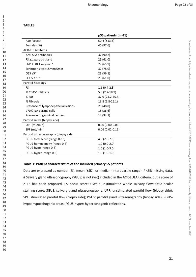
pSS patients (n=41)Age (years) 50.4 (±13.6)Females (%) 40 (97.6)
ACR-EULAR itemsAnti-SSA antibodies 37 (90.2)FS ≥1, parotid gland 25 (61.0)UWSF ≤0.1 mL/min* 27 (65.9)Schirmer’s test ≤5mm/5min 32 (78.0)OSS ≥5* 23 (56.1)SGUS ≥ 15# 25 (61.0)
Parotid histologyFS 1.1 (0.4-2.3)% CD45+ infiltrate 5.3 (2.2-18.9)% Fat 37.9 (24.2-45.8)% Fibrosis 19.8 (6.8-26.1)Presence of lymphoepithelial lesions 20 (48.8)≤70% IgA plasma cells 15 (36.6)Presence of germinal centers 14 (34.1)
Parotid saliva (biopsy side)UPF (mL/min) 0.00 (0.00-0.03)SPF (mL/min) 0.06 (0.02-0.11)
Parotid ultrasonography (biopsy side)PGUS total score (range 0-13) 4.0 (2.0-7.5)PGUS-homogeneity (range 0-3)PGUS-hypo (range 0-3)
1.0 (0.0-2.0)1.0 (1.0-3.0)
PGUS-hyper (range 0-3) 1.0 (1.0-1.0)
Table 1: Patient characteristics of the included primary SS patients
Data are expressed as number (%), mean (±SD), or median (interquartile range). * <5% missing data.
# Salivary gland ultrasonography (SGUS) is not (yet) included in the ACR-EULAR criteria, but a score of
≥ 15 has been proposed. FS: focus score; UWSF: unstimulated whole salivary flow; OSS: ocular
staining score; SGUS: salivary gland ultrasonography, UPF: unstimulated parotid flow (biopsy side);
SPF: stimulated parotid flow (biopsy side); PGUS: parotid gland ultrasonography (biopsy side); PGUS-
hypo: hypoechogenic areas; PGUS-hyper: hyperechogenic reflections.
Page 22 of 31Rheumatology
123456789101112131415161718192021222324252627282930313233343536373839404142434445464748495051525354555657585960
Dow
nloaded from https://academ
ic.oup.com/rheum
atology/advance-article/doi/10.1093/rheumatology/keab781/6407159 by U
niversity Library user on 03 Novem
ber 2021

22
Parotid gland histopathology Parotid salivary flow (biopsy side)
FS % CD45 % Fat % Fibrosis UPF (ml/min)
SPF(ml/min)
Parotid salivary flow
UPF (ml/min) 0.095 -0.226 0.228 -0.045 - -
SPF (ml/min) -0.123 -0.259 0.239 -0.133 - -
Parotid gland ultrasonography (biopsy side)
PGUS total score (biopsy side) (0-13) 0.510* 0.560* -0.110 0.077 0.069 -0.196
PGUS subscores of individual ultrasound components - Parenchymal echogenicity (0-1) 0.219 0.346* -0.186 0.177 0.134 0.015
- Homogeneity (0-3) 0.574* 0.633* 0.197 0.156 -0.002 -0.224
- Hypoechogenic areas (0-3) 0.523* 0.540* -0.114 0.034 0.042 -0.227
- Hyperechogenic reflections (0-3) 0.274 0.170 0.173 0.061 0.226 0.157
- Salivary gland posterior border (0-3) 0.091 0.231 0.028 -0.069 -0.027 -0.159
Table 2. Spearman correlations between parotid histological parameters, parotid salivary flow and
parotid gland ultrasonography
FS: focus score; UPF: unstimulated parotid flow; SPF: stimulated parotid flow; PGUS: parotid gland
ultrasonography. The range of individual ultrasound components is shown between brackets.
*p<0.05
Page 23 of 31 Rheumatology
123456789101112131415161718192021222324252627282930313233343536373839404142434445464748495051525354555657585960
Dow
nloaded from https://academ
ic.oup.com/rheum
atology/advance-article/doi/10.1093/rheumatology/keab781/6407159 by U
niversity Library user on 03 Novem
ber 2021

23
FIGURE LEGENDS
Figure 1. (Immuno-)histological analysis of parotid gland tissue from primary SS patients
Parotid gland biopsies from primary SS patients showing (A) a periductal focus on H&E staining with a
centrally located lymphoepithelial lesion. (B) High resolution image of the same lymphoepithelial
lesion, showing hyperplastic epithelium with intra-epithelial lymphocytes. (C) Presence of a germinal
center, as shown by a cluster of ≥5 Bcl6+ cells. (D) Dual staining for IgA (red) and IgG (brown) plasma
cells showing influx of IgG+ plasma cells. (E) CD45 staining of parotid gland tissue. (F) Digital image
analysis of the relative area of CD45+ infiltrate by using Qupath. (G) Parotid gland tissue stained with
a modified Masson stain in which dense connective tissue is colored blue. (H) Digital image analysis
of the modified Masson staining by using Qupath, in which red represents fibrotic tissue, yellow
represents fat cells and green represents glandular parenchyma.
Figure 2: Comparison between parotid ultrasound and parotid biopsy features
Hypoechogenic areas in the parotid gland compared with (A) focus score, (B) percentage of CD45+
infiltrate, (C) percentage of fat, and (D) percentage of fibrosis. Hyperechogenic reflections in the
parotid gland compared with (E) focus score, (F) percentage of CD45+ infiltrate, (G) percentage of fat,
and (H) percentage of fibrosis. Hypoechogenic areas were scored as follows: 0 = absent, 1 = a few
(less than 25%), 2 = several (between 25 and 50%) and 3 = numerous (more than 50%).
Hyperechogenic reflections were scored as follows: 0 = absent; 1 = a few, scattered (less than 25%); 2
= several (between 25 and 50%) and 3 = numerous (more than 50%). Dotted lines represent FS=1. *p<0.05.
Figure 3: PGUS total scores and PGUS-hypo scores compared with presence of lymphoepithelial
lesions, germinal centers and plasma cell shift
PGUS total scores in primary SS (pSS) patients with and without presence of (A) lymphoepithelial
lesions (LELs), (B) GCs, and (C) plasmacell shift. PGUS-hypo scores in pSS patients with and without
presence of (D) LELs, (E) GCs and (F) plasmacell shift. Horizontal bars respresent medians. *p<0.05.
GC: germinal centre; LEL: lymphoepithelial lesion; PGUS: parotid gland ultrasonography
Page 24 of 31Rheumatology
123456789101112131415161718192021222324252627282930313233343536373839404142434445464748495051525354555657585960
Dow
nloaded from https://academ
ic.oup.com/rheum
atology/advance-article/doi/10.1093/rheumatology/keab781/6407159 by U
niversity Library user on 03 Novem
ber 2021
24
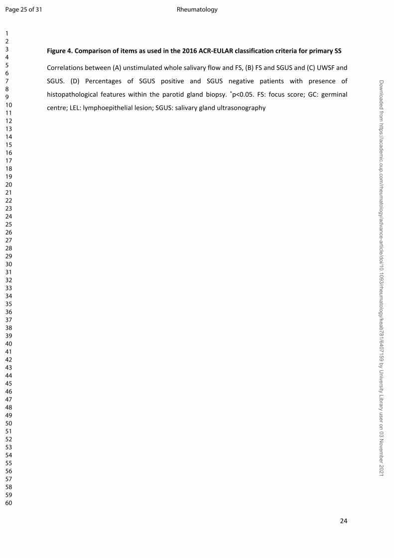
Figure 4. Comparison of items as used in the 2016 ACR-EULAR classification criteria for primary SS
Correlations between (A) unstimulated whole salivary flow and FS, (B) FS and SGUS and (C) UWSF and
SGUS. (D) Percentages of SGUS positive and SGUS negative patients with presence of
histopathological features within the parotid gland biopsy. *p<0.05. FS: focus score; GC: germinal
centre; LEL: lymphoepithelial lesion; SGUS: salivary gland ultrasonography
Page 25 of 31 Rheumatology
123456789101112131415161718192021222324252627282930313233343536373839404142434445464748495051525354555657585960
Dow
nloaded from https://academ
ic.oup.com/rheum
atology/advance-article/doi/10.1093/rheumatology/keab781/6407159 by U
niversity Library user on 03 Novem
ber 2021

Figure 1
338x190mm (300 x 300 DPI)
Page 26 of 31Rheumatology
123456789101112131415161718192021222324252627282930313233343536373839404142434445464748495051525354555657585960
Dow
nloaded from https://academ
ic.oup.com/rheum
atology/advance-article/doi/10.1093/rheumatology/keab781/6407159 by U
niversity Library user on 03 Novem
ber 2021

Figure 2
164x263mm (300 x 300 DPI)
Page 27 of 31 Rheumatology
123456789101112131415161718192021222324252627282930313233343536373839404142434445464748495051525354555657585960
Dow
nloaded from https://academ
ic.oup.com/rheum
atology/advance-article/doi/10.1093/rheumatology/keab781/6407159 by U
niversity Library user on 03 Novem
ber 2021

Figure 3
223x139mm (300 x 300 DPI)
Page 28 of 31Rheumatology
123456789101112131415161718192021222324252627282930313233343536373839404142434445464748495051525354555657585960
Dow
nloaded from https://academ
ic.oup.com/rheum
atology/advance-article/doi/10.1093/rheumatology/keab781/6407159 by U
niversity Library user on 03 Novem
ber 2021

Figure 4
228x156mm (300 x 300 DPI)
Page 29 of 31 Rheumatology
123456789101112131415161718192021222324252627282930313233343536373839404142434445464748495051525354555657585960
Dow
nloaded from https://academ
ic.oup.com/rheum
atology/advance-article/doi/10.1093/rheumatology/keab781/6407159 by U
niversity Library user on 03 Novem
ber 2021