x ray conference 2011.10.12
DESCRIPTION
報告者: fellow 1 陳筱惠. X ray conference 2011.10.12. Case 01. Patient Profile. Name: 游 O 琴 Sex: female Age: 56-year-old Occupation: 餐飲業 Chart number: 8970369 Date of admission: 2011/09/25. Chief Complaint. Right flank pain and black urine for 1 week. Present Illness. - PowerPoint PPT PresentationTRANSCRIPT

報告者: fellow 1 陳筱惠
Name: 游 O 琴Sex: femaleAge: 56-year-oldOccupation: 餐飲業Chart number: 8970369Date of admission: 2011/09/25
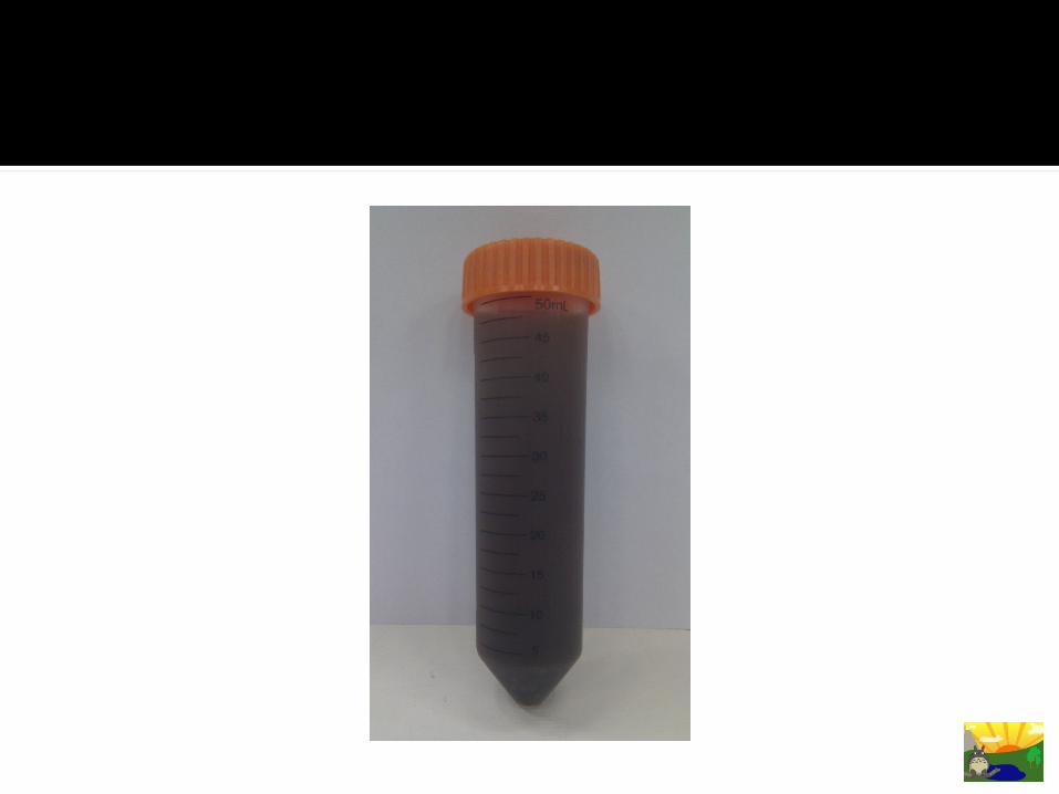
Right flank pain and black urine for 1 week

Small kidney with kidney stones was told at亞東 hospital 2~3 years ago. She received URS + SM then.
Right flank/low abdominal pain and black urine for 1 week; associated symptoms: dysuria, frequency, and urgency; no fever or hematuria
LMD visit twice, but no improvement under analgesic + oral antibiotic
At ER, foley was inserted for urine retention

Small kidney with kidney stones was told at 亞東 hospital 2~3 years ago. She received URS + SM then.
Urinary tract infection or chronic kidney diseases: denied
No hypertension, diabetes mellutis, heart, liver, or other significant systemic diseases
Current medicine: nil

Allergy: no known allergyAlcohol: denied; betel-nut: denied;
cigarette: deniedOver-the-counter medication or
chinese herb: nil

No family history of diabetes mellutis, malignancy, bleeding diathesis, heart, liver, kidney, or hereditary diseases
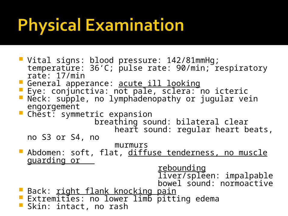
Vital signs: blood pressure: 142/81mmHg; temperature: 36‘C; pulse rate: 90/min; respiratory rate: 17/min
General apperance: acute ill looking Eye: conjunctiva: not pale, sclera: no icteric Neck: supple, no lymphadenopathy or jugular vein
engorgement Chest: symmetric expansion
breathing sound: bilateral clear heart sound: regular heart beats, no S3 or
S4, no murmurs Abdomen: soft, flat, diffuse tenderness, no muscle
guarding or rebounding liver/spleen: impalpable bowel sound: normoactive Back: right flank knocking pain Extremities: no lower limb pitting edema Skin: intact, no rash

WBC 8.7x1000/ul
Hgb 13.2 g/dl
Hct 38.2 %
MCV 89.3 fl
PLT 442 x1000/uL
Segment 43 %
Band 21 %
Urea N 17.6 mg/dl
Creatinine 0.75 mg/dl
GPT 53 IU/L
NA 138 mEq/L
K 3.8 mEq/L
Sugar 127 mg/dl
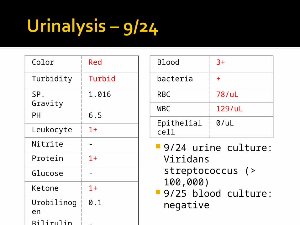
Color Red
Turbidity Turbid
SP. Gravity 1.016
PH 6.5
Leukocyte 1+
Nitrite -
Protein 1+
Glucose -
Ketone 1+
Urobilinogen 0.1
Bilirulin -
Blood 3+
bacteria +
RBC 78/uL
WBC 129/uL
Epithelial cell 0/uL
9/24 urine culture: Viridans streptococcus (> 100,000)
9/25 blood culture: negative

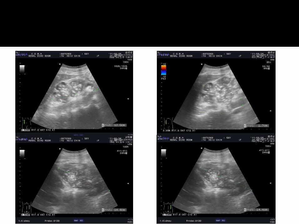

Left kidney Length: 11.2 cm Mild dilatation of the pelvocalcyeal systems A peri-pelvic echo-free lesion (2.0cm) in the
lower pole Right kidney Length: 14.4 cm
Irregular in contour, increased cortical echogenicity and decreased thickness
Severe dilatation of the pelvocalcyeal systems and ureter; multiple tiny hyperechoic lesions without acoustic shadow kidney and soft tissue-like density
Bladder: distended foley within it. A protruding mass (4.9x2.7cm) with
connection of a peri-bladder lumen near right vesicle-ureter junction, a iso-echoic lesion (1.2cm)


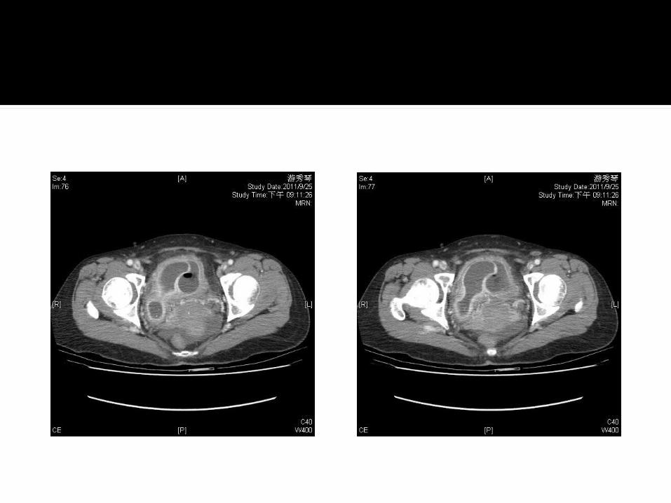
Right hydronpehrosis and hydroureter, due to ureterocele; complicated with infection and probably pyonephrosis and pyoyreter Multiple tiny stones inside
Left minimal hydronephrosisUrinary bladder mucosal thickening
and enhancement, suggesting chronic cystitis
Infected purulent urine in an obstructed collecting system
S/S: typically associated with fever, chills, and flank pain, although may be asymptomatic, too
Etiologies: Ascending infection of the urinary tract Hematogenous spread of a bacterial pathog
en

Incidence: relatively uncommon The risk of pyonephrosis is increased in patie
nts with upper urinary tract obstruction secondary to various causes (eg, stones, tumors, ureteropelvic junction [UPJ] obstruction
Pathogen: Escherichia coli, Enterococcus species, Can
dida species, Enterobacter species, Acteroides species, Staphylococcus species, Salmonella species, Tuberculosis
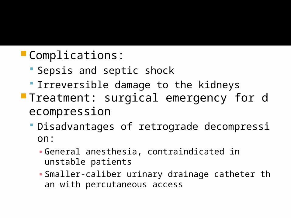
Complications: Sepsis and septic shock Irreversible damage to the kidneys
Treatment: surgical emergency for decompression Disadvantages of retrograde decompression:
▪ General anesthesia, contraindicated in unstable patients
▪ Smaller-caliber urinary drainage catheter than with percutaneous access

▪ Increased irritative urinary symptoms▪ Lack of antegrade access for radiologic studies or
inability to administer medications such as antibiotics via nephrostomy tube
▪ Bypassing the obstruction may not be possiblein some patients.
▪ Pyelovenous, pyelolymphatic, and pyelosinus backflow of infected urine into the systemic circulatory system

Ultrasonographic features of pyonephrosis: Dilated collecting system Echogenic debris in the in dependent
areas of collecting system▪ Strong echoes with acoustic shadowing▪ Change position when patient moves
Air can be seen in these infections.Ultrasonographic Evaluation of Renal InfectionsRadiol Clin N Am 44 (2006) 763–775


CT: depicts both hydronephrosis and often the underlying cause Contrast-enhanced imaging is more
desirable as in infection parenchymal and functional changes can be assessed.▪ Pelvic and ureteral wall thickness▪ Renal enlargement▪ Perinephric fat stranding▪ Fluid–fluid levels and gas within the collecting
system
Imaging of urinary tract infection in the adultEur Radiol (2004) 14:E168–E183

Name: 徐 O 華Sex: femaleAge: 63-year-oldOccupation: nilChart number: 6425429 Date of admission: 2011/09/05

Low abdominal pain for 4 days

Underlying diseases: rheumatoid arthritis, diabetes mellitus, and history of infectious spondylitis with left anterior epidural abscess post operation in 2011/03 (stool/urine incontinence under foley use and bedridden status since then)
Turbid urine, suprapubic and right flank pain for 4 days; associated symptoms: poor appetite, nausea/vomiting; no fever
Underlying diseases: Rheumatoid arthritis Hypertension Diabetes mellitus Osteoporosis Iatrogenic adrenal insufficiency History of infectious spondylitis with left
anterior epidural abscess post operation operation at 802 hospital in 2011/03
No heart, liver, or other significant systemic diseases
Current medicine: from our Rheuma OPD

Allergy: no known allergyAlcohol: denied; betel-nut: denied;
cigarette: deniedOver-the-counter medication or
chinese herb: nil

No family history of diabetes mellutis, malignancy, bleeding diathesis, heart, liver, kidney, or hereditary diseases
Vital signs: blood pressure: 124/92mmHg; temperature: 36.6‘C; pulse rate: 110/min; respiratory rate: 18/min
General apperance: acute ill looking Eye: conjunctiva: mild pale, sclera: no icteric Neck: supple, no lymphadenopathy or jugular vein
engorgement Chest: symmetric expansion
breathing sound: bilateral clear heart sound: regular heart beats, no S3 or
S4, no murmurs Abdomen: soft, flat, low abdominal tenderness, no
muscle guarding or rebounding liver/spleen: impalpable bowel sound: normoactive Back: right flank knocking pain Extremities: no lower limb pitting edema Skin: intact, no rash
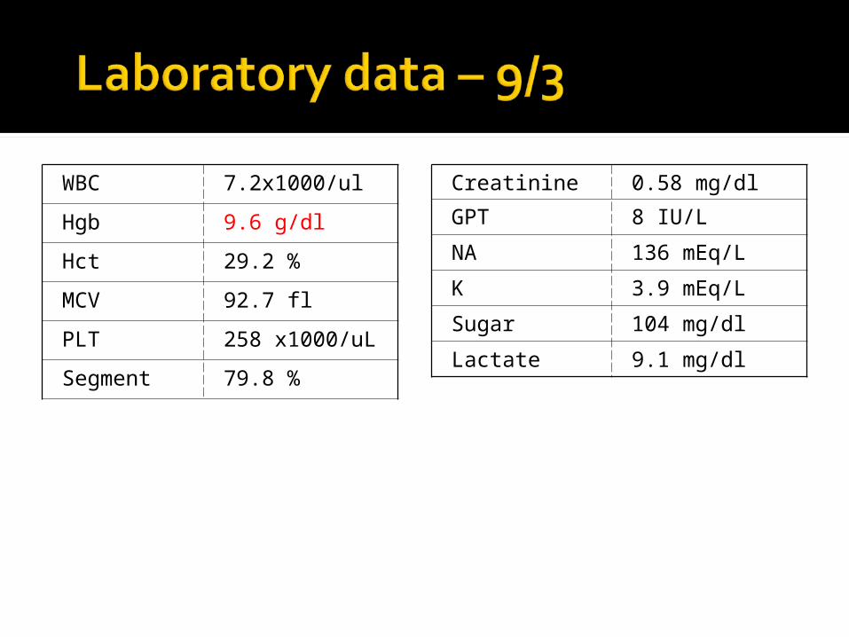
WBC 7.2x1000/ul
Hgb 9.6 g/dl
Hct 29.2 %
MCV 92.7 fl
PLT 258 x1000/uL
Segment 79.8 %
Creatinine 0.58 mg/dl
GPT 8 IU/L
NA 136 mEq/L
K 3.9 mEq/L
Sugar 104 mg/dl
Lactate 9.1 mg/dl

Color Yellow
Turbidity Cloudy
SP. Gravity 1.010
PH 8.5
Leukocyte 3+
Nitrite +
Protein Trace
Glucose -
Ketone -
Urobilinogen 0.1
Bilirulin -
Blood 2+
bacteria +
RBC 10/uL
WBC 65/uL
Epithelial cell 5/uL
9/3 urine culture: Proteus mirabilis (>100,000)
9/3 blood culture: negative


Left Kidney Length: 10.2 cm One isoechoic band extending from the
cortex to central sinus Right Kidney Length: 10.4 cm
One mass-like lesion (7.0x3.5cm) over middle portion
The both kidneys are normal in size and contour. The cortical echogenicity and thickness are normal.
No evidence of renal stone or cyst exists.
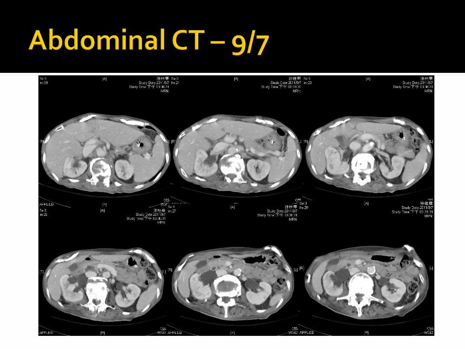
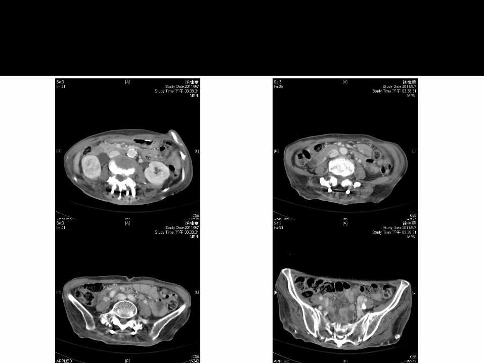

Multifocal ill-defined low denity of bilateral renal parenchyma, C/W acute pylonepheritis
Dilatation of bilateral renal pelvis and ureters to right middle ureter and left upper ureter level
No definite dilatation of bilateral renal calyces
No definite ureteral stones or tumor could be identified.
DDx: extrarenal pelvis, retroperitoneal fibrosis/ adhesion, or ureteral stricture
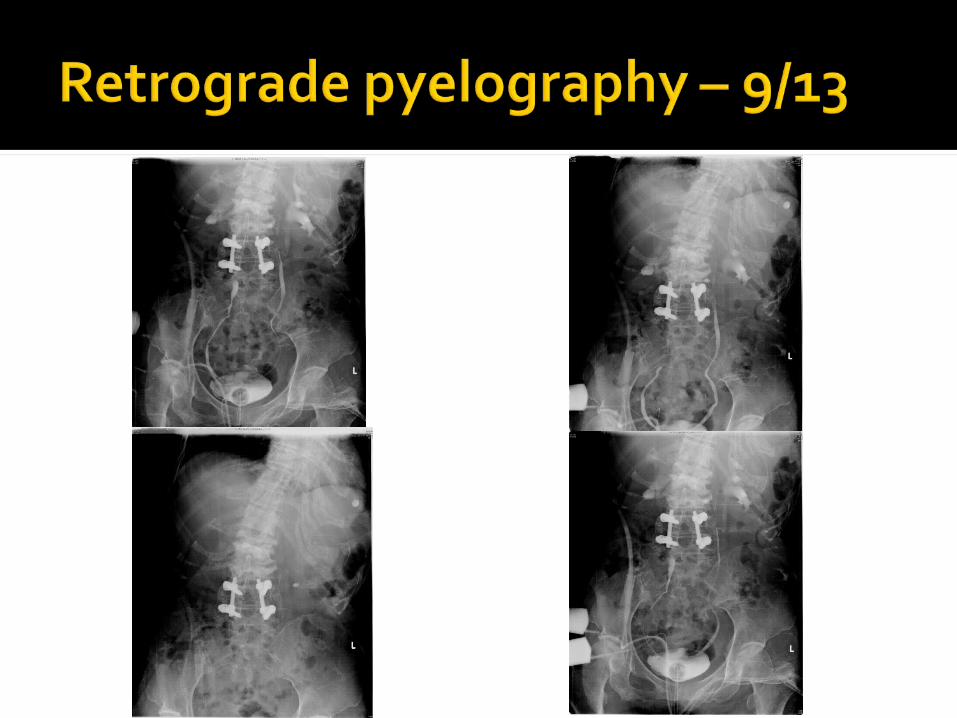

Ureteral catheter passing up to the left upper ureter at L4 level and right middle ureter at S3 level
Mild bilateral hydronephrosis. No obvious filling defect in the
collecting system. The right upper ureter and right renal collecting system are not well opacified.
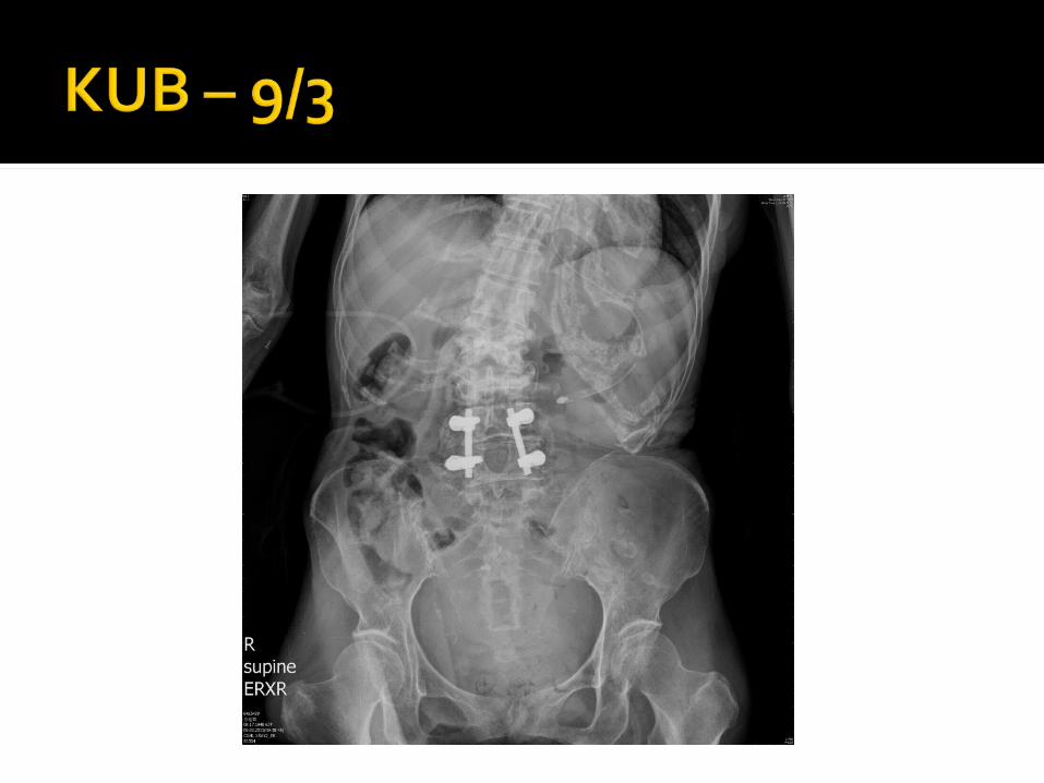
No definite radiopaque stone in the urinary tract
The presence of extrarenal calyces is a very rare anomaly of the upper urinary tract. First described in 1925 The total number of cases reported so far
is only 20. Kidney with extrarenal calyces is usually
associated with other anomalies like bifid kidney, renal ectopia, horseshoe kidney and renal dysplasia.Extrarenal calyces: A rare anomaly of the renal
collecting systemIndian J Pathol Microbiol. 2009 Jul-Sep;52(3):368-9.
The calyces were long and extrarenal in position. They drained into a cystic structure which represented either a grossly dilated pelvis (pelviureteric junction) or a ureteral cyst.
The exact cause of extrarenal calyces is not very clear. Hypothesis: a disparity resulting from slow
development of the metanephric tissue or to a relatively rapid development of the ureteric bud
Many cases of collecting system anomalies including extrarenal calyces are detected incidentally or may be diagnosed because of its complications.
Excretory urography often provides good anatomic information. A false impression of hydronephrosis or
chronic pyelonephritis
Rare disease, incidence of idiopathic form about 0.1~1.3 per 100,000 person-years
Etiology: Idiopathic form: 70%, 40 ~ 60 years of age, 2
to 3:1 male-to-female predominance Secondary form
▪ Drugs: ergot-derivatives, methysergide, bromocriptine, beta blockers, methyldopa, hydralazine, analgesics
▪ Malignancy: carcinoid, Hodgkin's and non-Hodgkin lymphoma, sarcomas

Infections: tuberculosis, histoplasmosis, actinomycosis
Radiation therapy for testicular seminoma, colon, pancreatic cancer
Surgery: lymphadenectomy, colectomy, aortic aneurysmectomy

Pathology: Macroscopically: a hard, white plaque of
varying thickness▪ Typically, around the abdominal aorta and
iliac vessels, as well as the inferior vena cava and the ureters
Microscopically: sclerosis and infiltration of mononuclear cells in varying proportions, depending on the stage of disease

Pathogenesis: chronic inflammation, fibroblast proliferation, and excessive extracellular matrix deposition
Clinical manifestations: Early stage: pain in the lower back, flank or
abdomen; characteristically dull, noncolicky, in a girdle distribution (> 90%)▪ Other nonspecific symptoms: weight loss, malaise,
anorexia, testicular pain, claudication, edema, and gross hematuria
Late stage: vessel compromise and characterized ureteral obstruction

Laboratory abnormalities: Elevated ESR and CRP Positive ANA (60%) Anemia, possibly related to renal
insufficiency or chronic inflammation No hematologic or biochemical
abnormalities diagnostic of this condition. The urinary sediment is most often
normal.
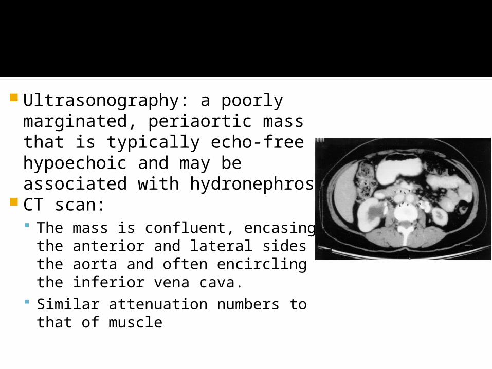
Ultrasonography: a poorly marginated, periaortic mass that is typically echo-free or hypoechoic and may be associated with hydronephrosis
CT scan: The mass is confluent, encasing
the anterior and lateral sides of the aorta and often encircling the inferior vena cava.
Similar attenuation numbers to that of muscle

Magnetic resonance imaging: comparable to those with CT scanning
Intravenous urography: proximal hydroureteronephrosis, medial deviation of the ureters, and extrinsic compression of the ureters
Retrograde or percutaneous antegrade pyelography: a smooth tapering of the ureters that is most pronounced at the level of the pelvic brim
Biopsy: no guidelines The location of the mass is atypical Clinical, laboratory or radiologic findings
suggest the presence of an underlying malignancy or infection
Local experience is limited The patient does not respond to initial
therapy.UpToDate