電腦斷層掃描儀 非年度品質保證測試 · 電腦斷層掃描儀...
TRANSCRIPT

電腦斷層掃描儀 非年度品質保證測試
陳建全
醫學物理師 醫事放射師
台灣醫學物理公司 wwwtmpinccomtw
陳建全
bull 學歷 ndash 陽明大學醫放系 學士
ndash 成功大學醫工所 碩士
bull 專業證書 ndash 教育部部定講師
ndash 放射診斷醫學物理師證書
ndash 醫事放射師證書
bull 研究成果 ndash SCI 第一作者1篇
ndash SCI 共同作者11篇
ndash 研究計畫主持人1件
ndash 研究計畫共同主持人6件
bull 經歷 ndash 台灣醫學物理公司
bull 總經理
ndash 長庚大學 bull 兼任講師
ndash 林口長庚紀念醫院 bull 磁振造影中心醫學物理師
bull 影像診療部醫學物理師
ndash 中華民國醫學物理學會 bull 常務監事
ndash 桃園縣醫事放射師公會 bull 理事
bull 總幹事
ndash 考試院醫事放射師檢覈考試 bull 命題審題委員
ndash 國健署乳篩計畫 bull 醫學物理組委員
ndash 原能會醫療曝露品質保證計畫 bull 講師
bull 命題及口試委員
bull Deterministic ndash visible documented confirmed within a relative short time ndash Skin erythema hair loss cataract infertility circulatory
disease bull Stochastic
ndash estimated years or decades to manifest ndash Cancer genetic effects
Health effects of ionizing radiation
bull Have thresholds that are typically quite high ndash Skin erythema ndash Hair loss ndash Cataracts (even in low doses of radiation)
bull 5 Sv for protracted exposures bull 2 Sv for acute exposures
bull Epidemiological evidence suggesting thresholds (equivalent dose) ndash Lens of eye 05 Gy ndash Circulatory system 05 Gy
Tissue reactions
bull Detriment-adjusted nominal risk coefficient at low dose rate ndash Cancer ndash 55 Sv ndash Genetic effects ndash 02 Sv (non-human species)
bull Cancer risks are estimated on the basis of
probability ndash Organ dose gt 100 mGy carcinogenic effects
bull Stochastic risks have no threshold
Stochastic effects
Tissue weighting factor of gonads 02 008 (ICRP 2007)
1 chest CT scan ~ 8 mSv 20 mGy to breast
5 ~ 15 CT scans carcinogenic effects
httpwwwrerfjpradefxlate_ecancriskhtml
For the average radiation exposure of survivors within 2500 meters (about 02 Gy)
the increase is about 10 above normal age-specific rates For a dose of 10 Gy
the corresponding cancer excess is about 50 (relative risk = 15)
The excess number of solid cancers is estimated as 848 (107)
The dose-response relationship appears to be linear without any apparent
threshold below which effects may not occur
The probability that an A-bomb survivor will have a cancer caused by A-bomb
radiation (excess lifetime risk) depends on the
dose received
age at exposure
sex
Other analyses (not shown) indicate that females have somewhat higher risks of
cancer from radiation exposure than males do
bull Patient-specific factors ndash Thickness of the body part in the beam
ndash Complexity of the procedure bull Complexity represents the mental and physical effort
required to perform a procedure
Common aspects of protection
bull Individual monitoring bull Individual monitoring of workers exposed to
ionizing radiation using film thermoluminescent dosimeters optically stimulated luminescence badges or other appropriate devices is used to verify the effectiveness of radiation control practices in the workplace
Whole body dose limit for workers of 20 mSvyear
(averaged over a defined 5-year period 100 mSv in 5
years)
1
2
3
1 necessary inside the apron
2 optional outside the apron at the collar level closest to x-ray tube
3 optional on the skin surface
醫療曝露品質保證的完整計畫
會診單 Protocol
HIS PACS
影像系統
品質保證
影像品管 產生影像
報告品管 產生報告
品質管制
資管人員 放射師 物理師 工程師
放射師 放射科醫師 臨床醫師
品管放射師 放射科醫師
放射科醫師 臨床醫師
確保硬體設備功能正常
確保檢查程序適當 確保影像符合標準 確保檢查正確適當
醫療曝露品質保證測試 (年度測試非年度測試)
httpwwwaecgovtwwebpageservicedownloaddownload_01_3-37pdf
醫療曝露之品質保證與品質控制
bull 品質保證 ndash 人員教育訓練
bull 醫師放射師護理師hellip
ndash 儀器設備 bull 保養維修
ndash 廠商自訂測試
bull 品保測試 ndash 接收測試
ndash 年度測試
ndash 非年度測試
ndash 標準化流程 bull 外部客戶
ndash 患者家屬
bull 內部客戶
bull 品質管制 ndash 外部客戶
bull 溝通衛教
bull 排程檢查效率
bull 病患安全舒適度
bull 輻射劑量合理抑低
bull 報告效率正確性
ndash 內部客戶 bull 排程檢查效率
bull 檢查正確性
bull 影像品質
bull 輻射劑量合理抑低
bull 報告效率正確性
執行醫療曝露品保測試的效益
bull 內部客戶
ndash 提供正確的影像 bull 正確的CT number
bull 無假影的影像
bull 無扭曲變形的影像
ndash 保證儀器功能正常 bull 以量化數據佐證
ndash 影像品質
ndash 輻射劑量
ndash 確保檢查正確執行 bull 檢查位置正確
bull 外部客戶
ndash 縮短檢查時間 bull 減少病患不適程度
ndash 減少重複曝露 bull 減少病患輻射劑量
bull 降低重照機率
bull 降低再次檢查機率
ndash 增進醫病關係 bull 提升醫院部門形象
bull 提升專業形象
bull 減少醫病糾紛
醫療曝露品保測試項目
bull 影像品質
ndash 空間解析度
ndash 對比解析度
ndash 雜訊
ndash 假影
ndash 幾何扭曲
bull 其他
ndash 像素值正確度
ndash 曝露指標正確度
bull 輻射相關
ndash 管電壓
ndash 曝露時間
ndash 常規標準檢查劑量
ndash 輻射輸出率
ndash 半值層
bull 組件相關
ndash 系統功能正常
ndash 組件完整安全
ndash 輻射防護設備
bull 影像品質-空間解析度
ndash High contrast spatial resolution
ndash 常用單位 bull Line-pair cm ( or mm)
ndash 影響因素 bull 焦斑大小
bull 偵測器尺寸
bull 影像重建後處理方法
bull 管球靶極傾斜角度
bull 測試物之幾何位置
ndash 另類測試 bull 調制轉換函數 (MTF ndash
modulation transfer function)
bull MTF的測量
Space Ms
Bar
Mb
MTF 2 lpmm = sd2 lpmm(Ms-Mb) 222
測試標準 MTF2 lpmm gt 58
微分
bull 高解析度影像的特點
ndash 影像儲存空間大傳輸速度慢 bull (甚至容易使電腦當機)
ndash 影像的訊號雜訊比(SNR)較低 bull 像素(pixel)尺寸小易受雜訊影響
bull 為維持訊號雜訊比需使用較高輻射劑量
bull 影像品質-對比解析度
ndash Low contrast detectability
ndash 常用單位 bull mm certain contrast
ndash 影響因素 bull 偵測器尺寸
bull 影像重建後處理方法
bull 輻射劑量
bull 影像像素尺寸
bull 射束品質半值層
ndash 另類測試 bull 對比雜訊比(CNR ndash contrast-
to-noise ratio)
Visual scoring
119862119873119877 =120583119887119886119888119896119892119903119900119906119899119889 minus 120583119905119886119903119892119890119905
119899119900119894119904119890
Target ROIμtarget = mean of pixel values inside ROI σtarget = standard deviation of pixel values inside ROI
Background ROI μbackground = mean of pixel values inside ROI σbackground = standard deviation of pixel values inside ROI
Beam hardening effect
119862119873119877 =120583119887119886119888119896119892119903119900119906119899119889 minus 120583119905119886119903119892119890119905
119899119900119894119904119890
noise = σbackground (σbackground + σtarget ) divide 2 [(σbackground 2 + σtarget
2) divide 2 ]05
Beam hardening effect
119868 = 1198680119890minus120583119909
119897119899119868
1198680= minus120583119909
119862119879 119899119906119898119887119890119903 119901119894119909119890119897 119907119886119897119906119890 =120583119905119894119904119904119906119890 minus 1205831198672119874
1205831198672119874times 1000
影像重建後處理方法的比較 bull 對比強化型
ndash Edge detail bone sharp hellip
ndash 適合用於偵測細微變化如微小骨裂
ndash 影像整體感覺變得較毛躁顆粒感重
bull 雜訊抑制型
ndash Smooth medium average hellip
ndash 適合用於觀察內臟類之軟組織的變異
ndash 影像整體感覺變得較溫和邊緣較模糊
times 1
9
Sharp kernel Smooth kernel
Image matrix
Sharp filtering Smooth filtering
Smooth kernel Sharp kernel
Histogram Equalization
For processing For presentation
The temporal changes of mammograms obtained from the same woman over seven years The top row illustrates the raw images processed to show density and the bottom row illustrates the ldquoFor Presentationrdquo images generated by the manufacturers (including Hologic Siemens and GE)
bull 影像品質-雜訊 ndash Noise
ndash 定義 bull 區域內所有像素值的標準差(σ)
ndash 影響因素
bull kV (photon energy)
bull mAs管球老化程度
bull 輻射劑量計位置
bull 輻射濾片種類厚度
bull 偵檢器校正因子
bull 影像重建模式
ndash FBPfiltered back projection
ndash IRiterative reconstruction
bull 影像重建法
ndash Standardbonesoftdetail hellip
當光子從光子源發出射入散射物質時 如果光子的能量相當低(與電子束縛能同數量級)則主要產生光電效應 如果光子的能量相當大(遠超過電子的束縛能)時則我們可以認為光子對自由電子發生散射而產生康普頓效應 如果光子能量極其大(gt1022百萬電子伏特)則足以轟擊原子核而生成一對粒子電子和正電子這個現象被稱為成對產生
120591 prop1198853~4
ℎ120584 3
120590 prop1
ℎ120584
120581 prop 1198852 ℎ120584
micro = τ + σ +κ
HZ = 1 C Z = 6 N Z = 7 O Z = 8 Na Z = 11 P Z = 15 K Z = 19 Ca Z = 20
bull 影像品質-雜訊 2015 2014
2016 重建基準值
bull 輻射相關-管電壓 曝露時間
ndash Tube voltage
ndash 常用單位 bull 準確性 (accuracy)
bull 再現性 (reproducibility)
ndash 影響因素 bull 高壓變壓器準確性與穩定性
ndash 另類測試 bull 介入性測量法
bull 輻射相關-常規標準檢查劑量
ndash Routine exam dose
ndash 常用單位
bull mSv
ndash 影響因素 bull kV (photon energy)
bull mAs
bull 管球老化程度
bull 輻射劑量計位置
bull 輻射寬度
bull 輻射濾片種類厚度
bull 影像品質
ndash 另類測試 bull 熱發光劑量計(TLD)測量法
TLD method
bull 輻射相關-輻射輸出率
ndash Radiation output rate
ndash 常用單位
bull mRs mGys
ndash 影響因素 bull kV (photon energy)
bull 管球老化程度
bull 輻射劑量計位置
bull 輻射寬度
bull 輻射濾片種類厚度
ndash 另類測試 bull 熱發光劑量計(TLD)測量法
bull 輻射相關-半值層
ndash Half value layer (beam quality)
ndash 常用單位
bull mm-Al
bull mm-Pb
ndash 影響因素 bull kV (photon energy)
bull 使用濾片種類
bull 射束大小
bull 管球老化程度
0 0
ln ln 2 2
HVL
ln
a bb a
a
b
E Et t
E E
E
E
Anode Heel effect
HVL小
Bow Tie filter of CT tube
bull 其他-像素正確度
ndash CT numbers accuracy
ndash 常用單位無
ndash 影響因素(對同一物質) bull kV (photon energy)
bull 管球老化程度
bull 輻射劑量計位置
bull 輻射濾片種類厚度
bull 偵檢器校正因子
bull 影像重建模式
ndash FBPfiltered back projection
ndash IRiterative reconstruction
bull 影像重建法
ndash Standardbonesoftdetail hellip
CT number or Hounsfield value
= 1000 (pixel - water ) water
項目名稱 頻率 診斷 治療 核醫 限值
目視檢查
日
V V V 各項檢查功能都正常
水假體影像CT值準確度及假影評估 V V V 無明顯假影水的CT值在plusmn7 HU
雷射與影像切面之相對位置一致性 V 三軸定位雷射中心軸位置偏差需在二毫米(mm)以下影像上需可看到孔洞或金屬記號
擷像工作站影像顯示評估
月
V V 依照SMPTE或TG-18測試圖像標準
檢查床水平檢測 V 縱向水平【基準值】宜為2度以下 縱向水平角度與其基準值差異為一度以下 橫向水平角度為零點五度以下
檢查床垂直與縱向移動位置準確性 V 二毫米(mm)以下
雷射與影像切面之相對軸向關係一致性 V 雷射在水平及垂直軸向方向差異為二毫米(mm)以下影像上需可以清楚看到標記
定位雷射與機架雷射間隔長度準確性 V
1機架雷射與定位雷射距離與原廠設定值差異為二毫米(mm)以下 2 定位雷射與機架雷射及電腦斷層掃描平面的間隔距離差異為二毫米(mm) 以下
定位雷射移動的準確性 V 移動誤差需二毫米(mm)以下
檢查床與影像切面軸向吻合性 V 誤差需二毫米(mm)以下
水假體影像均勻度及雜訊 V V 1 影像不均勻度差異為 5HU 以下 2 雜訊值與其基準值差異為百分之二十以下
CT 值準確性 V 1水的 CT 值為介於 -7 至+7 HU 之間 2 除了水以外其他物質之CT值與基準值差異為30HU
SPECTCT 或 PETCT 影像融合準確性 半年 V
GEFor PETCT ≦5mm For SPECT XYZ軸的absolute average≦3mm
PhilipsMaximum Distance 必須小於 5mm
Siemens 1檢查床之 CT 與 SPECT 位置(translation)誤差吻合性差異應≦plusmn5mm 2檢查床之 CT 與 SPECT 角度(rotation)誤差吻合性差異應≦plusmn1˚ 3檢查床之 CT 與 PET 位置差吻合性差異應≦5mm
診斷用CT非年度品保測試項目
bull 每日執行
ndash 目視檢查
ndash 水假體影像CT值準確度及假影評估
bull 每月執行
ndash 水假體影像均勻度及雜訊評估
ndash 擷像工作站影像顯示器評估
目視檢查
水假體影像CT值準確度及假影評估
ACR CT Accreditation Phantom
Philips CT phantom GE CT phantom
水假體影像CT值準確度及假影評估
醫事放射學會版
Scan 1
Scan 2
水CT值僅需分析假體中心之影像
每張影像均須分析假影
掃描方式
bull 選取rdquo常規成人腹部rdquo掃描protocol
bull 改為軸狀掃描模式
ndash 若不可行則
bull 記下kVpmAs掃描範圍(scan FOV)偵檢器組置影像重建法影像厚度
bull 另選一軸狀掃描protocol改以上述參數掃描
bull 配合假體大小變更影像照野範圍(FOV)
bull 執行掃瞄
水假體影像CT值準確度及假影評估
bull 理想的測試目的應包括
ndash 所有偵檢器功能正常
bull 所有排(column)列(row)的偵檢器
ndash 各種組合(N x T)均正常
bull 測試前應確認項目
ndash 假體內無任何雜質異物
ndash 所有X光掃描範圍內無任何顯影劑或異物
ndash 視情況執行空氣水校正(airwater calibration)
水的CT值在合理範圍內影像中無假影
程序中的常見問題
bull 常規成人腹部掃描條件
ndash 軸狀掃描與螺旋掃描的參數無法完全一致
bull 保持rdquo相同或最接近的切面厚度與偵檢器組置(N x T)rdquo
bull 在軸狀模式下使用相同或最接近的影像重建法
ndash 使用自動曝光控制(例如Auto mA CareDosehellip)
bull 挑選一組最常用的管電流(mA)與旋轉時間(s)
bull 適當照野範圍(大於假體直徑1公分)
ndash 假體不在影像中心
bull 床與機架對位錯誤
bull 矢狀面定位雷射偏移
廠牌(或含型號) Protocol 掃描次數 水CT值 假影評估
GE Abd 2 每張影像 每張影像
Hitachi Abd 2 每張影像 每張影像
NuroLogica CereTom Head 2 每張影像 每張影像
Philips MX8000 Dual Head 2 每張影像 每張影像
Philips Brilliance series Head 3 每張影像 每張影像
Toshiba Abd 3 中心影像 每張影像
Siemens Abd 3 每張影像 每張影像
每一家廠商不盡相同hellip
bull 該用何種版本
bull 可以自己決定用何種方式進行
bull 該用哪種方式進行
FAQs in QA Procedures
bull Phantom size
ndash 20 ~ 30 cm which is better
bull Scan FOV
ndash Not mentioned how to choose
bull Detector combination
ndash Max collimation min slice thickness enough
bull Scan Parameter
ndash Routine Abdomen protocol AEC
Ring artifacts
Ring artifact
Sinogram Reconstructed CT image
擷像工作站影像顯示器評估
974
984
1074
1024
Display settings window center(level) = 1024 window width = 100
979 1069
974 1074
Display settings window center(level) = 1024 window width = 100
128
384
640
3968
3712
(132 3125)
(332 9375)
(532 15625)
(3132 96875)
每一階層相差 256 pv 625
Display settings window center(level) = 2048 window width = 4096
用放大鏡看螢幕
影像螢幕顯示的縮放比
50 86 100 126
程序中的常見問題
bull 找不到標準影像(SMPTE或TG-18)
bull 窗寬(window width)窗高(window level or center)如何設定
bull 影像是否需要放大或縮小來觀察
bull 可否自行調整螢幕之亮度與對比
Toms DICOM Notes - Tom in Tech-Support
水假體影像均勻度及雜訊評估
醫事放射學會版
均勻度評估
雜訊評估
Standard Deviation (noise)
程序中的常見問題
bull 雜訊基準值
ndash 如何建立
ndash 何時該重建
ndash 超過標準如何處理
bull 均勻度
ndash 400mm2以外的區域會超過
ndash 在其他切面是否需要評估
Review your Annual QA Report
bull X-ray tube aging ndash Output rate (mRmAs mGymAs)
ndash CTDI (if protocol unchanged)
bull All detectors ok ndash Artifact evaluation test
bull Noise over-limit ndash Check noise test from other reconstruction
algorithms
ndash Output rate
CT非年度品保結果的影響
bull 水假體影像CT值準確度
ndash 可能原因 bull 管球老化偵測器異常
ndash 造成影響 bull 以CT值判定組織種類可
能發生錯誤
bull 組織對比度改變
bull 水假體影像假影評估
ndash 可能原因 bull 偵測器異常或需要校正
ndash 造成影響 bull 假影位置遮蔽組織病灶
bull 水假體影像均勻度
ndash 可能原因 bull 高壓產生器或偵測器異常
ndash 造成影響 bull 以CT值判定組織種類可能
發生錯誤
bull 組織對比度改變
bull 水假體影像雜訊評估
ndash 可能原因 bull 管球老化偵測器異常
ndash 造成影響 bull 低對比組織間鑑別度降低
治療用CT非年度品保測試項目 bull 每日執行
ndash 目視檢查 ndash 水假體影像CT值準確度及假影評估 ndash 雷射與影像切面之相對位置一致性
bull 每月執行
ndash 擷像工作站影像顯示器評估 ndash 水假體影像均勻度及雜訊評估 ndash 檢查床水平檢測 ndash 檢查床垂直與縱向移動位置準確性 ndash 雷射與影像切面之相對軸向關係一致性 ndash 定位雷射與機架雷射間隔長度準確性 ndash 定位雷射移動的準確性 ndash 檢查床與影像切面軸向吻合性 ndash CT 值準確性
定位雷射
機架雷射
固定距離
雷射與影像切面之相對位置一致性
1 三軸定位雷射中心軸位置偏差需在二毫米以下
2 影像上需可看到孔洞(圖三)或金屬記號(圖四)
檢查床水平檢測
1 縱向水平角度與其基準值差異為一度以下
2 橫向水平角度為零點五度以下
20 kg Step 1 Step 2 Step 4
檢查床垂直與縱向移動位置準確性
判定準則二毫米以下
測試步驟 1 將檢查床昇至適當位置打開兩側定位雷射在檢查床上垂直方向黏貼 固定一長尺
使其垂直於床面其原點與左右兩側定位雷射水平切齊(圖 七) 2 將檢查床垂直移動 30 公分檢視(長尺)與數位顯示(機架)讀值之移動距離差異 3 將檢查床昇至適當位置打開兩側定位雷射在檢查床上縱向方向黏貼 固定一長尺
使其平躺其原點與兩側定位雷射垂直切齊(圖八) 4 將檢查床縱向移動 80 公分檢視(長尺)與數位顯示(機架)讀值之移動距離差異 5 紀錄分析之結果確認符合效能判定準則
雷射與影像切面之相對軸向關係一致性 測試步驟 1 以方格紙或可執行相同測試目的之假體協助確認在射束(雷射)能清晰辨識的涵蓋範圍內定
位雷射與機架雷射在水平及垂直軸向方向的吻合性 2 取一在平面的不同位置內含兩個以上直徑 2 毫米圓形孔洞或標記之測試假體孔洞外緣延伸
標示孔洞中心軸位置(圖九)將此假體置於檢查床上調整假體位置使水平及垂直軸向定位雷射通過圓形孔洞中心移動檢查床固定距離使機架雷射通過圓形孔洞中心之標記將檢查床縱向位置顯示值歸零
3 使用最小射束寬度以適當管電壓及管電流乘積進行曝露設定掃描範圍(FOV)完整包括測試假體之圓形孔洞位置執行軸狀掃描(前後多掃描幾張影像)
4 檢視 CT 掃描影像選取圓形孔洞可以清楚的在螢幕上辨識的影像(圖十)紀錄該影像縱向座標位置
5 開啟 CT-Simulator CT 影像座標軸顯示功能將座標軸顯示於螢幕 6 分析影像縱向座標位置及座標軸原點座標值與圓形孔洞之差異確認影像中心參考座標與雷
射系統座標之差異 7 紀錄分析結果確認符合效能判定準則
1 雷射在水平及垂直軸向方向差異為二毫米以下
2 影像上需可以清楚看到標記
水平方向
垂直方向
定位雷射與機架雷射間隔長度準確性 測試步驟 1 於檢查床上平貼直尺長度大於原廠設定的定位雷射與機架雷射的間隔距離(例如 60公分)
目視檢查機架雷射與定位雷射距離是否為設定值(圖十一) 2 使用測試假體或專用假體(圖十二)調整假體位置使定位雷射通過圓形孔洞中心所在之平
面 3 檢查床往機架移動原廠設定的間隔距離使用最小射束寬度以適當管電壓及管電流乘積進
行曝露設定掃描範圍(FOV)完整包括測試假體之圓形孔洞位置執行軸狀掃描 4 檢視 CT 掃描影像圓形孔洞所在位置之影像是否可以清楚的從螢幕上辨識出每個圓形孔洞
的外觀確認定位雷射與機架雷射及 CT 掃描平面的間隔距離差異 5 紀錄分析之結果確認符合效能判定準則
1 機架雷射與定位雷射距離與原廠設定值差異為二毫米以下 2 定位雷射與機架雷射及電腦斷層掃描平面的間隔距離差異為二毫米以下
定位雷射移動的準確性 測試步驟 1 將方格紙或可執行同功能測試之假體置於檢查床上(圖十三)調整假體位置使水平及垂直
軸向定位雷射通過假體參考點中心設定天花板雷射向左及向右各移動若干固定距離(例如 plusmn 5plusmn10及 plusmn15公分建議總移動距離不小於20公分)檢查並記錄移動位置準確性
2 將尺直立於檢查床上(圖十四)設定兩側雷射向上及向下各移動若干固定距離(例如 plusmn 5及 plusmn10公分建議總移動距離不小於10公分)檢查並記錄移動位置準確性
3 紀錄分析之結果確認符合效能判定準則
移動誤差需二毫米以下
檢查床與影像切面軸向吻合性 測試步驟 1 在檢查床最前端位置放置一內含直徑 2 毫米圓形孔洞或標誌之測試假體調整測試假體位
置使圓形孔洞或標誌中心位於檢查床寬度正中央的位置打開天花板(定位)雷射紀錄雷射與標記的位置差異
2 將檢查床移至影像切面之位置並將縱向座標歸零使用最小射束寬度以適當管電壓及管電流乘積進行曝露設定掃描範圍(FOV)完整包括測試假體之圓形孔洞位置執行軸狀掃描並讀取影像中標記之座標值
3 相對步驟 1 之標記位置在相距 60公分處再放置另一直徑 2 毫米之金屬記號置於檢查床寬度正中央的位置紀錄天花板雷射與標記的位置差異
4 參考步驟 2 執行軸狀掃描並讀取影像中標記之座標值 5 記錄兩金屬座標並計算其差值紀錄分析前後兩標記與天花板雷射及影像中座標值之差異
其結果需符合效能判定準則
誤差需在二毫米以下
Step 1 Step 3
CT 值準確性
核醫用CT非年度品保測試項目
bull 每日執行
ndash 目視檢查
ndash 水假體影像CT值準確度及假影評估
bull 每半年執行
ndash SPECTCT 或 PETCT 影像融合準確性
Siemens SPECT-CT
重建lt基準值gt時機
bull 掃描參數變更
ndash kVp mA time pitch collimation slice thickness reconstruction kernel
bull 品保方式變更
ndash 更換假體變更分析方式
bull 系統軟硬體變更
ndash Repair replacement upgrade
bull 系統重新校正
ndash Airwater calibration detailmultiple calibration
Team Work in CT QA
bull 建立標準化
ndash 掃描方式 bull 掃描參數
bull 假體種類位置
ndash 分析方式 bull 假影評估
bull ROI 位置大小
bull 環境燈光照度
ndash 表單格式
ndash 填表內容 bull PF 或 數值
bull 簽名樣式
bull 建立共識
ndash 判定準則 bull 假影評估
bull 螢幕功能
ndash 處理方式 bull 再做一次
bull 臨床影響程度
bull 定期檢視
ndash 非年度QA bull 水CT值雜訊均勻度
ndash 年度QA bull CTDI雜訊對比度
電腦斷層的品質保證測試
ndash年度測試方式與結果分析
項目 名稱 診斷 核醫 治療
一 系統安全評估 (System Safety Evaluation) V V V
二 檢查床與機頭之對位(Alignment of Table to gantry )
V
三 切片位置準確性 (Slice Positioning Accuracy) V V V
四 切片厚度準確性(Slice thickness accuracy) V V V
五 CT值準確度與線性度 (CT number accuracy and linearity)
V V V
六 水假體影像評估 (Evaluation of image Uniformity noise and artifact)
V V V
七 空間解析度 (Spatial resolution) V V
八 低對比偵測度(Low contrast detectability ) V V
九 劑量評估(Dosimetry) V V V
十 輻射寬度 (Radiation width) V V V
十一 影像顯示器評估(Image display device evaluation)
V
測試項目
系統安全評估
組件檢查
項目 合格不合格 備註
1 整個電腦斷層掃描儀在機械方面是穩定的
2 所有可動的部分平穩動作沒有任何阻礙
3 病患或工作人員不會接觸到銳利粗糙邊緣或其它包括電
的危害
4 定位雷射燈功能正常
5 所有指示燈功能正常輻射使用中hellip等
6 指示病人的對講裝置功能正常
7 監控病人的攝影機與顯示器等功能正常
8 張貼警告標示於合適位置
注意輻射
懷孕婦女
9 張貼原能會認可文件設備人員
檢查床與機頭之對位 確認檢查床的長軸與掃描儀旋轉面的左右中心對齊
測試步驟
1 在檢查床左右中心附近貼上膠帶以尺決定中心位置並在膠帶上作記號
2 將檢查床貼膠帶處移至機頭內
3 用捲尺在機頭內橫向最長距離處決定左右中心位置並在檢查床上作記號
4 測量上述兩個記號間的距離
效能判定準則機頭的中心線應在檢查床中心線的+- 5公釐內 (AAPM TG39)或符合廠商規格標準
切片位置準確性
(一) 目的確認 1 定位雷射光線的準確性 (1)內部定位雷射光線 (2)外部定位雷射光線 2 使用掃描放射影像(例如Topo Scout Pilothellip) 決定切片位置的準確性 3 檢查床(Table)進出方向移動的準確性
切片定位雷射的準確性
使用掃描放射影像決定切片位置的準確性
使用掃描放射影像決定切片位置的準確性
Lateral projection of ACR Phantom
Definitions of slice indicators
1 Start position of slice
2 Center of slice
3 Total coverage of scan
檢查床進出切片方向移動的準確性
ImPACT Information Leaflet No 1
切片厚度準確性
(1) 將假體放置於檢查床上以常規成人腹部掃描模式進行測試設定組像範圍為 21 公分若常規成人
腹部掃描是以軸狀掃描則直接掃描切片厚度測試物的正中央區段
(2) 若常規成人腹部掃描是以螺旋掃描則將掃描模式改為軸狀掃描其他參數則維持固定不變若掃描儀為多切片機型但不能使用相同的偵檢器組置(N bull TT為一個資料通道在 Z軸方向上之寬度N為該
模式下的資料通道數目)進行軸狀掃描時則在維持相同的T設定下使用最大的N之偵檢器組置進行軸狀掃描
(3) 同上述步驟但改變切片厚度為高解析度肺部掃描時使用的厚度35 及7 毫米(若於步驟(1)中已有相同厚度則不需重複掃描)若無法達到前述設定則選取最接近的設定值進行測試
測試步驟
切片厚度準確性
CT值準確度與線性度
材料 空氣 聚乙烯 水 壓克力 骨頭 測量值 -97271 -8918 476 12547 92647
Attenuation Coefficient 0 01795 0187 0221 0373
y = 50842x - 9777 Rsup2 = 09989
-1500
-1000
-500
0
500
1000
1500
0 005 01 015 02 025 03 035 04
線性迴歸分析
線性迴歸分析
線性(線性迴歸分析)
Procedures
bull Scan uniform phantom
bull Place ROIs
bull Noise Standard Deviation of ROI
bull CT uniformity
- Diff between means of center and outer ROI
水假體影像評估
bull (1)水的CT值準確度(2)雜訊(3)影像均勻度與(4)假影
Foot
bull (2)雜訊
bull (3)影像均勻度
bull (4)假影
A streak B motion C beam-hardening DE ring F blooming
Incomplete projections ndash streaking artifacts
Helical Artifacts in the axial plane Single-Section Scanning
Helical artifacts
Partial volume effect
Cone beam effect
Axial scan Helical scan Corrected image
空間解析度
ACR phantom 8 Al bar patterns- 4 5 6 7 8 9 10 and 12 lpcm
Start here
The result is 9 lpcm
低對比偵測度 bull Four cylinder groups
bull 06 (6 HU) difference from a background material
bull mean CT number of approximately 90 HU
bull Cylinder-to-background contrast is energy-independent
bull cylinders diameters and spaces 2 3 4 5 and 6 mm
bull A 25-mm cylinder bull to verify the cylinder-to-background contrast level
119862119873119877 =120583119887119886119888119896119892119903119900119906119899119889 minus 120583119905119886119903119892119890119905
119899119900119894119904119890
noise = σbackground (σbackground + σtarget ) divide 2 [(σbackground 2 + σtarget
2) divide 2 ]05
項目十劑量評估
Dose report from CT scanner
Computed Tomography Dose Index
bull Weighted CTDI CTDIw
CTDI100center
CTDI100 P1
CTDI100 P2
CTDI100 P3
CTDI100 P4
CTDI100edge = 4
CTDI100 P1+P2+P3+P4
Dose Calculation
pcw CTDI3
2CTDI
3
1CTDI
wvol
CTDICTDI
Pitch
volDLP=CTDI total scan length
DLPE k
C P1
P2
P4
P3
DRL Diagnostic reference level
KJR 2012
Tsai HY Tung CJ Yu CC Tyan YS Survey of computed tomography scanners in Taiwan dose descriptors dose guidance levels and effective doses Med Phys 2007341234ndash1243
項目十一輻射寬度
bull Using packaged films bull placed at the isocenter surface during scans at
the different collimation thicknesses
bull The mAs technique is set to provide a maximum film density of between 10 and 20 OD
項目十二影像顯示器評估
SMPTE TG 18 QC
Display images on monitor
Image data Brightness on monitor
Display images on monitor
Standardized Display System
P-value P-value to
DDL
DDLdigital driving level
Display System
DDLs Luminance
P-value
DDL
DDL
Luminance
P-value
DDL
DDL
Luminance
P-value
Luminance
Grayscale Display Function
20181017
JNDs
bull Just-Noticeable Difference
001
01
1
10
100
1000
10000
0 200 400 600 800 1000
灰階值 實測亮度 JND 標準亮度 實測斜率 標準斜率 斜率差異 結果評定
128 123 80 122 00350 00346 117 Passed
384 242 114 240 00521 00526 -102 Passed
640 445 153 445 00761 00761 004 Passed
896 719 189 719 01038 01038 -002 Passed
1152 1072 223 1072 01419 01393 185 Passed
1408 1597 260 1587 01841 01860 -103 Passed
1664 2278 297 2275 02454 02451 011 Passed
1920 3186 334 3182 03265 03201 198 Passed
2176 4394 371 4367 04215 04212 007 Passed
2432 6122 412 6094 05478 05483 -009 Passed
2688 8094 448 8067 07090 07091 -001 Passed
2944 1093 488 10904 09250 09130 132 Passed
3200 1426 524 14190 11725 11737 -011 Passed
3456 1895 564 18885 15029 14993 024 Passed
3712 2421 599 24133 18750 18892 -075 Passed
3968 3096 635 30934 24282 24098 076 Passed
4224 4043 674 40332 31000 30897 033 Passed
4480 5221 712 52073
01
1
10
100
1000
00 1000 2000 3000 4000 5000 6000
Measured Data
Standard
100
1000
10000
100000
0 200 400 600
Measured Data
Standard
請多指教

1
2
3
1 necessary inside the apron
2 optional outside the apron at the collar level closest to x-ray tube
3 optional on the skin surface
醫療曝露品質保證的完整計畫
會診單 Protocol
HIS PACS
影像系統
品質保證
影像品管 產生影像
報告品管 產生報告
品質管制
資管人員 放射師 物理師 工程師
放射師 放射科醫師 臨床醫師
品管放射師 放射科醫師
放射科醫師 臨床醫師
確保硬體設備功能正常
確保檢查程序適當 確保影像符合標準 確保檢查正確適當
醫療曝露品質保證測試 (年度測試非年度測試)
httpwwwaecgovtwwebpageservicedownloaddownload_01_3-37pdf
醫療曝露之品質保證與品質控制
bull 品質保證 ndash 人員教育訓練
bull 醫師放射師護理師hellip
ndash 儀器設備 bull 保養維修
ndash 廠商自訂測試
bull 品保測試 ndash 接收測試
ndash 年度測試
ndash 非年度測試
ndash 標準化流程 bull 外部客戶
ndash 患者家屬
bull 內部客戶
bull 品質管制 ndash 外部客戶
bull 溝通衛教
bull 排程檢查效率
bull 病患安全舒適度
bull 輻射劑量合理抑低
bull 報告效率正確性
ndash 內部客戶 bull 排程檢查效率
bull 檢查正確性
bull 影像品質
bull 輻射劑量合理抑低
bull 報告效率正確性
執行醫療曝露品保測試的效益
bull 內部客戶
ndash 提供正確的影像 bull 正確的CT number
bull 無假影的影像
bull 無扭曲變形的影像
ndash 保證儀器功能正常 bull 以量化數據佐證
ndash 影像品質
ndash 輻射劑量
ndash 確保檢查正確執行 bull 檢查位置正確
bull 外部客戶
ndash 縮短檢查時間 bull 減少病患不適程度
ndash 減少重複曝露 bull 減少病患輻射劑量
bull 降低重照機率
bull 降低再次檢查機率
ndash 增進醫病關係 bull 提升醫院部門形象
bull 提升專業形象
bull 減少醫病糾紛
醫療曝露品保測試項目
bull 影像品質
ndash 空間解析度
ndash 對比解析度
ndash 雜訊
ndash 假影
ndash 幾何扭曲
bull 其他
ndash 像素值正確度
ndash 曝露指標正確度
bull 輻射相關
ndash 管電壓
ndash 曝露時間
ndash 常規標準檢查劑量
ndash 輻射輸出率
ndash 半值層
bull 組件相關
ndash 系統功能正常
ndash 組件完整安全
ndash 輻射防護設備
bull 影像品質-空間解析度
ndash High contrast spatial resolution
ndash 常用單位 bull Line-pair cm ( or mm)
ndash 影響因素 bull 焦斑大小
bull 偵測器尺寸
bull 影像重建後處理方法
bull 管球靶極傾斜角度
bull 測試物之幾何位置
ndash 另類測試 bull 調制轉換函數 (MTF ndash
modulation transfer function)
bull MTF的測量
Space Ms
Bar
Mb
MTF 2 lpmm = sd2 lpmm(Ms-Mb) 222
測試標準 MTF2 lpmm gt 58
微分
bull 高解析度影像的特點
ndash 影像儲存空間大傳輸速度慢 bull (甚至容易使電腦當機)
ndash 影像的訊號雜訊比(SNR)較低 bull 像素(pixel)尺寸小易受雜訊影響
bull 為維持訊號雜訊比需使用較高輻射劑量
bull 影像品質-對比解析度
ndash Low contrast detectability
ndash 常用單位 bull mm certain contrast
ndash 影響因素 bull 偵測器尺寸
bull 影像重建後處理方法
bull 輻射劑量
bull 影像像素尺寸
bull 射束品質半值層
ndash 另類測試 bull 對比雜訊比(CNR ndash contrast-
to-noise ratio)
Visual scoring
119862119873119877 =120583119887119886119888119896119892119903119900119906119899119889 minus 120583119905119886119903119892119890119905
119899119900119894119904119890
Target ROIμtarget = mean of pixel values inside ROI σtarget = standard deviation of pixel values inside ROI
Background ROI μbackground = mean of pixel values inside ROI σbackground = standard deviation of pixel values inside ROI
Beam hardening effect
119862119873119877 =120583119887119886119888119896119892119903119900119906119899119889 minus 120583119905119886119903119892119890119905
119899119900119894119904119890
noise = σbackground (σbackground + σtarget ) divide 2 [(σbackground 2 + σtarget
2) divide 2 ]05
Beam hardening effect
119868 = 1198680119890minus120583119909
119897119899119868
1198680= minus120583119909
119862119879 119899119906119898119887119890119903 119901119894119909119890119897 119907119886119897119906119890 =120583119905119894119904119904119906119890 minus 1205831198672119874
1205831198672119874times 1000
影像重建後處理方法的比較 bull 對比強化型
ndash Edge detail bone sharp hellip
ndash 適合用於偵測細微變化如微小骨裂
ndash 影像整體感覺變得較毛躁顆粒感重
bull 雜訊抑制型
ndash Smooth medium average hellip
ndash 適合用於觀察內臟類之軟組織的變異
ndash 影像整體感覺變得較溫和邊緣較模糊
times 1
9
Sharp kernel Smooth kernel
Image matrix
Sharp filtering Smooth filtering
Smooth kernel Sharp kernel
Histogram Equalization
For processing For presentation
The temporal changes of mammograms obtained from the same woman over seven years The top row illustrates the raw images processed to show density and the bottom row illustrates the ldquoFor Presentationrdquo images generated by the manufacturers (including Hologic Siemens and GE)
bull 影像品質-雜訊 ndash Noise
ndash 定義 bull 區域內所有像素值的標準差(σ)
ndash 影響因素
bull kV (photon energy)
bull mAs管球老化程度
bull 輻射劑量計位置
bull 輻射濾片種類厚度
bull 偵檢器校正因子
bull 影像重建模式
ndash FBPfiltered back projection
ndash IRiterative reconstruction
bull 影像重建法
ndash Standardbonesoftdetail hellip
當光子從光子源發出射入散射物質時 如果光子的能量相當低(與電子束縛能同數量級)則主要產生光電效應 如果光子的能量相當大(遠超過電子的束縛能)時則我們可以認為光子對自由電子發生散射而產生康普頓效應 如果光子能量極其大(gt1022百萬電子伏特)則足以轟擊原子核而生成一對粒子電子和正電子這個現象被稱為成對產生
120591 prop1198853~4
ℎ120584 3
120590 prop1
ℎ120584
120581 prop 1198852 ℎ120584
micro = τ + σ +κ
HZ = 1 C Z = 6 N Z = 7 O Z = 8 Na Z = 11 P Z = 15 K Z = 19 Ca Z = 20
bull 影像品質-雜訊 2015 2014
2016 重建基準值
bull 輻射相關-管電壓 曝露時間
ndash Tube voltage
ndash 常用單位 bull 準確性 (accuracy)
bull 再現性 (reproducibility)
ndash 影響因素 bull 高壓變壓器準確性與穩定性
ndash 另類測試 bull 介入性測量法
bull 輻射相關-常規標準檢查劑量
ndash Routine exam dose
ndash 常用單位
bull mSv
ndash 影響因素 bull kV (photon energy)
bull mAs
bull 管球老化程度
bull 輻射劑量計位置
bull 輻射寬度
bull 輻射濾片種類厚度
bull 影像品質
ndash 另類測試 bull 熱發光劑量計(TLD)測量法
TLD method
bull 輻射相關-輻射輸出率
ndash Radiation output rate
ndash 常用單位
bull mRs mGys
ndash 影響因素 bull kV (photon energy)
bull 管球老化程度
bull 輻射劑量計位置
bull 輻射寬度
bull 輻射濾片種類厚度
ndash 另類測試 bull 熱發光劑量計(TLD)測量法
bull 輻射相關-半值層
ndash Half value layer (beam quality)
ndash 常用單位
bull mm-Al
bull mm-Pb
ndash 影響因素 bull kV (photon energy)
bull 使用濾片種類
bull 射束大小
bull 管球老化程度
0 0
ln ln 2 2
HVL
ln
a bb a
a
b
E Et t
E E
E
E
Anode Heel effect
HVL小
Bow Tie filter of CT tube
bull 其他-像素正確度
ndash CT numbers accuracy
ndash 常用單位無
ndash 影響因素(對同一物質) bull kV (photon energy)
bull 管球老化程度
bull 輻射劑量計位置
bull 輻射濾片種類厚度
bull 偵檢器校正因子
bull 影像重建模式
ndash FBPfiltered back projection
ndash IRiterative reconstruction
bull 影像重建法
ndash Standardbonesoftdetail hellip
CT number or Hounsfield value
= 1000 (pixel - water ) water
項目名稱 頻率 診斷 治療 核醫 限值
目視檢查
日
V V V 各項檢查功能都正常
水假體影像CT值準確度及假影評估 V V V 無明顯假影水的CT值在plusmn7 HU
雷射與影像切面之相對位置一致性 V 三軸定位雷射中心軸位置偏差需在二毫米(mm)以下影像上需可看到孔洞或金屬記號
擷像工作站影像顯示評估
月
V V 依照SMPTE或TG-18測試圖像標準
檢查床水平檢測 V 縱向水平【基準值】宜為2度以下 縱向水平角度與其基準值差異為一度以下 橫向水平角度為零點五度以下
檢查床垂直與縱向移動位置準確性 V 二毫米(mm)以下
雷射與影像切面之相對軸向關係一致性 V 雷射在水平及垂直軸向方向差異為二毫米(mm)以下影像上需可以清楚看到標記
定位雷射與機架雷射間隔長度準確性 V
1機架雷射與定位雷射距離與原廠設定值差異為二毫米(mm)以下 2 定位雷射與機架雷射及電腦斷層掃描平面的間隔距離差異為二毫米(mm) 以下
定位雷射移動的準確性 V 移動誤差需二毫米(mm)以下
檢查床與影像切面軸向吻合性 V 誤差需二毫米(mm)以下
水假體影像均勻度及雜訊 V V 1 影像不均勻度差異為 5HU 以下 2 雜訊值與其基準值差異為百分之二十以下
CT 值準確性 V 1水的 CT 值為介於 -7 至+7 HU 之間 2 除了水以外其他物質之CT值與基準值差異為30HU
SPECTCT 或 PETCT 影像融合準確性 半年 V
GEFor PETCT ≦5mm For SPECT XYZ軸的absolute average≦3mm
PhilipsMaximum Distance 必須小於 5mm
Siemens 1檢查床之 CT 與 SPECT 位置(translation)誤差吻合性差異應≦plusmn5mm 2檢查床之 CT 與 SPECT 角度(rotation)誤差吻合性差異應≦plusmn1˚ 3檢查床之 CT 與 PET 位置差吻合性差異應≦5mm
診斷用CT非年度品保測試項目
bull 每日執行
ndash 目視檢查
ndash 水假體影像CT值準確度及假影評估
bull 每月執行
ndash 水假體影像均勻度及雜訊評估
ndash 擷像工作站影像顯示器評估
目視檢查
水假體影像CT值準確度及假影評估
ACR CT Accreditation Phantom
Philips CT phantom GE CT phantom
水假體影像CT值準確度及假影評估
醫事放射學會版
Scan 1
Scan 2
水CT值僅需分析假體中心之影像
每張影像均須分析假影
掃描方式
bull 選取rdquo常規成人腹部rdquo掃描protocol
bull 改為軸狀掃描模式
ndash 若不可行則
bull 記下kVpmAs掃描範圍(scan FOV)偵檢器組置影像重建法影像厚度
bull 另選一軸狀掃描protocol改以上述參數掃描
bull 配合假體大小變更影像照野範圍(FOV)
bull 執行掃瞄
水假體影像CT值準確度及假影評估
bull 理想的測試目的應包括
ndash 所有偵檢器功能正常
bull 所有排(column)列(row)的偵檢器
ndash 各種組合(N x T)均正常
bull 測試前應確認項目
ndash 假體內無任何雜質異物
ndash 所有X光掃描範圍內無任何顯影劑或異物
ndash 視情況執行空氣水校正(airwater calibration)
水的CT值在合理範圍內影像中無假影
程序中的常見問題
bull 常規成人腹部掃描條件
ndash 軸狀掃描與螺旋掃描的參數無法完全一致
bull 保持rdquo相同或最接近的切面厚度與偵檢器組置(N x T)rdquo
bull 在軸狀模式下使用相同或最接近的影像重建法
ndash 使用自動曝光控制(例如Auto mA CareDosehellip)
bull 挑選一組最常用的管電流(mA)與旋轉時間(s)
bull 適當照野範圍(大於假體直徑1公分)
ndash 假體不在影像中心
bull 床與機架對位錯誤
bull 矢狀面定位雷射偏移
廠牌(或含型號) Protocol 掃描次數 水CT值 假影評估
GE Abd 2 每張影像 每張影像
Hitachi Abd 2 每張影像 每張影像
NuroLogica CereTom Head 2 每張影像 每張影像
Philips MX8000 Dual Head 2 每張影像 每張影像
Philips Brilliance series Head 3 每張影像 每張影像
Toshiba Abd 3 中心影像 每張影像
Siemens Abd 3 每張影像 每張影像
每一家廠商不盡相同hellip
bull 該用何種版本
bull 可以自己決定用何種方式進行
bull 該用哪種方式進行
FAQs in QA Procedures
bull Phantom size
ndash 20 ~ 30 cm which is better
bull Scan FOV
ndash Not mentioned how to choose
bull Detector combination
ndash Max collimation min slice thickness enough
bull Scan Parameter
ndash Routine Abdomen protocol AEC
Ring artifacts
Ring artifact
Sinogram Reconstructed CT image
擷像工作站影像顯示器評估
974
984
1074
1024
Display settings window center(level) = 1024 window width = 100
979 1069
974 1074
Display settings window center(level) = 1024 window width = 100
128
384
640
3968
3712
(132 3125)
(332 9375)
(532 15625)
(3132 96875)
每一階層相差 256 pv 625
Display settings window center(level) = 2048 window width = 4096
用放大鏡看螢幕
影像螢幕顯示的縮放比
50 86 100 126
程序中的常見問題
bull 找不到標準影像(SMPTE或TG-18)
bull 窗寬(window width)窗高(window level or center)如何設定
bull 影像是否需要放大或縮小來觀察
bull 可否自行調整螢幕之亮度與對比
Toms DICOM Notes - Tom in Tech-Support
水假體影像均勻度及雜訊評估
醫事放射學會版
均勻度評估
雜訊評估
Standard Deviation (noise)
程序中的常見問題
bull 雜訊基準值
ndash 如何建立
ndash 何時該重建
ndash 超過標準如何處理
bull 均勻度
ndash 400mm2以外的區域會超過
ndash 在其他切面是否需要評估
Review your Annual QA Report
bull X-ray tube aging ndash Output rate (mRmAs mGymAs)
ndash CTDI (if protocol unchanged)
bull All detectors ok ndash Artifact evaluation test
bull Noise over-limit ndash Check noise test from other reconstruction
algorithms
ndash Output rate
CT非年度品保結果的影響
bull 水假體影像CT值準確度
ndash 可能原因 bull 管球老化偵測器異常
ndash 造成影響 bull 以CT值判定組織種類可
能發生錯誤
bull 組織對比度改變
bull 水假體影像假影評估
ndash 可能原因 bull 偵測器異常或需要校正
ndash 造成影響 bull 假影位置遮蔽組織病灶
bull 水假體影像均勻度
ndash 可能原因 bull 高壓產生器或偵測器異常
ndash 造成影響 bull 以CT值判定組織種類可能
發生錯誤
bull 組織對比度改變
bull 水假體影像雜訊評估
ndash 可能原因 bull 管球老化偵測器異常
ndash 造成影響 bull 低對比組織間鑑別度降低
治療用CT非年度品保測試項目 bull 每日執行
ndash 目視檢查 ndash 水假體影像CT值準確度及假影評估 ndash 雷射與影像切面之相對位置一致性
bull 每月執行
ndash 擷像工作站影像顯示器評估 ndash 水假體影像均勻度及雜訊評估 ndash 檢查床水平檢測 ndash 檢查床垂直與縱向移動位置準確性 ndash 雷射與影像切面之相對軸向關係一致性 ndash 定位雷射與機架雷射間隔長度準確性 ndash 定位雷射移動的準確性 ndash 檢查床與影像切面軸向吻合性 ndash CT 值準確性
定位雷射
機架雷射
固定距離
雷射與影像切面之相對位置一致性
1 三軸定位雷射中心軸位置偏差需在二毫米以下
2 影像上需可看到孔洞(圖三)或金屬記號(圖四)
檢查床水平檢測
1 縱向水平角度與其基準值差異為一度以下
2 橫向水平角度為零點五度以下
20 kg Step 1 Step 2 Step 4
檢查床垂直與縱向移動位置準確性
判定準則二毫米以下
測試步驟 1 將檢查床昇至適當位置打開兩側定位雷射在檢查床上垂直方向黏貼 固定一長尺
使其垂直於床面其原點與左右兩側定位雷射水平切齊(圖 七) 2 將檢查床垂直移動 30 公分檢視(長尺)與數位顯示(機架)讀值之移動距離差異 3 將檢查床昇至適當位置打開兩側定位雷射在檢查床上縱向方向黏貼 固定一長尺
使其平躺其原點與兩側定位雷射垂直切齊(圖八) 4 將檢查床縱向移動 80 公分檢視(長尺)與數位顯示(機架)讀值之移動距離差異 5 紀錄分析之結果確認符合效能判定準則
雷射與影像切面之相對軸向關係一致性 測試步驟 1 以方格紙或可執行相同測試目的之假體協助確認在射束(雷射)能清晰辨識的涵蓋範圍內定
位雷射與機架雷射在水平及垂直軸向方向的吻合性 2 取一在平面的不同位置內含兩個以上直徑 2 毫米圓形孔洞或標記之測試假體孔洞外緣延伸
標示孔洞中心軸位置(圖九)將此假體置於檢查床上調整假體位置使水平及垂直軸向定位雷射通過圓形孔洞中心移動檢查床固定距離使機架雷射通過圓形孔洞中心之標記將檢查床縱向位置顯示值歸零
3 使用最小射束寬度以適當管電壓及管電流乘積進行曝露設定掃描範圍(FOV)完整包括測試假體之圓形孔洞位置執行軸狀掃描(前後多掃描幾張影像)
4 檢視 CT 掃描影像選取圓形孔洞可以清楚的在螢幕上辨識的影像(圖十)紀錄該影像縱向座標位置
5 開啟 CT-Simulator CT 影像座標軸顯示功能將座標軸顯示於螢幕 6 分析影像縱向座標位置及座標軸原點座標值與圓形孔洞之差異確認影像中心參考座標與雷
射系統座標之差異 7 紀錄分析結果確認符合效能判定準則
1 雷射在水平及垂直軸向方向差異為二毫米以下
2 影像上需可以清楚看到標記
水平方向
垂直方向
定位雷射與機架雷射間隔長度準確性 測試步驟 1 於檢查床上平貼直尺長度大於原廠設定的定位雷射與機架雷射的間隔距離(例如 60公分)
目視檢查機架雷射與定位雷射距離是否為設定值(圖十一) 2 使用測試假體或專用假體(圖十二)調整假體位置使定位雷射通過圓形孔洞中心所在之平
面 3 檢查床往機架移動原廠設定的間隔距離使用最小射束寬度以適當管電壓及管電流乘積進
行曝露設定掃描範圍(FOV)完整包括測試假體之圓形孔洞位置執行軸狀掃描 4 檢視 CT 掃描影像圓形孔洞所在位置之影像是否可以清楚的從螢幕上辨識出每個圓形孔洞
的外觀確認定位雷射與機架雷射及 CT 掃描平面的間隔距離差異 5 紀錄分析之結果確認符合效能判定準則
1 機架雷射與定位雷射距離與原廠設定值差異為二毫米以下 2 定位雷射與機架雷射及電腦斷層掃描平面的間隔距離差異為二毫米以下
定位雷射移動的準確性 測試步驟 1 將方格紙或可執行同功能測試之假體置於檢查床上(圖十三)調整假體位置使水平及垂直
軸向定位雷射通過假體參考點中心設定天花板雷射向左及向右各移動若干固定距離(例如 plusmn 5plusmn10及 plusmn15公分建議總移動距離不小於20公分)檢查並記錄移動位置準確性
2 將尺直立於檢查床上(圖十四)設定兩側雷射向上及向下各移動若干固定距離(例如 plusmn 5及 plusmn10公分建議總移動距離不小於10公分)檢查並記錄移動位置準確性
3 紀錄分析之結果確認符合效能判定準則
移動誤差需二毫米以下
檢查床與影像切面軸向吻合性 測試步驟 1 在檢查床最前端位置放置一內含直徑 2 毫米圓形孔洞或標誌之測試假體調整測試假體位
置使圓形孔洞或標誌中心位於檢查床寬度正中央的位置打開天花板(定位)雷射紀錄雷射與標記的位置差異
2 將檢查床移至影像切面之位置並將縱向座標歸零使用最小射束寬度以適當管電壓及管電流乘積進行曝露設定掃描範圍(FOV)完整包括測試假體之圓形孔洞位置執行軸狀掃描並讀取影像中標記之座標值
3 相對步驟 1 之標記位置在相距 60公分處再放置另一直徑 2 毫米之金屬記號置於檢查床寬度正中央的位置紀錄天花板雷射與標記的位置差異
4 參考步驟 2 執行軸狀掃描並讀取影像中標記之座標值 5 記錄兩金屬座標並計算其差值紀錄分析前後兩標記與天花板雷射及影像中座標值之差異
其結果需符合效能判定準則
誤差需在二毫米以下
Step 1 Step 3
CT 值準確性
核醫用CT非年度品保測試項目
bull 每日執行
ndash 目視檢查
ndash 水假體影像CT值準確度及假影評估
bull 每半年執行
ndash SPECTCT 或 PETCT 影像融合準確性
Siemens SPECT-CT
重建lt基準值gt時機
bull 掃描參數變更
ndash kVp mA time pitch collimation slice thickness reconstruction kernel
bull 品保方式變更
ndash 更換假體變更分析方式
bull 系統軟硬體變更
ndash Repair replacement upgrade
bull 系統重新校正
ndash Airwater calibration detailmultiple calibration
Team Work in CT QA
bull 建立標準化
ndash 掃描方式 bull 掃描參數
bull 假體種類位置
ndash 分析方式 bull 假影評估
bull ROI 位置大小
bull 環境燈光照度
ndash 表單格式
ndash 填表內容 bull PF 或 數值
bull 簽名樣式
bull 建立共識
ndash 判定準則 bull 假影評估
bull 螢幕功能
ndash 處理方式 bull 再做一次
bull 臨床影響程度
bull 定期檢視
ndash 非年度QA bull 水CT值雜訊均勻度
ndash 年度QA bull CTDI雜訊對比度
電腦斷層的品質保證測試
ndash年度測試方式與結果分析
項目 名稱 診斷 核醫 治療
一 系統安全評估 (System Safety Evaluation) V V V
二 檢查床與機頭之對位(Alignment of Table to gantry )
V
三 切片位置準確性 (Slice Positioning Accuracy) V V V
四 切片厚度準確性(Slice thickness accuracy) V V V
五 CT值準確度與線性度 (CT number accuracy and linearity)
V V V
六 水假體影像評估 (Evaluation of image Uniformity noise and artifact)
V V V
七 空間解析度 (Spatial resolution) V V
八 低對比偵測度(Low contrast detectability ) V V
九 劑量評估(Dosimetry) V V V
十 輻射寬度 (Radiation width) V V V
十一 影像顯示器評估(Image display device evaluation)
V
測試項目
系統安全評估
組件檢查
項目 合格不合格 備註
1 整個電腦斷層掃描儀在機械方面是穩定的
2 所有可動的部分平穩動作沒有任何阻礙
3 病患或工作人員不會接觸到銳利粗糙邊緣或其它包括電
的危害
4 定位雷射燈功能正常
5 所有指示燈功能正常輻射使用中hellip等
6 指示病人的對講裝置功能正常
7 監控病人的攝影機與顯示器等功能正常
8 張貼警告標示於合適位置
注意輻射
懷孕婦女
9 張貼原能會認可文件設備人員
檢查床與機頭之對位 確認檢查床的長軸與掃描儀旋轉面的左右中心對齊
測試步驟
1 在檢查床左右中心附近貼上膠帶以尺決定中心位置並在膠帶上作記號
2 將檢查床貼膠帶處移至機頭內
3 用捲尺在機頭內橫向最長距離處決定左右中心位置並在檢查床上作記號
4 測量上述兩個記號間的距離
效能判定準則機頭的中心線應在檢查床中心線的+- 5公釐內 (AAPM TG39)或符合廠商規格標準
切片位置準確性
(一) 目的確認 1 定位雷射光線的準確性 (1)內部定位雷射光線 (2)外部定位雷射光線 2 使用掃描放射影像(例如Topo Scout Pilothellip) 決定切片位置的準確性 3 檢查床(Table)進出方向移動的準確性
切片定位雷射的準確性
使用掃描放射影像決定切片位置的準確性
使用掃描放射影像決定切片位置的準確性
Lateral projection of ACR Phantom
Definitions of slice indicators
1 Start position of slice
2 Center of slice
3 Total coverage of scan
檢查床進出切片方向移動的準確性
ImPACT Information Leaflet No 1
切片厚度準確性
(1) 將假體放置於檢查床上以常規成人腹部掃描模式進行測試設定組像範圍為 21 公分若常規成人
腹部掃描是以軸狀掃描則直接掃描切片厚度測試物的正中央區段
(2) 若常規成人腹部掃描是以螺旋掃描則將掃描模式改為軸狀掃描其他參數則維持固定不變若掃描儀為多切片機型但不能使用相同的偵檢器組置(N bull TT為一個資料通道在 Z軸方向上之寬度N為該
模式下的資料通道數目)進行軸狀掃描時則在維持相同的T設定下使用最大的N之偵檢器組置進行軸狀掃描
(3) 同上述步驟但改變切片厚度為高解析度肺部掃描時使用的厚度35 及7 毫米(若於步驟(1)中已有相同厚度則不需重複掃描)若無法達到前述設定則選取最接近的設定值進行測試
測試步驟
切片厚度準確性
CT值準確度與線性度
材料 空氣 聚乙烯 水 壓克力 骨頭 測量值 -97271 -8918 476 12547 92647
Attenuation Coefficient 0 01795 0187 0221 0373
y = 50842x - 9777 Rsup2 = 09989
-1500
-1000
-500
0
500
1000
1500
0 005 01 015 02 025 03 035 04
線性迴歸分析
線性迴歸分析
線性(線性迴歸分析)
Procedures
bull Scan uniform phantom
bull Place ROIs
bull Noise Standard Deviation of ROI
bull CT uniformity
- Diff between means of center and outer ROI
水假體影像評估
bull (1)水的CT值準確度(2)雜訊(3)影像均勻度與(4)假影
Foot
bull (2)雜訊
bull (3)影像均勻度
bull (4)假影
A streak B motion C beam-hardening DE ring F blooming
Incomplete projections ndash streaking artifacts
Helical Artifacts in the axial plane Single-Section Scanning
Helical artifacts
Partial volume effect
Cone beam effect
Axial scan Helical scan Corrected image
空間解析度
ACR phantom 8 Al bar patterns- 4 5 6 7 8 9 10 and 12 lpcm
Start here
The result is 9 lpcm
低對比偵測度 bull Four cylinder groups
bull 06 (6 HU) difference from a background material
bull mean CT number of approximately 90 HU
bull Cylinder-to-background contrast is energy-independent
bull cylinders diameters and spaces 2 3 4 5 and 6 mm
bull A 25-mm cylinder bull to verify the cylinder-to-background contrast level
119862119873119877 =120583119887119886119888119896119892119903119900119906119899119889 minus 120583119905119886119903119892119890119905
119899119900119894119904119890
noise = σbackground (σbackground + σtarget ) divide 2 [(σbackground 2 + σtarget
2) divide 2 ]05
項目十劑量評估
Dose report from CT scanner
Computed Tomography Dose Index
bull Weighted CTDI CTDIw
CTDI100center
CTDI100 P1
CTDI100 P2
CTDI100 P3
CTDI100 P4
CTDI100edge = 4
CTDI100 P1+P2+P3+P4
Dose Calculation
pcw CTDI3
2CTDI
3
1CTDI
wvol
CTDICTDI
Pitch
volDLP=CTDI total scan length
DLPE k
C P1
P2
P4
P3
DRL Diagnostic reference level
KJR 2012
Tsai HY Tung CJ Yu CC Tyan YS Survey of computed tomography scanners in Taiwan dose descriptors dose guidance levels and effective doses Med Phys 2007341234ndash1243
項目十一輻射寬度
bull Using packaged films bull placed at the isocenter surface during scans at
the different collimation thicknesses
bull The mAs technique is set to provide a maximum film density of between 10 and 20 OD
項目十二影像顯示器評估
SMPTE TG 18 QC
Display images on monitor
Image data Brightness on monitor
Display images on monitor
Standardized Display System
P-value P-value to
DDL
DDLdigital driving level
Display System
DDLs Luminance
P-value
DDL
DDL
Luminance
P-value
DDL
DDL
Luminance
P-value
Luminance
Grayscale Display Function
20181017
JNDs
bull Just-Noticeable Difference
001
01
1
10
100
1000
10000
0 200 400 600 800 1000
灰階值 實測亮度 JND 標準亮度 實測斜率 標準斜率 斜率差異 結果評定
128 123 80 122 00350 00346 117 Passed
384 242 114 240 00521 00526 -102 Passed
640 445 153 445 00761 00761 004 Passed
896 719 189 719 01038 01038 -002 Passed
1152 1072 223 1072 01419 01393 185 Passed
1408 1597 260 1587 01841 01860 -103 Passed
1664 2278 297 2275 02454 02451 011 Passed
1920 3186 334 3182 03265 03201 198 Passed
2176 4394 371 4367 04215 04212 007 Passed
2432 6122 412 6094 05478 05483 -009 Passed
2688 8094 448 8067 07090 07091 -001 Passed
2944 1093 488 10904 09250 09130 132 Passed
3200 1426 524 14190 11725 11737 -011 Passed
3456 1895 564 18885 15029 14993 024 Passed
3712 2421 599 24133 18750 18892 -075 Passed
3968 3096 635 30934 24282 24098 076 Passed
4224 4043 674 40332 31000 30897 033 Passed
4480 5221 712 52073
01
1
10
100
1000
00 1000 2000 3000 4000 5000 6000
Measured Data
Standard
100
1000
10000
100000
0 200 400 600
Measured Data
Standard
請多指教
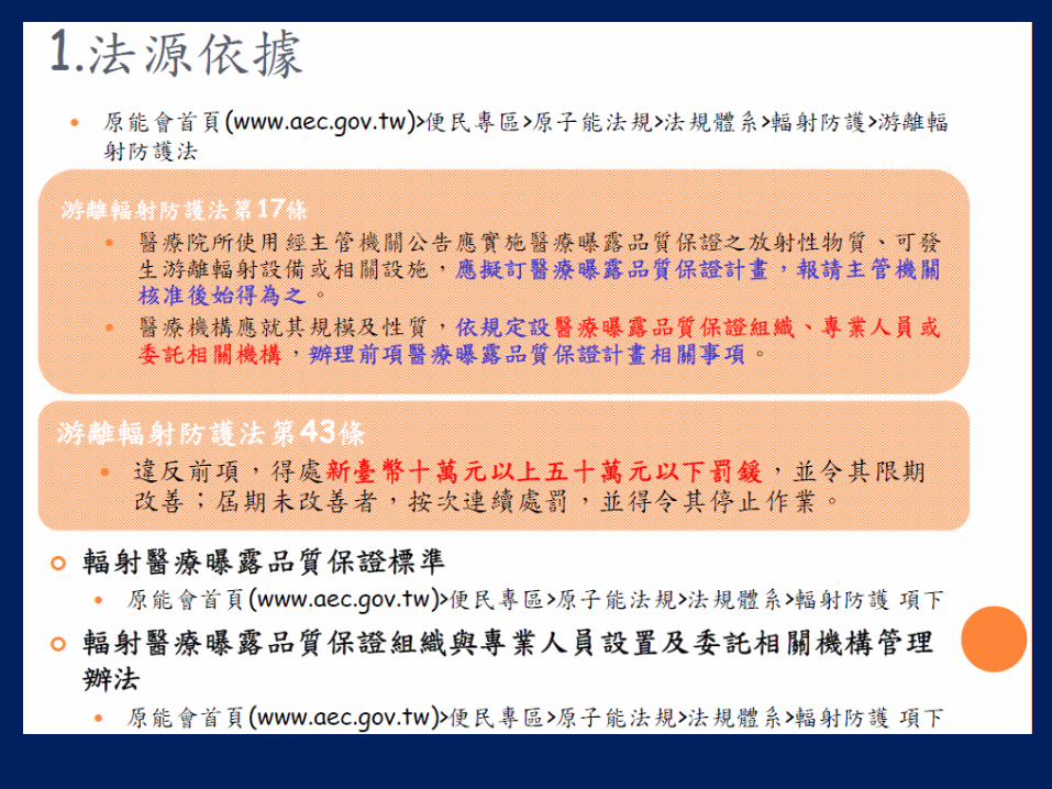

水假體影像CT值準確度及假影評估
ACR CT Accreditation Phantom
Philips CT phantom GE CT phantom
水假體影像CT值準確度及假影評估
醫事放射學會版
Scan 1
Scan 2
水CT值僅需分析假體中心之影像
每張影像均須分析假影
掃描方式
bull 選取rdquo常規成人腹部rdquo掃描protocol
bull 改為軸狀掃描模式
ndash 若不可行則
bull 記下kVpmAs掃描範圍(scan FOV)偵檢器組置影像重建法影像厚度
bull 另選一軸狀掃描protocol改以上述參數掃描
bull 配合假體大小變更影像照野範圍(FOV)
bull 執行掃瞄
水假體影像CT值準確度及假影評估
bull 理想的測試目的應包括
ndash 所有偵檢器功能正常
bull 所有排(column)列(row)的偵檢器
ndash 各種組合(N x T)均正常
bull 測試前應確認項目
ndash 假體內無任何雜質異物
ndash 所有X光掃描範圍內無任何顯影劑或異物
ndash 視情況執行空氣水校正(airwater calibration)
水的CT值在合理範圍內影像中無假影
程序中的常見問題
bull 常規成人腹部掃描條件
ndash 軸狀掃描與螺旋掃描的參數無法完全一致
bull 保持rdquo相同或最接近的切面厚度與偵檢器組置(N x T)rdquo
bull 在軸狀模式下使用相同或最接近的影像重建法
ndash 使用自動曝光控制(例如Auto mA CareDosehellip)
bull 挑選一組最常用的管電流(mA)與旋轉時間(s)
bull 適當照野範圍(大於假體直徑1公分)
ndash 假體不在影像中心
bull 床與機架對位錯誤
bull 矢狀面定位雷射偏移
廠牌(或含型號) Protocol 掃描次數 水CT值 假影評估
GE Abd 2 每張影像 每張影像
Hitachi Abd 2 每張影像 每張影像
NuroLogica CereTom Head 2 每張影像 每張影像
Philips MX8000 Dual Head 2 每張影像 每張影像
Philips Brilliance series Head 3 每張影像 每張影像
Toshiba Abd 3 中心影像 每張影像
Siemens Abd 3 每張影像 每張影像
每一家廠商不盡相同hellip
bull 該用何種版本
bull 可以自己決定用何種方式進行
bull 該用哪種方式進行
FAQs in QA Procedures
bull Phantom size
ndash 20 ~ 30 cm which is better
bull Scan FOV
ndash Not mentioned how to choose
bull Detector combination
ndash Max collimation min slice thickness enough
bull Scan Parameter
ndash Routine Abdomen protocol AEC
Ring artifacts
Ring artifact
Sinogram Reconstructed CT image
擷像工作站影像顯示器評估
974
984
1074
1024
Display settings window center(level) = 1024 window width = 100
979 1069
974 1074
Display settings window center(level) = 1024 window width = 100
128
384
640
3968
3712
(132 3125)
(332 9375)
(532 15625)
(3132 96875)
每一階層相差 256 pv 625
Display settings window center(level) = 2048 window width = 4096
用放大鏡看螢幕
影像螢幕顯示的縮放比
50 86 100 126
程序中的常見問題
bull 找不到標準影像(SMPTE或TG-18)
bull 窗寬(window width)窗高(window level or center)如何設定
bull 影像是否需要放大或縮小來觀察
bull 可否自行調整螢幕之亮度與對比
Toms DICOM Notes - Tom in Tech-Support
水假體影像均勻度及雜訊評估
醫事放射學會版
均勻度評估
雜訊評估
Standard Deviation (noise)
程序中的常見問題
bull 雜訊基準值
ndash 如何建立
ndash 何時該重建
ndash 超過標準如何處理
bull 均勻度
ndash 400mm2以外的區域會超過
ndash 在其他切面是否需要評估
Review your Annual QA Report
bull X-ray tube aging ndash Output rate (mRmAs mGymAs)
ndash CTDI (if protocol unchanged)
bull All detectors ok ndash Artifact evaluation test
bull Noise over-limit ndash Check noise test from other reconstruction
algorithms
ndash Output rate
CT非年度品保結果的影響
bull 水假體影像CT值準確度
ndash 可能原因 bull 管球老化偵測器異常
ndash 造成影響 bull 以CT值判定組織種類可
能發生錯誤
bull 組織對比度改變
bull 水假體影像假影評估
ndash 可能原因 bull 偵測器異常或需要校正
ndash 造成影響 bull 假影位置遮蔽組織病灶
bull 水假體影像均勻度
ndash 可能原因 bull 高壓產生器或偵測器異常
ndash 造成影響 bull 以CT值判定組織種類可能
發生錯誤
bull 組織對比度改變
bull 水假體影像雜訊評估
ndash 可能原因 bull 管球老化偵測器異常
ndash 造成影響 bull 低對比組織間鑑別度降低
治療用CT非年度品保測試項目 bull 每日執行
ndash 目視檢查 ndash 水假體影像CT值準確度及假影評估 ndash 雷射與影像切面之相對位置一致性
bull 每月執行
ndash 擷像工作站影像顯示器評估 ndash 水假體影像均勻度及雜訊評估 ndash 檢查床水平檢測 ndash 檢查床垂直與縱向移動位置準確性 ndash 雷射與影像切面之相對軸向關係一致性 ndash 定位雷射與機架雷射間隔長度準確性 ndash 定位雷射移動的準確性 ndash 檢查床與影像切面軸向吻合性 ndash CT 值準確性
定位雷射
機架雷射
固定距離
雷射與影像切面之相對位置一致性
1 三軸定位雷射中心軸位置偏差需在二毫米以下
2 影像上需可看到孔洞(圖三)或金屬記號(圖四)
檢查床水平檢測
1 縱向水平角度與其基準值差異為一度以下
2 橫向水平角度為零點五度以下
20 kg Step 1 Step 2 Step 4
檢查床垂直與縱向移動位置準確性
判定準則二毫米以下
測試步驟 1 將檢查床昇至適當位置打開兩側定位雷射在檢查床上垂直方向黏貼 固定一長尺
使其垂直於床面其原點與左右兩側定位雷射水平切齊(圖 七) 2 將檢查床垂直移動 30 公分檢視(長尺)與數位顯示(機架)讀值之移動距離差異 3 將檢查床昇至適當位置打開兩側定位雷射在檢查床上縱向方向黏貼 固定一長尺
使其平躺其原點與兩側定位雷射垂直切齊(圖八) 4 將檢查床縱向移動 80 公分檢視(長尺)與數位顯示(機架)讀值之移動距離差異 5 紀錄分析之結果確認符合效能判定準則
雷射與影像切面之相對軸向關係一致性 測試步驟 1 以方格紙或可執行相同測試目的之假體協助確認在射束(雷射)能清晰辨識的涵蓋範圍內定
位雷射與機架雷射在水平及垂直軸向方向的吻合性 2 取一在平面的不同位置內含兩個以上直徑 2 毫米圓形孔洞或標記之測試假體孔洞外緣延伸
標示孔洞中心軸位置(圖九)將此假體置於檢查床上調整假體位置使水平及垂直軸向定位雷射通過圓形孔洞中心移動檢查床固定距離使機架雷射通過圓形孔洞中心之標記將檢查床縱向位置顯示值歸零
3 使用最小射束寬度以適當管電壓及管電流乘積進行曝露設定掃描範圍(FOV)完整包括測試假體之圓形孔洞位置執行軸狀掃描(前後多掃描幾張影像)
4 檢視 CT 掃描影像選取圓形孔洞可以清楚的在螢幕上辨識的影像(圖十)紀錄該影像縱向座標位置
5 開啟 CT-Simulator CT 影像座標軸顯示功能將座標軸顯示於螢幕 6 分析影像縱向座標位置及座標軸原點座標值與圓形孔洞之差異確認影像中心參考座標與雷
射系統座標之差異 7 紀錄分析結果確認符合效能判定準則
1 雷射在水平及垂直軸向方向差異為二毫米以下
2 影像上需可以清楚看到標記
水平方向
垂直方向
定位雷射與機架雷射間隔長度準確性 測試步驟 1 於檢查床上平貼直尺長度大於原廠設定的定位雷射與機架雷射的間隔距離(例如 60公分)
目視檢查機架雷射與定位雷射距離是否為設定值(圖十一) 2 使用測試假體或專用假體(圖十二)調整假體位置使定位雷射通過圓形孔洞中心所在之平
面 3 檢查床往機架移動原廠設定的間隔距離使用最小射束寬度以適當管電壓及管電流乘積進
行曝露設定掃描範圍(FOV)完整包括測試假體之圓形孔洞位置執行軸狀掃描 4 檢視 CT 掃描影像圓形孔洞所在位置之影像是否可以清楚的從螢幕上辨識出每個圓形孔洞
的外觀確認定位雷射與機架雷射及 CT 掃描平面的間隔距離差異 5 紀錄分析之結果確認符合效能判定準則
1 機架雷射與定位雷射距離與原廠設定值差異為二毫米以下 2 定位雷射與機架雷射及電腦斷層掃描平面的間隔距離差異為二毫米以下
定位雷射移動的準確性 測試步驟 1 將方格紙或可執行同功能測試之假體置於檢查床上(圖十三)調整假體位置使水平及垂直
軸向定位雷射通過假體參考點中心設定天花板雷射向左及向右各移動若干固定距離(例如 plusmn 5plusmn10及 plusmn15公分建議總移動距離不小於20公分)檢查並記錄移動位置準確性
2 將尺直立於檢查床上(圖十四)設定兩側雷射向上及向下各移動若干固定距離(例如 plusmn 5及 plusmn10公分建議總移動距離不小於10公分)檢查並記錄移動位置準確性
3 紀錄分析之結果確認符合效能判定準則
移動誤差需二毫米以下
檢查床與影像切面軸向吻合性 測試步驟 1 在檢查床最前端位置放置一內含直徑 2 毫米圓形孔洞或標誌之測試假體調整測試假體位
置使圓形孔洞或標誌中心位於檢查床寬度正中央的位置打開天花板(定位)雷射紀錄雷射與標記的位置差異
2 將檢查床移至影像切面之位置並將縱向座標歸零使用最小射束寬度以適當管電壓及管電流乘積進行曝露設定掃描範圍(FOV)完整包括測試假體之圓形孔洞位置執行軸狀掃描並讀取影像中標記之座標值
3 相對步驟 1 之標記位置在相距 60公分處再放置另一直徑 2 毫米之金屬記號置於檢查床寬度正中央的位置紀錄天花板雷射與標記的位置差異
4 參考步驟 2 執行軸狀掃描並讀取影像中標記之座標值 5 記錄兩金屬座標並計算其差值紀錄分析前後兩標記與天花板雷射及影像中座標值之差異
其結果需符合效能判定準則
誤差需在二毫米以下
Step 1 Step 3
CT 值準確性
核醫用CT非年度品保測試項目
bull 每日執行
ndash 目視檢查
ndash 水假體影像CT值準確度及假影評估
bull 每半年執行
ndash SPECTCT 或 PETCT 影像融合準確性
Siemens SPECT-CT
重建lt基準值gt時機
bull 掃描參數變更
ndash kVp mA time pitch collimation slice thickness reconstruction kernel
bull 品保方式變更
ndash 更換假體變更分析方式
bull 系統軟硬體變更
ndash Repair replacement upgrade
bull 系統重新校正
ndash Airwater calibration detailmultiple calibration
Team Work in CT QA
bull 建立標準化
ndash 掃描方式 bull 掃描參數
bull 假體種類位置
ndash 分析方式 bull 假影評估
bull ROI 位置大小
bull 環境燈光照度
ndash 表單格式
ndash 填表內容 bull PF 或 數值
bull 簽名樣式
bull 建立共識
ndash 判定準則 bull 假影評估
bull 螢幕功能
ndash 處理方式 bull 再做一次
bull 臨床影響程度
bull 定期檢視
ndash 非年度QA bull 水CT值雜訊均勻度
ndash 年度QA bull CTDI雜訊對比度
電腦斷層的品質保證測試
ndash年度測試方式與結果分析
項目 名稱 診斷 核醫 治療
一 系統安全評估 (System Safety Evaluation) V V V
二 檢查床與機頭之對位(Alignment of Table to gantry )
V
三 切片位置準確性 (Slice Positioning Accuracy) V V V
四 切片厚度準確性(Slice thickness accuracy) V V V
五 CT值準確度與線性度 (CT number accuracy and linearity)
V V V
六 水假體影像評估 (Evaluation of image Uniformity noise and artifact)
V V V
七 空間解析度 (Spatial resolution) V V
八 低對比偵測度(Low contrast detectability ) V V
九 劑量評估(Dosimetry) V V V
十 輻射寬度 (Radiation width) V V V
十一 影像顯示器評估(Image display device evaluation)
V
測試項目
系統安全評估
組件檢查
項目 合格不合格 備註
1 整個電腦斷層掃描儀在機械方面是穩定的
2 所有可動的部分平穩動作沒有任何阻礙
3 病患或工作人員不會接觸到銳利粗糙邊緣或其它包括電
的危害
4 定位雷射燈功能正常
5 所有指示燈功能正常輻射使用中hellip等
6 指示病人的對講裝置功能正常
7 監控病人的攝影機與顯示器等功能正常
8 張貼警告標示於合適位置
注意輻射
懷孕婦女
9 張貼原能會認可文件設備人員
檢查床與機頭之對位 確認檢查床的長軸與掃描儀旋轉面的左右中心對齊
測試步驟
1 在檢查床左右中心附近貼上膠帶以尺決定中心位置並在膠帶上作記號
2 將檢查床貼膠帶處移至機頭內
3 用捲尺在機頭內橫向最長距離處決定左右中心位置並在檢查床上作記號
4 測量上述兩個記號間的距離
效能判定準則機頭的中心線應在檢查床中心線的+- 5公釐內 (AAPM TG39)或符合廠商規格標準
切片位置準確性
(一) 目的確認 1 定位雷射光線的準確性 (1)內部定位雷射光線 (2)外部定位雷射光線 2 使用掃描放射影像(例如Topo Scout Pilothellip) 決定切片位置的準確性 3 檢查床(Table)進出方向移動的準確性
切片定位雷射的準確性
使用掃描放射影像決定切片位置的準確性
使用掃描放射影像決定切片位置的準確性
Lateral projection of ACR Phantom
Definitions of slice indicators
1 Start position of slice
2 Center of slice
3 Total coverage of scan
檢查床進出切片方向移動的準確性
ImPACT Information Leaflet No 1
切片厚度準確性
(1) 將假體放置於檢查床上以常規成人腹部掃描模式進行測試設定組像範圍為 21 公分若常規成人
腹部掃描是以軸狀掃描則直接掃描切片厚度測試物的正中央區段
(2) 若常規成人腹部掃描是以螺旋掃描則將掃描模式改為軸狀掃描其他參數則維持固定不變若掃描儀為多切片機型但不能使用相同的偵檢器組置(N bull TT為一個資料通道在 Z軸方向上之寬度N為該
模式下的資料通道數目)進行軸狀掃描時則在維持相同的T設定下使用最大的N之偵檢器組置進行軸狀掃描
(3) 同上述步驟但改變切片厚度為高解析度肺部掃描時使用的厚度35 及7 毫米(若於步驟(1)中已有相同厚度則不需重複掃描)若無法達到前述設定則選取最接近的設定值進行測試
測試步驟
切片厚度準確性
CT值準確度與線性度
材料 空氣 聚乙烯 水 壓克力 骨頭 測量值 -97271 -8918 476 12547 92647
Attenuation Coefficient 0 01795 0187 0221 0373
y = 50842x - 9777 Rsup2 = 09989
-1500
-1000
-500
0
500
1000
1500
0 005 01 015 02 025 03 035 04
線性迴歸分析
線性迴歸分析
線性(線性迴歸分析)
Procedures
bull Scan uniform phantom
bull Place ROIs
bull Noise Standard Deviation of ROI
bull CT uniformity
- Diff between means of center and outer ROI
水假體影像評估
bull (1)水的CT值準確度(2)雜訊(3)影像均勻度與(4)假影
Foot
bull (2)雜訊
bull (3)影像均勻度
bull (4)假影
A streak B motion C beam-hardening DE ring F blooming
Incomplete projections ndash streaking artifacts
Helical Artifacts in the axial plane Single-Section Scanning
Helical artifacts
Partial volume effect
Cone beam effect
Axial scan Helical scan Corrected image
空間解析度
ACR phantom 8 Al bar patterns- 4 5 6 7 8 9 10 and 12 lpcm
Start here
The result is 9 lpcm
低對比偵測度 bull Four cylinder groups
bull 06 (6 HU) difference from a background material
bull mean CT number of approximately 90 HU
bull Cylinder-to-background contrast is energy-independent
bull cylinders diameters and spaces 2 3 4 5 and 6 mm
bull A 25-mm cylinder bull to verify the cylinder-to-background contrast level
119862119873119877 =120583119887119886119888119896119892119903119900119906119899119889 minus 120583119905119886119903119892119890119905
119899119900119894119904119890
noise = σbackground (σbackground + σtarget ) divide 2 [(σbackground 2 + σtarget
2) divide 2 ]05
項目十劑量評估
Dose report from CT scanner
Computed Tomography Dose Index
bull Weighted CTDI CTDIw
CTDI100center
CTDI100 P1
CTDI100 P2
CTDI100 P3
CTDI100 P4
CTDI100edge = 4
CTDI100 P1+P2+P3+P4
Dose Calculation
pcw CTDI3
2CTDI
3
1CTDI
wvol
CTDICTDI
Pitch
volDLP=CTDI total scan length
DLPE k
C P1
P2
P4
P3
DRL Diagnostic reference level
KJR 2012
Tsai HY Tung CJ Yu CC Tyan YS Survey of computed tomography scanners in Taiwan dose descriptors dose guidance levels and effective doses Med Phys 2007341234ndash1243
項目十一輻射寬度
bull Using packaged films bull placed at the isocenter surface during scans at
the different collimation thicknesses
bull The mAs technique is set to provide a maximum film density of between 10 and 20 OD
項目十二影像顯示器評估
SMPTE TG 18 QC
Display images on monitor
Image data Brightness on monitor
Display images on monitor
Standardized Display System
P-value P-value to
DDL
DDLdigital driving level
Display System
DDLs Luminance
P-value
DDL
DDL
Luminance
P-value
DDL
DDL
Luminance
P-value
Luminance
Grayscale Display Function
20181017
JNDs
bull Just-Noticeable Difference
001
01
1
10
100
1000
10000
0 200 400 600 800 1000
灰階值 實測亮度 JND 標準亮度 實測斜率 標準斜率 斜率差異 結果評定
128 123 80 122 00350 00346 117 Passed
384 242 114 240 00521 00526 -102 Passed
640 445 153 445 00761 00761 004 Passed
896 719 189 719 01038 01038 -002 Passed
1152 1072 223 1072 01419 01393 185 Passed
1408 1597 260 1587 01841 01860 -103 Passed
1664 2278 297 2275 02454 02451 011 Passed
1920 3186 334 3182 03265 03201 198 Passed
2176 4394 371 4367 04215 04212 007 Passed
2432 6122 412 6094 05478 05483 -009 Passed
2688 8094 448 8067 07090 07091 -001 Passed
2944 1093 488 10904 09250 09130 132 Passed
3200 1426 524 14190 11725 11737 -011 Passed
3456 1895 564 18885 15029 14993 024 Passed
3712 2421 599 24133 18750 18892 -075 Passed
3968 3096 635 30934 24282 24098 076 Passed
4224 4043 674 40332 31000 30897 033 Passed
4480 5221 712 52073
01
1
10
100
1000
00 1000 2000 3000 4000 5000 6000
Measured Data
Standard
100
1000
10000
100000
0 200 400 600
Measured Data
Standard
請多指教

水假體影像CT值準確度及假影評估
醫事放射學會版
Scan 1
Scan 2
水CT值僅需分析假體中心之影像
每張影像均須分析假影
掃描方式
bull 選取rdquo常規成人腹部rdquo掃描protocol
bull 改為軸狀掃描模式
ndash 若不可行則
bull 記下kVpmAs掃描範圍(scan FOV)偵檢器組置影像重建法影像厚度
bull 另選一軸狀掃描protocol改以上述參數掃描
bull 配合假體大小變更影像照野範圍(FOV)
bull 執行掃瞄
水假體影像CT值準確度及假影評估
bull 理想的測試目的應包括
ndash 所有偵檢器功能正常
bull 所有排(column)列(row)的偵檢器
ndash 各種組合(N x T)均正常
bull 測試前應確認項目
ndash 假體內無任何雜質異物
ndash 所有X光掃描範圍內無任何顯影劑或異物
ndash 視情況執行空氣水校正(airwater calibration)
水的CT值在合理範圍內影像中無假影
程序中的常見問題
bull 常規成人腹部掃描條件
ndash 軸狀掃描與螺旋掃描的參數無法完全一致
bull 保持rdquo相同或最接近的切面厚度與偵檢器組置(N x T)rdquo
bull 在軸狀模式下使用相同或最接近的影像重建法
ndash 使用自動曝光控制(例如Auto mA CareDosehellip)
bull 挑選一組最常用的管電流(mA)與旋轉時間(s)
bull 適當照野範圍(大於假體直徑1公分)
ndash 假體不在影像中心
bull 床與機架對位錯誤
bull 矢狀面定位雷射偏移
廠牌(或含型號) Protocol 掃描次數 水CT值 假影評估
GE Abd 2 每張影像 每張影像
Hitachi Abd 2 每張影像 每張影像
NuroLogica CereTom Head 2 每張影像 每張影像
Philips MX8000 Dual Head 2 每張影像 每張影像
Philips Brilliance series Head 3 每張影像 每張影像
Toshiba Abd 3 中心影像 每張影像
Siemens Abd 3 每張影像 每張影像
每一家廠商不盡相同hellip
bull 該用何種版本
bull 可以自己決定用何種方式進行
bull 該用哪種方式進行
FAQs in QA Procedures
bull Phantom size
ndash 20 ~ 30 cm which is better
bull Scan FOV
ndash Not mentioned how to choose
bull Detector combination
ndash Max collimation min slice thickness enough
bull Scan Parameter
ndash Routine Abdomen protocol AEC
Ring artifacts
Ring artifact
Sinogram Reconstructed CT image
擷像工作站影像顯示器評估
974
984
1074
1024
Display settings window center(level) = 1024 window width = 100
979 1069
974 1074
Display settings window center(level) = 1024 window width = 100
128
384
640
3968
3712
(132 3125)
(332 9375)
(532 15625)
(3132 96875)
每一階層相差 256 pv 625
Display settings window center(level) = 2048 window width = 4096
用放大鏡看螢幕
影像螢幕顯示的縮放比
50 86 100 126
程序中的常見問題
bull 找不到標準影像(SMPTE或TG-18)
bull 窗寬(window width)窗高(window level or center)如何設定
bull 影像是否需要放大或縮小來觀察
bull 可否自行調整螢幕之亮度與對比
Toms DICOM Notes - Tom in Tech-Support
水假體影像均勻度及雜訊評估
醫事放射學會版
均勻度評估
雜訊評估
Standard Deviation (noise)
程序中的常見問題
bull 雜訊基準值
ndash 如何建立
ndash 何時該重建
ndash 超過標準如何處理
bull 均勻度
ndash 400mm2以外的區域會超過
ndash 在其他切面是否需要評估
Review your Annual QA Report
bull X-ray tube aging ndash Output rate (mRmAs mGymAs)
ndash CTDI (if protocol unchanged)
bull All detectors ok ndash Artifact evaluation test
bull Noise over-limit ndash Check noise test from other reconstruction
algorithms
ndash Output rate
CT非年度品保結果的影響
bull 水假體影像CT值準確度
ndash 可能原因 bull 管球老化偵測器異常
ndash 造成影響 bull 以CT值判定組織種類可
能發生錯誤
bull 組織對比度改變
bull 水假體影像假影評估
ndash 可能原因 bull 偵測器異常或需要校正
ndash 造成影響 bull 假影位置遮蔽組織病灶
bull 水假體影像均勻度
ndash 可能原因 bull 高壓產生器或偵測器異常
ndash 造成影響 bull 以CT值判定組織種類可能
發生錯誤
bull 組織對比度改變
bull 水假體影像雜訊評估
ndash 可能原因 bull 管球老化偵測器異常
ndash 造成影響 bull 低對比組織間鑑別度降低
治療用CT非年度品保測試項目 bull 每日執行
ndash 目視檢查 ndash 水假體影像CT值準確度及假影評估 ndash 雷射與影像切面之相對位置一致性
bull 每月執行
ndash 擷像工作站影像顯示器評估 ndash 水假體影像均勻度及雜訊評估 ndash 檢查床水平檢測 ndash 檢查床垂直與縱向移動位置準確性 ndash 雷射與影像切面之相對軸向關係一致性 ndash 定位雷射與機架雷射間隔長度準確性 ndash 定位雷射移動的準確性 ndash 檢查床與影像切面軸向吻合性 ndash CT 值準確性
定位雷射
機架雷射
固定距離
雷射與影像切面之相對位置一致性
1 三軸定位雷射中心軸位置偏差需在二毫米以下
2 影像上需可看到孔洞(圖三)或金屬記號(圖四)
檢查床水平檢測
1 縱向水平角度與其基準值差異為一度以下
2 橫向水平角度為零點五度以下
20 kg Step 1 Step 2 Step 4
檢查床垂直與縱向移動位置準確性
判定準則二毫米以下
測試步驟 1 將檢查床昇至適當位置打開兩側定位雷射在檢查床上垂直方向黏貼 固定一長尺
使其垂直於床面其原點與左右兩側定位雷射水平切齊(圖 七) 2 將檢查床垂直移動 30 公分檢視(長尺)與數位顯示(機架)讀值之移動距離差異 3 將檢查床昇至適當位置打開兩側定位雷射在檢查床上縱向方向黏貼 固定一長尺
使其平躺其原點與兩側定位雷射垂直切齊(圖八) 4 將檢查床縱向移動 80 公分檢視(長尺)與數位顯示(機架)讀值之移動距離差異 5 紀錄分析之結果確認符合效能判定準則
雷射與影像切面之相對軸向關係一致性 測試步驟 1 以方格紙或可執行相同測試目的之假體協助確認在射束(雷射)能清晰辨識的涵蓋範圍內定
位雷射與機架雷射在水平及垂直軸向方向的吻合性 2 取一在平面的不同位置內含兩個以上直徑 2 毫米圓形孔洞或標記之測試假體孔洞外緣延伸
標示孔洞中心軸位置(圖九)將此假體置於檢查床上調整假體位置使水平及垂直軸向定位雷射通過圓形孔洞中心移動檢查床固定距離使機架雷射通過圓形孔洞中心之標記將檢查床縱向位置顯示值歸零
3 使用最小射束寬度以適當管電壓及管電流乘積進行曝露設定掃描範圍(FOV)完整包括測試假體之圓形孔洞位置執行軸狀掃描(前後多掃描幾張影像)
4 檢視 CT 掃描影像選取圓形孔洞可以清楚的在螢幕上辨識的影像(圖十)紀錄該影像縱向座標位置
5 開啟 CT-Simulator CT 影像座標軸顯示功能將座標軸顯示於螢幕 6 分析影像縱向座標位置及座標軸原點座標值與圓形孔洞之差異確認影像中心參考座標與雷
射系統座標之差異 7 紀錄分析結果確認符合效能判定準則
1 雷射在水平及垂直軸向方向差異為二毫米以下
2 影像上需可以清楚看到標記
水平方向
垂直方向
定位雷射與機架雷射間隔長度準確性 測試步驟 1 於檢查床上平貼直尺長度大於原廠設定的定位雷射與機架雷射的間隔距離(例如 60公分)
目視檢查機架雷射與定位雷射距離是否為設定值(圖十一) 2 使用測試假體或專用假體(圖十二)調整假體位置使定位雷射通過圓形孔洞中心所在之平
面 3 檢查床往機架移動原廠設定的間隔距離使用最小射束寬度以適當管電壓及管電流乘積進
行曝露設定掃描範圍(FOV)完整包括測試假體之圓形孔洞位置執行軸狀掃描 4 檢視 CT 掃描影像圓形孔洞所在位置之影像是否可以清楚的從螢幕上辨識出每個圓形孔洞
的外觀確認定位雷射與機架雷射及 CT 掃描平面的間隔距離差異 5 紀錄分析之結果確認符合效能判定準則
1 機架雷射與定位雷射距離與原廠設定值差異為二毫米以下 2 定位雷射與機架雷射及電腦斷層掃描平面的間隔距離差異為二毫米以下
定位雷射移動的準確性 測試步驟 1 將方格紙或可執行同功能測試之假體置於檢查床上(圖十三)調整假體位置使水平及垂直
軸向定位雷射通過假體參考點中心設定天花板雷射向左及向右各移動若干固定距離(例如 plusmn 5plusmn10及 plusmn15公分建議總移動距離不小於20公分)檢查並記錄移動位置準確性
2 將尺直立於檢查床上(圖十四)設定兩側雷射向上及向下各移動若干固定距離(例如 plusmn 5及 plusmn10公分建議總移動距離不小於10公分)檢查並記錄移動位置準確性
3 紀錄分析之結果確認符合效能判定準則
移動誤差需二毫米以下
檢查床與影像切面軸向吻合性 測試步驟 1 在檢查床最前端位置放置一內含直徑 2 毫米圓形孔洞或標誌之測試假體調整測試假體位
置使圓形孔洞或標誌中心位於檢查床寬度正中央的位置打開天花板(定位)雷射紀錄雷射與標記的位置差異
2 將檢查床移至影像切面之位置並將縱向座標歸零使用最小射束寬度以適當管電壓及管電流乘積進行曝露設定掃描範圍(FOV)完整包括測試假體之圓形孔洞位置執行軸狀掃描並讀取影像中標記之座標值
3 相對步驟 1 之標記位置在相距 60公分處再放置另一直徑 2 毫米之金屬記號置於檢查床寬度正中央的位置紀錄天花板雷射與標記的位置差異
4 參考步驟 2 執行軸狀掃描並讀取影像中標記之座標值 5 記錄兩金屬座標並計算其差值紀錄分析前後兩標記與天花板雷射及影像中座標值之差異
其結果需符合效能判定準則
誤差需在二毫米以下
Step 1 Step 3
CT 值準確性
核醫用CT非年度品保測試項目
bull 每日執行
ndash 目視檢查
ndash 水假體影像CT值準確度及假影評估
bull 每半年執行
ndash SPECTCT 或 PETCT 影像融合準確性
Siemens SPECT-CT
重建lt基準值gt時機
bull 掃描參數變更
ndash kVp mA time pitch collimation slice thickness reconstruction kernel
bull 品保方式變更
ndash 更換假體變更分析方式
bull 系統軟硬體變更
ndash Repair replacement upgrade
bull 系統重新校正
ndash Airwater calibration detailmultiple calibration
Team Work in CT QA
bull 建立標準化
ndash 掃描方式 bull 掃描參數
bull 假體種類位置
ndash 分析方式 bull 假影評估
bull ROI 位置大小
bull 環境燈光照度
ndash 表單格式
ndash 填表內容 bull PF 或 數值
bull 簽名樣式
bull 建立共識
ndash 判定準則 bull 假影評估
bull 螢幕功能
ndash 處理方式 bull 再做一次
bull 臨床影響程度
bull 定期檢視
ndash 非年度QA bull 水CT值雜訊均勻度
ndash 年度QA bull CTDI雜訊對比度
電腦斷層的品質保證測試
ndash年度測試方式與結果分析
項目 名稱 診斷 核醫 治療
一 系統安全評估 (System Safety Evaluation) V V V
二 檢查床與機頭之對位(Alignment of Table to gantry )
V
三 切片位置準確性 (Slice Positioning Accuracy) V V V
四 切片厚度準確性(Slice thickness accuracy) V V V
五 CT值準確度與線性度 (CT number accuracy and linearity)
V V V
六 水假體影像評估 (Evaluation of image Uniformity noise and artifact)
V V V
七 空間解析度 (Spatial resolution) V V
八 低對比偵測度(Low contrast detectability ) V V
九 劑量評估(Dosimetry) V V V
十 輻射寬度 (Radiation width) V V V
十一 影像顯示器評估(Image display device evaluation)
V
測試項目
系統安全評估
組件檢查
項目 合格不合格 備註
1 整個電腦斷層掃描儀在機械方面是穩定的
2 所有可動的部分平穩動作沒有任何阻礙
3 病患或工作人員不會接觸到銳利粗糙邊緣或其它包括電
的危害
4 定位雷射燈功能正常
5 所有指示燈功能正常輻射使用中hellip等
6 指示病人的對講裝置功能正常
7 監控病人的攝影機與顯示器等功能正常
8 張貼警告標示於合適位置
注意輻射
懷孕婦女
9 張貼原能會認可文件設備人員
檢查床與機頭之對位 確認檢查床的長軸與掃描儀旋轉面的左右中心對齊
測試步驟
1 在檢查床左右中心附近貼上膠帶以尺決定中心位置並在膠帶上作記號
2 將檢查床貼膠帶處移至機頭內
3 用捲尺在機頭內橫向最長距離處決定左右中心位置並在檢查床上作記號
4 測量上述兩個記號間的距離
效能判定準則機頭的中心線應在檢查床中心線的+- 5公釐內 (AAPM TG39)或符合廠商規格標準
切片位置準確性
(一) 目的確認 1 定位雷射光線的準確性 (1)內部定位雷射光線 (2)外部定位雷射光線 2 使用掃描放射影像(例如Topo Scout Pilothellip) 決定切片位置的準確性 3 檢查床(Table)進出方向移動的準確性
切片定位雷射的準確性
使用掃描放射影像決定切片位置的準確性
使用掃描放射影像決定切片位置的準確性
Lateral projection of ACR Phantom
Definitions of slice indicators
1 Start position of slice
2 Center of slice
3 Total coverage of scan
檢查床進出切片方向移動的準確性
ImPACT Information Leaflet No 1
切片厚度準確性
(1) 將假體放置於檢查床上以常規成人腹部掃描模式進行測試設定組像範圍為 21 公分若常規成人
腹部掃描是以軸狀掃描則直接掃描切片厚度測試物的正中央區段
(2) 若常規成人腹部掃描是以螺旋掃描則將掃描模式改為軸狀掃描其他參數則維持固定不變若掃描儀為多切片機型但不能使用相同的偵檢器組置(N bull TT為一個資料通道在 Z軸方向上之寬度N為該
模式下的資料通道數目)進行軸狀掃描時則在維持相同的T設定下使用最大的N之偵檢器組置進行軸狀掃描
(3) 同上述步驟但改變切片厚度為高解析度肺部掃描時使用的厚度35 及7 毫米(若於步驟(1)中已有相同厚度則不需重複掃描)若無法達到前述設定則選取最接近的設定值進行測試
測試步驟
切片厚度準確性
CT值準確度與線性度
材料 空氣 聚乙烯 水 壓克力 骨頭 測量值 -97271 -8918 476 12547 92647
Attenuation Coefficient 0 01795 0187 0221 0373
y = 50842x - 9777 Rsup2 = 09989
-1500
-1000
-500
0
500
1000
1500
0 005 01 015 02 025 03 035 04
線性迴歸分析
線性迴歸分析
線性(線性迴歸分析)
Procedures
bull Scan uniform phantom
bull Place ROIs
bull Noise Standard Deviation of ROI
bull CT uniformity
- Diff between means of center and outer ROI
水假體影像評估
bull (1)水的CT值準確度(2)雜訊(3)影像均勻度與(4)假影
Foot
bull (2)雜訊
bull (3)影像均勻度
bull (4)假影
A streak B motion C beam-hardening DE ring F blooming
Incomplete projections ndash streaking artifacts
Helical Artifacts in the axial plane Single-Section Scanning
Helical artifacts
Partial volume effect
Cone beam effect
Axial scan Helical scan Corrected image
空間解析度
ACR phantom 8 Al bar patterns- 4 5 6 7 8 9 10 and 12 lpcm
Start here
The result is 9 lpcm
低對比偵測度 bull Four cylinder groups
bull 06 (6 HU) difference from a background material
bull mean CT number of approximately 90 HU
bull Cylinder-to-background contrast is energy-independent
bull cylinders diameters and spaces 2 3 4 5 and 6 mm
bull A 25-mm cylinder bull to verify the cylinder-to-background contrast level
119862119873119877 =120583119887119886119888119896119892119903119900119906119899119889 minus 120583119905119886119903119892119890119905
119899119900119894119904119890
noise = σbackground (σbackground + σtarget ) divide 2 [(σbackground 2 + σtarget
2) divide 2 ]05
項目十劑量評估
Dose report from CT scanner
Computed Tomography Dose Index
bull Weighted CTDI CTDIw
CTDI100center
CTDI100 P1
CTDI100 P2
CTDI100 P3
CTDI100 P4
CTDI100edge = 4
CTDI100 P1+P2+P3+P4
Dose Calculation
pcw CTDI3
2CTDI
3
1CTDI
wvol
CTDICTDI
Pitch
volDLP=CTDI total scan length
DLPE k
C P1
P2
P4
P3
DRL Diagnostic reference level
KJR 2012
Tsai HY Tung CJ Yu CC Tyan YS Survey of computed tomography scanners in Taiwan dose descriptors dose guidance levels and effective doses Med Phys 2007341234ndash1243
項目十一輻射寬度
bull Using packaged films bull placed at the isocenter surface during scans at
the different collimation thicknesses
bull The mAs technique is set to provide a maximum film density of between 10 and 20 OD
項目十二影像顯示器評估
SMPTE TG 18 QC
Display images on monitor
Image data Brightness on monitor
Display images on monitor
Standardized Display System
P-value P-value to
DDL
DDLdigital driving level
Display System
DDLs Luminance
P-value
DDL
DDL
Luminance
P-value
DDL
DDL
Luminance
P-value
Luminance
Grayscale Display Function
20181017
JNDs
bull Just-Noticeable Difference
001
01
1
10
100
1000
10000
0 200 400 600 800 1000
灰階值 實測亮度 JND 標準亮度 實測斜率 標準斜率 斜率差異 結果評定
128 123 80 122 00350 00346 117 Passed
384 242 114 240 00521 00526 -102 Passed
640 445 153 445 00761 00761 004 Passed
896 719 189 719 01038 01038 -002 Passed
1152 1072 223 1072 01419 01393 185 Passed
1408 1597 260 1587 01841 01860 -103 Passed
1664 2278 297 2275 02454 02451 011 Passed
1920 3186 334 3182 03265 03201 198 Passed
2176 4394 371 4367 04215 04212 007 Passed
2432 6122 412 6094 05478 05483 -009 Passed
2688 8094 448 8067 07090 07091 -001 Passed
2944 1093 488 10904 09250 09130 132 Passed
3200 1426 524 14190 11725 11737 -011 Passed
3456 1895 564 18885 15029 14993 024 Passed
3712 2421 599 24133 18750 18892 -075 Passed
3968 3096 635 30934 24282 24098 076 Passed
4224 4043 674 40332 31000 30897 033 Passed
4480 5221 712 52073
01
1
10
100
1000
00 1000 2000 3000 4000 5000 6000
Measured Data
Standard
100
1000
10000
100000
0 200 400 600
Measured Data
Standard
請多指教

程序中的常見問題
bull 常規成人腹部掃描條件
ndash 軸狀掃描與螺旋掃描的參數無法完全一致
bull 保持rdquo相同或最接近的切面厚度與偵檢器組置(N x T)rdquo
bull 在軸狀模式下使用相同或最接近的影像重建法
ndash 使用自動曝光控制(例如Auto mA CareDosehellip)
bull 挑選一組最常用的管電流(mA)與旋轉時間(s)
bull 適當照野範圍(大於假體直徑1公分)
ndash 假體不在影像中心
bull 床與機架對位錯誤
bull 矢狀面定位雷射偏移
廠牌(或含型號) Protocol 掃描次數 水CT值 假影評估
GE Abd 2 每張影像 每張影像
Hitachi Abd 2 每張影像 每張影像
NuroLogica CereTom Head 2 每張影像 每張影像
Philips MX8000 Dual Head 2 每張影像 每張影像
Philips Brilliance series Head 3 每張影像 每張影像
Toshiba Abd 3 中心影像 每張影像
Siemens Abd 3 每張影像 每張影像
每一家廠商不盡相同hellip
bull 該用何種版本
bull 可以自己決定用何種方式進行
bull 該用哪種方式進行
FAQs in QA Procedures
bull Phantom size
ndash 20 ~ 30 cm which is better
bull Scan FOV
ndash Not mentioned how to choose
bull Detector combination
ndash Max collimation min slice thickness enough
bull Scan Parameter
ndash Routine Abdomen protocol AEC
Ring artifacts
Ring artifact
Sinogram Reconstructed CT image
擷像工作站影像顯示器評估
974
984
1074
1024
Display settings window center(level) = 1024 window width = 100
979 1069
974 1074
Display settings window center(level) = 1024 window width = 100
128
384
640
3968
3712
(132 3125)
(332 9375)
(532 15625)
(3132 96875)
每一階層相差 256 pv 625
Display settings window center(level) = 2048 window width = 4096
用放大鏡看螢幕
影像螢幕顯示的縮放比
50 86 100 126
程序中的常見問題
bull 找不到標準影像(SMPTE或TG-18)
bull 窗寬(window width)窗高(window level or center)如何設定
bull 影像是否需要放大或縮小來觀察
bull 可否自行調整螢幕之亮度與對比
Toms DICOM Notes - Tom in Tech-Support
水假體影像均勻度及雜訊評估
醫事放射學會版
均勻度評估
雜訊評估
Standard Deviation (noise)
程序中的常見問題
bull 雜訊基準值
ndash 如何建立
ndash 何時該重建
ndash 超過標準如何處理
bull 均勻度
ndash 400mm2以外的區域會超過
ndash 在其他切面是否需要評估
Review your Annual QA Report
bull X-ray tube aging ndash Output rate (mRmAs mGymAs)
ndash CTDI (if protocol unchanged)
bull All detectors ok ndash Artifact evaluation test
bull Noise over-limit ndash Check noise test from other reconstruction
algorithms
ndash Output rate
CT非年度品保結果的影響
bull 水假體影像CT值準確度
ndash 可能原因 bull 管球老化偵測器異常
ndash 造成影響 bull 以CT值判定組織種類可
能發生錯誤
bull 組織對比度改變
bull 水假體影像假影評估
ndash 可能原因 bull 偵測器異常或需要校正
ndash 造成影響 bull 假影位置遮蔽組織病灶
bull 水假體影像均勻度
ndash 可能原因 bull 高壓產生器或偵測器異常
ndash 造成影響 bull 以CT值判定組織種類可能
發生錯誤
bull 組織對比度改變
bull 水假體影像雜訊評估
ndash 可能原因 bull 管球老化偵測器異常
ndash 造成影響 bull 低對比組織間鑑別度降低
治療用CT非年度品保測試項目 bull 每日執行
ndash 目視檢查 ndash 水假體影像CT值準確度及假影評估 ndash 雷射與影像切面之相對位置一致性
bull 每月執行
ndash 擷像工作站影像顯示器評估 ndash 水假體影像均勻度及雜訊評估 ndash 檢查床水平檢測 ndash 檢查床垂直與縱向移動位置準確性 ndash 雷射與影像切面之相對軸向關係一致性 ndash 定位雷射與機架雷射間隔長度準確性 ndash 定位雷射移動的準確性 ndash 檢查床與影像切面軸向吻合性 ndash CT 值準確性
定位雷射
機架雷射
固定距離
雷射與影像切面之相對位置一致性
1 三軸定位雷射中心軸位置偏差需在二毫米以下
2 影像上需可看到孔洞(圖三)或金屬記號(圖四)
檢查床水平檢測
1 縱向水平角度與其基準值差異為一度以下
2 橫向水平角度為零點五度以下
20 kg Step 1 Step 2 Step 4
檢查床垂直與縱向移動位置準確性
判定準則二毫米以下
測試步驟 1 將檢查床昇至適當位置打開兩側定位雷射在檢查床上垂直方向黏貼 固定一長尺
使其垂直於床面其原點與左右兩側定位雷射水平切齊(圖 七) 2 將檢查床垂直移動 30 公分檢視(長尺)與數位顯示(機架)讀值之移動距離差異 3 將檢查床昇至適當位置打開兩側定位雷射在檢查床上縱向方向黏貼 固定一長尺
使其平躺其原點與兩側定位雷射垂直切齊(圖八) 4 將檢查床縱向移動 80 公分檢視(長尺)與數位顯示(機架)讀值之移動距離差異 5 紀錄分析之結果確認符合效能判定準則
雷射與影像切面之相對軸向關係一致性 測試步驟 1 以方格紙或可執行相同測試目的之假體協助確認在射束(雷射)能清晰辨識的涵蓋範圍內定
位雷射與機架雷射在水平及垂直軸向方向的吻合性 2 取一在平面的不同位置內含兩個以上直徑 2 毫米圓形孔洞或標記之測試假體孔洞外緣延伸
標示孔洞中心軸位置(圖九)將此假體置於檢查床上調整假體位置使水平及垂直軸向定位雷射通過圓形孔洞中心移動檢查床固定距離使機架雷射通過圓形孔洞中心之標記將檢查床縱向位置顯示值歸零
3 使用最小射束寬度以適當管電壓及管電流乘積進行曝露設定掃描範圍(FOV)完整包括測試假體之圓形孔洞位置執行軸狀掃描(前後多掃描幾張影像)
4 檢視 CT 掃描影像選取圓形孔洞可以清楚的在螢幕上辨識的影像(圖十)紀錄該影像縱向座標位置
5 開啟 CT-Simulator CT 影像座標軸顯示功能將座標軸顯示於螢幕 6 分析影像縱向座標位置及座標軸原點座標值與圓形孔洞之差異確認影像中心參考座標與雷
射系統座標之差異 7 紀錄分析結果確認符合效能判定準則
1 雷射在水平及垂直軸向方向差異為二毫米以下
2 影像上需可以清楚看到標記
水平方向
垂直方向
定位雷射與機架雷射間隔長度準確性 測試步驟 1 於檢查床上平貼直尺長度大於原廠設定的定位雷射與機架雷射的間隔距離(例如 60公分)
目視檢查機架雷射與定位雷射距離是否為設定值(圖十一) 2 使用測試假體或專用假體(圖十二)調整假體位置使定位雷射通過圓形孔洞中心所在之平
面 3 檢查床往機架移動原廠設定的間隔距離使用最小射束寬度以適當管電壓及管電流乘積進
行曝露設定掃描範圍(FOV)完整包括測試假體之圓形孔洞位置執行軸狀掃描 4 檢視 CT 掃描影像圓形孔洞所在位置之影像是否可以清楚的從螢幕上辨識出每個圓形孔洞
的外觀確認定位雷射與機架雷射及 CT 掃描平面的間隔距離差異 5 紀錄分析之結果確認符合效能判定準則
1 機架雷射與定位雷射距離與原廠設定值差異為二毫米以下 2 定位雷射與機架雷射及電腦斷層掃描平面的間隔距離差異為二毫米以下
定位雷射移動的準確性 測試步驟 1 將方格紙或可執行同功能測試之假體置於檢查床上(圖十三)調整假體位置使水平及垂直
軸向定位雷射通過假體參考點中心設定天花板雷射向左及向右各移動若干固定距離(例如 plusmn 5plusmn10及 plusmn15公分建議總移動距離不小於20公分)檢查並記錄移動位置準確性
2 將尺直立於檢查床上(圖十四)設定兩側雷射向上及向下各移動若干固定距離(例如 plusmn 5及 plusmn10公分建議總移動距離不小於10公分)檢查並記錄移動位置準確性
3 紀錄分析之結果確認符合效能判定準則
移動誤差需二毫米以下
檢查床與影像切面軸向吻合性 測試步驟 1 在檢查床最前端位置放置一內含直徑 2 毫米圓形孔洞或標誌之測試假體調整測試假體位
置使圓形孔洞或標誌中心位於檢查床寬度正中央的位置打開天花板(定位)雷射紀錄雷射與標記的位置差異
2 將檢查床移至影像切面之位置並將縱向座標歸零使用最小射束寬度以適當管電壓及管電流乘積進行曝露設定掃描範圍(FOV)完整包括測試假體之圓形孔洞位置執行軸狀掃描並讀取影像中標記之座標值
3 相對步驟 1 之標記位置在相距 60公分處再放置另一直徑 2 毫米之金屬記號置於檢查床寬度正中央的位置紀錄天花板雷射與標記的位置差異
4 參考步驟 2 執行軸狀掃描並讀取影像中標記之座標值 5 記錄兩金屬座標並計算其差值紀錄分析前後兩標記與天花板雷射及影像中座標值之差異
其結果需符合效能判定準則
誤差需在二毫米以下
Step 1 Step 3
CT 值準確性
核醫用CT非年度品保測試項目
bull 每日執行
ndash 目視檢查
ndash 水假體影像CT值準確度及假影評估
bull 每半年執行
ndash SPECTCT 或 PETCT 影像融合準確性
Siemens SPECT-CT
重建lt基準值gt時機
bull 掃描參數變更
ndash kVp mA time pitch collimation slice thickness reconstruction kernel
bull 品保方式變更
ndash 更換假體變更分析方式
bull 系統軟硬體變更
ndash Repair replacement upgrade
bull 系統重新校正
ndash Airwater calibration detailmultiple calibration
Team Work in CT QA
bull 建立標準化
ndash 掃描方式 bull 掃描參數
bull 假體種類位置
ndash 分析方式 bull 假影評估
bull ROI 位置大小
bull 環境燈光照度
ndash 表單格式
ndash 填表內容 bull PF 或 數值
bull 簽名樣式
bull 建立共識
ndash 判定準則 bull 假影評估
bull 螢幕功能
ndash 處理方式 bull 再做一次
bull 臨床影響程度
bull 定期檢視
ndash 非年度QA bull 水CT值雜訊均勻度
ndash 年度QA bull CTDI雜訊對比度
電腦斷層的品質保證測試
ndash年度測試方式與結果分析
項目 名稱 診斷 核醫 治療
一 系統安全評估 (System Safety Evaluation) V V V
二 檢查床與機頭之對位(Alignment of Table to gantry )
V
三 切片位置準確性 (Slice Positioning Accuracy) V V V
四 切片厚度準確性(Slice thickness accuracy) V V V
五 CT值準確度與線性度 (CT number accuracy and linearity)
V V V
六 水假體影像評估 (Evaluation of image Uniformity noise and artifact)
V V V
七 空間解析度 (Spatial resolution) V V
八 低對比偵測度(Low contrast detectability ) V V
九 劑量評估(Dosimetry) V V V
十 輻射寬度 (Radiation width) V V V
十一 影像顯示器評估(Image display device evaluation)
V
測試項目
系統安全評估
組件檢查
項目 合格不合格 備註
1 整個電腦斷層掃描儀在機械方面是穩定的
2 所有可動的部分平穩動作沒有任何阻礙
3 病患或工作人員不會接觸到銳利粗糙邊緣或其它包括電
的危害
4 定位雷射燈功能正常
5 所有指示燈功能正常輻射使用中hellip等
6 指示病人的對講裝置功能正常
7 監控病人的攝影機與顯示器等功能正常
8 張貼警告標示於合適位置
注意輻射
懷孕婦女
9 張貼原能會認可文件設備人員
檢查床與機頭之對位 確認檢查床的長軸與掃描儀旋轉面的左右中心對齊
測試步驟
1 在檢查床左右中心附近貼上膠帶以尺決定中心位置並在膠帶上作記號
2 將檢查床貼膠帶處移至機頭內
3 用捲尺在機頭內橫向最長距離處決定左右中心位置並在檢查床上作記號
4 測量上述兩個記號間的距離
效能判定準則機頭的中心線應在檢查床中心線的+- 5公釐內 (AAPM TG39)或符合廠商規格標準
切片位置準確性
(一) 目的確認 1 定位雷射光線的準確性 (1)內部定位雷射光線 (2)外部定位雷射光線 2 使用掃描放射影像(例如Topo Scout Pilothellip) 決定切片位置的準確性 3 檢查床(Table)進出方向移動的準確性
切片定位雷射的準確性
使用掃描放射影像決定切片位置的準確性
使用掃描放射影像決定切片位置的準確性
Lateral projection of ACR Phantom
Definitions of slice indicators
1 Start position of slice
2 Center of slice
3 Total coverage of scan
檢查床進出切片方向移動的準確性
ImPACT Information Leaflet No 1
切片厚度準確性
(1) 將假體放置於檢查床上以常規成人腹部掃描模式進行測試設定組像範圍為 21 公分若常規成人
腹部掃描是以軸狀掃描則直接掃描切片厚度測試物的正中央區段
(2) 若常規成人腹部掃描是以螺旋掃描則將掃描模式改為軸狀掃描其他參數則維持固定不變若掃描儀為多切片機型但不能使用相同的偵檢器組置(N bull TT為一個資料通道在 Z軸方向上之寬度N為該
模式下的資料通道數目)進行軸狀掃描時則在維持相同的T設定下使用最大的N之偵檢器組置進行軸狀掃描
(3) 同上述步驟但改變切片厚度為高解析度肺部掃描時使用的厚度35 及7 毫米(若於步驟(1)中已有相同厚度則不需重複掃描)若無法達到前述設定則選取最接近的設定值進行測試
測試步驟
切片厚度準確性
CT值準確度與線性度
材料 空氣 聚乙烯 水 壓克力 骨頭 測量值 -97271 -8918 476 12547 92647
Attenuation Coefficient 0 01795 0187 0221 0373
y = 50842x - 9777 Rsup2 = 09989
-1500
-1000
-500
0
500
1000
1500
0 005 01 015 02 025 03 035 04
線性迴歸分析
線性迴歸分析
線性(線性迴歸分析)
Procedures
bull Scan uniform phantom
bull Place ROIs
bull Noise Standard Deviation of ROI
bull CT uniformity
- Diff between means of center and outer ROI
水假體影像評估
bull (1)水的CT值準確度(2)雜訊(3)影像均勻度與(4)假影
Foot
bull (2)雜訊
bull (3)影像均勻度
bull (4)假影
A streak B motion C beam-hardening DE ring F blooming
Incomplete projections ndash streaking artifacts
Helical Artifacts in the axial plane Single-Section Scanning
Helical artifacts
Partial volume effect
Cone beam effect
Axial scan Helical scan Corrected image
空間解析度
ACR phantom 8 Al bar patterns- 4 5 6 7 8 9 10 and 12 lpcm
Start here
The result is 9 lpcm
低對比偵測度 bull Four cylinder groups
bull 06 (6 HU) difference from a background material
bull mean CT number of approximately 90 HU
bull Cylinder-to-background contrast is energy-independent
bull cylinders diameters and spaces 2 3 4 5 and 6 mm
bull A 25-mm cylinder bull to verify the cylinder-to-background contrast level
119862119873119877 =120583119887119886119888119896119892119903119900119906119899119889 minus 120583119905119886119903119892119890119905
119899119900119894119904119890
noise = σbackground (σbackground + σtarget ) divide 2 [(σbackground 2 + σtarget
2) divide 2 ]05
項目十劑量評估
Dose report from CT scanner
Computed Tomography Dose Index
bull Weighted CTDI CTDIw
CTDI100center
CTDI100 P1
CTDI100 P2
CTDI100 P3
CTDI100 P4
CTDI100edge = 4
CTDI100 P1+P2+P3+P4
Dose Calculation
pcw CTDI3
2CTDI
3
1CTDI
wvol
CTDICTDI
Pitch
volDLP=CTDI total scan length
DLPE k
C P1
P2
P4
P3
DRL Diagnostic reference level
KJR 2012
Tsai HY Tung CJ Yu CC Tyan YS Survey of computed tomography scanners in Taiwan dose descriptors dose guidance levels and effective doses Med Phys 2007341234ndash1243
項目十一輻射寬度
bull Using packaged films bull placed at the isocenter surface during scans at
the different collimation thicknesses
bull The mAs technique is set to provide a maximum film density of between 10 and 20 OD
項目十二影像顯示器評估
SMPTE TG 18 QC
Display images on monitor
Image data Brightness on monitor
Display images on monitor
Standardized Display System
P-value P-value to
DDL
DDLdigital driving level
Display System
DDLs Luminance
P-value
DDL
DDL
Luminance
P-value
DDL
DDL
Luminance
P-value
Luminance
Grayscale Display Function
20181017
JNDs
bull Just-Noticeable Difference
001
01
1
10
100
1000
10000
0 200 400 600 800 1000
灰階值 實測亮度 JND 標準亮度 實測斜率 標準斜率 斜率差異 結果評定
128 123 80 122 00350 00346 117 Passed
384 242 114 240 00521 00526 -102 Passed
640 445 153 445 00761 00761 004 Passed
896 719 189 719 01038 01038 -002 Passed
1152 1072 223 1072 01419 01393 185 Passed
1408 1597 260 1587 01841 01860 -103 Passed
1664 2278 297 2275 02454 02451 011 Passed
1920 3186 334 3182 03265 03201 198 Passed
2176 4394 371 4367 04215 04212 007 Passed
2432 6122 412 6094 05478 05483 -009 Passed
2688 8094 448 8067 07090 07091 -001 Passed
2944 1093 488 10904 09250 09130 132 Passed
3200 1426 524 14190 11725 11737 -011 Passed
3456 1895 564 18885 15029 14993 024 Passed
3712 2421 599 24133 18750 18892 -075 Passed
3968 3096 635 30934 24282 24098 076 Passed
4224 4043 674 40332 31000 30897 033 Passed
4480 5221 712 52073
01
1
10
100
1000
00 1000 2000 3000 4000 5000 6000
Measured Data
Standard
100
1000
10000
100000
0 200 400 600
Measured Data
Standard
請多指教

程序中的常見問題
bull 找不到標準影像(SMPTE或TG-18)
bull 窗寬(window width)窗高(window level or center)如何設定
bull 影像是否需要放大或縮小來觀察
bull 可否自行調整螢幕之亮度與對比
Toms DICOM Notes - Tom in Tech-Support
水假體影像均勻度及雜訊評估
醫事放射學會版
均勻度評估
雜訊評估
Standard Deviation (noise)
程序中的常見問題
bull 雜訊基準值
ndash 如何建立
ndash 何時該重建
ndash 超過標準如何處理
bull 均勻度
ndash 400mm2以外的區域會超過
ndash 在其他切面是否需要評估
Review your Annual QA Report
bull X-ray tube aging ndash Output rate (mRmAs mGymAs)
ndash CTDI (if protocol unchanged)
bull All detectors ok ndash Artifact evaluation test
bull Noise over-limit ndash Check noise test from other reconstruction
algorithms
ndash Output rate
CT非年度品保結果的影響
bull 水假體影像CT值準確度
ndash 可能原因 bull 管球老化偵測器異常
ndash 造成影響 bull 以CT值判定組織種類可
能發生錯誤
bull 組織對比度改變
bull 水假體影像假影評估
ndash 可能原因 bull 偵測器異常或需要校正
ndash 造成影響 bull 假影位置遮蔽組織病灶
bull 水假體影像均勻度
ndash 可能原因 bull 高壓產生器或偵測器異常
ndash 造成影響 bull 以CT值判定組織種類可能
發生錯誤
bull 組織對比度改變
bull 水假體影像雜訊評估
ndash 可能原因 bull 管球老化偵測器異常
ndash 造成影響 bull 低對比組織間鑑別度降低
治療用CT非年度品保測試項目 bull 每日執行
ndash 目視檢查 ndash 水假體影像CT值準確度及假影評估 ndash 雷射與影像切面之相對位置一致性
bull 每月執行
ndash 擷像工作站影像顯示器評估 ndash 水假體影像均勻度及雜訊評估 ndash 檢查床水平檢測 ndash 檢查床垂直與縱向移動位置準確性 ndash 雷射與影像切面之相對軸向關係一致性 ndash 定位雷射與機架雷射間隔長度準確性 ndash 定位雷射移動的準確性 ndash 檢查床與影像切面軸向吻合性 ndash CT 值準確性
定位雷射
機架雷射
固定距離
雷射與影像切面之相對位置一致性
1 三軸定位雷射中心軸位置偏差需在二毫米以下
2 影像上需可看到孔洞(圖三)或金屬記號(圖四)
檢查床水平檢測
1 縱向水平角度與其基準值差異為一度以下
2 橫向水平角度為零點五度以下
20 kg Step 1 Step 2 Step 4
檢查床垂直與縱向移動位置準確性
判定準則二毫米以下
測試步驟 1 將檢查床昇至適當位置打開兩側定位雷射在檢查床上垂直方向黏貼 固定一長尺
使其垂直於床面其原點與左右兩側定位雷射水平切齊(圖 七) 2 將檢查床垂直移動 30 公分檢視(長尺)與數位顯示(機架)讀值之移動距離差異 3 將檢查床昇至適當位置打開兩側定位雷射在檢查床上縱向方向黏貼 固定一長尺
使其平躺其原點與兩側定位雷射垂直切齊(圖八) 4 將檢查床縱向移動 80 公分檢視(長尺)與數位顯示(機架)讀值之移動距離差異 5 紀錄分析之結果確認符合效能判定準則
雷射與影像切面之相對軸向關係一致性 測試步驟 1 以方格紙或可執行相同測試目的之假體協助確認在射束(雷射)能清晰辨識的涵蓋範圍內定
位雷射與機架雷射在水平及垂直軸向方向的吻合性 2 取一在平面的不同位置內含兩個以上直徑 2 毫米圓形孔洞或標記之測試假體孔洞外緣延伸
標示孔洞中心軸位置(圖九)將此假體置於檢查床上調整假體位置使水平及垂直軸向定位雷射通過圓形孔洞中心移動檢查床固定距離使機架雷射通過圓形孔洞中心之標記將檢查床縱向位置顯示值歸零
3 使用最小射束寬度以適當管電壓及管電流乘積進行曝露設定掃描範圍(FOV)完整包括測試假體之圓形孔洞位置執行軸狀掃描(前後多掃描幾張影像)
4 檢視 CT 掃描影像選取圓形孔洞可以清楚的在螢幕上辨識的影像(圖十)紀錄該影像縱向座標位置
5 開啟 CT-Simulator CT 影像座標軸顯示功能將座標軸顯示於螢幕 6 分析影像縱向座標位置及座標軸原點座標值與圓形孔洞之差異確認影像中心參考座標與雷
射系統座標之差異 7 紀錄分析結果確認符合效能判定準則
1 雷射在水平及垂直軸向方向差異為二毫米以下
2 影像上需可以清楚看到標記
水平方向
垂直方向
定位雷射與機架雷射間隔長度準確性 測試步驟 1 於檢查床上平貼直尺長度大於原廠設定的定位雷射與機架雷射的間隔距離(例如 60公分)
目視檢查機架雷射與定位雷射距離是否為設定值(圖十一) 2 使用測試假體或專用假體(圖十二)調整假體位置使定位雷射通過圓形孔洞中心所在之平
面 3 檢查床往機架移動原廠設定的間隔距離使用最小射束寬度以適當管電壓及管電流乘積進
行曝露設定掃描範圍(FOV)完整包括測試假體之圓形孔洞位置執行軸狀掃描 4 檢視 CT 掃描影像圓形孔洞所在位置之影像是否可以清楚的從螢幕上辨識出每個圓形孔洞
的外觀確認定位雷射與機架雷射及 CT 掃描平面的間隔距離差異 5 紀錄分析之結果確認符合效能判定準則
1 機架雷射與定位雷射距離與原廠設定值差異為二毫米以下 2 定位雷射與機架雷射及電腦斷層掃描平面的間隔距離差異為二毫米以下
定位雷射移動的準確性 測試步驟 1 將方格紙或可執行同功能測試之假體置於檢查床上(圖十三)調整假體位置使水平及垂直
軸向定位雷射通過假體參考點中心設定天花板雷射向左及向右各移動若干固定距離(例如 plusmn 5plusmn10及 plusmn15公分建議總移動距離不小於20公分)檢查並記錄移動位置準確性
2 將尺直立於檢查床上(圖十四)設定兩側雷射向上及向下各移動若干固定距離(例如 plusmn 5及 plusmn10公分建議總移動距離不小於10公分)檢查並記錄移動位置準確性
3 紀錄分析之結果確認符合效能判定準則
移動誤差需二毫米以下
檢查床與影像切面軸向吻合性 測試步驟 1 在檢查床最前端位置放置一內含直徑 2 毫米圓形孔洞或標誌之測試假體調整測試假體位
置使圓形孔洞或標誌中心位於檢查床寬度正中央的位置打開天花板(定位)雷射紀錄雷射與標記的位置差異
2 將檢查床移至影像切面之位置並將縱向座標歸零使用最小射束寬度以適當管電壓及管電流乘積進行曝露設定掃描範圍(FOV)完整包括測試假體之圓形孔洞位置執行軸狀掃描並讀取影像中標記之座標值
3 相對步驟 1 之標記位置在相距 60公分處再放置另一直徑 2 毫米之金屬記號置於檢查床寬度正中央的位置紀錄天花板雷射與標記的位置差異
4 參考步驟 2 執行軸狀掃描並讀取影像中標記之座標值 5 記錄兩金屬座標並計算其差值紀錄分析前後兩標記與天花板雷射及影像中座標值之差異
其結果需符合效能判定準則
誤差需在二毫米以下
Step 1 Step 3
CT 值準確性
核醫用CT非年度品保測試項目
bull 每日執行
ndash 目視檢查
ndash 水假體影像CT值準確度及假影評估
bull 每半年執行
ndash SPECTCT 或 PETCT 影像融合準確性
Siemens SPECT-CT
重建lt基準值gt時機
bull 掃描參數變更
ndash kVp mA time pitch collimation slice thickness reconstruction kernel
bull 品保方式變更
ndash 更換假體變更分析方式
bull 系統軟硬體變更
ndash Repair replacement upgrade
bull 系統重新校正
ndash Airwater calibration detailmultiple calibration
Team Work in CT QA
bull 建立標準化
ndash 掃描方式 bull 掃描參數
bull 假體種類位置
ndash 分析方式 bull 假影評估
bull ROI 位置大小
bull 環境燈光照度
ndash 表單格式
ndash 填表內容 bull PF 或 數值
bull 簽名樣式
bull 建立共識
ndash 判定準則 bull 假影評估
bull 螢幕功能
ndash 處理方式 bull 再做一次
bull 臨床影響程度
bull 定期檢視
ndash 非年度QA bull 水CT值雜訊均勻度
ndash 年度QA bull CTDI雜訊對比度
電腦斷層的品質保證測試
ndash年度測試方式與結果分析
項目 名稱 診斷 核醫 治療
一 系統安全評估 (System Safety Evaluation) V V V
二 檢查床與機頭之對位(Alignment of Table to gantry )
V
三 切片位置準確性 (Slice Positioning Accuracy) V V V
四 切片厚度準確性(Slice thickness accuracy) V V V
五 CT值準確度與線性度 (CT number accuracy and linearity)
V V V
六 水假體影像評估 (Evaluation of image Uniformity noise and artifact)
V V V
七 空間解析度 (Spatial resolution) V V
八 低對比偵測度(Low contrast detectability ) V V
九 劑量評估(Dosimetry) V V V
十 輻射寬度 (Radiation width) V V V
十一 影像顯示器評估(Image display device evaluation)
V
測試項目
系統安全評估
組件檢查
項目 合格不合格 備註
1 整個電腦斷層掃描儀在機械方面是穩定的
2 所有可動的部分平穩動作沒有任何阻礙
3 病患或工作人員不會接觸到銳利粗糙邊緣或其它包括電
的危害
4 定位雷射燈功能正常
5 所有指示燈功能正常輻射使用中hellip等
6 指示病人的對講裝置功能正常
7 監控病人的攝影機與顯示器等功能正常
8 張貼警告標示於合適位置
注意輻射
懷孕婦女
9 張貼原能會認可文件設備人員
檢查床與機頭之對位 確認檢查床的長軸與掃描儀旋轉面的左右中心對齊
測試步驟
1 在檢查床左右中心附近貼上膠帶以尺決定中心位置並在膠帶上作記號
2 將檢查床貼膠帶處移至機頭內
3 用捲尺在機頭內橫向最長距離處決定左右中心位置並在檢查床上作記號
4 測量上述兩個記號間的距離
效能判定準則機頭的中心線應在檢查床中心線的+- 5公釐內 (AAPM TG39)或符合廠商規格標準
切片位置準確性
(一) 目的確認 1 定位雷射光線的準確性 (1)內部定位雷射光線 (2)外部定位雷射光線 2 使用掃描放射影像(例如Topo Scout Pilothellip) 決定切片位置的準確性 3 檢查床(Table)進出方向移動的準確性
切片定位雷射的準確性
使用掃描放射影像決定切片位置的準確性
使用掃描放射影像決定切片位置的準確性
Lateral projection of ACR Phantom
Definitions of slice indicators
1 Start position of slice
2 Center of slice
3 Total coverage of scan
檢查床進出切片方向移動的準確性
ImPACT Information Leaflet No 1
切片厚度準確性
(1) 將假體放置於檢查床上以常規成人腹部掃描模式進行測試設定組像範圍為 21 公分若常規成人
腹部掃描是以軸狀掃描則直接掃描切片厚度測試物的正中央區段
(2) 若常規成人腹部掃描是以螺旋掃描則將掃描模式改為軸狀掃描其他參數則維持固定不變若掃描儀為多切片機型但不能使用相同的偵檢器組置(N bull TT為一個資料通道在 Z軸方向上之寬度N為該
模式下的資料通道數目)進行軸狀掃描時則在維持相同的T設定下使用最大的N之偵檢器組置進行軸狀掃描
(3) 同上述步驟但改變切片厚度為高解析度肺部掃描時使用的厚度35 及7 毫米(若於步驟(1)中已有相同厚度則不需重複掃描)若無法達到前述設定則選取最接近的設定值進行測試
測試步驟
切片厚度準確性
CT值準確度與線性度
材料 空氣 聚乙烯 水 壓克力 骨頭 測量值 -97271 -8918 476 12547 92647
Attenuation Coefficient 0 01795 0187 0221 0373
y = 50842x - 9777 Rsup2 = 09989
-1500
-1000
-500
0
500
1000
1500
0 005 01 015 02 025 03 035 04
線性迴歸分析
線性迴歸分析
線性(線性迴歸分析)
Procedures
bull Scan uniform phantom
bull Place ROIs
bull Noise Standard Deviation of ROI
bull CT uniformity
- Diff between means of center and outer ROI
水假體影像評估
bull (1)水的CT值準確度(2)雜訊(3)影像均勻度與(4)假影
Foot
bull (2)雜訊
bull (3)影像均勻度
bull (4)假影
A streak B motion C beam-hardening DE ring F blooming
Incomplete projections ndash streaking artifacts
Helical Artifacts in the axial plane Single-Section Scanning
Helical artifacts
Partial volume effect
Cone beam effect
Axial scan Helical scan Corrected image
空間解析度
ACR phantom 8 Al bar patterns- 4 5 6 7 8 9 10 and 12 lpcm
Start here
The result is 9 lpcm
低對比偵測度 bull Four cylinder groups
bull 06 (6 HU) difference from a background material
bull mean CT number of approximately 90 HU
bull Cylinder-to-background contrast is energy-independent
bull cylinders diameters and spaces 2 3 4 5 and 6 mm
bull A 25-mm cylinder bull to verify the cylinder-to-background contrast level
119862119873119877 =120583119887119886119888119896119892119903119900119906119899119889 minus 120583119905119886119903119892119890119905
119899119900119894119904119890
noise = σbackground (σbackground + σtarget ) divide 2 [(σbackground 2 + σtarget
2) divide 2 ]05
項目十劑量評估
Dose report from CT scanner
Computed Tomography Dose Index
bull Weighted CTDI CTDIw
CTDI100center
CTDI100 P1
CTDI100 P2
CTDI100 P3
CTDI100 P4
CTDI100edge = 4
CTDI100 P1+P2+P3+P4
Dose Calculation
pcw CTDI3
2CTDI
3
1CTDI
wvol
CTDICTDI
Pitch
volDLP=CTDI total scan length
DLPE k
C P1
P2
P4
P3
DRL Diagnostic reference level
KJR 2012
Tsai HY Tung CJ Yu CC Tyan YS Survey of computed tomography scanners in Taiwan dose descriptors dose guidance levels and effective doses Med Phys 2007341234ndash1243
項目十一輻射寬度
bull Using packaged films bull placed at the isocenter surface during scans at
the different collimation thicknesses
bull The mAs technique is set to provide a maximum film density of between 10 and 20 OD
項目十二影像顯示器評估
SMPTE TG 18 QC
Display images on monitor
Image data Brightness on monitor
Display images on monitor
Standardized Display System
P-value P-value to
DDL
DDLdigital driving level
Display System
DDLs Luminance
P-value
DDL
DDL
Luminance
P-value
DDL
DDL
Luminance
P-value
Luminance
Grayscale Display Function
20181017
JNDs
bull Just-Noticeable Difference
001
01
1
10
100
1000
10000
0 200 400 600 800 1000
灰階值 實測亮度 JND 標準亮度 實測斜率 標準斜率 斜率差異 結果評定
128 123 80 122 00350 00346 117 Passed
384 242 114 240 00521 00526 -102 Passed
640 445 153 445 00761 00761 004 Passed
896 719 189 719 01038 01038 -002 Passed
1152 1072 223 1072 01419 01393 185 Passed
1408 1597 260 1587 01841 01860 -103 Passed
1664 2278 297 2275 02454 02451 011 Passed
1920 3186 334 3182 03265 03201 198 Passed
2176 4394 371 4367 04215 04212 007 Passed
2432 6122 412 6094 05478 05483 -009 Passed
2688 8094 448 8067 07090 07091 -001 Passed
2944 1093 488 10904 09250 09130 132 Passed
3200 1426 524 14190 11725 11737 -011 Passed
3456 1895 564 18885 15029 14993 024 Passed
3712 2421 599 24133 18750 18892 -075 Passed
3968 3096 635 30934 24282 24098 076 Passed
4224 4043 674 40332 31000 30897 033 Passed
4480 5221 712 52073
01
1
10
100
1000
00 1000 2000 3000 4000 5000 6000
Measured Data
Standard
100
1000
10000
100000
0 200 400 600
Measured Data
Standard
請多指教
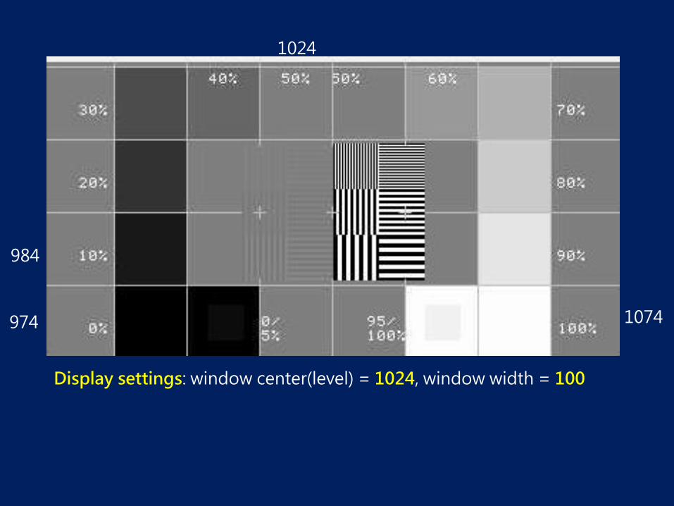
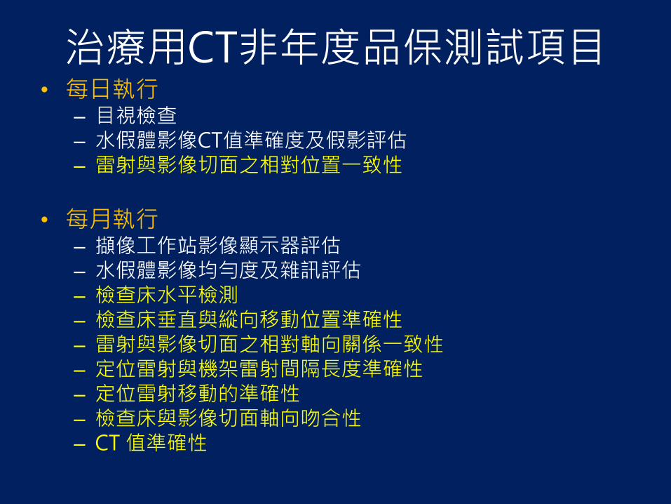

使用掃描放射影像決定切片位置的準確性
使用掃描放射影像決定切片位置的準確性
Lateral projection of ACR Phantom
Definitions of slice indicators
1 Start position of slice
2 Center of slice
3 Total coverage of scan
檢查床進出切片方向移動的準確性
ImPACT Information Leaflet No 1
切片厚度準確性
(1) 將假體放置於檢查床上以常規成人腹部掃描模式進行測試設定組像範圍為 21 公分若常規成人
腹部掃描是以軸狀掃描則直接掃描切片厚度測試物的正中央區段
(2) 若常規成人腹部掃描是以螺旋掃描則將掃描模式改為軸狀掃描其他參數則維持固定不變若掃描儀為多切片機型但不能使用相同的偵檢器組置(N bull TT為一個資料通道在 Z軸方向上之寬度N為該
模式下的資料通道數目)進行軸狀掃描時則在維持相同的T設定下使用最大的N之偵檢器組置進行軸狀掃描
(3) 同上述步驟但改變切片厚度為高解析度肺部掃描時使用的厚度35 及7 毫米(若於步驟(1)中已有相同厚度則不需重複掃描)若無法達到前述設定則選取最接近的設定值進行測試
測試步驟
切片厚度準確性
CT值準確度與線性度
材料 空氣 聚乙烯 水 壓克力 骨頭 測量值 -97271 -8918 476 12547 92647
Attenuation Coefficient 0 01795 0187 0221 0373
y = 50842x - 9777 Rsup2 = 09989
-1500
-1000
-500
0
500
1000
1500
0 005 01 015 02 025 03 035 04
線性迴歸分析
線性迴歸分析
線性(線性迴歸分析)
Procedures
bull Scan uniform phantom
bull Place ROIs
bull Noise Standard Deviation of ROI
bull CT uniformity
- Diff between means of center and outer ROI
水假體影像評估
bull (1)水的CT值準確度(2)雜訊(3)影像均勻度與(4)假影
Foot
bull (2)雜訊
bull (3)影像均勻度
bull (4)假影
A streak B motion C beam-hardening DE ring F blooming
Incomplete projections ndash streaking artifacts
Helical Artifacts in the axial plane Single-Section Scanning
Helical artifacts
Partial volume effect
Cone beam effect
Axial scan Helical scan Corrected image
空間解析度
ACR phantom 8 Al bar patterns- 4 5 6 7 8 9 10 and 12 lpcm
Start here
The result is 9 lpcm
低對比偵測度 bull Four cylinder groups
bull 06 (6 HU) difference from a background material
bull mean CT number of approximately 90 HU
bull Cylinder-to-background contrast is energy-independent
bull cylinders diameters and spaces 2 3 4 5 and 6 mm
bull A 25-mm cylinder bull to verify the cylinder-to-background contrast level
119862119873119877 =120583119887119886119888119896119892119903119900119906119899119889 minus 120583119905119886119903119892119890119905
119899119900119894119904119890
noise = σbackground (σbackground + σtarget ) divide 2 [(σbackground 2 + σtarget
2) divide 2 ]05
項目十劑量評估
Dose report from CT scanner
Computed Tomography Dose Index
bull Weighted CTDI CTDIw
CTDI100center
CTDI100 P1
CTDI100 P2
CTDI100 P3
CTDI100 P4
CTDI100edge = 4
CTDI100 P1+P2+P3+P4
Dose Calculation
pcw CTDI3
2CTDI
3
1CTDI
wvol
CTDICTDI
Pitch
volDLP=CTDI total scan length
DLPE k
C P1
P2
P4
P3
DRL Diagnostic reference level
KJR 2012
Tsai HY Tung CJ Yu CC Tyan YS Survey of computed tomography scanners in Taiwan dose descriptors dose guidance levels and effective doses Med Phys 2007341234ndash1243
項目十一輻射寬度
bull Using packaged films bull placed at the isocenter surface during scans at
the different collimation thicknesses
bull The mAs technique is set to provide a maximum film density of between 10 and 20 OD
項目十二影像顯示器評估
SMPTE TG 18 QC
Display images on monitor
Image data Brightness on monitor
Display images on monitor
Standardized Display System
P-value P-value to
DDL
DDLdigital driving level
Display System
DDLs Luminance
P-value
DDL
DDL
Luminance
P-value
DDL
DDL
Luminance
P-value
Luminance
Grayscale Display Function
20181017
JNDs
bull Just-Noticeable Difference
001
01
1
10
100
1000
10000
0 200 400 600 800 1000
灰階值 實測亮度 JND 標準亮度 實測斜率 標準斜率 斜率差異 結果評定
128 123 80 122 00350 00346 117 Passed
384 242 114 240 00521 00526 -102 Passed
640 445 153 445 00761 00761 004 Passed
896 719 189 719 01038 01038 -002 Passed
1152 1072 223 1072 01419 01393 185 Passed
1408 1597 260 1587 01841 01860 -103 Passed
1664 2278 297 2275 02454 02451 011 Passed
1920 3186 334 3182 03265 03201 198 Passed
2176 4394 371 4367 04215 04212 007 Passed
2432 6122 412 6094 05478 05483 -009 Passed
2688 8094 448 8067 07090 07091 -001 Passed
2944 1093 488 10904 09250 09130 132 Passed
3200 1426 524 14190 11725 11737 -011 Passed
3456 1895 564 18885 15029 14993 024 Passed
3712 2421 599 24133 18750 18892 -075 Passed
3968 3096 635 30934 24282 24098 076 Passed
4224 4043 674 40332 31000 30897 033 Passed
4480 5221 712 52073
01
1
10
100
1000
00 1000 2000 3000 4000 5000 6000
Measured Data
Standard
100
1000
10000
100000
0 200 400 600
Measured Data
Standard
請多指教

使用掃描放射影像決定切片位置的準確性
Lateral projection of ACR Phantom
Definitions of slice indicators
1 Start position of slice
2 Center of slice
3 Total coverage of scan
檢查床進出切片方向移動的準確性
ImPACT Information Leaflet No 1
切片厚度準確性
(1) 將假體放置於檢查床上以常規成人腹部掃描模式進行測試設定組像範圍為 21 公分若常規成人
腹部掃描是以軸狀掃描則直接掃描切片厚度測試物的正中央區段
(2) 若常規成人腹部掃描是以螺旋掃描則將掃描模式改為軸狀掃描其他參數則維持固定不變若掃描儀為多切片機型但不能使用相同的偵檢器組置(N bull TT為一個資料通道在 Z軸方向上之寬度N為該
模式下的資料通道數目)進行軸狀掃描時則在維持相同的T設定下使用最大的N之偵檢器組置進行軸狀掃描
(3) 同上述步驟但改變切片厚度為高解析度肺部掃描時使用的厚度35 及7 毫米(若於步驟(1)中已有相同厚度則不需重複掃描)若無法達到前述設定則選取最接近的設定值進行測試
測試步驟
切片厚度準確性
CT值準確度與線性度
材料 空氣 聚乙烯 水 壓克力 骨頭 測量值 -97271 -8918 476 12547 92647
Attenuation Coefficient 0 01795 0187 0221 0373
y = 50842x - 9777 Rsup2 = 09989
-1500
-1000
-500
0
500
1000
1500
0 005 01 015 02 025 03 035 04
線性迴歸分析
線性迴歸分析
線性(線性迴歸分析)
Procedures
bull Scan uniform phantom
bull Place ROIs
bull Noise Standard Deviation of ROI
bull CT uniformity
- Diff between means of center and outer ROI
水假體影像評估
bull (1)水的CT值準確度(2)雜訊(3)影像均勻度與(4)假影
Foot
bull (2)雜訊
bull (3)影像均勻度
bull (4)假影
A streak B motion C beam-hardening DE ring F blooming
Incomplete projections ndash streaking artifacts
Helical Artifacts in the axial plane Single-Section Scanning
Helical artifacts
Partial volume effect
Cone beam effect
Axial scan Helical scan Corrected image
空間解析度
ACR phantom 8 Al bar patterns- 4 5 6 7 8 9 10 and 12 lpcm
Start here
The result is 9 lpcm
低對比偵測度 bull Four cylinder groups
bull 06 (6 HU) difference from a background material
bull mean CT number of approximately 90 HU
bull Cylinder-to-background contrast is energy-independent
bull cylinders diameters and spaces 2 3 4 5 and 6 mm
bull A 25-mm cylinder bull to verify the cylinder-to-background contrast level
119862119873119877 =120583119887119886119888119896119892119903119900119906119899119889 minus 120583119905119886119903119892119890119905
119899119900119894119904119890
noise = σbackground (σbackground + σtarget ) divide 2 [(σbackground 2 + σtarget
2) divide 2 ]05
項目十劑量評估
Dose report from CT scanner
Computed Tomography Dose Index
bull Weighted CTDI CTDIw
CTDI100center
CTDI100 P1
CTDI100 P2
CTDI100 P3
CTDI100 P4
CTDI100edge = 4
CTDI100 P1+P2+P3+P4
Dose Calculation
pcw CTDI3
2CTDI
3
1CTDI
wvol
CTDICTDI
Pitch
volDLP=CTDI total scan length
DLPE k
C P1
P2
P4
P3
DRL Diagnostic reference level
KJR 2012
Tsai HY Tung CJ Yu CC Tyan YS Survey of computed tomography scanners in Taiwan dose descriptors dose guidance levels and effective doses Med Phys 2007341234ndash1243
項目十一輻射寬度
bull Using packaged films bull placed at the isocenter surface during scans at
the different collimation thicknesses
bull The mAs technique is set to provide a maximum film density of between 10 and 20 OD
項目十二影像顯示器評估
SMPTE TG 18 QC
Display images on monitor
Image data Brightness on monitor
Display images on monitor
Standardized Display System
P-value P-value to
DDL
DDLdigital driving level
Display System
DDLs Luminance
P-value
DDL
DDL
Luminance
P-value
DDL
DDL
Luminance
P-value
Luminance
Grayscale Display Function
20181017
JNDs
bull Just-Noticeable Difference
001
01
1
10
100
1000
10000
0 200 400 600 800 1000
灰階值 實測亮度 JND 標準亮度 實測斜率 標準斜率 斜率差異 結果評定
128 123 80 122 00350 00346 117 Passed
384 242 114 240 00521 00526 -102 Passed
640 445 153 445 00761 00761 004 Passed
896 719 189 719 01038 01038 -002 Passed
1152 1072 223 1072 01419 01393 185 Passed
1408 1597 260 1587 01841 01860 -103 Passed
1664 2278 297 2275 02454 02451 011 Passed
1920 3186 334 3182 03265 03201 198 Passed
2176 4394 371 4367 04215 04212 007 Passed
2432 6122 412 6094 05478 05483 -009 Passed
2688 8094 448 8067 07090 07091 -001 Passed
2944 1093 488 10904 09250 09130 132 Passed
3200 1426 524 14190 11725 11737 -011 Passed
3456 1895 564 18885 15029 14993 024 Passed
3712 2421 599 24133 18750 18892 -075 Passed
3968 3096 635 30934 24282 24098 076 Passed
4224 4043 674 40332 31000 30897 033 Passed
4480 5221 712 52073
01
1
10
100
1000
00 1000 2000 3000 4000 5000 6000
Measured Data
Standard
100
1000
10000
100000
0 200 400 600
Measured Data
Standard
請多指教

切片厚度準確性
CT值準確度與線性度
材料 空氣 聚乙烯 水 壓克力 骨頭 測量值 -97271 -8918 476 12547 92647
Attenuation Coefficient 0 01795 0187 0221 0373
y = 50842x - 9777 Rsup2 = 09989
-1500
-1000
-500
0
500
1000
1500
0 005 01 015 02 025 03 035 04
線性迴歸分析
線性迴歸分析
線性(線性迴歸分析)
Procedures
bull Scan uniform phantom
bull Place ROIs
bull Noise Standard Deviation of ROI
bull CT uniformity
- Diff between means of center and outer ROI
水假體影像評估
bull (1)水的CT值準確度(2)雜訊(3)影像均勻度與(4)假影
Foot
bull (2)雜訊
bull (3)影像均勻度
bull (4)假影
A streak B motion C beam-hardening DE ring F blooming
Incomplete projections ndash streaking artifacts
Helical Artifacts in the axial plane Single-Section Scanning
Helical artifacts
Partial volume effect
Cone beam effect
Axial scan Helical scan Corrected image
空間解析度
ACR phantom 8 Al bar patterns- 4 5 6 7 8 9 10 and 12 lpcm
Start here
The result is 9 lpcm
低對比偵測度 bull Four cylinder groups
bull 06 (6 HU) difference from a background material
bull mean CT number of approximately 90 HU
bull Cylinder-to-background contrast is energy-independent
bull cylinders diameters and spaces 2 3 4 5 and 6 mm
bull A 25-mm cylinder bull to verify the cylinder-to-background contrast level
119862119873119877 =120583119887119886119888119896119892119903119900119906119899119889 minus 120583119905119886119903119892119890119905
119899119900119894119904119890
noise = σbackground (σbackground + σtarget ) divide 2 [(σbackground 2 + σtarget
2) divide 2 ]05
項目十劑量評估
Dose report from CT scanner
Computed Tomography Dose Index
bull Weighted CTDI CTDIw
CTDI100center
CTDI100 P1
CTDI100 P2
CTDI100 P3
CTDI100 P4
CTDI100edge = 4
CTDI100 P1+P2+P3+P4
Dose Calculation
pcw CTDI3
2CTDI
3
1CTDI
wvol
CTDICTDI
Pitch
volDLP=CTDI total scan length
DLPE k
C P1
P2
P4
P3
DRL Diagnostic reference level
KJR 2012
Tsai HY Tung CJ Yu CC Tyan YS Survey of computed tomography scanners in Taiwan dose descriptors dose guidance levels and effective doses Med Phys 2007341234ndash1243
項目十一輻射寬度
bull Using packaged films bull placed at the isocenter surface during scans at
the different collimation thicknesses
bull The mAs technique is set to provide a maximum film density of between 10 and 20 OD
項目十二影像顯示器評估
SMPTE TG 18 QC
Display images on monitor
Image data Brightness on monitor
Display images on monitor
Standardized Display System
P-value P-value to
DDL
DDLdigital driving level
Display System
DDLs Luminance
P-value
DDL
DDL
Luminance
P-value
DDL
DDL
Luminance
P-value
Luminance
Grayscale Display Function
20181017
JNDs
bull Just-Noticeable Difference
001
01
1
10
100
1000
10000
0 200 400 600 800 1000
灰階值 實測亮度 JND 標準亮度 實測斜率 標準斜率 斜率差異 結果評定
128 123 80 122 00350 00346 117 Passed
384 242 114 240 00521 00526 -102 Passed
640 445 153 445 00761 00761 004 Passed
896 719 189 719 01038 01038 -002 Passed
1152 1072 223 1072 01419 01393 185 Passed
1408 1597 260 1587 01841 01860 -103 Passed
1664 2278 297 2275 02454 02451 011 Passed
1920 3186 334 3182 03265 03201 198 Passed
2176 4394 371 4367 04215 04212 007 Passed
2432 6122 412 6094 05478 05483 -009 Passed
2688 8094 448 8067 07090 07091 -001 Passed
2944 1093 488 10904 09250 09130 132 Passed
3200 1426 524 14190 11725 11737 -011 Passed
3456 1895 564 18885 15029 14993 024 Passed
3712 2421 599 24133 18750 18892 -075 Passed
3968 3096 635 30934 24282 24098 076 Passed
4224 4043 674 40332 31000 30897 033 Passed
4480 5221 712 52073
01
1
10
100
1000
00 1000 2000 3000 4000 5000 6000
Measured Data
Standard
100
1000
10000
100000
0 200 400 600
Measured Data
Standard
請多指教
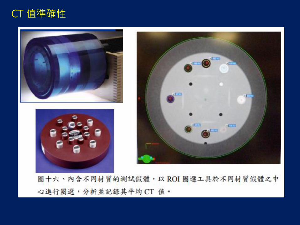
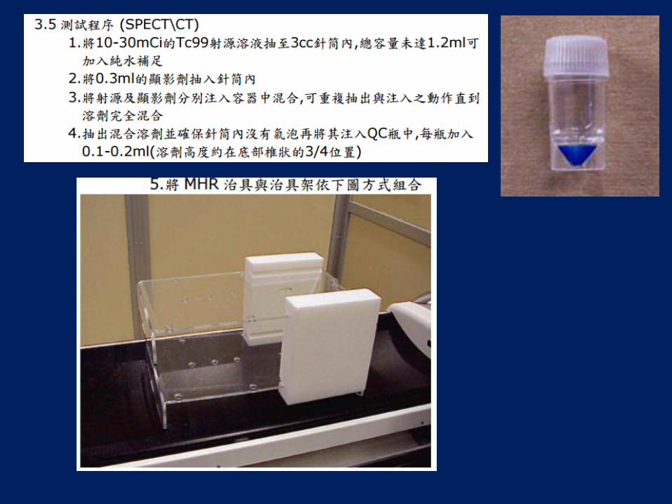
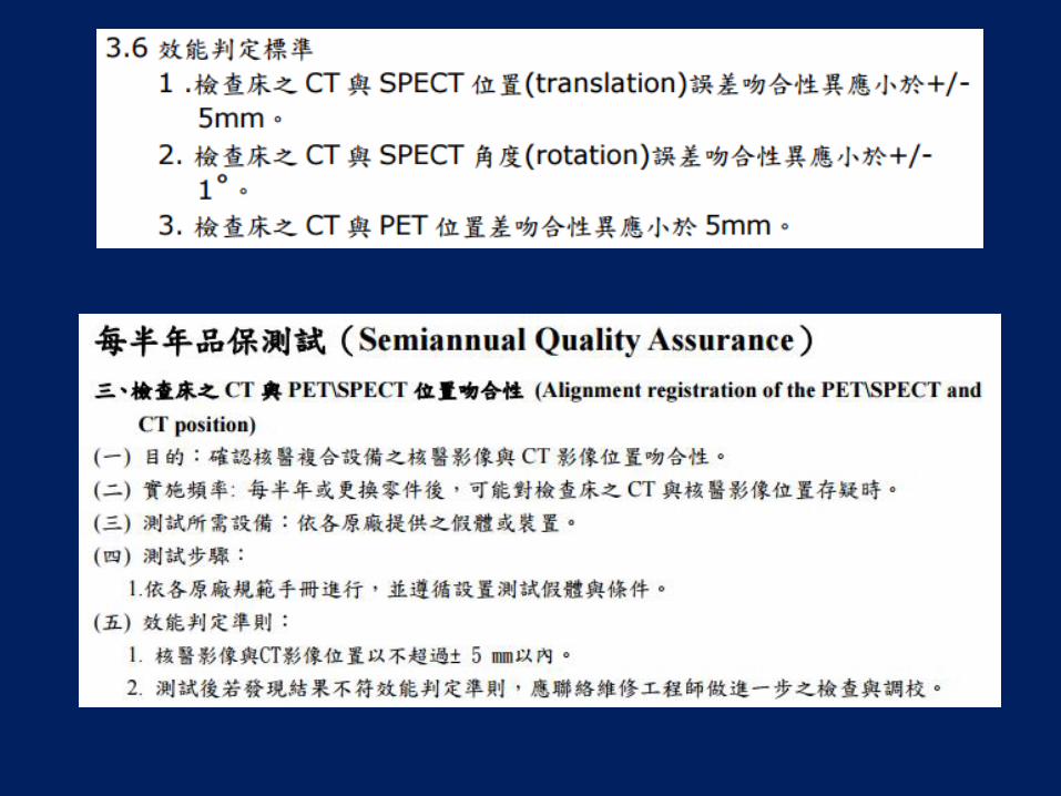
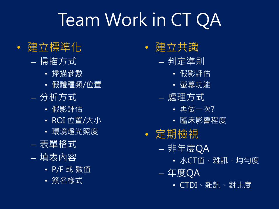

Display images on monitor
Standardized Display System
P-value P-value to
DDL
DDLdigital driving level
Display System
DDLs Luminance
P-value
DDL
DDL
Luminance
P-value
DDL
DDL
Luminance
P-value
Luminance
Grayscale Display Function
20181017
JNDs
bull Just-Noticeable Difference
001
01
1
10
100
1000
10000
0 200 400 600 800 1000
灰階值 實測亮度 JND 標準亮度 實測斜率 標準斜率 斜率差異 結果評定
128 123 80 122 00350 00346 117 Passed
384 242 114 240 00521 00526 -102 Passed
640 445 153 445 00761 00761 004 Passed
896 719 189 719 01038 01038 -002 Passed
1152 1072 223 1072 01419 01393 185 Passed
1408 1597 260 1587 01841 01860 -103 Passed
1664 2278 297 2275 02454 02451 011 Passed
1920 3186 334 3182 03265 03201 198 Passed
2176 4394 371 4367 04215 04212 007 Passed
2432 6122 412 6094 05478 05483 -009 Passed
2688 8094 448 8067 07090 07091 -001 Passed
2944 1093 488 10904 09250 09130 132 Passed
3200 1426 524 14190 11725 11737 -011 Passed
3456 1895 564 18885 15029 14993 024 Passed
3712 2421 599 24133 18750 18892 -075 Passed
3968 3096 635 30934 24282 24098 076 Passed
4224 4043 674 40332 31000 30897 033 Passed
4480 5221 712 52073
01
1
10
100
1000
00 1000 2000 3000 4000 5000 6000
Measured Data
Standard
100
1000
10000
100000
0 200 400 600
Measured Data
Standard
請多指教

P-value
DDL
DDL
Luminance
P-value
Luminance
Grayscale Display Function
20181017
JNDs
bull Just-Noticeable Difference
001
01
1
10
100
1000
10000
0 200 400 600 800 1000
灰階值 實測亮度 JND 標準亮度 實測斜率 標準斜率 斜率差異 結果評定
128 123 80 122 00350 00346 117 Passed
384 242 114 240 00521 00526 -102 Passed
640 445 153 445 00761 00761 004 Passed
896 719 189 719 01038 01038 -002 Passed
1152 1072 223 1072 01419 01393 185 Passed
1408 1597 260 1587 01841 01860 -103 Passed
1664 2278 297 2275 02454 02451 011 Passed
1920 3186 334 3182 03265 03201 198 Passed
2176 4394 371 4367 04215 04212 007 Passed
2432 6122 412 6094 05478 05483 -009 Passed
2688 8094 448 8067 07090 07091 -001 Passed
2944 1093 488 10904 09250 09130 132 Passed
3200 1426 524 14190 11725 11737 -011 Passed
3456 1895 564 18885 15029 14993 024 Passed
3712 2421 599 24133 18750 18892 -075 Passed
3968 3096 635 30934 24282 24098 076 Passed
4224 4043 674 40332 31000 30897 033 Passed
4480 5221 712 52073
01
1
10
100
1000
00 1000 2000 3000 4000 5000 6000
Measured Data
Standard
100
1000
10000
100000
0 200 400 600
Measured Data
Standard
請多指教

01
1
10
100
1000
00 1000 2000 3000 4000 5000 6000
Measured Data
Standard
100
1000
10000
100000
0 200 400 600
Measured Data
Standard
請多指教

100
1000
10000
100000
0 200 400 600
Measured Data
Standard
請多指教

請多指教