electrolytes*and*acid*base* - nigel fong · hyponatremia–pathophysiologic* ... malignancy*...
TRANSCRIPT

Electrolytes and Acid Base
Yii Zheng-‐Wei
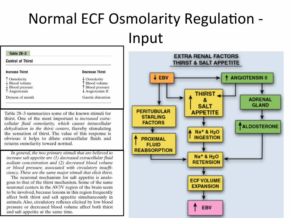
Normal ECF Osmolarity Regula>on -‐ Input
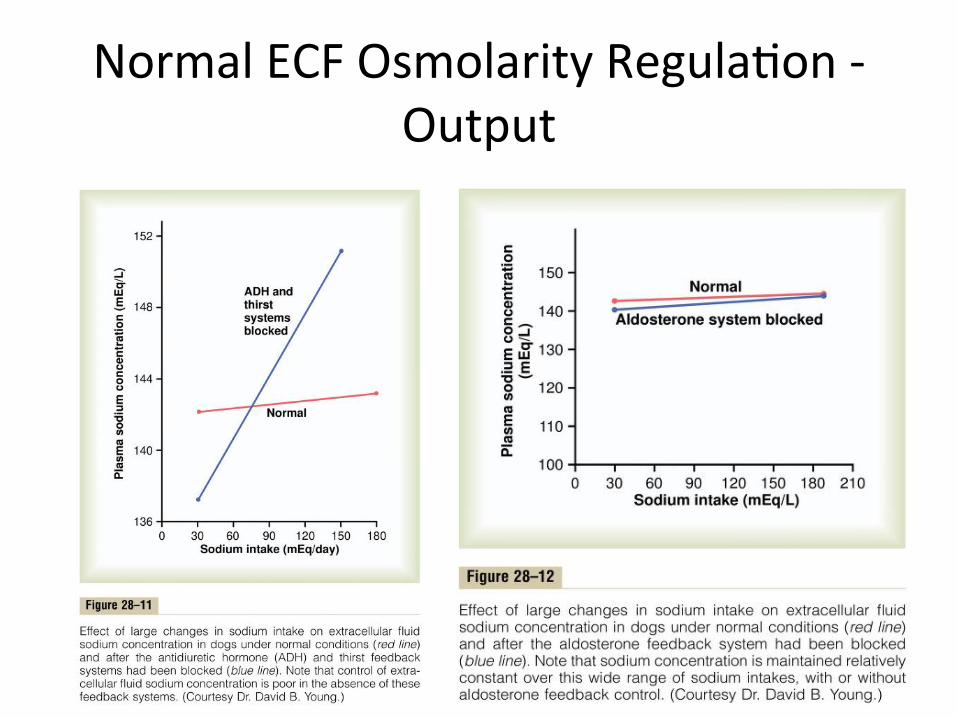
Normal ECF Osmolarity Regula>on -‐ Output
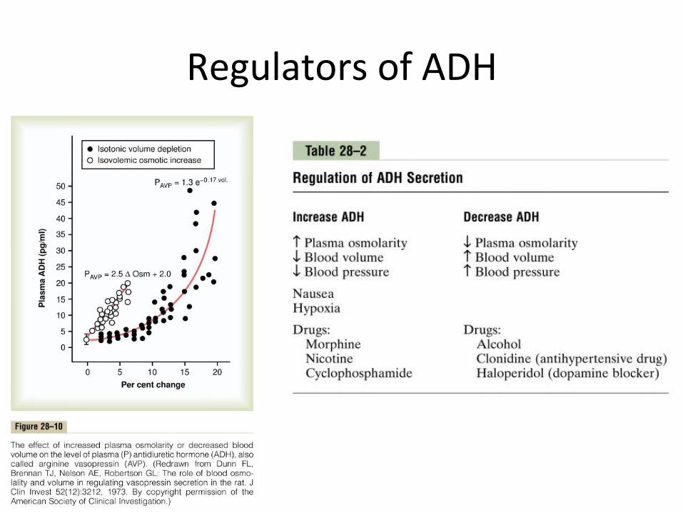
Regulators of ADH
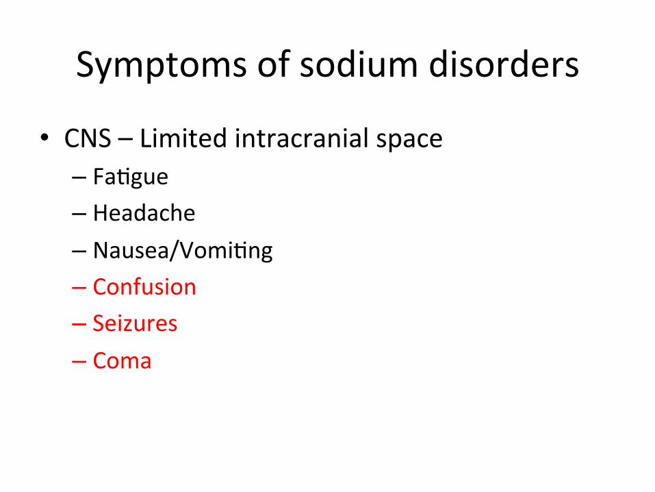
Symptoms of sodium disorders
• CNS – Limited intracranial space – Fa>gue – Headache – Nausea/Vomi>ng – Confusion – Seizures – Coma

Hyponatremia – Pathophysiologic Approach
ADH High ADH Low
Chronic Low BP SIADH Endocrinopathy
Volume Deple>on (Clasically Diure>cs/Osmo>c Diuresis/GI
Loss)
Malignancy Hypothyroidism (Crazy rare)
Advanced Renal Failure
Heart Failure CNS bleeds Primary Polydipsia (Generally psych
pa>ents)
Cirrhosis Lung Low-‐nutrient diet
Nephro>c Syndrome Drug SE
Adrenal Insufficiency Pain
Cerebral Salt Was>ng
Reset Osmostat
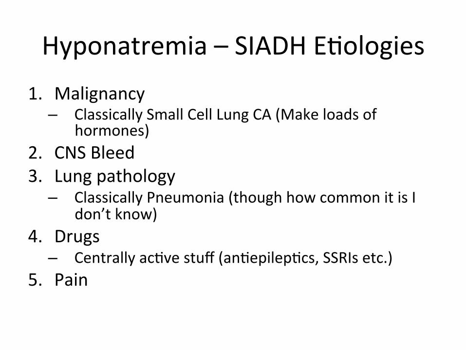
Hyponatremia – SIADH E>ologies 1. Malignancy – Classically Small Cell Lung CA (Make loads of
hormones) 2. CNS Bleed 3. Lung pathology – Classically Pneumonia (though how common it is I
don’t know) 4. Drugs – Centrally ac>ve stuff (an>epilep>cs, SSRIs etc.)
5. Pain
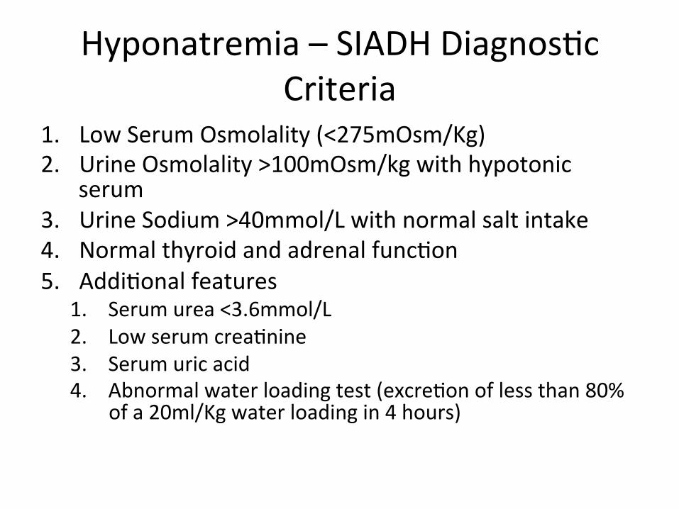
Hyponatremia – SIADH Diagnos>c Criteria
1. Low Serum Osmolality (<275mOsm/Kg) 2. Urine Osmolality >100mOsm/kg with hypotonic
serum 3. Urine Sodium >40mmol/L with normal salt intake 4. Normal thyroid and adrenal func>on 5. Addi>onal features
1. Serum urea <3.6mmol/L 2. Low serum crea>nine 3. Serum uric acid 4. Abnormal water loading test (excre>on of less than 80%
of a 20ml/Kg water loading in 4 hours)

Hyponatremia – Clinical Approach Urine Sodium
Volume Status Low High
Hypovolemic 1. Actual Hypovolemia (usually GI Loss)
1. Diure>cs 2. Primary Adrenal
Insufficiency
Euvolemic 1. Primary Polydipsia 2. Nutrient poor diet
1. SIADH 2. CSW
3. Hypothyroidism 4. Secondary Adrenal
Insufficiency
Hypervolemic 1. CHF 2. Cirrhosis 3. Nephro>c
1. Advanced Renal Failure

Hyponatremia – Correc>on
• Less than 9mEq/L per 24hr • Possibly can correct faster IF hyponatremia is less than 48h dura>on (but this is NOT your call)

Hyponatremia -‐ Cases
• hjp://www.turner-‐white.com/pdf/brm_IM_V10P1.pdf
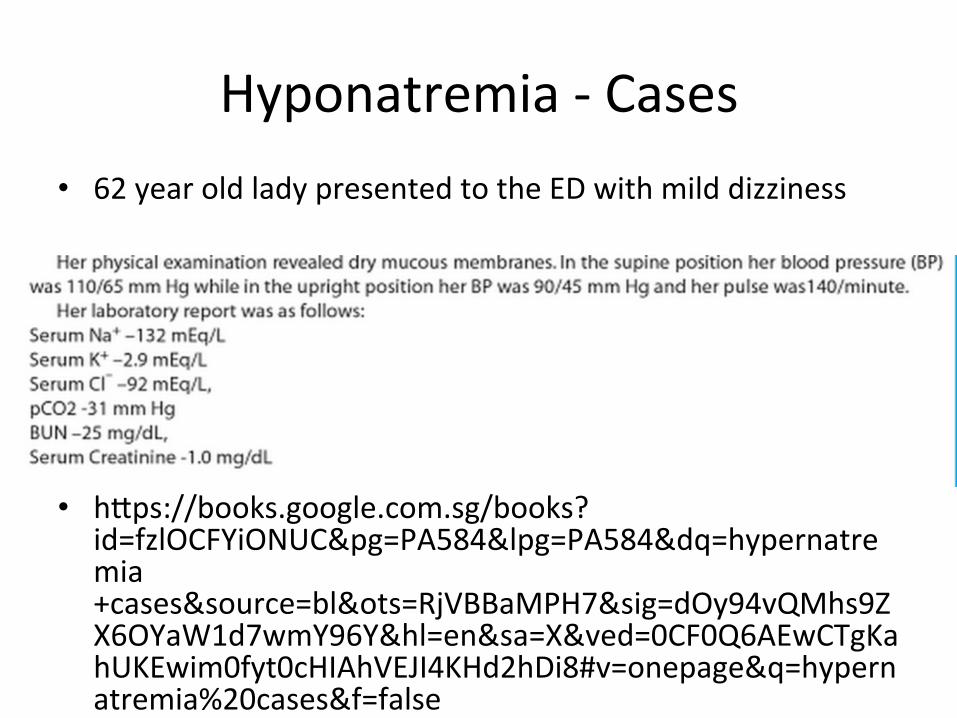
Hyponatremia -‐ Cases • 62 year old lady presented to the ED with mild dizziness
• hjps://books.google.com.sg/books?id=fzlOCFYiONUC&pg=PA584&lpg=PA584&dq=hypernatremia+cases&source=bl&ots=RjVBBaMPH7&sig=dOy94vQMhs9ZX6OYaW1d7wmY96Y&hl=en&sa=X&ved=0CF0Q6AEwCTgKahUKEwim0fyt0cHIAhVEJI4KHd2hDi8#v=onepage&q=hypernatremia%20cases&f=false

BREAK
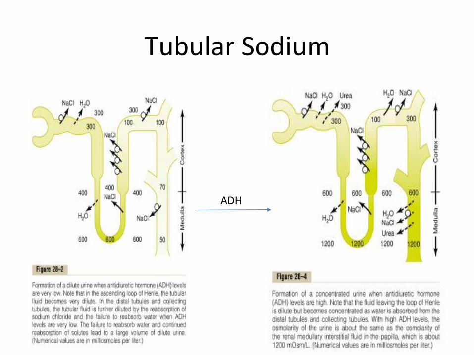
Tubular Sodium
ADH
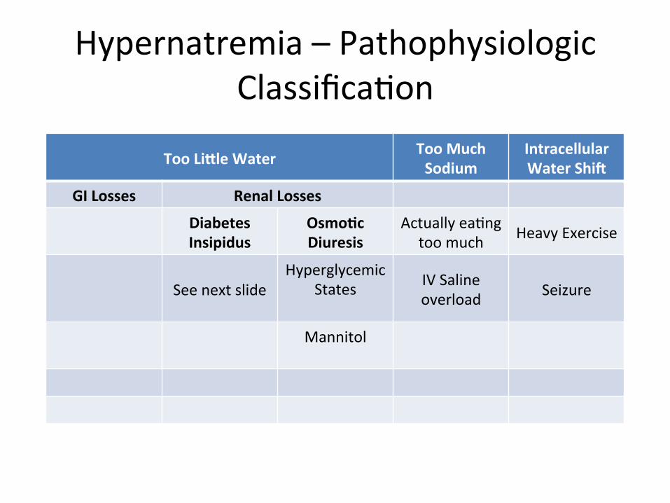
Hypernatremia – Pathophysiologic Classifica>on
Too Li9le Water Too Much Sodium
Intracellular Water Shi>
GI Losses Renal Losses
Diabetes Insipidus
OsmoDc Diuresis
Actually ea>ng too much Heavy Exercise
See next slide Hyperglycemic
States
IV Saline overload Seizure
Mannitol
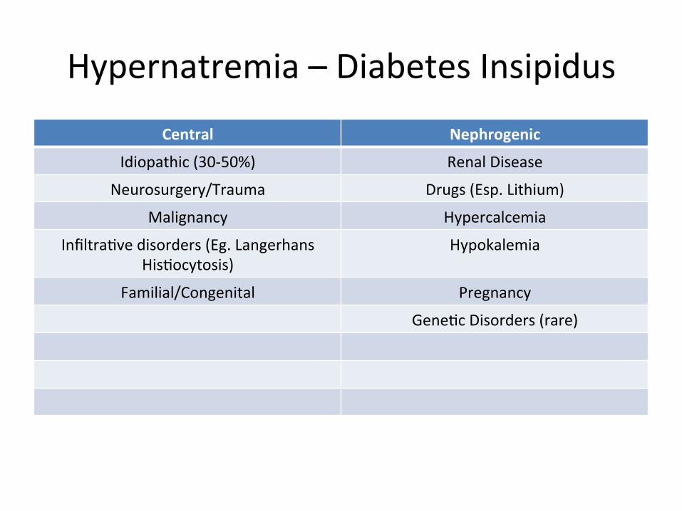
Hypernatremia – Diabetes Insipidus
Central Nephrogenic
Idiopathic (30-‐50%) Renal Disease
Neurosurgery/Trauma Drugs (Esp. Lithium)
Malignancy Hypercalcemia
Infiltra>ve disorders (Eg. Langerhans His>ocytosis)
Hypokalemia
Familial/Congenital Pregnancy
Gene>c Disorders (rare)
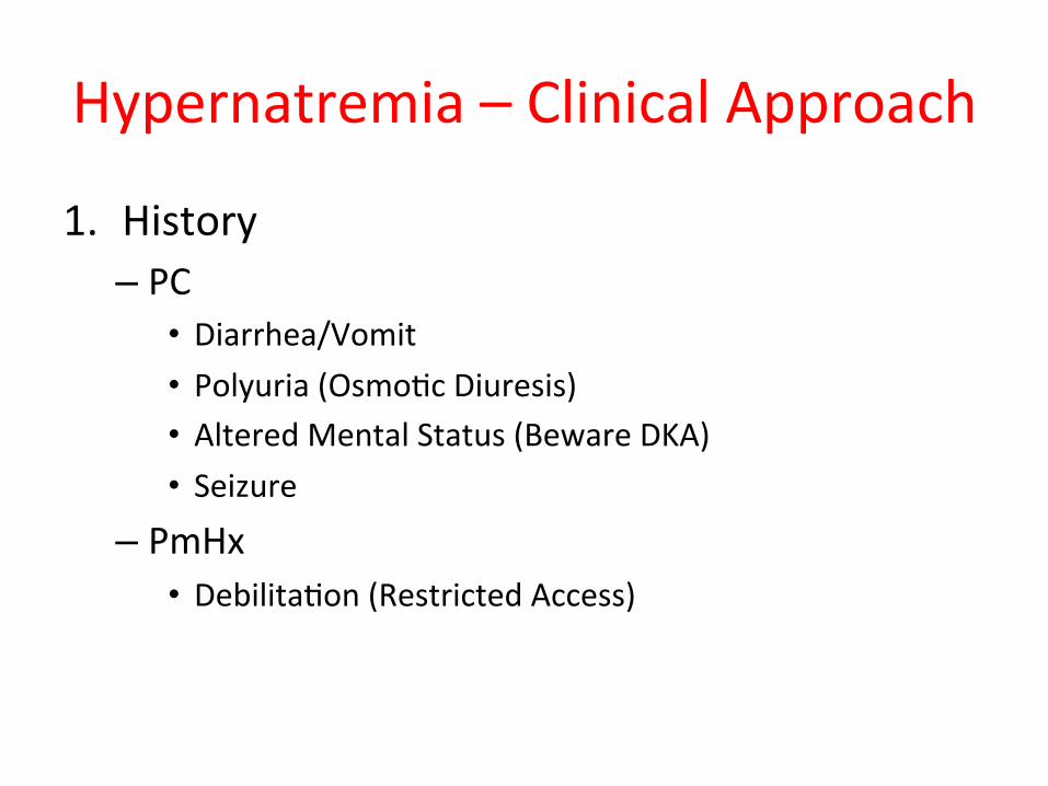
Hypernatremia – Clinical Approach
1. History – PC
• Diarrhea/Vomit • Polyuria (Osmo>c Diuresis) • Altered Mental Status (Beware DKA) • Seizure
– PmHx • Debilita>on (Restricted Access)

Hypernatremia – Diagnosing Diabetes Insipidus
1. Plasma Sodium 2. Plasma Osmolality + Urine Osmolality – Urine Osmolality <300mOsm/kg = Likely DI
• Urine < Plasma: Compete DI • Urine > Plasma: Par>al DI
– Urine Osmolality >600mOsm/kg = Not enough water – Urine Osmolality 300-‐600mOsm/kg = inconclusive
3. Urine Osmolality response to DDAVP – <50% increase awer administra>on: Nephrogenic DI – >50% increase awer administra>on: Central DI
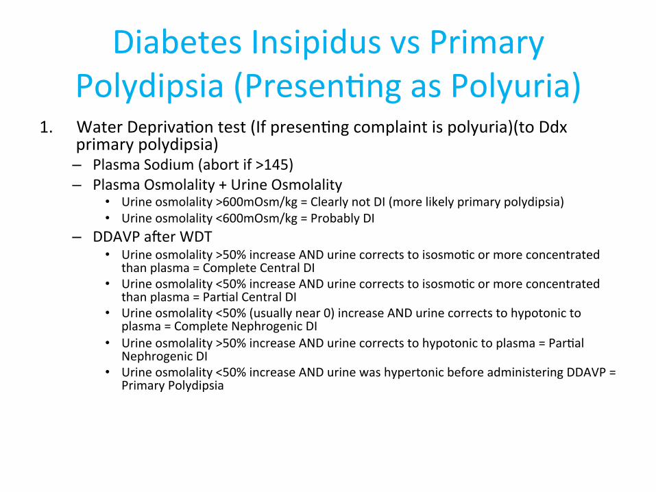
Diabetes Insipidus vs Primary Polydipsia (Presen>ng as Polyuria)
1. Water Depriva>on test (If presen>ng complaint is polyuria)(to Ddx primary polydipsia) – Plasma Sodium (abort if >145) – Plasma Osmolality + Urine Osmolality
• Urine osmolality >600mOsm/kg = Clearly not DI (more likely primary polydipsia) • Urine osmolality <600mOsm/kg = Probably DI
– DDAVP awer WDT • Urine osmolality >50% increase AND urine corrects to isosmo>c or more concentrated
than plasma = Complete Central DI • Urine osmolality <50% increase AND urine corrects to isosmo>c or more concentrated
than plasma = Par>al Central DI • Urine osmolality <50% (usually near 0) increase AND urine corrects to hypotonic to
plasma = Complete Nephrogenic DI • Urine osmolality >50% increase AND urine corrects to hypotonic to plasma = Par>al
Nephrogenic DI • Urine osmolality <50% increase AND urine was hypertonic before administering DDAVP =
Primary Polydipsia

Hypernatremia – Cases
• hjp://highered.mheduca>on.com/sites/0071402357/student_view0/11__kidney__amp__urinary/clinical_case_2.html

BREAK

Potassium Homeostasis – Renal
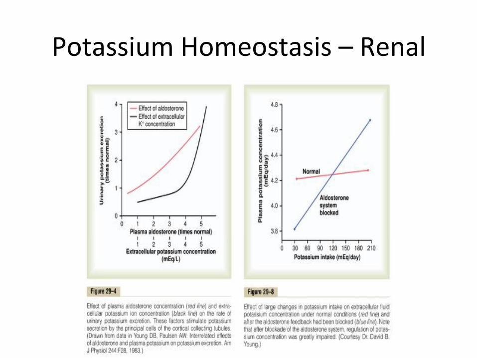
Potassium Homeostasis – Renal

Potassium Homeostasis – Cellular
• Acidosis/Alkalosis – H+ in, K+ out (Electrical neutrality)
• Insulin/Adrenaline – Cathecholamines + Insulin ac>vate Na/K ATPase to shiw potassium intracellularly

Hypokalemia – Symptoms
• Muscular generally – Weakness – Arrythmia

Hypokalemia – ECG Changes
• T wave flajening • U wave development • ST depression • Long PR interval • If not corrected, Torsade

Hypokalemia – Pathophysiologic classifica>on
Low Input High output Transcellular Shi>
GI Loss Renal Loss
Starva>on Vomi>ng Diure>cs Alkalosis
Anorexia Diarrhea Polyuria Insulin
Laxa>ve abuse Hyperaldosteronism
Cathecholamines
Ureterosigmoidostomy
RTA 1/2 Rapid Cell Prolifera>on
Salt was>ng nephropathy
Hypomagnesemia
Amphotericin B
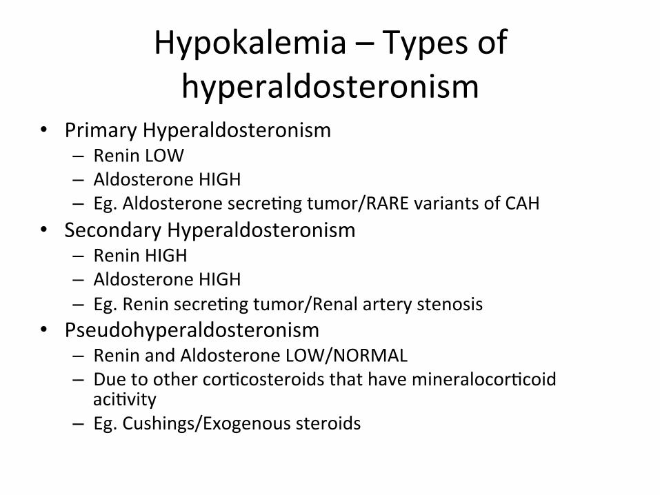
Hypokalemia – Types of hyperaldosteronism
• Primary Hyperaldosteronism – Renin LOW – Aldosterone HIGH – Eg. Aldosterone secre>ng tumor/RARE variants of CAH
• Secondary Hyperaldosteronism – Renin HIGH – Aldosterone HIGH – Eg. Renin secre>ng tumor/Renal artery stenosis
• Pseudohyperaldosteronism – Renin and Aldosterone LOW/NORMAL – Due to other cor>costeroids that have mineralocor>coid aci>vity
– Eg. Cushings/Exogenous steroids
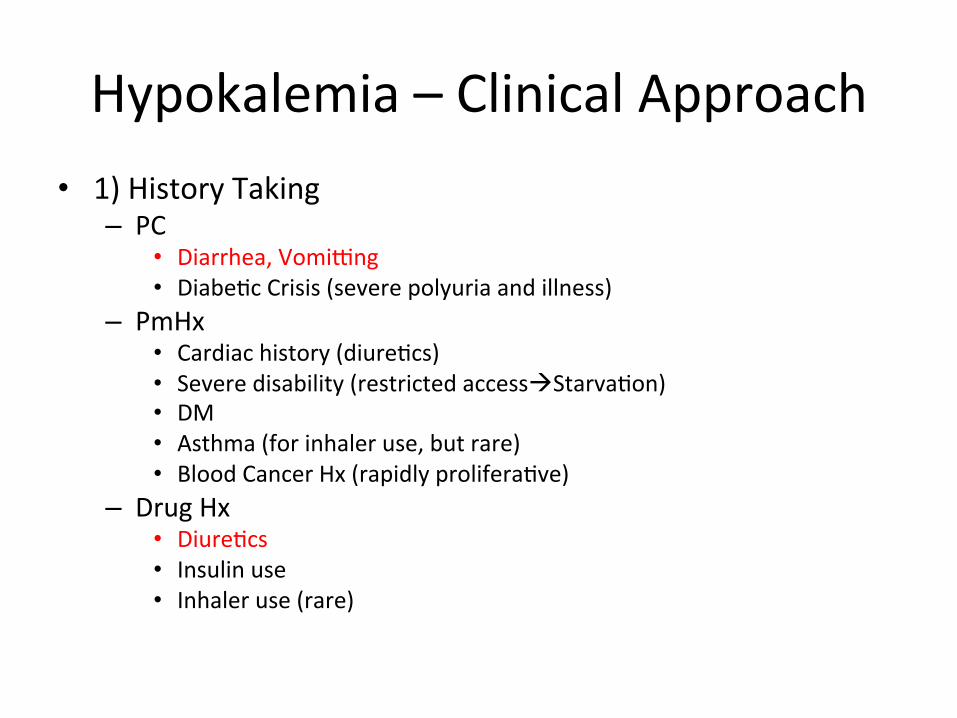
Hypokalemia – Clinical Approach • 1) History Taking
– PC • Diarrhea, Vomiyng • Diabe>c Crisis (severe polyuria and illness)
– PmHx • Cardiac history (diure>cs) • Severe disability (restricted accessàStarva>on) • DM • Asthma (for inhaler use, but rare) • Blood Cancer Hx (rapidly prolifera>ve)
– Drug Hx • Diure>cs • Insulin use • Inhaler use (rare)
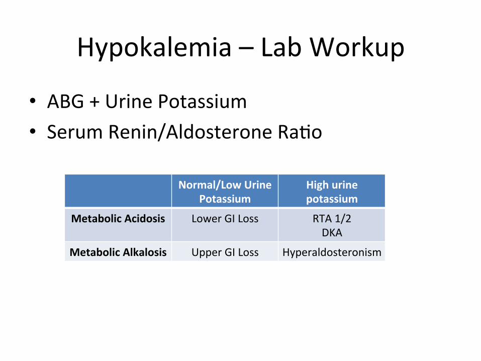
Hypokalemia – Lab Workup
• ABG + Urine Potassium • Serum Renin/Aldosterone Ra>o
Normal/Low Urine Potassium
High urine potassium
Metabolic Acidosis Lower GI Loss RTA 1/2 DKA
Metabolic Alkalosis Upper GI Loss Hyperaldosteronism

Hypokalemia – Treatment
• ECG + Ajend to pa>ent STAT • Oral vs IV Potassium – Oral is preferred if possible and potassium is mildly lowered
– IV is usually used, then swapped to oral later (Burns like crazy, more the faster the infusion runs)

BREAK

Hyperkalemia – Symptoms
• Muscular/Cardiac generally – Weakness – Arrythmia

Hyperkalemia – ECG changes
• String

Hyperkalemia – Pathophysiologic Approach
Input Transcellular Shi> Output
Aldosterone Problems Renal DysfuncDon
IV Drip Cell Breakdown Hypoaldosteronism Acute Kidney Injury
Medica>ons Aldosterone Resistance
Chronic Kidney Disease
Acidosis
Insulin Deficiency
Hyperkalemic Periodic Paralysis

Hyperkalemia – Transcellular Shiws
• Cell Breakdown 1. Tumor Lysis Syndrome 2. Severe hemolysis 3. Rhabdomyolysis
• Theore>cally, can be due to heavy exercise • Medica>ons
1. Beta blockers (Inhibits Na/K ATPase) 2. Digoxin Toxicity (Inhibits Na/K ATPase) 3. Succinylcholine

Hyperkalemia – Aldosterone Problems
• Hyporeninemic Hypoaldosteronism – Renal Dysfunc>on (Decreased Renin Produc>on) – NSAID use
• Normoreninemic Hypoaldosteronism – ACE Inhibitors – Primary Adrenal Insufficiency – Gene>c Disorders
• Aldosterone Resistance – Aldosterone Receptor Blockers – Bactrim – Gene>c Disorders

Hyperkalemia – Clinical Approach 1. ECG + Ajend to pa>ent STAT 2. Exclude Pseudohyperkalemia (Hemolysed sample)
– Repeat Bloods 3. History Taking (Focus on renal func>on and drugs)
– PC • Muscle Pain (Rhabdo) • Trauma (Rhabdo) • Anemic Symptoms • Altered Mental Status (Acidosis) • Hypotension (AKI)
– PmHx • Cardiac Hx (Digoxin, other medica>ons) • Adrenal Insufficiency • DM (DKA) • CKD
– Drug Hx • Digoxin • ACE-‐I • ARBs • NSAIDs • Bactrim
4. Bloods – Renin, Aldosterone, Cor>sol

Hyperkalemia – Treatment
• 7 Treatments for Hyperkalemia courtesy of Dr Seow CJ 1. IV Calcium gluconate (stabilise cardiac membrane) 2. IV Insulin + D50 (Force intracellular shiw) 3. Resonium (Sodium Polystyrene Sulfonate)(Binds K+
in GI tract) 4. Inhaled Beta Agonists (Ac>vate Na/K ATPase) 5. Non-‐potassium sparing diure>cs 6. Alkalinise blood (please don’t, this is theore>cal) 7. Hemodialysis

Potassium Case Studies
• hjps://www.netce.com/casestudies.php?courseid=1199

BREAK
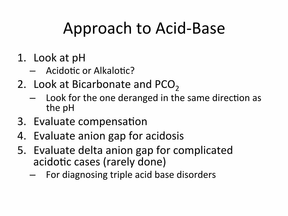
Approach to Acid-‐Base 1. Look at pH – Acido>c or Alkalo>c?
2. Look at Bicarbonate and PCO2 – Look for the one deranged in the same direc>on as
the pH 3. Evaluate compensa>on 4. Evaluate anion gap for acidosis 5. Evaluate delta anion gap for complicated
acido>c cases (rarely done) – For diagnosing triple acid base disorders
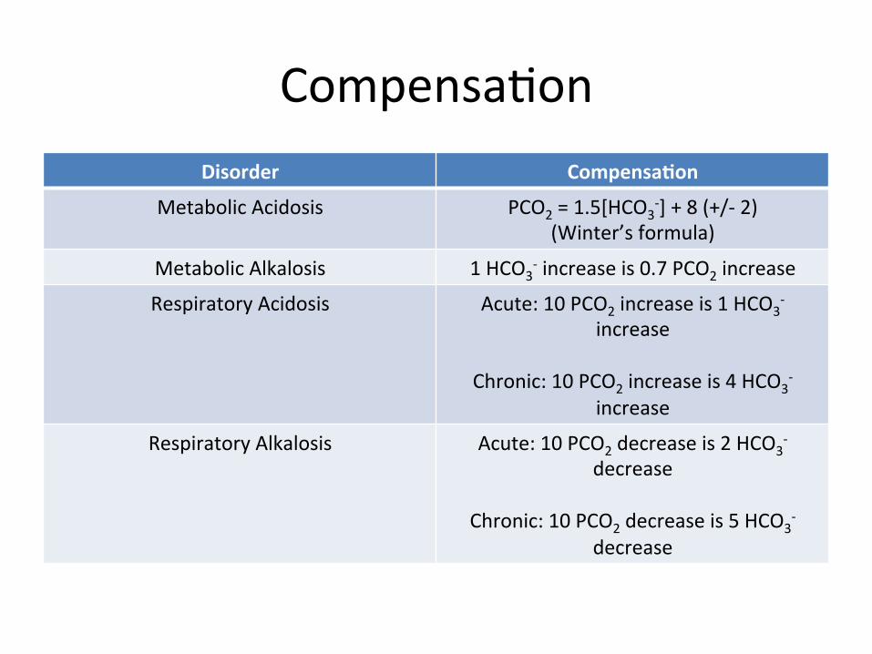
Compensa>on Disorder CompensaDon
Metabolic Acidosis PCO2 = 1.5[HCO3-‐] + 8 (+/-‐ 2)
(Winter’s formula)
Metabolic Alkalosis 1 HCO3-‐ increase is 0.7 PCO2 increase
Respiratory Acidosis Acute: 10 PCO2 increase is 1 HCO3-‐
increase
Chronic: 10 PCO2 increase is 4 HCO3-‐
increase
Respiratory Alkalosis Acute: 10 PCO2 decrease is 2 HCO3-‐
decrease
Chronic: 10 PCO2 decrease is 5 HCO3-‐
decrease
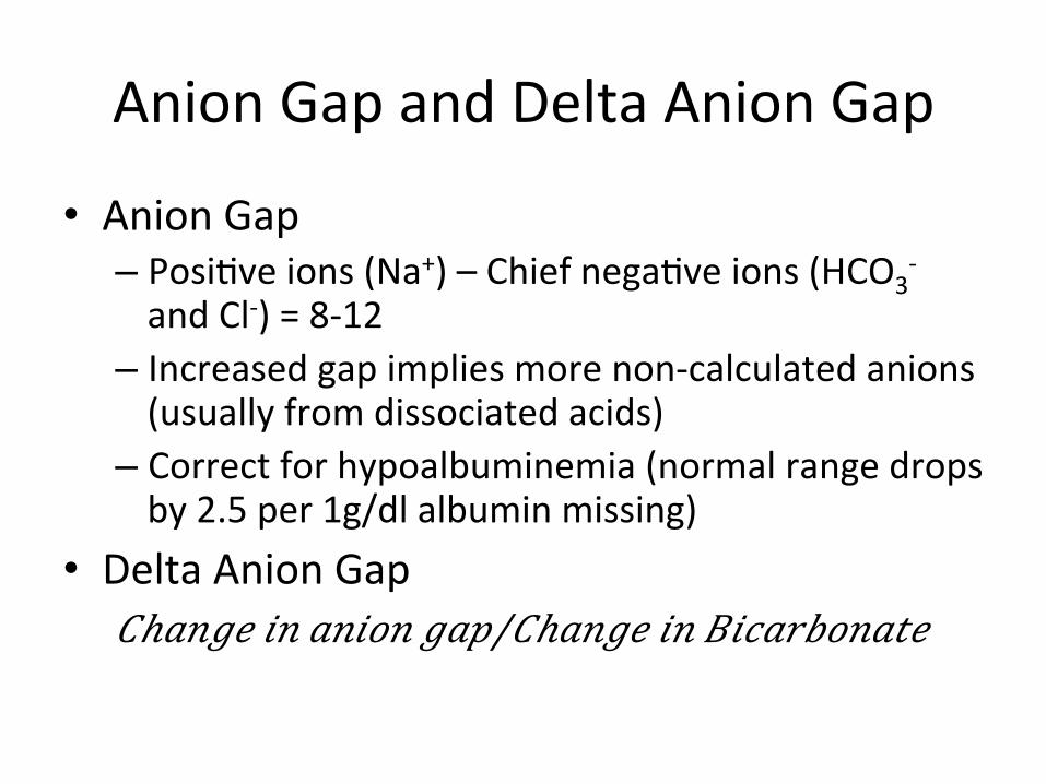
Anion Gap and Delta Anion Gap
• Anion Gap – Posi>ve ions (Na+) – Chief nega>ve ions (HCO3
-‐ and Cl-‐) = 8-‐12
– Increased gap implies more non-‐calculated anions (usually from dissociated acids)
– Correct for hypoalbuminemia (normal range drops by 2.5 per 1g/dl albumin missing)
• Delta Anion Gap 𝐶ℎ𝑎𝑛𝑔𝑒 𝑖𝑛 𝑎𝑛𝑖𝑜𝑛 𝑔𝑎𝑝/𝐶ℎ𝑎𝑛𝑔𝑒 𝑖𝑛 𝐵𝑖𝑐𝑎𝑟𝑏𝑜𝑛𝑎𝑡𝑒
Delta Anion Gap
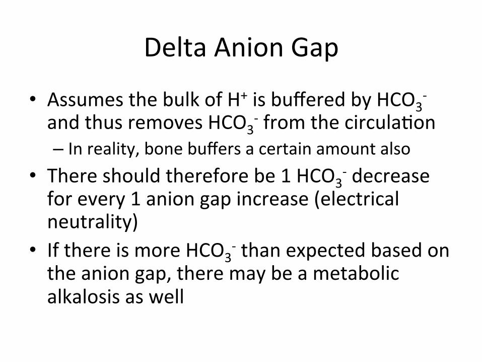
• Assumes the bulk of H+ is buffered by HCO3-‐
and thus removes HCO3-‐ from the circula>on
– In reality, bone buffers a certain amount also • There should therefore be 1 HCO3
-‐ decrease for every 1 anion gap increase (electrical neutrality)
• If there is more HCO3-‐ than expected based on
the anion gap, there may be a metabolic alkalosis as well

Osmolal Gap
• Difference between measured serum osmolality and calculated serum osmolality – Serum osmolality made up of sodium, chloride, bicarb, urea, glucose
• Calculated serum osmolality = 2[Na+ in mmol/L] + [glucose in mmol/L] + [urea in mmol/L]
• A gap of ≥20 suggests weird things in the blood – Small, osmo>cally ac>ve molecules
• Usually alcohols of some sort

HAGMA Mnemonics
• Ketoacidosis • Uremia • Lac>c acidosis – ?Me}ormin
• Toxins – Esp. Salicylates
• Glycols • Oxoproline • L-‐lactate • D-‐lactate • Methanol • Aspirin • Renal failure • Ketoacids

Ketoacidosis
• DM (DKA) • Starva>on • Chronic Alcoholism (both ketoacidosis and lac>c acidosis) – Ethanol à Acetaldehyde + NADH (NADH s>mulates betahydroxybutyrate produc>on)
– Acetaldehyde à Acetate à Acetyl-‐CoA • High Acetyl-‐CoA and NADH shunt pyruvate to lactate (Lac>c acidosis)
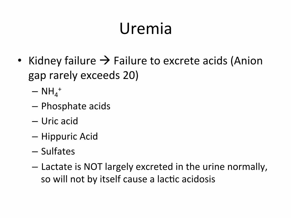
Uremia
• Kidney failure à Failure to excrete acids (Anion gap rarely exceeds 20) – NH4
+ – Phosphate acids – Uric acid – Hippuric Acid – Sulfates – Lactate is NOT largely excreted in the urine normally, so will not by itself cause a lac>c acidosis
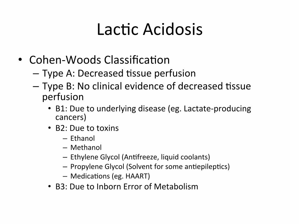
Lac>c Acidosis • Cohen-‐Woods Classifica>on – Type A: Decreased >ssue perfusion – Type B: No clinical evidence of decreased >ssue perfusion • B1: Due to underlying disease (eg. Lactate-‐producing cancers)
• B2: Due to toxins – Ethanol – Methanol – Ethylene Glycol (An>freeze, liquid coolants) – Propylene Glycol (Solvent for some an>epilep>cs) – Medica>ons (eg. HAART)
• B3: Due to Inborn Error of Metabolism

Other HAGMAs • Salicylates – Acutely, Metabolic acidosis with Respiratory alkalosis (s>mulates respiratory centre)
– Awer awhile, both Metabolic and Respiratory acidosis (later respiratory suppression)
• Toluene (glue sniffing) – Metabolised to Hippuric Acid
• 5-‐Oxoproline – Almost Exclusively chronic Acetaminophen use
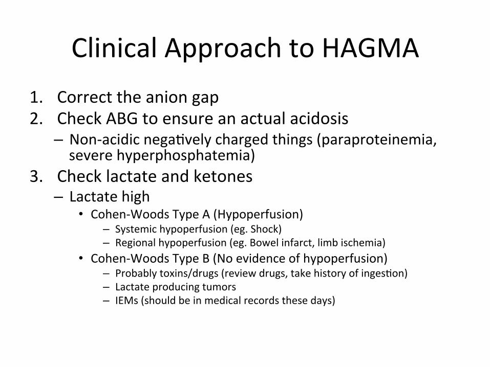
Clinical Approach to HAGMA 1. Correct the anion gap 2. Check ABG to ensure an actual acidosis – Non-‐acidic nega>vely charged things (paraproteinemia, severe hyperphosphatemia)
3. Check lactate and ketones – Lactate high
• Cohen-‐Woods Type A (Hypoperfusion) – Systemic hypoperfusion (eg. Shock) – Regional hypoperfusion (eg. Bowel infarct, limb ischemia)
• Cohen-‐Woods Type B (No evidence of hypoperfusion) – Probably toxins/drugs (review drugs, take history of inges>on) – Lactate producing tumors – IEMs (should be in medical records these days)
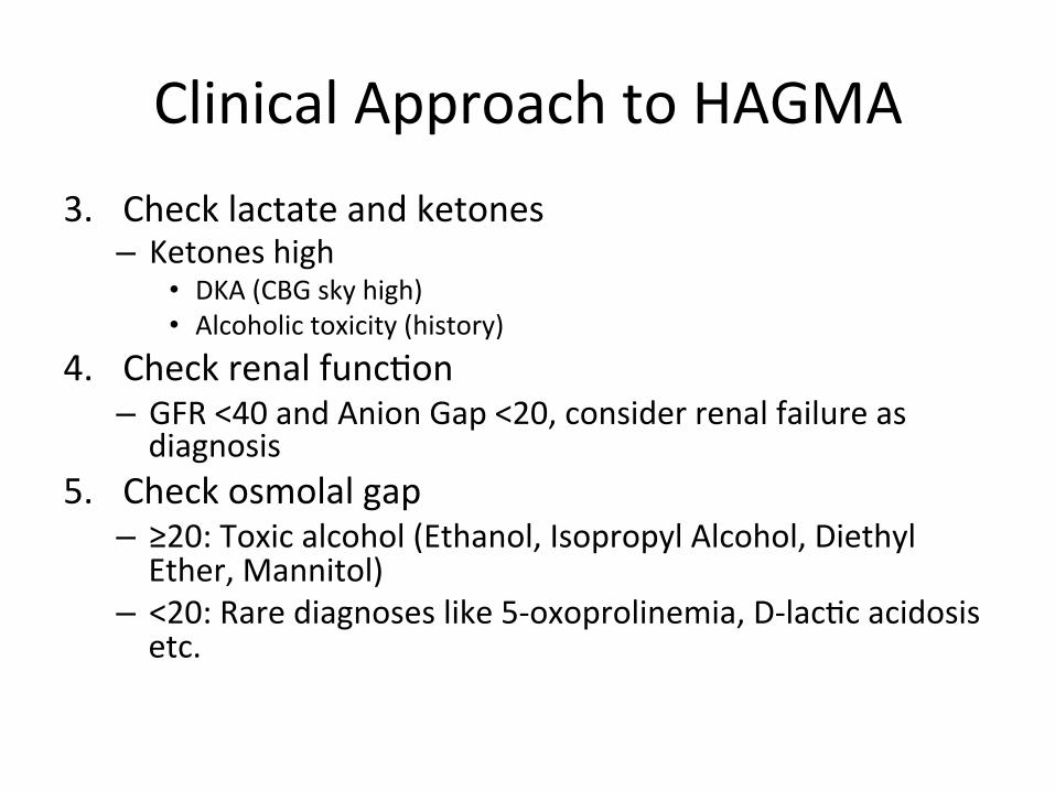
Clinical Approach to HAGMA 3. Check lactate and ketones – Ketones high
• DKA (CBG sky high) • Alcoholic toxicity (history)
4. Check renal func>on – GFR <40 and Anion Gap <20, consider renal failure as diagnosis
5. Check osmolal gap – ≥20: Toxic alcohol (Ethanol, Isopropyl Alcohol, Diethyl Ether, Mannitol)
– <20: Rare diagnoses like 5-‐oxoprolinemia, D-‐lac>c acidosis etc.


BREAK

NAGMA GI Renal
Gain of H+ Hyperalimenta>on
Type 1 and 4 RTA
Renal Failure
Hyperkalemia
Loss of HCO3-‐
Diarrhea
Type 2 RTA Cholestyramine
Ureteral Diversion
Others: Normal Saline Infusion
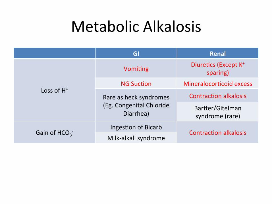
Metabolic Alkalosis GI Renal
Loss of H+
Vomi>ng Diure>cs (Except K+ sparing)
NG Suc>on Mineralocor>coid excess
Rare as heck syndromes (Eg. Congenital Chloride
Diarrhea)
Contrac>on alkalosis
Barjer/Gitelman syndrome (rare)
Gain of HCO3-‐
Inges>on of Bicarb Contrac>on alkalosis
Milk-‐alkali syndrome

BREAK

Respiratory Control of Acid-‐Base
• O2: Diffusion-‐dependent • CO2: Ven>la>on dependent • Ven>la>on control – Central – Neuromuscular – Chest-‐wall compliance

Respiratory Failure
• Type 1: PO2 low (Diffusion problem) • Type 2: PO2 low and PCO2 high (Ven>la>on problem)

Respiratory Acidosis -‐ Symptoms
• CNS symptoms due to hypercapnia and pH changes – Anxiety – Confusion – Somnolence – Delirium – Obtunda>on

Respiratory Acidosis
Lung Problem Central Control Problem
Neuromuscular weakness
Compliance problem
COPD Drugs C3, 4, 5 lesion Kyphoscoliosis
Asthma Brainstem (BEWARE CONING) GBS
Pulmonary Edema Myasthenic Crisis
ILD DMD
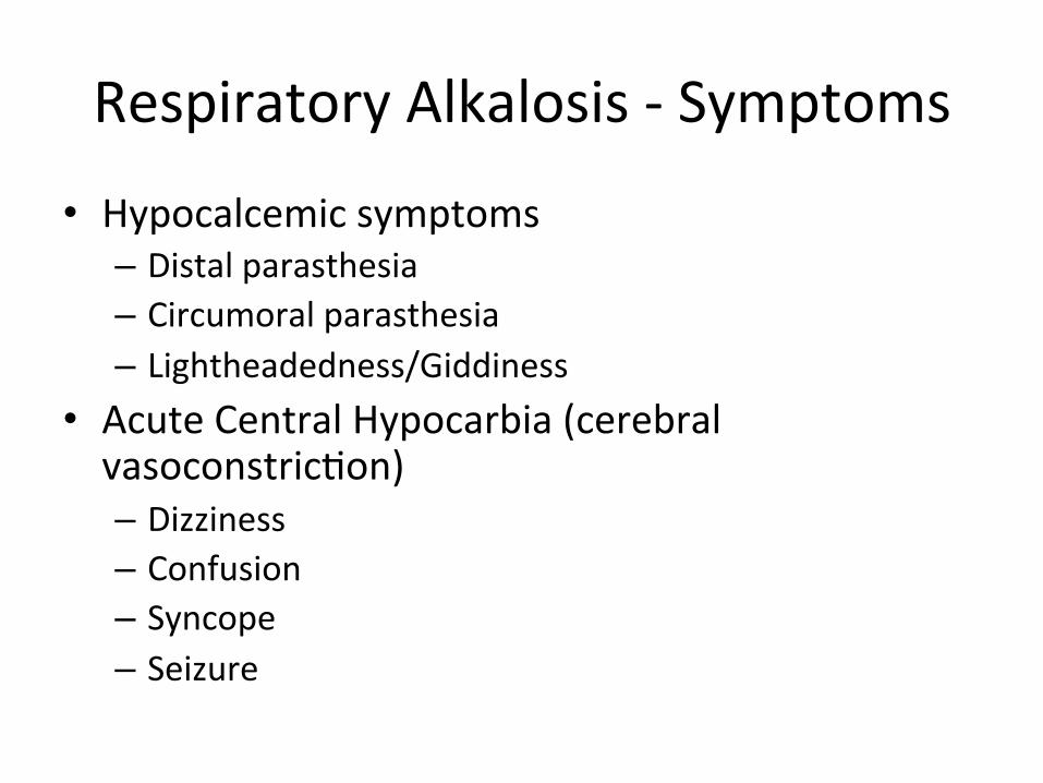
Respiratory Alkalosis -‐ Symptoms
• Hypocalcemic symptoms – Distal parasthesia – Circumoral parasthesia – Lightheadedness/Giddiness
• Acute Central Hypocarbia (cerebral vasoconstric>on) – Dizziness – Confusion – Syncope – Seizure
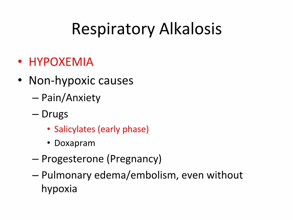
Respiratory Alkalosis
• HYPOXEMIA • Non-‐hypoxic causes – Pain/Anxiety – Drugs
• Salicylates (early phase) • Doxapram
– Progesterone (Pregnancy) – Pulmonary edema/embolism, even without hypoxia