zulaa大学院h26 4
TRANSCRIPT
Neuronal responses to intragastric Neuronal responses to intragastric administration of amino acids in the administration of amino acids in the
rat amygdala and lateral rat amygdala and lateral hypothalamic areahypothalamic area
アミノ酸溶液の胃内投与に対する扁桃体およびアミノ酸溶液の胃内投与に対する扁桃体および視床下部外側野ニューロンの応答様式視床下部外側野ニューロンの応答様式
Davaasuren Munkhzul
System Emotional Science, Graduate School of Medical and
Pharmaceutical Science, University of Toyama, Toyama, Japan,
A.B.
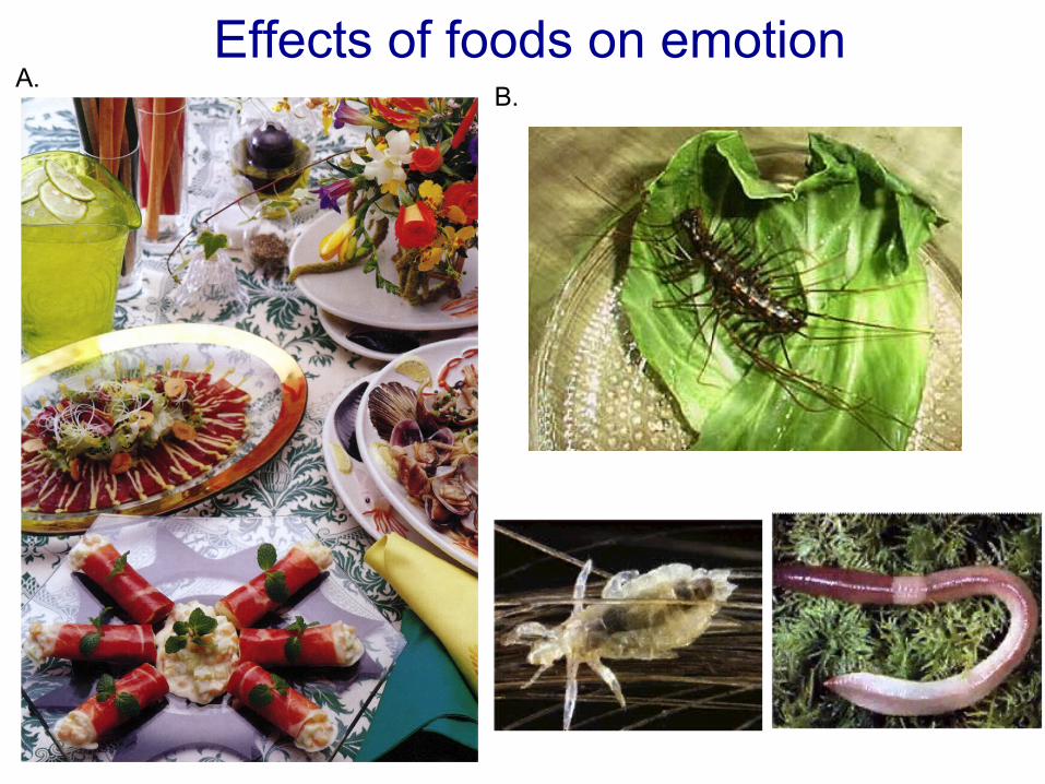
Effects of foods on emotion

Behavioral responses to a bitter taste solution
Another example of emotion induced by foods (2)Another example of emotion induced by foods (2)
Negative emotion
Speculation from these photos suggests that exteroceptive sensory signals, especially taste sensation, induce emotion; taste sensation of palatable foods induces pleasant emotion, and consequently promotes feeding behavior. So, feeding behavior is a kind of appetitive behavior driven by positive emotion.

Pleasant feeling; Taste good.
Pleasant feeling; A sense of well-being or satisfaction?
Exteroceptive sensory signals
Schematic illustration of sites of actions of foods
Visceral sensory signals
Recent studies reported that visceral sensory signals induce not only satiation but also rewarding effects (Sclafani, 2008).
Thus, foods stimulate both the taste system and gastrointestinal (GI) tract to induce emotion. In the same way, umami compounds in the foods are also supposed to stimulate both the taste system and GI tract. I will show you this possibility in the next slide.
IntroductionIntroduction
1. 1. Intragastric infusion of umami substances, monosodium glutamate (MSG) and inosine monophosphate (IMP), altered activities of the vagus nerve and limbic system (Tsurugizawa et al., 2010; Kitamura et al., 2011), and is reported to be rewarding (Uematsu et al., 2009).
2. 2. On the other hand, recent studies suggest that abnormality of vagal functions is related to socio-emotional disturbance including depression, social disturbance, aggression, etc. in humans.
3. 3. We hypothesized that visceral sensory signals might induce emotion such as a sense of well-being, and that deficits in this process would induce socio-emotional disturbance.
4. 4. These findings also suggest that umami compounds might affect socio-emotional behaviors through the vagus nerve.
IntroductionIntroduction
• On the other hand, the amygdale (CeA) and lateral hypothalamic
area (LHA), which are major structures in the limbic system, play
an important role in emotion, and receive strong inputs from the
nucleus of solitary tract (NTS) that receives afferent inputs from
the vagus nerve (Berthoud et al. 2000).
• In the present study, we recorded neuronal activity from the
central amygdala (CeA) and lateral hypothalamic area (LHA), and
investigated changes in neuronal activity during intragastric
administration of amino acids including umami substance,
glutamate.

Purpose of present studyPurpose of present study
• To investigate the gut-brain communication, neuronal activity was recorded from central nucleus amygdala (CeA) and lateral hypothalamic area (LHA) during intragastric infusion of amino acids and umami substance.

Materials and MethodMaterials and Method
Subjects:• 15 adult male Wistar rats, 270-320g
Surgery:• Chronic electrode implatantion • Intragastric cannula implantation
Umami solutions: • Glutamate solution group: GA (glutamate arginine), MSG
(monosodium glutamate), MSG+IMP, and Dashi (dried bonito solution) (60mM, 12, 5, 50%, 83.5%)
• Other solution group: AH (arginine HCl), NaCl, Saline (60mM, 0.9%)
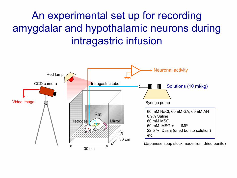
An experimental set up for recording amygdalar and hypothalamic neurons during
intragastric infusion
Syringe pump
Solutions (10 ml/kg)
30 cm
30 cm
Video image
Red lamp
CCD camera
Neuronal activity
Mirror
60 mM NaCl, 60mM GA, 60mM AH0.9% Saline60 mM MSG60 mM MSG + IMP22.5 % Dashi (dried bonito solution)etc.
Intragastric tube
Tetrodes
Rat
(Japanese soup stock made from dried bonito)

• Recording schedule
…
… Amino acid injection
Saline injection(Checking stretch receptor-related responses)
. PAUSE-5min Saline-injection-30sec .
-10 0 10 min 50 -5 0 5 min
Materials and MethodMaterials and Method
10 min – 20minInject 1ml/min/kg (60 mM) solution for 10min (GA, AH, Saline, MSG or
NaCl. One solution per day)

Data Analysing method• Single unit activities were sorted by the Offline Sorter program. • In each cluster of neuronal activities, interspike interval histogram was
constructed to confirm that absolute refractory period was longer than 1 ms. • Finally, superimposed waveforms of the isolated units were drawn to check
wave form consistency throughout the recording sessions, and then were transferred to the Neuroexplorer program.
Materials and MethodMaterials and MethodPC 2
PC 1
Rang
e +/-
2 mV
Unit a Unit b

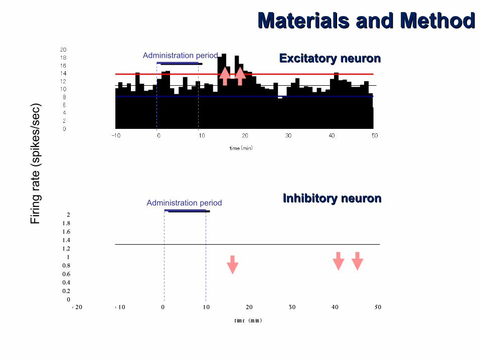
Data Analysis• Pre and post administration period were defined as 10 min before and after the onset of
the administration, respectively.• Perievent histograms aligned with each administration of solution were constructed in each
neuron (bin size=60s). • The baseline firing rate of a neuron was calculated from the pre administration period. • Significant responses were defined as those with activities exceeding 2 standard
deviations above or below the mean firing rates. • In each solution, latency and duration of the neuronal response were computed.
00.51
1.52
2.53
3.54
4.55
- 20 - 10 0 10 20 30 40 50
Latency
Period 1 Period 2 Period 3 Period 4 Period 5
0 50minTime course
Inhibitory duration
Firi
ng r
ate
(spi
kes/
sec)
Administration period
Exc duration
Materials and MethodMaterials and Method
0
0.2
0.4
0.6
0.8
1
1 .2
1 .4
1 .6
1 .8
2
- 20 - 1 0 0 1 0 20 30 40 50
t im e (m in )
Excitatory neuronExcitatory neuron
Inhibitory neuronInhibitory neuron
Firi
ng r
ate
(spi
kes/
sec)
Administration period
Administration period
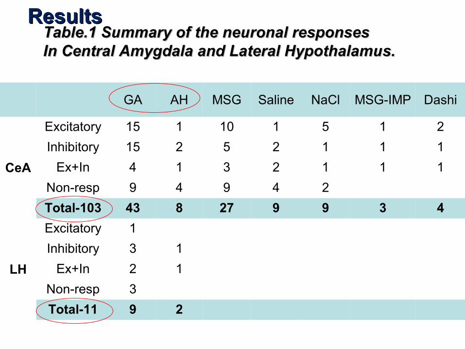
Table.1 Summary of the neuronal responsesTable.1 Summary of the neuronal responsesIn Central Amygdala and Lateral Hypothalamus.In Central Amygdala and Lateral Hypothalamus.
GA AH MSG Saline NaCl MSG-IMP Dashi
CeA
Excitatory 15 1 10 1 5 1 2
Inhibitory 15 2 5 2 1 1 1
Ex+In 4 1 3 2 1 1 1
Non-resp 9 4 9 4 2
Total-103 43 8 27 9 9 3 4
LH
Excitatory 1
Inhibitory 3 1
Ex+In 2 1
Non-resp 3
Total-11 9 2
ResultsResults
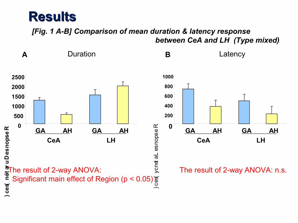
0
500
1000
1500
2000
2500
CeA LH
0
200
400
600
800
1000
GA AH
Response Latency (sec)
Resp
on
se Du
ration
(sec)
Duration Latency
[Fig. 1 A-B] Comparison of mean duration & latency response between CeA and LH (Type mixed)
The result of 2-way ANOVA: Significant main effect of Region (p < 0.05)
The result of 2-way ANOVA: n.s.
GA AH
CeA LH
GA AH GA AH
A B
ResultsResults

0
1
2
3
4
-20 -10 0 10 20 30 40 50time (min)
A. CeA responses
0
10
20
30
40
-20 -10 0 10 20 30 40 50
time (min)
Fig.2 Examples of CeA and LH neuronal responsesResultsResults
B. B. LHA responses
Intragastric infusion (60 mM GA, 10 ml/kg)
Intragastric infusion (60 mM GA, 10 ml/kg)Firi
ng r
ate
(spi
kes/
sec)

[Fig 3] Mean duration of Excitatory responses in each time window in each Solution
****** ************
****
(**p<0.01, ***p<0.001, Bonferroni's correction)
The result of 2-way repeated measured ANOVA: Significant main effect of Time (p < 0.001), Significant interaction of Time x Solution (p < 0.05)
0
100
200
300
400
500
600
GAMSGNaClMSG+IMPDashi
0 - 10 min 10 – 20 min 20 – 30 min 30 – 40 min 40 – 50 min
Response D
uration (sec)
ResultsResults
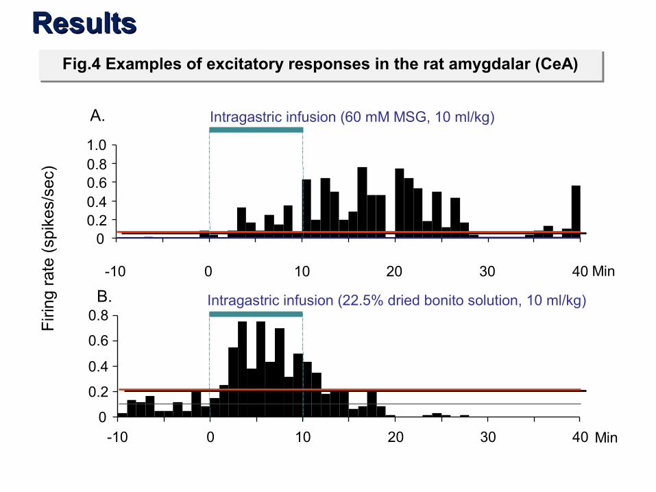
Fig.4 Examples of excitatory responses in the rat amygdalar (CeA)Fig.4 Examples of excitatory responses in the rat amygdalar (CeA)
B.
A. Intragastric infusion (60 mM MSG, 10 ml/kg)
00.20.40.60.81.0
Intragastric infusion (22.5% dried bonito solution, 10 ml/kg)
0
0.2
0.4
0.6
0.8
-10 0 10 20 30 40 Min
-10 0 10 20 30 40 Min
Firi
ng r
ate
(spi
kes/
sec)
ResultsResults
0
50
100
150
200
250
300
350
400
450 GAAHMSGSaline
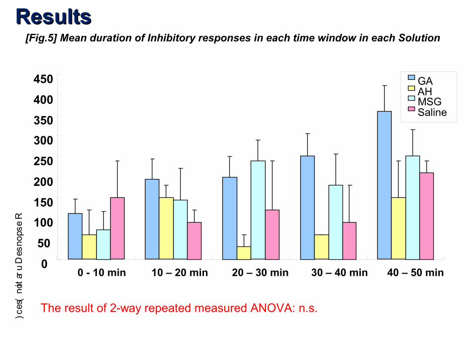
[Fig.5] Mean duration of Inhibitory responses in each time window in each Solution
0 - 10 min 10 – 20 min 20 – 30 min 30 – 40 min 40 – 50 min
Response D
uration (sec)
The result of 2-way repeated measured ANOVA: n.s.
ResultsResults

01020304050
-20 -10 0 10 20 30 40 50time (min)
01234
-20 -10 0 10 20 30 40 50time (min)
Fig.6 An example of Inhibitory response in the CeA
ResultsResults
Intragastric infusion (60 mM MSG, 10 ml/kg)
Intragastric infusion (0,9% saline, 10 ml/kg)
Firi
ng r
ate
(spi
kes/
sec)
0
500
1000
1500
2000
GA AH MSG Saline NaCl MSG+IMP Dashi
Resp
on
se D
uratio
n (sec)
Solution
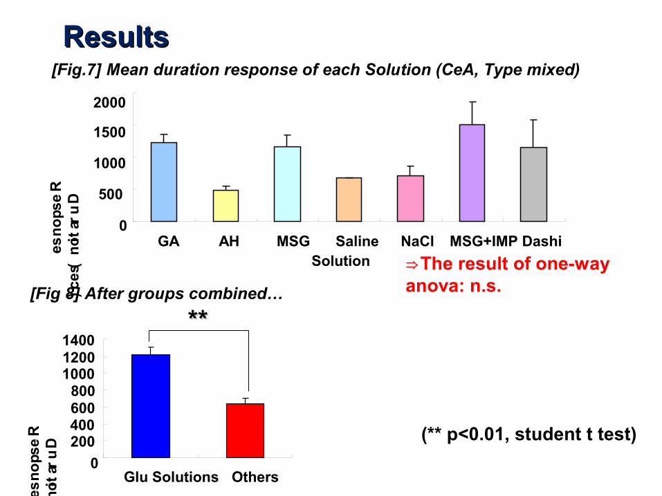
[Fig.7] Mean duration response of each Solution (CeA, Type mixed)
⇒The result of one-way anova: n.s.
0
200400600800
100012001400
Glu Solutions Others
[Fig 8] After groups combined…
Resp
on
se D
uratio
n (sec)
****
(** p<0.01, student t test)
ResultsResults
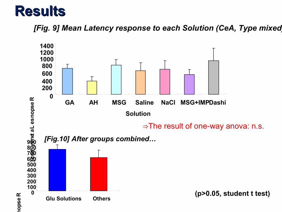
[Fig. 9] Mean Latency response to each Solution (CeA, Type mixed)
0200400600800
100012001400
GA AH MSG Saline NaCl MSG+IMPDashi
Resp
on
se La
tenc
y (sec)
Solution
⇒The result of one-way anova: n.s.
0100200300400500600700800900
Glu Solutions Others
[Fig.10] After groups combined…
Resp
on
se Laten
cy (sec)
(p>0.05, student t test)
ResultsResults
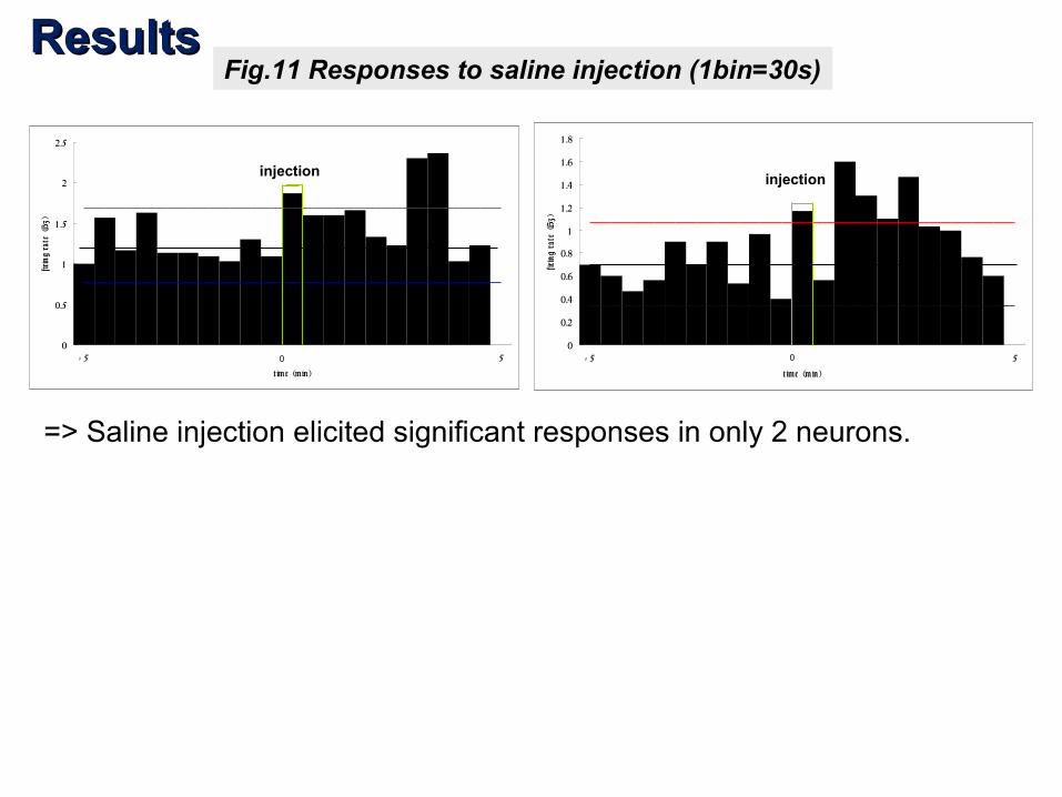
=> Saline injection elicited significant responses in only 2 neurons.
Fig.11 Responses to saline injection (1bin=30s)
0
0.5
1
1 .5
2
2.5
- 5 0 5
t ime (min )
firin
g ra
te (H
z)
0
0.2
0.4
0.6
0.8
1
1 .2
1 .4
1 .6
1 .8
- 5 0 5
t ime (m in )
firin
g ra
te (H
z)
injectioninjection
00
ResultsResults
Result SummaryResult Summary
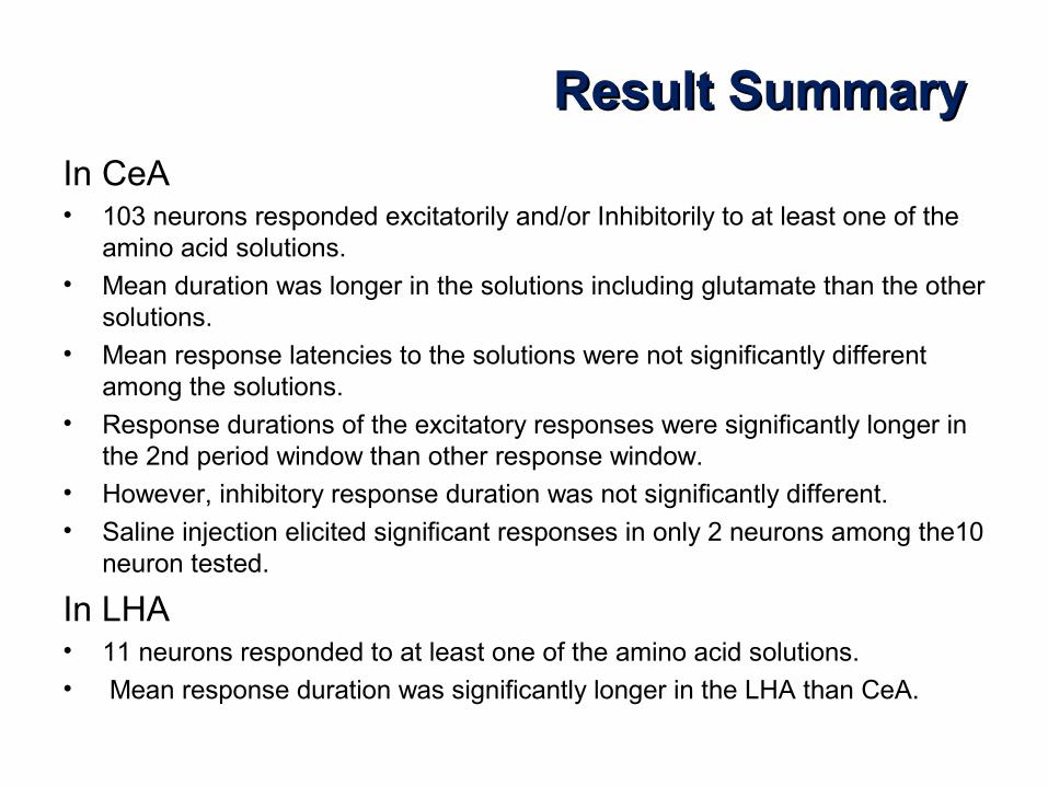
In CeA• 103 neurons responded excitatorily and/or Inhibitorily to at least one of the
amino acid solutions.• Mean duration was longer in the solutions including glutamate than the other
solutions.• Mean response latencies to the solutions were not significantly different
among the solutions.• Response durations of the excitatory responses were significantly longer in
the 2nd period window than other response window.• However, inhibitory response duration was not significantly different.• Saline injection elicited significant responses in only 2 neurons among the10
neuron tested.
In LHA• 11 neurons responded to at least one of the amino acid solutions.• Mean response duration was significantly longer in the LHA than CeA.

• The results suggest that the LHA and CeA receive interoreciptive information from the gut.
• More CeA neurons responded to the solutions, suggesting that CeA is more involved in gut-derived (interoceptive) information processing than LHA.
• Saline injection result suggest that CeA’s neuronal responses were not ascribed to stretch receptor of the vagus, but to chemical information from the gut.
DiscussionDiscussion

Thank you …Thank you …