chest & heart journal
TRANSCRIPT
Chest & HeartJournal
A Journal and
Official Organof the Chest &
Heart Associationof Bangladesh
Volume 42 Number 02 July 2018
ISSN 1562 - 5044
http://chabjournal.org
Indexed & Member: Cross Ref.
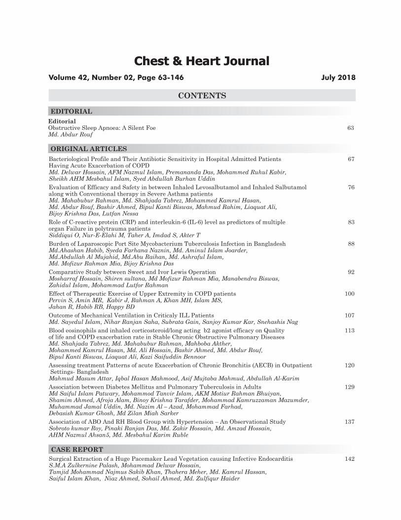
Bacteriological Profile and Their Antibiotic Sensitivity in Hospital Admitted Patients 67Having Acute Exacerbation of COPD Md. Delwar Hossain, AFM Nazmul Islam, Premananda Das, Mohammed Ruhul Kabir, Sheikh AHM Mesbahul Islam, Syed Abdullah Burhan Uddin
Evaluation of Efficacy and Safety in between Inhaled Levosalbutamol and Inhaled Salbutamol 76along with Conventional therapy in Severe Asthma patients Md. Mahabubur Rahman, Md. Shahjada Tabrez, Mohammed Kamrul Hasan, Md. Abdur Rouf, Bashir Ahmed, Bipul Kanti Biswas, Mahmud Rahim, Liaquat Ali, Bijoy Krishna Das, Lutfan Nessa
Role of C-reactive protein (CRP) and interleukin-6 (IL-6) level as predictors of multiple 83organ Failure in polytrauma patients Siddiqui O, Nur-E-Elahi M, Taher A, Imdad S, Akter T
Burden of Laparoscopic Port Site Mycobacterium Tuberculosis Infection in Bangladesh 88Md.Ahashan Habib, Syeda Farhana Naznin, Md. Aminul Islam Joarder, Md.Abdullah Al Mujahid, Md.Abu Raihan, Md. Ashraful Islam, Md. Mofizur Rahman Mia, Bijoy Krishna Das
Comparative Study between Sweet and Ivor Lewis Operation 92Mosharraf Hossain, Shiren sultana, Md Mofizur Rahman Mia, Manabendra Biswas, Zahidul Islam, Mohammad Lutfor Rahman
Effect of Therapeutic Exercise of Upper Extremity in COPD patients 100Pervin S, Amin MR, Kabir J, Rahman A, Khan MH, Islam MS, Jahan R, Habib RB, Happy BD
Outcome of Mechanical Ventilation in Criticaly ILL Patients 107Md. Sayedul Islam, Nihar Ranjan Saha, Subrata Gain, Sanjoy Kumar Kar, Snehashis Nag
Blood eosinophils and inhaled corticosteroid/long acting b2 agonist efficacy on Quality 113of life and COPD exacerbation rate in Stable Chronic Obstructive Pulmonary Diseases Md. Shahjada Tabrez, Md. Mahabubur Rahman, Mahboba Akther, Mohammed Kamrul Hasan, Md. Ali Hossain, Bashir Ahmed, Md. Abdur Rouf,Bipul Kanti Biswas, Liaquat Ali, Kazi Saifuddin Bennoor
Assessing treatment Patterns of acute Exacerbation of Chronic Bronchitis (AECB) in Outpatient 120 Settings- Bangladesh Mahmud Masum Attar, Iqbal Hasan Mahmood, Asif Mujtaba Mahmud, Abdullah Al-Karim
Association between Diabetes Mellitus and Pulmonary Tuberculosis in Adults 129Md Saiful Islam Patwary, Mohammod Tanvir Islam, AKM Motiur Rahman Bhuiyan,Shamim Ahmed, Afroja Alam, Binoy Krishna Tarafder, Mohammad Kamruzzaman Mazumder, Muhammad Jamal Uddin, Md. Nazim Al – Azad, Mohammad Farhad,Debasish Kumar Ghosh, Md Zilan Miah Sarker
Association of ABO And RH Blood Group with Hypertension – An Observational Study 137Sobroto kumar Roy, Pinaki Ranjan Das, Md. Zakir Hossain, Md. Amzad Hossain, AHM Nazmul Ahsan5, Md. Mesbahul Karim Ruble
CONTENTS
Chest & Heart JournalChest & Heart Journal
Volume 42, Number 02, Page 63-146 July 2018
EDITORIAL
ORIGINAL ARTICLES
CASE REPORT
EditorialObstructive Sleep Apnoea: A Silent Foe 63Md. Abdur Rouf
Surgical Extraction of a Huge Pacemaker Lead Vegetation causing Infective Endocarditis 142S.M.A Zulkernine Palash, Mohammad Delwar Hossain, Tamjid Mohammad Najmus Sakib Khan, Thahera Meher, Md. Kamrul Hassan, Saiful Islam Khan, Niaz Ahmed, Sohail Ahmed, Md. Zulfiqur Haider
Chest & Heart Journalchabjournal.org
Publication of The Chest & Heart Association of Bangladesh
Dedicated to Scientific & Professional Development of Pulmonologist & Cardiologist
ISSN: 1562-5044
Chest & Heart Journalchabjournal.org
Publication of The Chest & Heart Association of Bangladesh
Dedicated to Scientific & Professional Development of Pulmonologist & Cardiologist
ISSN: 1562-5044
This publication is a dedication of
The Chest & Heart Association of
Bangladesh towards knowledge &
professional development of
Pulmonologist and Cardiologist
practice in Bangladesh & the whole
world. It is published biannually
and accepts original article, review
article and case reports. We try to
accommodate any content which
may help in promotion of
knowledge, quality of patient care
and research potential amongst
concerned personnel. While every
effort is always made by the
Editorial Board to avoid inaccurate
or misleading information
appearing in the Journal,
information within the individual
articles are the responsibility of its
author(s). The Chest and Heart
Journal, its Editorial Board accept
no liability whatsoever for the
consequences of any such inaccurate
and misleading information, opinion
or statement.
Professor KMHS Sirajul Haque
Professor Mirza Mohammad Hiron
Professor Shafiqul Ahsan
Professor A.K.M Mustafa Hussain
Professor Biswas Akhtar Hossain
Professor Md. Abdur Rouf
Professor Uttam Kumar Barua
Professor S.M. Mustafa Zaman
Professor Md. Atiqur Rahman
Professor Md. Shamiul Islam
Editor in Chief
on behalf of the Chest and Heart
Association of Bangladesh
Chairman
Professor KMHS Sirajul Haque
Co-Chairman
Professor Md. Shahedur Rahman Khan
Editor in Chief
Dr. Md. Sayedul Islam
Assistant Editor
Dr. S.M. Abdur Razzaque
Dr. Md. Khairul Anam
Dr. Md. Shamim Ahmed
http://chabjournal.org.
http://www.chabjournal/writer/register
ADVISORY BOARDEDITORIAL BOARD
The Editor in Chief, The Chest and Heart Journal.
Association Secretariat, Administrative Block, Institute of Diseases of the Chest & Hospital.
Mohakhali, Dhaka-1212, Phone/Fax: +88-02-55067145
E-mail: [email protected] Website: www.chestheart.org
CORRESPONDENCE
ONLINE
Asian Colour Printing
130 DIT Extension Road
Fakirerpool, Dhaka-1000, Bangladesh
Phone: 49357726, 58313186
E-mail: [email protected]
PRINTED BY:
PUBLISHED BY:
INDEX
Member: Cross Ref.
Indexed in: Cross Ref.
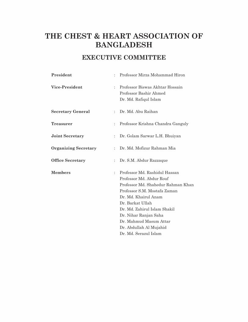
THE CHEST & HEART ASSOCIATION OF
BANGLADESH
EXECUTIVE COMMITTEE
President : Professor Mirza Mohammad Hiron
Vice-President : Professor Biswas Akhtar Hossain
Professor Bashir Ahmed
Dr. Md. Rafiqul Islam
Secretary General : Dr. Md. Abu Raihan
Treasurer : Professor Krishna Chandra Ganguly
Joint Secretary : Dr. Golam Sarwar L.H. Bhuiyan
Organizing Secretary : Dr. Md. Mofizur Rahman Mia
Office Secretary : Dr. S.M. Abdur Razzaque
Members : Professor Md. Rashidul Hassan
Professor Md. Abdur Rouf
Professor Md. Shahedur Rahman Khan
Professor S.M. Mostafa Zaman
Dr. Md. Khairul Anam
Dr. Barkat Ullah
Dr. Md. Zahirul Islam Shakil
Dr. Nihar Ranjan Saha
Dr. Mahmud Masum Attar
Dr. Abdullah Al Mujahid
Dr. Md. Serazul Islam
The Chest and Heart Journal is published twice in a year in the months of January and July. The
journal publishes original papers, reviews concerned with recent practice and case report of exceptional
merits. Papers are accepted for publication with an understanding that they are subject to editorial
revision. A covering letter signed by all authors must state that the data have not been published
elsewhere in whole or in part and all authors agree their publication in Chest and Heart Journal. All
submitted manuscripts are reviewed by the editors and rejected manuscripts will not be returned.
Ethical aspects will be considered in the assessment of the paper. Three typed copies of the article and
one soft copy in CD or Pen Drive processed all MS Word 6.0 should be submitted to the editor.
Preparation of Manuscripts
Manuscripts should be typed on one side of good quality paper, with margins of at least 25mm and
using double space throughout. Each component of the manuscript should begin on a new page in the
sequence of title page, abstract, text, references, tables, and legend for illustrations. The title page
should include the title of the paper, name of the author(s), name of the departments) to which work
should be attributed. The text should be presented in the form of Introduction, Materials and Methods,
Results, and Discussion. The text should not exceed 2500 words and a word count should be supplied.
Abstracts/Summary
Provide on a separate page an abstract of not more than 250 words. This abstract should consist of four
paragraphs, labeled Background, Methods, Results and Conclusions. They should briefly describe the
problem being addressed in the study, how the study was performed, the salient results, and what the
authors conclude from the results.
Table
Each table should be typed in on separate sheet. Table should have brief title for each, should be
numbered consecutively using Roman numbers and be cited in the consecutive order, internal
horizontal and vertical rules should not be used.
Results should be presented in logical sequence in the text, tables or illustration. Do not repeat in the
text all data in the tables or illustrations; emphasize or summarize only important observations.
Drug Names
Genetic names should generally be used. When proprietary brands are used in research, include the
brand name in parentheses in the Methods section.
Illustrations
Figure should be professionally designed symbols, lettering, and numbering should be clear and large.
The back of each figure should include the sequence number and the proper orientation (e.g. “top”).
Photographs and photomicrographs should be supplied as glossy black and white prints unmounted.
Legend for each illustration should be submitted in separate sheets. All photographs, graphs and
diagrams should be referred to as figures numbered consecutively in the text in Roman numerals.
Discussion
Emphasize the new and important aspects of the study and the conclusions that follow from them. The
detail data or other material given in the Introduction or the Results section should not be repeated.
The implications of the findings and their limitations, including implication for future research should
be included in the Discussion section. The observations should be compared and related to other
relevant studies, new hypothesis is appreciated, and however they should be clearly labeled as such.
Recommendations may be included only when appropriate.
INSTRUCTION TO AUTHORS ABOUT
UNIFORM MANUSCRIPT WRITING
References
References should be numbered consecutively in the order in which they are first mentioned in the
text. Identify references in text, tables, and legend by Roman numerals in parenthesis. Use the styles
of the example below, which are based on the formats used by the US National Library of Medicine
(NLM) in the Index Medicus.
Avoid using abstracts as references. References to paper accepted but not yet published should be
designated as “in press” or “forthcoming”; authors should obtain written permission to cite such papers
as well as verification that they have been accepted for publication. Information from manuscripts
submitted but not accepted should be cited as “unpublished observations” with written permission
from the source. Avoid using a “personal communication” unless it provides essential information not
available from a public source. For scientific articles, authors should obtain written permission and
confirmation of accuracy from the source of a personal communication.
The references must be verified by the authors(s) against the original documents.
1. Articles in Journal
a) List all six authors when six or less;
Connors JP, Roper CL, Ferguson TB. Transbronchial Catheterisation of Pulmonary Abscess. Ann
Thorac Surg 1975; 19 : 254-7.
b) When seven or more, list the first three and then add et al;
Karalus NC, Cursons RT, Leng RA, et al. Community acquired pneumonia: aetiology and
prognostic Index evaluation. Thorax 1991; 46 : 413-12.
c) No author given;
Cancer in South Africa (editorial). S Afr Med J 1994; 84-15.
d) Organization as author
The Cardiac Society of Australia and New Zealand. Clinical exercise stress training. Safety and
performance guideline. Med J Aust 1996; 164 : 282-4.
2. Books and Other Manuscripts
a) Personal author
Tierney LM, -McPhee SJ, Papakadis MA. Current Medical Diagnosis and Treatment. Lange
Medical books/Mcgrow Hill 2000.
b) Editor(s), complier(s) as author
Baum GL, Wolinsky E, editor. Text Book of Pulmonary diseases. 5th ed. New York: Little Brown
Co. 1994.
c) Organization as author and publisher
World Health Organization, Ethical Criteria for Medical Drug Promotion. Geneva: World Health
Organization; 1988.
d) Chapter in a book
Macnee W. Chronic bronchitis and emphysema. Seaton A, Seaton D, editors. Crofton and
Douglas’s Respiratory Diseases. 5th ed. UK. The Blackwell Science; 2000; p.616-95.
e) Dissertation
Kaplan SJ. Post-hospital home health care: the elderly’s access and utilization (dissertation). St.
Louis (MO). Washington Univ; 1995.
3. Other published material
a) Newspaper article
Lee G. Hospitalizations tied to ozone pollution: study estimates 50,000 admissions annually. The
Washington Post 1996, June 21; Sect. A : 3(col. 5).
b) Dictionary and similar references
Student’s medical dictionary. 26th ed. Baltimore: Williams & Wilkins; 1995. Apraxia; p.119-20.
4. Unpublished Material
a) In press
Leshner AI. Molecular mechanisms of cocaine addition. N Engl J Med In Press 1997.
5. Electronic Material
a) Journal articles in electronic format
Morse SS. Factors in the emergence of infectious diseases. Emerg Infect Dis Serial online I 1995
Jan-Mar I cited 1996 June 5 I; 1(1): 24 screens I
Available from: URL: http://www.cdc.gov/ncidod/E[D/eid.htm
Nomenclature and Abbreviation
1. Abbreviations and symbols must be standard and SI units should be used thoughtout.
2. Terms such as electrocardiogram, ultrasonogram etc. should when mentioned first, be written in
full followed by accepted abbreviations (ECG, USG etc.)
Permissions
A written statement must accompany materials taken from other sources from both author and
publisher giving permission to the Journal for reproduction. Obtain permission in writing from at
least on a author of papers still in press, unpublished data, and personal communications.
Review and Action
Manuscripts are examined by the editorial staff and are usually sent to reviewers, but we reserve the
right of final selection.
Proof
Two marked copies of the proofs may be sent to the principal author, which should be read carefully
for error. One corrected copy must be returned to the editor within the next three days. Major
alteration in the text can not be accepted.
Editorial Mail
Manuscripts and other communication for the editors should be addressed to
The Editor in Chief
Chest and Heart Journal
Association Secretariat, Administrative Block, Institute of Diseases of the Chest & Hospital.
Mohakhali, Dhaka-1212, Phone/Fax: 8851668
Chest Heart Journal
Vol. 42, No. 2, July 2018
EDITORIAL
[Chest Heart Journal 2018; 42(2) : 63-65]
DOI: http://dx.doi.org/10.33316/chab.j.v42i2.2019580
Obstructive sleep apnoea is an under recognized
and under diagnosed medical condition, with a
myriad of negative consequences on patients
health and society as a whole. Symptoms include
daytime sleepiness, loud snoring, and restless
sleep. While the “gold standard” of diagnosis is
by polysomnography, a detailed history and
focused physical examination may help uncover
previously undiagnosed cases. Undetected
obstructive sleep apnea can lead to hypertension,
heart disease, depression, and even death.
Several modalities exist for treating obstructive
sleep apnea, including continuous positive airway
pressure, oral appliances, and several surgical
procedures. However, conservative approaches,
such as weight loss and alcohol and tobacco
cessation, are also strongly encouraged in the
patient with obstructive sleep apnea.
Epidemiology
It is believed that more than 85% of patients
with clinically significant OSA have never been
diagnosed.1 This is thought to reflect the fact
that many patients with symptoms of OSA are
not aware of their heavy snoring and nocturnal
arousals. It is estimated that as many as 1 of 5
adults has at least mild symptoms of obstructive
sleep apnea, while 1 of 15 has moderate to severe
symptoms. Most population-based studies
support the existence of a twofold to threefold
greater risk of OSA in men than in women.
Patients aged 65 through 95 years are also at
significantly increased risk of developing
symptoms.
Pathophysiology
OSA is caused by repetitive bouts of upper
airway obstruction during sleep as a result of
the narrowing of respiratory passages.3 The
most common site of obstruction is the
nasopharynx.3 It is important to differentiate
OSA from the less common central sleep apnea,
which is caused by an imbalance in the brain’s
respiratory control centers during sleep. While
the pathogenesis of OSA is thought to be
multifactorial, anatomic defects are thought to
play a major role.
Certain physical characteristics that may
contribute to OSA include obesity, thickened
lateral pharyngeal walls, nasal congestion,
enlarged uvula, facial malformations,
micrognathia, macroglossia, and tonsillar
hypertrophy. Obesity contributes to airway
narrowing through fatty infiltration of the
tongue, soft palate, or other areas surrounding
the airway.
As the patient falls asleep, muscles of the
nasopharynx begin to relax and the surrounding
tissue collapses, causing compromise of the
airway. As oxygen levels in the body start to drop
and carbon dioxide levels rise, the patient is
aroused from sleep; this causes an increase in
sympathetic tone and subsequent contraction of
nasopharyngeal tissue, which allows alleviation
of the obstruction.1
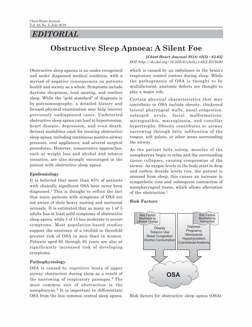
Risk Factors
Obstructive Sleep Apnoea: A Silent Foe
OSA
Risk FactorsModifiable by
Lifestyle Change
Risk FactorsModifiable byIntervention
Obesity
Tobacco Use
Nasal Congestion
Alcohol Consumption
Diabetes
Pregnancy
Menopause
Hypothyroidism
Craniofacial Anatomy
Risk factors for obstructive sleep apnea (OSA).
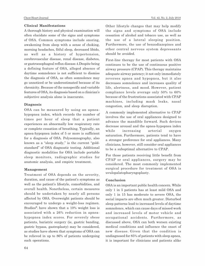
Clinical Manifestations
A thorough history and physical examination will
often elucidate some of the signs and symptoms
of OSA. Common symptoms include snoring,
awakening from sleep with a sense of choking,
morning headaches, fitful sleep, decreased libido,
as well as a history of hypertension,
cerebrovascular disease, renal disease, diabetes,
or gastroesophageal reflux disease.4.Despite being
a defining feature of OSA, alleged absence of
daytime somnolence is not sufficient to dismiss
the diagnosis of OSA, as often somnolence may
go unnoticed or be underestimated because of its
chronicity. Because of the nonspecific and variable
features of OSA, its diagnosis based on a clinician’s
subjective analysis alone is inaccurate.
Diagnosis
OSA can be measured by using an apnea-
hypopnea index, which records the number of
times per hour of sleep that a patient
experiences an abnormally low respiratory rate
or complete cessation of breathing. Typically, an
apnea-hypopnea index of 5 or more is sufficient
for a diagnosis of OSA. Polysomnography, also
known as a “sleep study,” is the current “gold-
standard” of OSA diagnostic testing. Additional
diagnostic modalities for OSA include portable
sleep monitors, radiographic studies for
anatomic analysis, and empiric treatment.
Management
Treatment of OSA depends on the severity,
duration, and cause of the patient’s symptoms as
well as the patient’s lifestyle, comorbidities, and
overall health. Nonetheless, certain measures
should be undertaken by nearly all persons
affected by OSA. Overweight patients should be
encouraged to undergo a weight-loss regimen.
Studies6. have shown that a 10% weight loss is
associated with a 26% reduction in apnea-
hypopnea index scores. For severely obese
patients, bariatric surgery (ie, gastric banding,
gastric bypass, gastroplasty) may be considered,
as studies have shown that symptoms of OSA can
be relieved in up to 86% of patients undergoing
such operations.
Other lifestyle changes that may help modify
the signs and symptoms of OSA include
cessation of alcohol and tobacco use, as well as
the use of a lateral sleeping position.
Furthermore, the use of benzodiazepines and
other central nervous system depressants
should be avoided.
First-line therapy for most patients with OSA
continues to be the use of continuous positive
airway pressure (CPAP). This therapy maintains
adequate airway patency; it not only immediately
reverses apnea and hypopnea, but it also
decreases somnolence and increases quality of
life, alertness, and mood. However, patient
compliance levels average only 50% to 60%
because of the frustrations associated with CPAP
machines, including mask leaks, nasal
congestion, and sleep disruption.
A commonly implemented alternative to CPAP
involves the use of oral appliances designed to
advance the mandible forward. Such devices
decrease arousal and the apnea-hypopnea index
while increasing arterial oxygen
saturation. Furthermore, patients tend to have
a stronger preference for oral appliances. Many
clinicians, however, still consider oral appliances
to be a suboptimal alternative to CPAP.
For those patients receiving little benefit from
CPAP or oral appliances, surgery may be
considered. The most commonly implemented
surgical procedure for treatment of OSA is
uvulopalatopharyngoplasty.
Conclusion
OSA is an important public health concern. While
only 1 in 5 patients has at least mild OSA and
only 1 in 15 has moderate to severe OSA, the
social impacts are often much greater. Disturbed
sleep patterns lead to increased levels of daytime
somnolence, which can cause days of missed work
and increased levels of motor vehicle and
occupational accidents. Furthermore, as
discussed above, OSA can both worsen existing
medical conditions and influence the onset of
new disease. Given that the condition is
undiagnosed for 85% of patients with sleep apnea,
it is important for clinicians and patients alike
64
Chest Heart Journal Vol. 42, No. 2, July 2018
to recognize and deal with the early signs and
symptoms of obstructive sleep apnea.
Prof. Dr Md. Abdur Rouf
Professor of Respiratory Medicine,
National Institute of Disease of the Chest and
Hospital, Dhaka, Bangladesh
Mobile: 01711487002
E-mail: [email protected]
References
1. Kato M., Adachi T., Koshino Y., Somers V.
K. Obstructive sleep apnea and
cardiovascular disease. Circ J. 2009; 73(8):
1363–1370.
2. Young T., Peppard P. E., Gottlieb D. J.
Epidemiology of obstructive sleep apnea: a
population health perspective. Am J Respir
Crit Care Med. 2002;165(9):1217–1239.
3. Morrison D. L., Launois S. H., Isono S., Feroah
T. R., Whitelaw W. A., Remmers J. E.
Pharyngeal narrowing and closing pressures
in patients with obstructive sleep apnea. Am
Rev Respir Dis. 1993;148(3):606–611.
4. Senn O., Brack T., Russi E. W., Bloch K. E.
A continuous positive airway pressure trial
as a novel approach to the diagnosis of the
obstructive sleep apnea syndrome.
Chest. 2006;129(1):67–75.
5. Peppard P. E., Young T., Palta M., et al.
Longitudinal study of moderate weight
change and sleep-disoriented breathing.
JAMA. 2000;284(23):3015–3021.
6. Buchwald H., Avidor Y., Braunwald E., et
al. Bariatric surgery: a systematic review
and meta-analysis. JAMA. 2004; 292(14):
1724–1737. Erratum in: JAMA. 2005;
293(14):1728.
65
Obstructive Sleep Apnoea: A Silent Foe Md. Abdur Rouf
Introduction
Chronic Obstructive Pulmonary Disease (COPD),
a common preventable and treatable disease is
a leading cause of morbidity and mortality. It
exerts substantial and increasing economic and
social burden worldwide.1 In Bangladesh the
Chest Heart Journal
Vol. 42, No. 2, July 2018
ORIGINAL ARTICLE
1. Registrar, Department of Neurology, Sylhet MAG Osmani Medical College, Sylhet, Bangladesh.
2. Associate Professor, Department of Respiratory Medicine, Sylhet MAG Osmani Medical College, Sylhet.
3. ,Registrar, Dept. of Medicine, Sylhet MAG Osmani Medical College, Sylhet, Bangladesh.
4. Assistant Professor, Department of Microbiology, Sylhet MAG Osmani Medical College, Sylhet, Bangladesh.
5. Professor, Department of Medicine, Sylhet MAG Osmani Medical College, Sylhet, Bangladesh.
6. Assistant Professor of Respiratory Medicine, Sylhet MAG Osmani Medical College, Sylhet, Bangladesh.
Correspondence to: Dr. Md. Delwar Hossain, Registrar, Department of Neurology, Sylhet MAG
Osmani Medical College, Sylhet, Bangladesh.
Submission on: 15 May, 2018 Accepted for Publication: 10 June, 2018
Available at http.//www.chabjournal.org
Bacteriological Profile and Their Antibiotic
Sensitivity in Hospital Admitted Patients Having
Acute Exacerbation of COPDMd. Delwar Hossain1, AFM Nazmul Islam2, Premananda Das3, Mohammed Ruhul Kabir4,
Sheikh AHM Mesbahul Islam5, Syed Abdullah Burhan Uddin6
Abstract:
Background: The course of COPD is punctuated by episodes of “acute exacerbations”
which is responsible increase in mortality and morbidity. Majority of exacerbations are
infectious and bacteria are responsible for 30-50% of cases. This study was designed to
know the bacteria predominantly responsible for Acute Exacerbation of COPD (AECOPD)
in hospital admitted patients and their antibiogram. This may help to formulate a cost
effective antibiotic strategy and reducing the emergence of drug resistance.
Materials and Methods: This cross sectional descriptive study was carried out in Sylhet
MAG Osmani Medical College Hospital, from 1st January 2016 to 31st December 2017. 86
patients with AECOPD were consecutively enrolled. The sputum and blood serology were
studied and causative organisms with their antibiogram were identified by standard
microbiological techniques.
Results: The mean age of the patients was 63.94 (SD±10.54) years (range, 42 to 90 years)
and 93.0% of them were male. In 38.4% of cases positive growth of organisms were detected
in sputum and predominant isolated bacteria were: P. aeruginosa(11.6%), K. pneumoniae
(9.3%), E. coli(7.0%), M. catarrhalis (3.5%), Acinetobacter spp (2.3%), Enterobacter
(1.2%), S.pneumoniae(1.2%), S. pyogenes(1.2%), S. aureus (1.2%). Gram negative bacteria
(90.9%) were more than Gram positive (9.1%) (p<0.001). Levofloxacin was the most
sensitive antibiotic (75.8%), followed by gentamicin (72.7%), ceftriaxone (69.7%), imipenem
(69.7%) and moxifloxacin (54.5%). Mycoplasma IgM and Chlamydia IgM antibodies were
positive in blood serology of 7.0% and 10.5% cases respectively .
Conclusions: P.aeruginosa and K.pneumoniae are the commonest pathogens responsible
for AECOPD in hospital admitted patients. Levofloxacin is the sensitive to majority of
the organisms. So, levofloxacin could be the first choice as empirical antibiotic in patients
with AECOPD. However, gentamicin instead of quinolones may be used in admitted
patients due to the high prevalence of tuberculosis in this region.
[Chest Heart Journal 2018; 42(2) : 67-75]
DOI: http://dx.doi.org/10.33316/chab.j.v42i2.2019581
prevalence of COPD is 5.9% in hospital admitted
patients (≥30 years).2
The course of COPD is punctuated by episodes
of acute deterioration in respiratory health,
termed ‘exacerbations’. Acute exacerbation
results deterioration of lung function, health
related quality of life and acceleration in disease
progression.3,4 and accounts for 50%–75% of the
cost of healthcare services for COPD.5 The
mortality rate of hospital admitted AECOPD is
up to 24% but it reaches to 43% in patients
needing artificial ventilation.6 Prompt antibiotic
treatment shortens the duration of exacerbations
and may prevent hospital admission and further
lung damage.7
At least 50% of COPD exacerbations are
responsible to pathogenic bacteria.8 A hospital
based study of acute exacerbation showed that
Gram negative organisms outnumbered Gram
positive organisms and Haemophilus influenzae
and Pseudomonas aeruginosa were the most
common in sputum culture.9 However, in
another study, microbial patterns corresponded
to community-acquired pathogens (S.pneumoniae,
H.influenzae, and M.catarrhalis) in 56% and
Pseudomonas and Stenotrophomonas spp. in
44% of isolates.10 In Bangladesh, Bari et al. found
that 65% of sputum of AECOPD showed positive
culture for bacteria and the common organisms
were Pseudomonas and Klebsiella.11 Infections
with Pseudomonas spp, Stenotrophomonas spp,
and Gram negative bacteria occur in more severe
exacerbations, affecting the most debilitated
patients.12
Prevalent flora and their antimicrobial
resistance pattern may vary from region to
region.13 and the sensitivity pattern also
continues to change.14 So, the choice of the
antibiotic should be based on the local bacterial
resistance pattern .Knowledge of local
bacteriological profile and antibiogram will help
to reduce the failure cases with empirical
treatment in AECOPD.
The present work is designed to find out the
causative bacteria and their antibiotic sensitivity
pattern in AECOPD patients in our perspective.
.Materials and Methods:
This cross-sectional descriptive study was
conducted during the period from 1st January
2016 to 31st December 2017 in the inpatient
department of Medicine, Sylhet MAG Osmani
Medical College Hospital (SOMCH). A total of
86 patients were recruited as study population
with the inclusion and exclusion criteria.
Inclusion Criteria: 1. All cases of acute
exacerbation of COPD. 2. Able to produce
adequate sputum containing <10 squamous
epithelial cells and >25 pus cell.
Exclusion criteria: 1. Patients having bronchial
asthma, bronchiectasis, interstitial lung disease
(ILD), Tuberculosis, Pneumonia, Lung abscess,
Malignancy or other evident diseases on chest
X-ray. 2. Treatment with antibiotic in previous
7 days. 3. Spirometric finding not suggesting
COPD in stable state.
Procedure of Data Collection:
After taking Informed written consent, following
information were recorded from the study
subjects : age, sex, BMI, smoking history, onset
of respiratory distress, duration and stage of
COPD, previous spirometry report (if available),
baseline dyspnoea (MRC dyspnoea scale),
exacerbation severity, exacerbations frequency
in the last one year period. A spirometry was
performed in all cases when patient becomes
clinically stable before discharge on a
computerized spirometer (Helios 401 PC based
Spirometer, RMS, India). The FEV1/FVC less
than 0.70 (70%), after salbutamol inhalation, was
considered COPD.
Sputum collection:
One early morning sputum was collected in a
sterile container after rinsing the mouth twice
with pure drinking water. Patients were
instructed to collect deep coughed sputum into
a sterile wide mouth container with a screw cap.
At the same time, 5 ml blood was collected for
serological tests. Samples were labeled for proper
identification and carried immediately to the
Department of Microbiology, SOMCH for
microbiological and serological analysis.
Microscopy and Culture:
Sputum smears were prepared for Gram’s stain
from the area of maximal purulence and
examined for presence of neutrophils on low
power field (x100) and organisms in high power
68
Chest Heart Journal Vol. 42, No. 2, July 2018
field (x1000). The criteria for an acceptable
sputum sample for analysis were: <10 epithelial
cells and >25 leukocytes per low power field
(according to a Murray -Washington and
Heineman criteria).
Another documented purulent portion of sputum
was used for culture. Before inoculation and
incubation, the specimen was homogenized by
agitation with an equal volume of 0.9% NaCl for
1 minute. The sputum samples were cultured
on Blood agar (5% sheep blood) for isolation of
haemolytic organisms, MacConkey’s agar for
isolation and differentiation of Gram negative
bacilli and Chocolate agar for Hemophillus and
Neisseria species.
The agar plates were kept in an incubator at
37ºC and examined after 24 and 48 hours.
Characteristic features of colonies’ morphology
on positive culture plates were observed. The
cultures were assessed semi-quantitatively and
was considered “positive” (proving bacterial
infection) when bacterial growth occupied more
than 2 quadrants (>106 CFU) of agar plate. All
isolated microorganisms were identified through
standard laboratory methods.
Antibiotic Sensitivity: Antibiotic sensitivity test
of the isolates were performed on Mueller-Hinton
agar by the disc diffusion method of Kirby-Bauer.
Serology: The qualitative immune-enzymatic
determination of IgM-class antibodies against
Chlamydia pneumoniae and Mycoplasma
pneumoniae were done by ELISA technique by
reagent from DRG International, Inc., USA. The
result was interpreted as positive or negative
according to manufacturer’s given cut off values.
Statistical Analysis: Data were processed and were
analyzed manually and by using SPSS (Statistical
Package for Social Sciences) Version 22.0.
Ethical Consideration:
• Informed consent was taken after discussing
purpose of the study in detail.
• An approval of the study protocol was
obtained from the ethical committee of
SOMC, Sylhet before the commencement of
the study.
Results
From 200 (two hundred) patients with acute
exacerbation of COPD 86 (Eighty six) patients
were included in the study. The main causes of
exclusion were: prior antibiotics ingestion, x-ray
abnormality and inability to provide adequate
sputum. Among the study group, 80 were males
(93.0%) and 6 were females (7%), with a mean
age of 63.94 (SD ± 10.54) years (range, 42 to 90
years). Most of the patients (n= 34; 39.5%) were
between 61-70 years and 5 (5.8%) patients were
above 80 years. 61.6% of them were current
smokers.
Table-I shows the distribution of the patients
according to type of exacerbation by Winnipeg
criteria. Type of exacerbation by Winnipeg
criteria was type 2 exacerbation (two major
symptoms) in 65.1% and type 1 exacerbation
(three major symptoms) in 34.9% of patients.
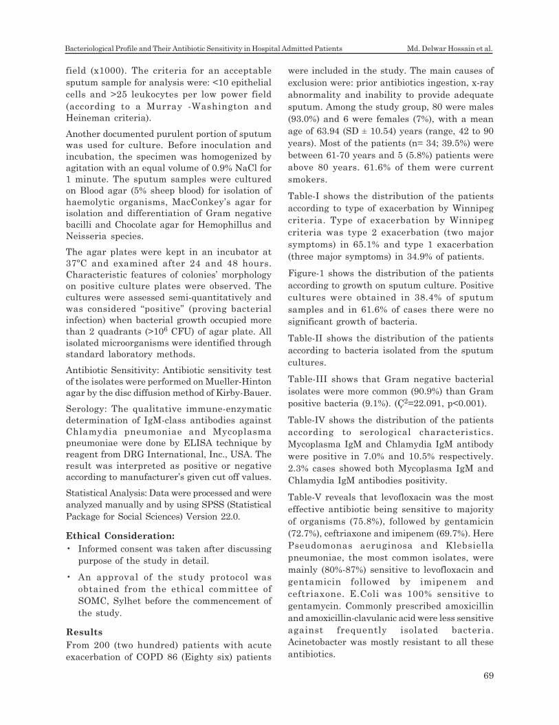
Figure-1 shows the distribution of the patients
according to growth on sputum culture. Positive
cultures were obtained in 38.4% of sputum
samples and in 61.6% of cases there were no
significant growth of bacteria.
Table-II shows the distribution of the patients
according to bacteria isolated from the sputum
cultures.
Table-III shows that Gram negative bacterial
isolates were more common (90.9%) than Gram
positive bacteria (9.1%). (Ç2=22.091, p<0.001).
Table-IV shows the distribution of the patients
according to serological characteristics.
Mycoplasma IgM and Chlamydia IgM antibody
were positive in 7.0% and 10.5% respectively.
2.3% cases showed both Mycoplasma IgM and
Chlamydia IgM antibodies positivity.
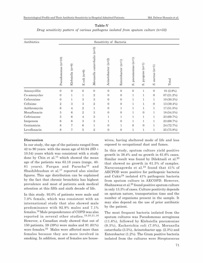
Table-V reveals that levofloxacin was the most
effective antibiotic being sensitive to majority
of organisms (75.8%), followed by gentamicin
(72.7%), ceftriaxone and imipenem (69.7%). Here
Pseudomonas aeruginosa and Klebsiella
pneumoniae, the most common isolates, were
mainly (80%-87%) sensitive to levofloxacin and
gentamicin followed by imipenem and
ceftriaxone. E.Coli was 100% sensitive to
gentamycin. Commonly prescribed amoxicillin
and amoxicillin-clavulanic acid were less sensitive
against frequently isolated bacteria.
Acinetobacter was mostly resistant to all these
antibiotics.
69
Bacteriological Profile and Their Antibiotic Sensitivity in Hospital Admitted Patients Md. Delwar Hossain et al.
Positive
33 (38.4%)
Negative
53 (61.69%)
Table-I
Distribution of the patients according to type
of exacerbation by Winnipeg criteria (n=86)
Type of exacerbation Frequency Percentage
Type 1 (Three major symptoms) 30 34.9
Type 2 (Two major symptoms) 56 65.1
Type 3 (One major with any 0 0.0
minor symptoms)
Major symptoms: Increased sputum purulence ,
Increased sputum volume, Increased dyspnea
and minor symptoms (a) Upper respiratory
infection in the past 5 days, (b) Fever without
other apparent cause, (c) Increased wheezing,
Table-II
Distribution of the patients according to bacteria isolated from the sputum cultures (n=86)
Isolated bacteria Frequency Percentage
Gram negative
Pseudomonas aeruginosa 10 11.6
Klebsiella pneumoniae 8 9.3
Escherichia coli 6 7.0
Moraxella catarrhalis 3 3.5
Acinetobacter 2 2.3
Enterobacter 1 1.2
Gram positive
Streptococcus pneumoniae 1 1.2
Streptococcus pyogenes 1 1.2
Staphylococcus aureus 1 1.2
Table-III
Distribution of the patients by status of Gram negative and
Gram positive bacterial isolates (n=86)
Bacterial isolates Frequency Percentage p-value
Gram Negative 30 90.9
Gram Positive 3 9.1 p<0.001
Total 33 100
Table-IV
Distribution of the patients according to serological characteristics (n=86)
Serological characteristics Frequency Percentage
Mycoplasma IgM Positive 6 7.0
Chlamydia IgM Positive 9 10.5
Both Mycoplasma and Chlamydia 2 2.3
IgM Positive
Serology negative 69 80.2
Fig.-1: Distribution of the patients according to
growth on sputum culture (n=86)
(d) Increased cough, (e) Respiratory rate or
Heart rate increased 20% above baseline.
70
Chest Heart Journal Vol. 42, No. 2, July 2018
Discussion
In our study, the age of the patients ranged from
42 to 90 years with the mean age of 63.94 (SD ±
10.54) years which was consistent with a study
done by Chin et al.15 which showed the mean
age of the patients was 63.18 years (range, 40-
81 years). Furqan and Paracha16 and
Shashibhushan et al.17 reported also similar
figures. This age distribution can be explained
by the fact that chronic bronchitis has highest
prevalence and most of patients seek medical
attention at this fifth and sixth decade of life.
In this study. 93.0% of patients were male and
7.0% female, which was consistent with an
international study that also showed male
predominance with 92.9% males and 7.1%
females.18 Male preponderance of COPD was also
reported in several other studies.19,20,21,16
However, a Canadian study showed that out of
150 patients, 59 (39%) were males and 91 (61%)
were females.22 Males were affected more than
females because they are more involved in
smoking. In addition, most of females are house-
wives, having sheltered mode of life and less
exposed to occupational dust and fumes.
In this study, sputum culture yield positive
growth in 38.4% and no growth in 61.6% cases.
Similar result was found by Dilektasli et al.20
that showed no growth in 61.5% of samples.
Narayanagowda et al.23 found that 41% of
AECPOD were positive for pathogenic bacteria
and Cukic24 isolated 41% pathogenic bacteria
from sputum culture in AECOPD. However,
Shahnawaz et al.25 found positive sputum culture
in only 13.3% of cases. Culture positivity depends
on sputum nature, transportation time and the
number of organisms present in the sample. It
may also depend on the use of prior antibiotic
by the patient.
The most frequent bacteria isolated from the
sputum cultures was Pseudomonas aeruginosa
(11.6%), followed by Klebsiella pneumoniae
(9.3%), Escherichia coli (7.0%), Moraxella
catarrhalis (3.5%), Acinetobacter spp. (2.3%) and
Enterobacter (1.2%). The Gram positive bacteria
isolated from the cultures were Streptococcus
Table-V
Drug sensitivity pattern of various pathogens isolated from sputum culture (n=33)
Amoxycillin 0 0 0 0 0 0 0 1 0 01 (2.9%)
Co-amoxyclav 0 1 1 2 0 0 1 1 0 07 (21.2%)
Cefuroxime 0 1 3 3 0 0 1 1 1 10 (30.3%)
Cefixime 2 3 3 2 0 0 1 1 0 13 (39.4%)
Azithromycin 6 4 2 1 0 1 1 1 1 17 (51.5%)
Moxafloxacin 5 6 2 3 0 0 1 0 1 18 (54.5%)
Ceftriaxone 5 6 4 3 1 1 1 1 1 23 (69.7%)
Imipenem 6 6 3 3 1 0 1 1 1 23 (69.7%)
Gentamicin 8 7 6 1 0 1 0 0 1 24 (72.7%)
Levofloxacin 8 7 5 2 0 0 1 1 1 25 (75.8%)
Antibiotics Sensitivity of Bacteria
Pse
ud
om
on
as
(n=
10)
Kle
bsi
ell
a (
n=
8)
E c
oli
(n
=6
)
Mora
xel
la c
ata
rrh
ali
s (n
=3)
Aci
neto
ba
cter
(n=
2)
En
tero
bact
er
(n=
1)
S. p
neu
mon
iae (
n=
1)
S.
pyogen
e (
n=
1)
S. a
ure
us
(n=
1)
Tota
l
71
Bacteriological Profile and Their Antibiotic Sensitivity in Hospital Admitted Patients Md. Delwar Hossain et al.
pneumoniae (1.2%), Streptococcus pyogenes
(1.2%), and Staphylococcus aureus (1.2%). Bari
et al.12 found almost similar picture with
predominant bacteria as Pseudomonas (25%),
Klebsiella (13.33%), Acinetobacter (6.66%) along
with Moraxella (3.33%) and Enterobacter (1.66%)
in his study. Basu et al 26 in Kolkata found
predominant organisms as Klebsiella
pneumoniae (33.33%), Pseudomonas aeruginosa
(19.05%), Escherichia coli (9.51%) and
Acinetobacter spp (9.51%). But Shahnawaz et
al.25 found Pseudomonas aeruginosa (8.35%),
Staphylococcus aureus (3.33%) and
Streptococcus pyogenes (1.66%). Similar results
were also seen in an Indian study by Chawla et
al.27 P. aeruginosa was the predominant isolate
(25.92%) amongst the hospitalized patients
followed by S.pneumoniae and Acinetobacter spp
(18.51% each), Klebsiella spp. and M.catarrhalis
(14.80% each). Borthakur and Deb,28 reported
Klebsiella pneumoniae as the most commonly
isolated bacteria followed by Staphylococcus
aureus, Pseudomonas aeruginosa and
Acinetobacter species.
In this study the prevalence of Gram negative
bacteria were more common than Gram positive
bacteria (90.9% compared to 9.1%) in the acute
exacerbations of COPD patients (p<0.001).
Madhavi et al.29 also reported that Gram
negative bacilli were isolated more than Gram
positive cocci, which was consistent with this
study. Borthakur and Deb,30 reported that the
prevalence of Gram negative isolates were 62.6%.
Aleemullah et al.31 also reported that Gram
negative organisms were isolated more (62.39%),
than Gram positive organisms (37.61%).
However, according to the study conducted by
ElFeky et al.32 Gram positive bacteria
represented 80% of isolates, while Gram negative
bacteria represent the remaining 20%.
A change in the microbial pathogens is seen
during infective exacerbations and infection with
Gram negative bacteria including Pseudomonas
spp occur more severe exacerbations, affecting
the most debilitated patients.12 The cases in our
study were hospital admitted patients of
AECOPD, who were mostly suffering from
moderate to severe exacerbations and most of
them were frequent exacerbators,hence, Gram-
negative pathogens such as Pseudomonas and
Klebsiella w ere more prevalent and can explain
the lower numbers of Gram positive bacteria
isolation, as severe COPD benefits the
enterobacteriaceae and P. aeruginosa
colonization.9
Serum IgM antibody against Mycoplasma
pneumoniae was positive in 7.0%, IgM antibody
against Chlamydia pneumoniae was positive in
10.5% and both antibodies were positive in 2.3%
subjects. Mycoplasma pneumoniae and
Chlamydia pneumoniae may be responsible for
less than 10% of exacerbations reported in
several studies.10,33
This study revealed that levofloxacin was the
most effective antibiotic being sensitive to
majority of organisms (75.8%), followed by
gentamicin (72.7%), ceftriaxone and imipenem
(69.7% each) and moxafloxacin (54.5%). Sharan
et al.34 found both levofloxacin and
aminoglycosides were effective on Gram positive
cocci and Gram negative bacilli combindly,
whereas meropenem was most effective mainly
on Gram negative organisms. Sheng-Hsiang
LIN19 in Taiwan also found that levofloxacin was
76.5 % sensitive in his study. Chawla et al.27 in
2008 found quinolones were most effective
whereas Patel et al.35 showed
piperacillin+tazobactum more effective than
quiniolones. Borthakur and Deb,30 found
quinolones were less effective. Levofloxacin was
resistant to 33.33% of patients having infection
with Gram negative organisms. Co-amoxyclav
was resistant to 34.62% and Amoxycillin was
resistant to 46.15% of the patients having Gram
positive organisms. Among macrolides,
azithromycin was the most effective drug against
Gram positive organisms having resistance of
only 26.92%. These dissimilarities may be due
the fact that prevalent flora and their antibiotic
sensitivity pattern continues to change over time
and also shows regional variation.13,14
Conclusion:
Pseudomonas aeruginosa and Klebsiella
pneumoniae are the most commonly responsible
for hospital admitted patients of AECOPD.
Levofloxacin is the most effective antibiotic being
sensitive to majority of organisms. It is followed
by gentamicin, ceftriaxone and imepenem. So,
72
Chest Heart Journal Vol. 42, No. 2, July 2018
levofloxacin could be the first choice as empirical
antibiotic in patients with AECOPD. However,
gentamicin instead of quinolones may be used
to treat acute exacerbation in hospital admitted
patients due to the high prevalence of
tuberculosis in this region.
Limitations
It was a cross-sectional, single-centered, small
sample sized study and only one sputum sample
was investigated from each subject which may
not give actual impression of the overall disease
spectrum.
Sputum study for AFB by ZN stain, culture and
sensitivity were not performed for detecting
Mycobacterium tuberculosis.
Some of the subjects may have reported
incorrectly about their disease status and
antibiotic ingestion. The duration of antibiotic
free period may have an impact on culture
positivity of sputum samples.
Recommendation:
Antibiotics should be used based on clinical
judgment of individual patient as In hospital
inpatient department, more than 50% of the
sputum did not yield any pathogenic bacteria.
Although levofloxacin was the most sensitive
antibiotic, fluroquinolones (eg. levofloxacin,
moxifloxacin) can mask the diagnosis of
tuberculosis and moxifloxacin should be reserved
for treatment of multi-drug resistant
tuberculosis in this region.
More studies like this are required at regular
interval to formulate an antibiotic policy in acute
exacerbation of COPD which would help in
preventing mortality and morbidity of COPD.
References:
1. Global Strategy for the Diagnosis,
Management and Prevention of COPD,
Global Initiative for Chronic Obstructive
Lung Disease (GOLD) 2016. [Cited 2016 Aug
30]. Available from: http://goldcopd.org
2. Zaman MM, Nargis N, Perucic AM, Rahman
K, editors. Impact of Tobacco-related
illnesses in Bangladesh. Table 9, Prevalence
of eight tobacco-related diseases among
patients aged 30 and above of four medical
college hospitals: SEARO, WHO, New Delhi;
2007.p.30.
3. Cote CG, Dordelly LJ, Celli BR. Impact of
COPD exacerbations on patient-centered
outcomes. Chest.2007; 131(3): 696-704.
4. Tanabe N, Muro S, Hirai T, Oguma T,
Terada K, Marumo S, et al. Impact of
Exacerbations on Emphysema Progression
in Chronic Obstructive Pulmonary Disease.
Am J RespirCrit Care Med. 2011; 183:
1653–9.
5. Celli BR, MacNee W, Agusti A, Anzueto A,
Berg B, Buist A S et al. ATS/ERS Task
Force. Standards for the diagnosis and
treatment of patients with COPD: A
summary of the ATS/ERS position paper.
Eur Respir J. 2004; 23:932–46.
6. Johnston A K, Mannino D M. Epidemiology
of COPD Exacerbations. September, 2008,
Available at: http://works.bepress.com/
david_mannino/216/
7. Frew AJ, Holgate ST. Respiratory disease.
In: Kumar P, Clark M, editors. Clinical
Medicine. Philadelphia: Elsevier Saunders;
2012. p.812-8.
8. Sethi S, Murphy TF. Bacterial infection in
chronic obstructive pulmonary disease in
2000: a state-of-the-art review. Clinical
Microbiology reviews.2001;14(2):336–63.
9. Boixeda R , Rabella N, Sauca G, Delgado
M, Martínez-Costa X, Mauri M et al.
Microbiological study of patients
hospitalized for acute exacerbation of
chronic obstructive pulmonary disease (AE-
COPD) and the usefulness of analytical and
clinical parameters in its identification
(VIRAE study). International Journal of
COPD.2012;7: 327-35.
10. Soler N, Torres A, Ewig S, Gonzalez J,Celis
R, El-Ebiary M et al. Bronchial Microbial
Patterns in Severe COPD Exacerbations.
Am J Respir Crit Care Med. 1998;157:1498–
505.
11. Bari MR, Hiron MM, Zaman SM, Rahman
MM, Ganguly KC. Microbes Responsible For
Acute Exacerbation of COPD. Mymensingh
Med J. 2010; 19(4): 576-85.
73
Bacteriological Profile and Their Antibiotic Sensitivity in Hospital Admitted Patients Md. Delwar Hossain et al.
12. Sapey E, Stockley RA.COPD exacerbations.
2: aetiology.Thorax.2006; 61(3):250-8.
13. Lakshmi V. Need for national/regional
guidelines and policies in India to combat
antibiotic resistance. Indian J Med
Microbiol. 2008; 26:105-7.
14. Brunton S, Carmichael B, Colgan R, Feeney
A, Fendrick A, Quintiliani R, et al. Acute
exacerbation of chronic bronchitis: a
primary care consensus guideline. Am J
Manag Care. 2004; 10: 689-96.
15. Chin CL, Manzel LJ, Lehman EE, Humlicek
AL, Shi L, Starner TD, et al. Haemophilus
influenzae from Patients with Chronic
Obstructive Pulmonary Disease
Exacerbation Induce More Inflammation
than Colonizers. Am J Respir Critical Care
Med 2005; 172: 85-91.
16. Furqan S, Paracha SAU. Frequency of
Streptococcus pneumonia and Haemophilus
influenza in acute exacerbation of chronic
obstructive airway disease and their
sensitivity to levofloxacin.JPMA.
2014;64:399-402.
17. Shashibhushan BL, Nagaraja C, Arun BJ,
Nagaraj N. Bacteriological profile and
antibiotic sensitivity pattern in sputum
culture of chronic obstructive pulmonary
disease patients. Int J Adv Med.
2016;3(3):671.
18. Reechaipichitkul W. Precipitating causes
and outcomes of chronic obstructive
pulmonary disease exacerbation at a
tertiary care center in Northeast Thailand.
Asian Biomed. 2014;8(2):229-36.
19. Lin SH, Kuo PH, Hsueh PR, Yang PC, Kuo
SH. Sputum bacteriology in hospitalized
patients with acute exacerbation of chronic
obstructive pulmonary disease in Taiwan
with an emphasis on Klebsiella pneumoniae
and Pseudomonas aeruginosa. Respirology.
2007;12:81-7.
20. Dilektasli AG, Cetinoglu ED, Ozturk NAA,
Coskun F, Ozkaya G, Ursavas A, et al.
Bacterial etiology in acute hospitalized
chronic obstructive pulmonary disease
exacerbations. Eur Res J 2016; 2(2): 99-106.
21. Alamoudi OS. Bacterial infection and risk
factors in outpatients with acute
exacerbation of chronic obstructive
pulmonary disease: A 2-year prospective
study. Respirology. 2007;12:283–7.
22. Goddard RD, Mcneil SA, Slayter KL, Mclvor
RA. Antimicrobials in acute exacerbations
of chronic obstructive pulmonary disease-
An analysis of the time to next exacerbation
before and after the implementation of
standing orders. Can J Infect Dis.
2003;14:254-9.
23. Narayanagowda DS, Golia S, Jaiswal J,
Manasa SS. A bacteriological study of acute
exacerbation of chronic obstructive
pulmonary disease over a period of one year.
Int J Res Med Sci. 2015;3(11):3141-6.
24. Cukic V. The Most Common Detected
Bacteria in Sputum of Patients with the
Acute Exacerbation of COPD.Mater
Sociomed.2013; 25(4): 226-9.
25. Shahnawaz A, Saleem SM, Bhat MA, Bhat
G, Dhobi GN. Bacteriological profile in acute
exacerbation of chronic obstructive
pulmonary disease (COPD).JK Practitioner.
2003;10:185-7.
26. Basu S, Mukherjee S, Samanta A.
Epidemiological study of bacterial
microbiology in acute exacerbation of
chronic obstructive pulmonary disease
patients of Kolkata, India. Asian journal of
pharmaceutical and Clinical Research.2013;
6:112-16.
27. Chawla K, Mukhopadhay C, Majumdar M,
Bairy I. Bacteriological profile and their
antibiogram from cases of acute
exacerbations of chronic obstructive
pulmonary disease: A hospital based study.
Journal of Clinical and Diagnostic Research.
2008;2:612-6
28. Borthakur AK, Deb C. Antibacterial
Evaluation of Common Bacteriological
Profile (Aerobic) in Acute Exacerbation of
Chronic Obstructive Pulmonary Disease
(AECOPD) in Tertiary Care Hospital
(Silchar Medical College & Hospital).
International Journal of Science and
Research (IJSR). 2017; 6(3): 648-52.
74
Chest Heart Journal Vol. 42, No. 2, July 2018
29. Madhavi S, Ramarao MV, Janardhanrao R.
Bacterial etiology of acute exacerbations of
chronic obstructive pulmonary disease.
Journal of Microbiology and Biotechnology
Research. 2012; 2: 440-4.
30. Rakesh G, Kasturi T, Yuvarajan S. Bacterial
agents causing acute exacerbations in
Chronic Obstructive Pulmonary Disease
(COPD) patients, their antibiograms to
Extended Spectrum Beta-Lactamases
(ESBL) production in a tertiary care
hospital, India. Int J Curr Microbiol App
Sci. 2013;2(11):273-82.
31. Aleemullah MF, Krishnamurthy V, Harish
M, Akeel CA. Bacteriological Profile of
Patients with AECOPD- Hospital Based
Study. Int J Curr Microbiol App Sci. 2016;
5(4): 84-90.
32. ElFeky DF, Elmandory HM, Galal M, Hakim
MA. 2016. Sputum Bacteriology in Patients
with Acute Exacerbation of Chronic
Obstructive Pulmonary Disease. Int J Curr
Microbiol App Sci. 2016; 5(1): 289–305.
33. Miyashita N, Niki Y, Nakajima M et al.
Chlamydia pneumonia infections in patients
with diffuse panbronchiolitis and COPD.
Chest.1998; 157: 1498–505.
34. Sharan H. Aerobic Bacteriological Study of
Acute Exacerbations of Chronic Obstructive
Pulmonary Disease. J Clin Diag Res. 2015;
9(8): DC10–2.
35. Patel AK, Luhadia AS, Luhadia SK.
Sputum Bacteriology and Antibiotic
Sensitivity Pattern of Patients Having Acute
Exacerbation of COPD in India – A
Preliminary Study. J Pulm Respir Med.
2015;5:238.
75
Bacteriological Profile and Their Antibiotic Sensitivity in Hospital Admitted Patients Md. Delwar Hossain et al.
Chest Heart Journal
Vol. 42, No. 2, July 2018
ORIGINAL ARTICLE
1. Medical Officer, National Institute of Diseases of the Chest & Hospital (NIDCH), Mohakhali, Dhaka,
Bangladesh.
2. Medical Officer, NIDCH, Mohakhali, Dhaka, Bangladesh.
3. MD (Chest Diseases),Thesis part student, NIDCH, Mohakhali, Dhaka, Bangladesh.
4. Professor of Respiratory Medicine, NIDCH, Mohakhali, Dhaka, Bangladesh.
5. Associate Professor of Respiratory Medicine, NIDCH, Mohakhali, Dhaka, Bangladesh.
6. Professor of Biochemistry and Cell Biology, BUHS, Mirpur-1, Dhaka, Bangladesh.
7. Assistant Professor, Respiratory Medicine,AMUMC, Noakhali, Bangladesh.
8. Associate Professor, Pediatrics, AMUMC, Noakhali, Bangladesh.
Address of correspondence: Dr. Md. Mahabubur Rahman , Medical Officer, National Institute of Diseases of
the Chest & Hospital (NIDCH), Mohakhali, Dhaka, Bangladesh. Cell: 01732661412, e-mail: k57drmabd @gmail.com
Correspondence to:
Submission on: 18 May, 2018 Accepted for Publication: 15 June, 2018
Available at http.//www.chabjournal.org
Evaluation of Efficacy and Safety in between
Inhaled Levosalbutamol and Inhaled Salbutamol
along with Conventional therapy in Severe
Asthma patientsMd. Mahabubur Rahman1, Md. Shahjada Tabrez2, Mohammed Kamrul Hasan3,
Md. Abdur Rouf4, Bashir Ahmed4 , Bipul Kanti Biswas5, Mahmud Rahim5, Liaquat Ali6,
Bijoy Krishna Das7, Lutfan Nessa8
Abstract:
Background: Levosalbutamol causes more bronchodilatation with less side effects as
compared to salbutamol in asthma patients during acute relief of asthma symptoms,
symptoms relieve during maintenance treatment of asthma and protection against exercise
-induced asthma. Aims: To explore the efficacy and safety of Inhaled Levosalbutamol
(group A) 300 µg/day (50µg 2 puffs thrice daily) compared with Inhaled Salbutamol
(group B) 600µg/day (100µg 2 puffs thrice daily) along with Conventional therapy in
case of treatment of severe asthma patients. Methods: This interventional study was carried
out in the Department of Respiratory Medicine in NIDCH, Mohakhali, Dhaka, during
November, 2016 to October, 2017. Severe (FEV1=<50 to 30% predicted) Asthma patients
with agee”12years, both sexes, non smoker, who are not known case of COPD,
Bronchiectasis, GERD were enrolled in this study. A total no. of 85 patients were included
in this study. Among them 43 patients were treated with Levosalbutamol inhaler and 42
patients were treated with Salbutamol inhaler along with conventional therapy for severe
Asthma. Results: In this study, in case of severe asthma, in group A (Levosalbutamol
inhaler 300µg/day), FEV1 was increased from 38.84 ± 5.52 to 49.53 ± 7.63 (p<.001) and
in group B (Salbutamol inhaler 600µg/day), FEV1 was increased from 37.22 ± 5.13 to
43.71 ± 6.79 (p<.001), which were highly significant but group A showed significant
improvement than group B (p<0.05). In group A, FVC was increased from 52.61 ± 6.96 to
63.93 ± 8.33 (p<.001) and In group B, FVC was increased from 49.24 ± 6.52 to 57.28 ±7.87
(p<.001) which were highly significant but group A showed significant improvement
than group B (p<0.05). In group A, Heart rate was increased from 78.56 ± 10.56 to 86.42
± 9.21 (p<.001) and in group B, Heart rate was increased from 77.06 ± 9.46 to 93.71 ±8.18
(p<.001) which were highly significant but group B(Salbutamol) showed more tachycardia
Introduction:
Asthma is a common and potentially serious
chronic disease that imposes a substantial burden
on patients, their families and the community.
It causes respiratory symptoms, limitation of
activity, and exacerbation that require urgent
health care and may be fatal if not addressed
properly. Asthma causes symptoms such as
wheezing, shortness of breath, chest tightness
and cough that vary over time in occurence,
frequency and intensity. These symptoms are
associated with variable expiratory airflow,i.e.
difficulty in breathing air out of the lungs due
to bronchoconstriction, airway wall thickening,
and increased mucus.1 β2-Agonists drugs are the
most commonly used bronchodilators for the
treatment of asthma to relieve bronchospasm.2,3
Bronchodilation may be the result of β2-receptor
stimulation as induced by a β2-receptor agonist.
Salbutamol is the most widely used short-acting
²2-agonist in the symptomatic relief of asthma.4,5
Racemic Salbutamol has been the mainstay of
treatment for bronchial smooth muscle
contraction since 1982. Salbutamol are racemic
drugs containing both ‘R’ (Levo) and ‘S’ (Dextro)
optical isomers. Only R-isomer fits into three-
dimensional conformation of β2-adrenoceptor
Proteins.4-6 So (R)- and RS- salbutamol have a
2:1 potency ratio for improvement in FEV1 in
asthmatic patients and shows that (S)-
salbutamol is clinically inactive or little active.
Because the RS - salbutamol mixture contains
only 50% (R)-salbutamol, it is clear that the
clinical effect of salbutamol resides with the (R)-
enantiomer. 7,8 Consequently, “Levosalbutamol”
was approved by FDA (Food and Drug
Administration) in 1999 as a purified single
isomer for clinical use in asthma patients.5
Moreover, (S)-salbutamol appears to be
preferentially retained in the lungs in comparison
with (R)–salbutamol.4 So, that this slower
metabolism increases the proportion of (S)–
salbutamol than levosalbutamol in vivo and
exposes the patient to relatively more potential
adverse effects of (S)–salbutamol than
levosalbutamol like hypokalemia, tremor,
tachycardia.6,8,9,10,11,12 So, the efficacy and safety
of Levosalbutamol Inhaler is better than
Salbutamol Inhaler in asthma patients.This study
will be undertaken to test this hypothesis.
Materials & Methods:
This was a randomized Clinical Trial carried
out in inpatient department of National Institute
of Diseases of the Chest and Hospital (NIDCH),
Mohakhali, Dhaka during the period from
November, 2016 to October, 2017 for one year.
All patients over 12 years of both sexes suffering
from asthma were taken as study population
and those who fulfill the inclusion and exclusion
criteria were recruited as study sample.
A total number of 96 patients were taken as
study sample that means severe (FEV1=<50 to
30% predicted) asthma. A semi-structured
questionnaire was followed by face to face
interview on the basis of objective of study.
Eligible patients were allocated randomly into
than group A( Levosalbutamol) (p<0.05). In group A, Tremor was increased from 4.20 ±
1.56 to 5.73 ± 2.07 (p<.001) and in group B, Tremor was increased from 3.80 ± 1.48 to 7.65
± 2.63 (p<.001) which were highly significant but group B(Salbutamol) showed more tremor
than group A( Levosalbutamol) (p<0.05). In group A, Serum potassium level was decreased
from 4.02 ± .42 to 3.79 ± .36 (p<.001) and in group B, Serum potassium level was decreased
from 4.14 ± .51 to 3.38 ± .56 (p<.001) which were highly significant but group B(Salbutamol)
showed more hypokalemia than group A (Levosalbutamol) (p<0.05). Conclusion: The
present study concluded that Levosalbutamol inhaler appears to be more efficacious
than Salbutamol inhaler in terms of improvement in lung functions (FEV1 and FVC)
while adverse events like tachycardia, tremor and hypokalemia are seen less with
Levosalbutamol inhaler than Salbutamol inhaler in case of severe asthma patients.
Keywords: Levosalbutamol, Salbutamol, FEV1, FVC , Heart rate, Tremor, Serum
potassium(S. K+).
[Chest Heart Journal 2018; 42(2) : 76-82]
DOI: http://dx.doi.org/10.33316/chab.j.v42i2.2019582
77
Evaluation of Efficacy and Safety in between Inhaled Levosalbutamol Md. Mahabubur Rahman et al.
two groups. Of them 48 patients were in group
A and 48 patients were in group B. Group A
was treated with Levosalbutamol inhaler (300µg/
day) 50 µg 2 puffs thrice daily and group B was
treated with Salbutamol inhaler (600µg/day) 100
µg 2 puffs thrice daily for 4 weeks along with
other with conventional therapy for asthma.
Each subject was evaluated with history and
symptoms regarding the presentation. Patients
age, occupation, working environment, smoking
history, past medical history, current
medications were asked. Patients were asked
about the dyspnoea, wheezing, chest tightness,
cough, sputum production, daytime symptoms,
night time symptoms, triggering factor, activity
level, associated diseases. They were examined
and spirometry, in addition, to the other
necessary baseline investigation (including CBC
with ESR, Serum electrolytes, Chest X-ray P/A
view, RBS, ECG, Sputum for AFB, Sputum for
eosinophil count etc.) were done.
Baseline Lung function tests (FEV1, FVC), ECG
(Heart rate), Tremor assessment, Serum
potassium level ( S. K+) were obtained before
the day of discharge after cessation of the
following respiratory medications: oxygen
therapy, nebulization and other injectable
medications.These parameters were again done
during follow up after 4 weeks. Tremor
assessment was observed by TRG (Tremor
research group) Essential Tremor Rating
Assessment Scale (TETRAS) which was the
performance measures of head, upper limb and
lower limb tremor.
In group A ,1 patient did not come to follow up,
2 patient need Levosalbutamol inhaler more than
thrice daily and 2 patient need less than thrice
daily. In group B, 2 patient did not come to
follow up, 2 patient need Salbutamol inhaler
more than thrice daily and 2 patient need less
than thrice daily.
Finally total 85 patients were included(43
patients in group A and 42 patients in group B).
All the informations were properly documented
in the prescribed forms. All interviewed
questionnaire were checked for completeness,
accuracy and consistency to exclude missing or
inconsistent data. Data were checked, cleaned
and edited properly. Quantitative data were
expressed as mean and standard deviation and
comparison done by paired and unpaired t-test.
Qualitative data were expressed as frequency
and percentage and comparison was carried by
Chi-square (Ç2) test. 95% confidence limit was
taken. A probability value (p) of less than 0.05
was considered to indicate statistical significance.
All patients/legal guardians were briefed about
the study. Informed and written consent were
taken from all study population.
Results:
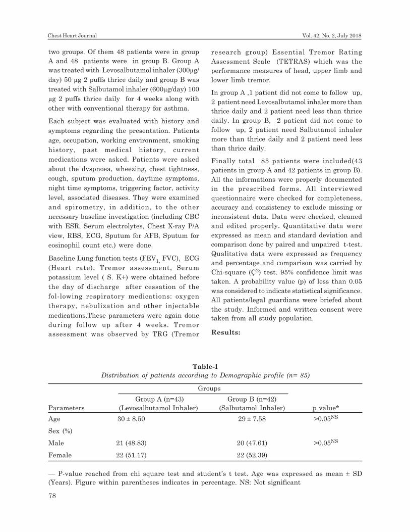
Table-I
Distribution of patients according to Demographic profile (n= 85)
Groups
Group A (n=43) Group B (n=42)
Parameters (Levosalbutamol Inhaler) (Salbutamol Inhaler) p value*
Age 30 ± 8.50 29 ± 7.58 >0.05NS
Sex (%)
Male 21 (48.83) 20 (47.61) >0.05NS
Female 22 (51.17) 22 (52.39)
— P-value reached from chi square test and student’s t test. Age was expressed as mean ± SD
(Years). Figure within parentheses indicates in percentage. NS: Not significant
78
Chest Heart Journal Vol. 42, No. 2, July 2018
Table-II
Mean ± SD of FEV1 by groups of severe asthma (n-85)
Groups
Group A (n=43) Group B (n=42)
(Levosalbutamol Inhaler) (Salbutamol Inhaler) p value*
Baseline 38.84 ± 5.52 37.22 ± 5.13 .333 NS
After 4 weeks 49.53 ± 7.63 43.71 ± 6.79 .013 S
(p<.001 S ) (p<.001 S )
NS: Not Significant, S: Significant; p value reached from both paired and unpaired t-test.
Table-III
Mean ± SD of FVC by groups of severe asthma (n-85)
Groups
Group A (n=43) Group B (n=42)
(Levosalbutamol Inhaler) (Salbutamol Inhaler) p value*
Baseline 52.61 ± 6.96 49.24 ± 6.52 .172 NS
After 4 weeks 63.93 ± 8.33 57.28 ±7.87 .012 S
(p<.001 S ) (p<.001 S )
NS: Not Significant, S: Significant; p value reached from both paired and unpaired t-test.
Table-IV
Mean ± SDof Heart rate by groups of severe asthma (n-85)
Groups
Group A (n=43) Group B (n=42)
(Levosalbutamol Inhaler) (Salbutamol Inhaler) p value*
Baseline 78.56 ± 10.56 77.06 ± 9.46 .633 NS
After 4 weeks 86.42 ± 9.21 93.71 ±8.18 .010 S
(p<.001 S) (p<.001 S)
NS: Not Significant, S: Significant; p value reached from both paired and unpaired t-test.
Table-V
Mean ± SD of Tremor by groups of severe asthma (n-85)
Groups
Group A (n=43) Group B (n=42)
(Levosalbutamol Inhaler) (Salbutamol Inhaler) p value*
Baseline 4.20 ± 1.56 3.80 ± 1.48 .411 NS
After 4 weeks 5.73 ± 2.07 7.65 ± 2.63 .013 S
(p<.001 S ) (p<.001 S )
NS: Not Significant,S: Significant; p value reached from both paired and unpaired t-test.
79
Evaluation of Efficacy and Safety in between Inhaled Levosalbutamol Md. Mahabubur Rahman et al.
Discussion:
This prospective interventional study was
carried out with an aim to explore the efficacy
and safety of Inhaled Levosalbutamol compared
with Inhaled Salbutamol along with Conventional
therapy in case of treatment of severe asthma
patients. The patients of either group were
evaluated at base line and after 4 weeks. Lung
function tests (FEV1, FVC), Heart rate, Tremor
assessment, Serum potassium level ( S. K+) level
were done at base line and during follow up after
4 weeks. Finally 43 patients in group A and 42
patients in group B were included.
In this study among 85 cases in both
Levosalbutamol and salbutamol inhaler groups
majority were at or below 35 years of age. Similar
study was reported by Chen et al., 2003; Schatz
et al., 2006 and mentioned that younger age
group are the most prevalent in asthma attack.13,14 The distribution of the study population
according to sex was shown in this study.
Females (51.76%) with asthma were a bit
predominant than male (48.23%). Similar result
was reported by Schatz et al., 2006 and added
that female is more commonly affected by
asthma than male. 14
In this study, as shown in table II, base line FEV1
was not statistically significant in both groups
(p>0.05). With therapy, after 4 weeks FEV1 were
increased to 49.53 ± 7.63 (p<0.05) and 43.71 ±
6.79 (p<0.05) in Levosalbutamol and salbutamol
inhaler group respectively.This differences
between base line and after 4 weeks were
statistically significant. So, both the drugs were
effective in improving FEV1 in asthma patient.
Again after 4 weeks it was found that
Levosalbutamol Inhaler causes more FEV1
improvement than salbutamol Inhaler because
the mean differences of improvement between
two groups were statistically significant (p<0.05).
Nowak et al., 2006; Milgrom et al., 2001 found
similar results by some other studies. 9,11 But
Rathore K et al., 2012 showed in a study that
significant improvement of FEV1 occurred in
case of Levosalbutamol Inhaler but improvement
of FEV1 in case of salbutamol Inhaler was not
statistically significant. 8
As shown in table III, Base line FVC was not
statistically significant in both groups
(p>0.05).With therapy,after 4 weeks FVC were
increased to 63.93 ± 8.33 (p<0.05) and 57.28 ±7.87
(p<0.05) in Levosalbutamol and salbutamol
inhaler group respectively. This differences
between base line and after 4 weeks were
statistically significant. So, both the drugs were
effective in improving FVC in asthma patient.
Again after 4 weeks it was found that
Levosalbutamol Inhaler causes more FVC
improvement than salbutamol Inhaler because
the mean differences of improvement between
two groups were statistically significant (p<0.05).
Rathore K et al., 2012 found similar results by
some other studies. 8
As shown in table IV, Base line Heart rate was
not statistically significant in both groups
(p>0.05). With therapy, after 4 weeks Heart rate
were increased to 86.42 ± 9.21 (p<0.05) and 93.71
±8.18 (p<0.05) in Levosalbutamol and salbutamol
inhaler group respectively.This differences
between base line and after 4 weeks were
statistically significant. So, both the drugs were
effective in improving Heart rate in asthma
patient. Again after 4 weeks it was found that
Levosalbutamol Inhaler causes more Heart rate
improvement than salbutamol Inhaler because
mean the differences of improvement between
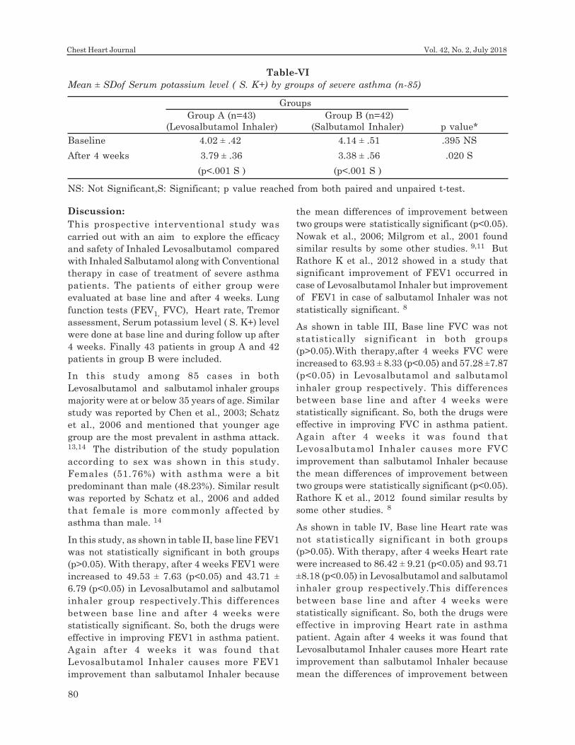
Table-VI
Mean ± SDof Serum potassium level ( S. K+) by groups of severe asthma (n-85)
Groups
Group A (n=43) Group B (n=42)
(Levosalbutamol Inhaler) (Salbutamol Inhaler) p value*
Baseline 4.02 ± .42 4.14 ± .51 .395 NS
After 4 weeks 3.79 ± .36 3.38 ± .56 .020 S
(p<.001 S ) (p<.001 S )
NS: Not Significant,S: Significant; p value reached from both paired and unpaired t-test.
80
Chest Heart Journal Vol. 42, No. 2, July 2018
two groups were statistically significant (p<0.05).
This results were consistent with Punj et al.,
2009; Lotvall et al., 2001. 6,7 But Rathore K et
al., 2012 showed similar result but heart rate
differences between two groups were not
significant.8
As shown in table V, Base line Tremor was not
statistically significant in both groups (p>0.05).
With therapy, after 4 weeks Tremor was
increased to 5.73 ± 2.07 (p<0.05) and 7.65 ± 2.63
(p<0.05) in Levosalbutamol and salbutamol
inhaler group respectively. This differences
between base line and after 4 weeks were
statistically significant. So, both the drugs were
responsible for Tremor in asthma patient. Again
after 4 weeks it was found that salbutamol
Inhaler causes more Tremor than
Levosalbutamol Inhaler because the mean
differences of increased Tremor between two
groups were statistically significant (p<0.05).
Cazzola et al., 2012 also reported occurrence of
tremor in case of using both drugs. 12
As shown in table VI, Base line Serum potassium
level ( S. K+) level was not statistically significant
in both groups (p>0.05).With therapy, after 4
weeks Serum potassium level ( S. K+) level were
were decreased to 3.79 ± .36 (p<0.05) and 3.38 ±
.56 (p<0.05) in Levosalbutamol and salbutamol
inhaler group respectively.This differences
between base line and after 4 weeks were
statistically significant. So,both the drugs were
responsible for decreasing Serum potassium
level(S. K+) level in asthma patient. Again after
4 weeks it was found that salbutamol Inhaler
decreases more Serum potassium level(S. K+)
level than Levosalbutamol Inhaler because the
mean differences of decreased Serum potassium
level (S. K+) level between two groups were
statistically significant (p<0.05). Similar results
were observed by Milgrom et al., 2001; Lotvall
et al., 2001; Punj et al., 2009; Rathore K et al.,
2012. 6,7,8,11
Conclusion:
There were significant improvements of FEV1,
FVC in Levosalbutamol inhaler while adverse
events like tachycardia, tremor and hypokalemia
are seen less with Levosalbutamol inhaler than
Salbutamol inhaler in case of severe asthma
patients. So treatment with Levosalbutamol
inhaler is a better bronchodilator in terms of
efficacy and safety than Salbutamol inhaler in
patients with severe asthma along with
Conventional therapy for symptomatic relief.
Acknowledgement:
Author Contribution: Dr. Md. Mahabubur
Rahman had full access to all the data in the
study and takes responsibility for the integrity
of the data and accuracy of the data analysis.
Dr. Md. Mahabubur Rahman contributed to
study conception and design, data collection,
analysis and interpretation of data, drafting of
the manuscript and critical revisions of the
article.
Prof. Dr. Md. Abdur Rouf, Prof. Dr. Bashir
Ahmed, Prof. Dr. Liaquat Ali, Associate Prof.
Dr. Bipul Kanti Biswas, Associate Prof. Dr.
Mahmud Rahim, Dr. Shahjada Tabrez , Dr.
Mohammed Kamrul Hasan contributed to
concept and design of the study, interpretation
of data, critical revisions and editing of the
manuscript.
Conflict of Interest: The authors of this paper
have declared that there is no conflict of interest
to any of the authors.
Funding Agency: This study has been performed
without any funding from outside else.
References:
1. GINA,2018, A Pocket Guide for asthma
management & prevention.
2. Ezeamuzie CI, Mary A..Differential effects
of salbutamol and salmetrol on human
eosinophil responses. J. Pharmacol. 1998;
Exp. Ther. 284: 25-31.
3. Carl JC, Myers TR, Kirchner HL, Kercsmar
CM. Comparison of racemic albuterol and
levalbuterol for treatment of acute asthma.
J Pediatr. 2003;143:731-6.
4. Dhand R,Goode M, Reid R, Fink JB, Fahey
PJ, Tobin MJ. Preferential pulmonary
retention of (S)-albuterol after inhalation
of racemic albuterol. Am J Respir Crit Care
Med. 1999; 160(4):1136–41.
5. Punj A, Prakash A, Bhasin A.
Levosalbutamol vs racemic salbutamol in
the treatment of acute exacerbation of
81
Evaluation of Efficacy and Safety in between Inhaled Levosalbutamol Md. Mahabubur Rahman et al.
asthma. Indian J Pediatr. 2009;76(11):
1131-1135.
6. Penn RB, Frielle T, McCullough FR, Aberg
G, Benovic J. Comparison of R-, S- and RS-
albuterol interaction with human ²1-and ²2
adrenergic receptors. Clin Rev Allergy
Immunol. 1996;14: 37-45.
7. Lotvall J, Palmqvist M, Arvidsson P,
Maloney A, Ventresca GP, Ward J. The
therapeutic ratio of R-albuterol is
comparable with that of RS-albuterol in
asthmatic patients. J. Allergy Clin.
Immunol. 2001;108:726-731.
8. Rathore K, Sharma TK, Aseri ML, Mathur
SK, Gupta RC, Vardey SK, et al.
Comparative study of pulmonary functions
after administration of albuterol and
levalbuterol in patients with moderate to
severe bronchial asthma. International
Journal of Medicine and Medical Sciences.
2012; 4(2): 39 – 44.
9. Nowak RM, Emerman CL, Schaefer K,
Disantosterfano RL, Vaickus L, Roach JM.
Levalbuterol compared with racemic
albuterol in the treatment of acute asthma:
results of a pilot study. Am J Emerg Med.
2004; 22 : 29-36.
10. Nelson HS, Bensch G, Pleskow WW, Di
Santostefano R, DeGraw S, Reasner DS.
Improved bronchodilation with levalbuterol
compared with racemic albuterol in patients
with asthma. J Allergy Clin Immunol. 1998;
102: 943-952.
11. Milgrom H, Skoner DP, Benscn G, Kim KT,
Claus R, Rudolf BA. Low-dose Levalbuterol
in children with asthma safety and efficacy
in comparison with placebo and racemic
albuterol. J Allergy Clin Immunol. 2001;108:
938-45.
12. M. Cazzola, M.G. Matera Tremor and ²2
adrenergic agents: Is it a real clinical
problem? Pulmonary Pharmacology &
Therapeutics. 2012, 25 (4): 4.
13. Chen, Y., P. Stewart, et al. Sex difference
in hospitalization due to asthma in relation
to age.¼ Journal of clinical epidemiology.
2003;56(2):180-187.
14. Schatz M, Clark S, Camargo CA Jr. Sex
differences in the presentation and course
of asthma hospitalizations. Chest. J. 2006;
129(1):50-55.
82
Chest Heart Journal Vol. 42, No. 2, July 2018
Introduction:
With the advent of time though improvement of
therapeutic concepts has decreased trauma
related fatalities1 still ‘Organ Dysfunction’
remains a frequent and severe complication
during clinical course and the sole cause of high
mortality. Survival rate of ‘Polytrauma’ patients
has improved but the frequency of development
of organ dysfunction remains static.2,3
To formulate a prudent combat strategy against
the stress response of Polytrauma it requires to
identify patients with a high risk for
Chest Heart Journal
Vol. 42, No. 2, July 2018
ORIGINAL ARTICLE
1. Omar Siddiqui, Junior Consultant, DGHS (OSD), BSMMU (Deputation), Bangladesh.
2. Md. Nur-E-Elahi, Assistant Professor, Department of Hepatobiliary, Pancreatic & Liver Transplant Surgery,
BSMMU, Bangladesh.
3. Md. Abu Taher, Associate Professor, Department of Colorectal Surgery, BSMMU, Bangladesh.
4. Sadia Imdad, Junior Consultant, Department of Surgery, Shaheed Suhrawardi Medical College Hospital,
Bangladesh.
5. Tasnuva Akter, HMO, Department of Gyn & OBS, SSMCMH, Bangladesh.
Correspondence to: Dr. Omar Siddiqui, Junior Consultant (Surgery), DGHS (OSD), Bangabandhu Sheikh Mujib
Medical University (Deputed), Bangladesh. Contact No. : +880-1705322990, E-mail: [email protected]
Submission on: 10 May, 2018 Accepted for Publication: 20 June, 2018
Available at http.//www.chabjournal.org
Role of C-reactive protein (CRP) and interleukin-
6 (IL-6) level as predictors of multiple organ
Failure in polytrauma patients.Siddiqui O1, Nur-E-Elahi M 2, Taher A3, Imdad S4, Akter T5
Abstract:
Background: The initial pathophysiological events immediately after the ‘trauma’ event
has been found to play key role for future development of multiple organ failure (MOF).
Objective: This study was aimed to assess the role of C-reactive protein (CRP) and
interleukin-6 (IL-6) as predictors of Multiple Organ Failure (MOF).
Methodology: This prospective study concluded 283 Polytrauma patients as sample
who were treated in Department of Casualty of Sir Salimullah Medical College Mitford
Hospital, Dhaka from January 2013 to January, 2018. Patients arriving within 24 hours
of trauma, age ranging from 16-65 years, surviving more than 48 hours and with Injury
Severity Score ee 18 (‘Polytrauma”) were included. We excluded all patients with injury
to CNS.
Result: A statistically significant difference was noted in average CRP levels between
patients with and without MOF on all days. IL-6 concentration also followed decreasing
trend being highest initially.
Conclusion: The most important parameters of MOF development are serum IL-6
concentration on the first day of hospitalization and the number of positive SIRS criteria
on the fourth day of hospitalization.
Key words: Polytrauma, Multiple Organ Failure (MOF), CRP, IL-6
[Chest Heart Journal 2018; 42(2) : 83-87]
DOI: http://dx.doi.org/10.33316/chab.j.v42i2.2019583
posttraumatic complications within very short
interval after the assault. Polytrauma to date
possess a great challenge for evaluation of
clinical state and prognosis as many clinical
parameters such as blood pressure, pH or heart
rate failed to assess the posttraumatic situation.4
A tight correlation been noted between cytokine
plasma level, development of MOF and mortality
rate.5 Thus immune monitoring can be our
answer to help in the selection of patients at
risk for posttraumatic complications.
The aim of this study was to assess the kinetics
of C-reactive protein (CRP) and interleukin-6
(IL-6) as predictors of MOF in Polytrauma cases.
Methodology:
This prospective study concluded 297 Polytrauma
patients as sample who were treated in
Department of Casualty of Sir Salimullah
Medical College Mitford Hospital, Dhaka from
January 2012 to January, 2016. Patients arriving
within 24 hours of trauma, age ranging from 16-
65 years, surviving more than 48 hours and with
Injury Severity Score e” 18 (‘Polytrauma’) were
included. We excluded all patients with injury
to CNS. Development of MOF in the first 24
hour and lethal outcome in the first 48 hours
made us to exclude 14 patients. The remaining
283 cases were followed up for next 10 days.
Markers and mediators of inflammation (CRP
& IL-6) were first assessed within 24 hours of
injury and then consecutively on every day till
the tenth day. Concentration of CRP was
standardized with a normal value being < 9 mg/
ml and reference range for IL-6 was 0–8 pg/mL.
The SAPS II has 17 variables, 12 of which assess
physiology, age and type of treatment and 3 of
which identify the presence of chronic diseases
such as AIDS, metastatic cancer or hematologic
malignancy. For the 12 physiologic variables, the
poorest values in the first 24 hours upon
admission (those with the highest number of
points) were taken into account.
We determined the Injury Severity Score (ISS)
from the formula (ISS = Abbreviated Injury
Score [AIS]2 + AIS2 + AIS2) where
AIS2 represents squared single highest scores
from 3 regions of the body in the first 24 hours.
We also applied the combined MOF/MODS score
that defines organ damage according to the
presence of 1 or more variables.1,6,7 Respiratory
failure was deemed present when mechanical
ventilation was necessary for at least 72 hours,
when PaO2/FiO2 < 37.3 kPa positive end-
expiratory pressure (PEEP) was over 8 cm H2O
and when there was radiographically confirmed
acute respiratory distress syndrome(ARDS) or
a respiratory rate d” 5/min or e” 49/min. Hepatic
failure was defined as bilirubinemia over 51 ìmol/
L for at least 48 hours. Renal insufficiency was
defined as serum creatinine over 177 ìmol/L for
at least 48 hours. Heart failure was defined as
cardiac index < 3.0 × min–1 × m–2, obligatory
application of inotropic therapy, heart rate d”
54/min, present ventricular tachycardia and/or
fibrillation and mean arterial pressure d” 49 mm
Hg or pH d” 7.24. Hematologic insufficiency was
defined as a platelet count < 20000 or a white
blood cell count < 1 × 10⁻⁹/L. We analyzed these
parameters in all patients included in our study,
and the worst results in the first 24 hours were
taken into account when we qualified for the
existence of organ insufficiency. The presence
of at least 1 of the mentioned criteria during
hospitalization defined existing organ damage.
MOF duration was not significant because it
represents an “all-or-nothing” event.
For statistical analysis help from a statistician
was sought. ÷2, Student’s t test and the Mann–
Whitney U test were used as deemed
appropriate for intergroup comparisons. A
logistic regression coefficient was used to
investigate the correlation between the number
of SIRS criteria per hospital days and the
development of MOF. Values of p < 0.05 or p <
0.01 were considered significant. Each variable
was assessed individually with univariate
analysis as a resulting variable for MOF.
Parameters that were found to be statistically
significant predictors with univariate analysis
were then included in a multivariate model.
Results:
The MOF patients’ average age was 32.1
(standard deviation [SD] 1.9) years. Most
patients had blunt injuries (192 patients, 67.84%),
whereas 55 (19.43.9%) had open wounds. Most
patients had triple (50.7%), double (27.3%) or
single (19.7%) organ insufficiency. Only 8.7 % of
patients had 4 or more insufficient organs. The
male: female ratio was 4.05:1, which is common
for traumatic injuries. The average ISS score
was 21.79 ± 0.6 (SD), and the average SAPS II
score was 41.12 ± 1.5(SD).
84
Chest Heart Journal Vol. 42, No. 2, July 2018
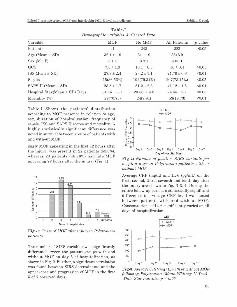
Table-I Shows the patients’ distribution
according to MOF presence in relation to age,
sex, duration of hospitalization, frequency of
sepsis, ISS and SAPS II scores and mortality. A
highly statistically significant difference was
noted in survival between groups of patients with
and without MOF.
Early MOF appearing in the first 72 hours after
the injury, was present in 22 patients (53.6%),
whereas 20 patients (48.78%) had late MOF
appearing 72 hours after the injury. (Fig. 1)
Average CRP (mg/L) and IL-6 (pg/mL) on the
first, second, third, seventh and tenth day after
the injury are shown in Fig. 3 & 4. During the
entire follow-up period, a statistically significant
difference in average CRP level was noted
between patients with and without MOF.
Concentrations of IL-6 significantly varied on all
days of hospitalization.
Table-I
Demographic variables & General Data
Variable MOF No MOF All Patients p value
Patients 41 242 283 >0.05
Age (Mean ± SD) 32.1 ± 1.9 31.1±.9 35±3.9
Sex (M : F) 5.1:1 3.9:1 4.05:1
GCS 7.5 ± 1.9 10.1 ± 0.3 10 ± 0.4 >0.05
ISS(Mean ± SD) 27.9 ± 2.4 23.2 ± 1.1 21.79 ± 0.6 <0.01
Sepsis 15(36.56%) 192(79.34%) 207(73.15%) <0.05
SAPS II (Mean ± SD) 23.8 ± 1.7 51.2 ± 2.3 41.12 ± 1.5 <0.01
Hospital Stay(Mean ± SD) Days 31.15 ± 3.1 23.56 ± 4.3 24.65 ± 2.7 <0.05
Mortality (%) 29(70.73) 24(9.91) 53(18.73) <0.01
The number of SIRS variables was significantly
different between the patient groups with and
without MOF on day 5 of hospitalization, as
shown in Fig. 2. Further, a significant correlation
was found between SIRS determinants and the
appearance and progression of MOF in the first
5 of 7 observed days.
Fig.-1: Onset of MOF after injury in Polytrauma
patients.
1
2,8
3,13
4,11
5,5
6,17,2
Onwards,1
0
2
4
6
Nu
mb
er
of P
atie
nts
8
10
12
14
2 3 4 5 6 7 Onwards
Days of hospital stay
0Day 1 Day 2 Day 3
Day of Hospital Stay
Day 4 Day 5 Day 6 Day 7
0.5
0.1
Num
ber
of ‘S
IRS
’ var
iabl
es
1.5
2
2.5
3
MOF+
MOF-
Fig-2: Number of positive SIRS variable per
hospital days in Polytrauma patients with or
without MOF.
Fig-3: Average CRP (mg/L) with or without MOF
following Polytrauma (Mann-Whitney U Test)
White Star indicates p < 0.05
MOF+
MOF-
300
250
200
150
100
50
0Day 1 Day 2 Day 3 Day 7 Day 10
CRP
85
Role of C-reactive protein (CRP) and interleukin-6 (IL-6) level as predictors Siddiqui O et al.
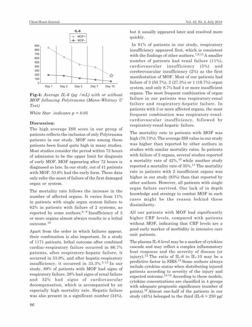
Discussion:
The high average ISS score in our group of
patients reflects the inclusion of only Polytrauma
patients in our study. MOF rate among these
patients been found quite high in many studies.
Most studies consider the period within 72 hours
of admission to be the upper limit for diagnosis
of early MOF. MOF appearing after 72 hours is
diagnosed as late. In our study, out of 41 patients
with MOF, 53.6% had the early form. These data
only refer the onset of failure of the first damaged
organ or system.
The mortality rate follows the increase in the
number of affected organs. It varies from 11%
in patients with single organ system failure to
62% in patients with failure of 2 systems, as
reported by some authors.8, 9 Insufficiency of 3
or more organs almost always results in a lethal
outcome.10
Apart from the order in which failures appear,
their combination is also important. In a study
of 1171 patients, lethal outcome after combined
cardiac-respiratory failure occurred in 66.7%
patients, after respiratory-hepatic failure, it
occurred in 53.9%, and after hepatic-respiratory
insufficiency, it occurred in 33.3%.5-13 In our
study, 89% of patients with MOF had signs of
respiratory failure, 28% had signs of renal failure
and 32% had signs of cardiovascular
decompensation, which is accompanied by an
especially high mortality rate. Hepatic failure
was also present in a significant number (34%),
but it usually appeared later and resolved more
quickly.
In 81% of patients in our study, respiratory
insufficiency appeared first, which is consistent
with the findings of other authors.7,8,11 A smaller
number of patients had renal failure (11%),
cardiovascular insufficiency (5%) and
cerebrovascular insufficiency (2%) as the first
manifestation of MOF. Most of our patients had
failure of 3 (50.7%), 2 (27.3%) or 1 (19.7%) organ
system, and only 8.7% had 4 or more insufficient
organs. The most frequent combination of organ
failure in our patients was respiratory-renal
failure and respiratory-hepatic failure. In
patients with 3 or more affected organs, the most
frequent combination was respiratory-renal-
cardiovascular insufficiency, followed by
respiratory-renal-hepatic failure.
The mortality rate in patients with MOF was
high (70.73%). The average ISS value in our study
was higher than reported by other authors in
studies with similar mortality rates. In patients
with failure of 2 organs, several studies reported
a mortality rate of 42%,10 while another study
reported a mortality rate of 35%.11 The mortality
rate in patients with 2 insufficient organs was
higher in our study (83%) than that reported by
other authors. However, all patients with single
organ failure survived. Our lack of in depth
knowledge and strategy to combat MOF in such
cases might be the reason behind these
dissimilarity.
All our patients with MOF had significantly
higher CRP levels, compared with patients
without MOF, indicating that CRP levels are a
good early marker of morbidity in intensive care
unit patients.
The plasma IL-6 level may be a marker of cytokine
cascade and may reflect a complex inflammatory
host response and the severity of disease (or
injury).12 The ratio of IL-6 to IL-10 may be a
predictive factor in SIRS.13 Some authors always
include cytokine status when distributing injured
patients according to severity of the injury and
expected outcome.13-15 According to these models,
cytokine concentrations are classified in 4 groups
with adequate prognostic significance (number of
points).16 Almost one-half of the patients in our
study (45%) belonged to the third (IL-6 > 250 pg/
Fig-4: Average IL-6 (pg /mL) with or without
MOF following Polytrauma (Mann-Whitney U
Test)
White Star indicates p < 0.05
900
800
700
600
500
400
300
200
100
0Day 1 Day 2 Day 3 Day 7 Day 10
MOF+
MOF-
IL-6
86
Chest Heart Journal Vol. 42, No. 2, July 2018
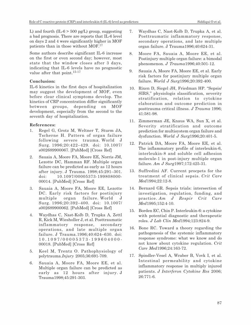
L) and fourth (IL-6 > 500 pg/L) group, suggesting
a bad prognosis. There are reports that IL-6 level
on days 2 and 4 were significantly higher in MOF
patients than in those without MOF.17
Some authors describe significant IL-6 increase
on the first or even second day; however, most
state that the window closes after 3 days,
indicating that IL-6 levels have no prognostic
value after that point.15-17
Conclusion:
IL-6 kinetics in the first days of hospitalization
may suggest the development of MOF, even
before clear clinical symptoms develop. The
kinetics of CRP concentration differ significantly
between groups, depending on MOF
development, especially from the second to the
seventh day of hospitalization.
References:
1. Regel G, Grotz M, Weltner T, Sturm JA,
Tscherne H. Pattern of organ failurefollowing severe trauma. World J
Surg. 1996;20:422–429. doi: 10.1007/
s002689900067. [PubMed] [Cross Ref]
2. Sauaia A, Moore FA, Moore EE, Norris JM,
Lezotte DC, Hamman RF. Multiple organfailure can be predicted as early as 12 hours
after injury. J Trauma. 1998;45:291–301.doi: 10.1097/00005373-199808000-
00014. [PubMed] [Cross Ref]
3. Sauaia A, Moore FA, Moore EE, LezotteDC. Early risk factors for postinjury
multiple organ failure. World JSurg. 1996;20:392–400. doi: 10.1007/
s002689900062. [PubMed] [Cross Ref]
4. Waydhas C, Nast-Kolb D, Trupka A, ZettlR, Kick M, Wiesholler J, et al. Posttraumatic
inflammatory response, secondaryoperations, and late multiple organ
failure. J Trauma.1996;40:624–630. doi:
1 0 . 1 0 9 7 / 0 0 0 0 5 3 7 3 - 1 9 9 6 0 4 0 0 0 -00018. [PubMed] [Cross Ref]
5. Keel M, Trentz O. Pathophysiology ofpolytrauma.Injury 2005;36:691-709.
6. Sauaia A, Moore FA, Moore EE, et al.
Multiple organ failure can be predicted asearly as 12 hours after injury. J
Trauma1998;45:291-303.
7. Waydhas C, Nast-Kolb D, Trupka A, et al.
Posttraumatic inflammatory response,
secondary operations, and late multiple
organ failure. J Trauma1996;40:624-31.
8. Moore FA, Sauaia A, Moore EE, et al.
Postinjury multiple organ failure: a bimodal
phenomenon. J Trauma1996;40:501-12.
9. Sauaia A, Moore FA, Moore EE, et al. Early
risk factors for postinjury multiple organ
failure. World J Surg1996;20:392-400.
10. Rixen D, Siegel JH, Friedman HP. “Sepsis/
SIRS,’’ physiologis slassification, severity
stratification, relation to cytokine
elaboration and outcome prediction in
posttrauma critical illness. J Trauma 1996;
41:581-98.
11. Zimmerman JE, Knaus WA, Sun X, et al.
Severity stratification and outcome
prediction for multisystem organ failure and
dysfunction. World J Surg1996;20:401-5.
12. Patrick DA, Moore FA, Moore EE, et al.
The inflammatory profile of interleukin-6,
interleukin-8 and soluble cell adhesion
molecule-1 in post-injury multiple organ
failure. Am J Surg1997;172:425-31.
13. Suffredini AF. Current procpets for the
treatment of clinical sepsis. Crit Care
Med1994;22:12-8.
14. Bernard GR. Sepsis trials: intersection of
investigation, regulation, funding, and
practice. Am J Respir Crit Care
Med1995;152:4-10.
15. Borden EC, Chin P. Interleukin-6: a cytokine
with potential diagnostic and therapeutic
roles. J Lab Clin Med1994;123:824-9.
16. Bone RC. Toward a theory regarding the
pathogenesis of the systemic inflammatory
response syndrome: what we know and do
not know about cytokine regulation. Crit
Care Med1996;24:163-72.
17. Spindler-Vesel A, Wraber B, Vovk I, et al.
Intestinal permeability and cytokine
inflammatory response in multiply injured
patients. J Interferon Cytokine Res 2006;
26:771-6.
87
Role of C-reactive protein (CRP) and interleukin-6 (IL-6) level as predictors Siddiqui O et al.
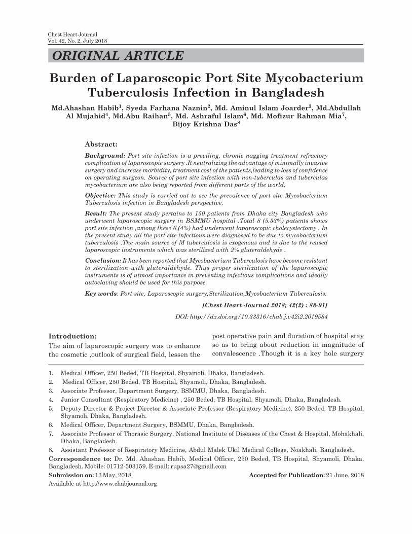
Introduction:
The aim of laparoscopic surgery was to enhance
the cosmetic ,outlook of surgical field, lessen the
Chest Heart Journal
Vol. 42, No. 2, July 2018
ORIGINAL ARTICLE
post operative pain and duration of hospital stay
so as to bring about reduction in magnitude of
convalescence .Though it is a key hole surgery
1. Medical Officer, 250 Beded, TB Hospital, Shyamoli, Dhaka, Bangladesh.
2. Medical Officer, 250 Beded, TB Hospital, Shyamoli, Dhaka, Bangladesh.
3. Associate Professor, Department Surgery, BSMMU, Dhaka, Bangladesh.
4. Junior Consultant (Respiratory Medicine) , 250 Beded, TB Hospital, Shyamoli, Dhaka, Bangladesh.
5. Deputy Director & Project Director & Associate Professor (Respiratory Medicine), 250 Beded, TB Hospital,
Shyamoli, Dhaka, Bangladesh.
6. Medical Officer, Department Surgery, BSMMU, Dhaka, Bangladesh.
7. Associate Professor of Thorasic Surgery, National Institute of Diseases of the Chest & Hospital, Mohakhali,
Dhaka, Bangladesh.
8. Assistant Professor of Respiratory Medicine, Abdul Malek Ukil Medical College, Noakhali, Bangladesh.
Correspondence to: Dr. Md. Ahashan Habib, Medical Officer, 250 Beded, TB Hospital, Shyamoli, Dhaka,
Bangladesh. Mobile: 01712-503159, E-mail: [email protected]
Submission on: 13 May, 2018 Accepted for Publication: 21 June, 2018
Available at http.//www.chabjournal.org
Burden of Laparoscopic Port Site Mycobacterium
Tuberculosis Infection in BangladeshMd.Ahashan Habib1, Syeda Farhana Naznin2, Md. Aminul Islam Joarder3, Md.Abdullah
Al Mujahid4, Md.Abu Raihan5, Md. Ashraful Islam6, Md. Mofizur Rahman Mia7,
Bijoy Krishna Das8
Abstract:
Background: Port site infection is a previling, chronic nagging treatment refractory
complication of laparoscopic surgery .It neutralizing the advantage of minimally invasive
surgery and increase morbidity, treatment cost of the patients,leading to loss of confidence
on operating surgeon. Source of port site infection with non-tuberculas and tuberculas
mycobacterium are also being reported from different parts of the world.
Objective: This study is carried out to see the prevalence of port site Mycobacterium
Tuberculosis infection in Bangladesh perspective.
Result: The present study pertains to 150 patients from Dhaka city Bangladesh who
underwent laparoscopic surgery in BSMMU hospital .Total 8 (5.33%) patients shows
port site infection ,among these 6 (4%) had underwent laparoscopic cholecystectomy . In
the present study all the port site infections were diagnosed to be due to mycobacterium
tuberculosis .The main source of M tuberculosis is exogenous and is due to the reused
laparoscopic instruments which was sterilized with 2% gluteraldehyde .
Conclusion: It has been reported that Mycobacterium Tuberculosis have become resistant
to sterilization with gluteraldehyde. Thus proper sterilization of the laparoscopic
instruments is of utmost importance in preventing infectious complications and ideally
autoclaving should be used for this purpose.
Key words: Port site, Laparoscopic surgery,Sterilization,Mycobacterium Tuberculosis.
[Chest Heart Journal 2018; 42(2) : 88-91]
DOI: http://dx.doi.org/10.33316/chab.j.v42i2.2019584
but when these small wound get coursed with
infections with a dragging and indolent course,
the entire purpose of decreasing morbidity goes
in to vain.In Bangladesh tuberculosis is one of
the major health problem, among which 1/5th are
extra pulmonary. It has been noted that port
site TB in usually due to improper sterilization
of the laparoscopic instruments ,but few case
report publish which has attributed port site TB
to an endogenous source1.Port site TB has been
reported following laparoscopic cholecystectomy2,3,
appendectomy4, urological surgery5.Review of
literature shows that the problem of port site
infection is a global one and not restricted to
developing countries only ,no doubt the incidence
in developed countries is far less in copmparison
to developing nations6.In the present study 150
cases of laparoscopic surgeries, 8 cases are being
reported with port site infection by
mycobacterium tuberculosis.
Materials and methods:
The present study includes 150 patients who
underwent laparoscopic surgeries for various
indications in a BSMMU hospital in Dhaka
city.This study was conducted for a period of 24
months march 2014 to February 2016.All those
patients who underwent laparoscopic surgery
during this period were included in the study
and those patients ,who were converted to open
procedure were excluded from the study . In all
the patients the pre-operative skin preparation
done by on table shaving method and use
providone iodine wash.The patients were
admitted in the morning of the day of surgery
and one pre-operative dose of ceftriaxone 1 gm
at the time of induction and two subsequent
postoperative dose of the same were given 12
hourly and then it convert in to oral cefixime
preparation for total 7 days. All surgeries were
done under general anesthesia. Pneumo-
peritonium was created through open method
in supra or infra umbilical transverse incision.
Through the incision 10mm primary trocar was
introduced in to peritoneal cavity.The time
duration from first incision to end of the
procedure was calculated.All the specimens of
gall bladder ,appendix,and ovarian cyst were
extracted through the umbilical port,and used
endobag in ovarian cyst and appendix
extraction.All laparoscopic instruments were
sterilized by 2% gluteraldehyde solution with a
contact time of 20 minutes .Before surgery all
the instruments were washed with normal
saline.Gluteraldehyde solution was replaced
after every 15 days.
All patients were monitored for superficial and
deep port site infections .Port site infections
were included those occurring within 30 days of
an operation7.Wounds were assed clinically on
3rd post operative day and after 7days of
operation .In case of infection had occurred ,
pus was sent for culture and sensitivity ,dressing
and cleaning of wounds were done regularly and
a course of empirical antibiotic was started till
the culture sensitivity report was received .The
infected wounds were re examined once weekly
for four weeks or more ,depending on the
response of antibiotic ,if no response was seen
,pus/discharge was sent for AFB staining. In case
of AFB positive ,pus was sent for culture in L J
media to rule out mycobacterium tuberculosis.In
case of sinuses in addition to above, excision of
the sinuses tract were done and sent for
histopathological examination.
Result:
Out of 150 laparoscopic surgeries ,142 were of
laparoscopic cholecystectomy, 2 of laparoscopic
appendectomy,and 6 of laparoscopic ovarian
cystectomy .Only 8(5.3%) of patients developed
port site infections ,of which 6 undergone
laparoscopic cholecystcetomy and 1 underwent
appendicectomy and another 1 underwent
ovarian cystectomy .Superficial infection was
seen 6 cases (75%) and deep infection seen in
2(25%) patients .All patients present with pus
discharge,erythrema ,induration with mild
tenderness but no fever .Nodule and discharging
sinus were only seen in deep infections.The
sinuses involved the muscle plane and did not
involve the peritoneum.The infections did not
respond to second or third generation
cephalosporins.
The umbilical port through which all specimens
were extracted ,shows infection in 5(62.5%) cases
thus being the most frequent.The infection
involving in epigastric port 2(25%) patients and
both lateral port in 1(12.5%) patient.The
operative findings in cases of port site infection
89
Burden of Laparoscopic Port Site Mycobacterium Tuberculosis Infection Md.Ahashan Habib et al.
in laparoscopic cholecystectomies,
appendectomies and ovarian cystectomies
,including acute cholecystitis in 2(25%),chronic
cholecystitis with thick walled gall bladder in
4(50%) patients and acute appendicitis in
1(12.5%) and ovarian cyst in 1(12.5%) patient .In
all patients with port site infection or without
port site infection the operative time varied from
45-50minutes,except ovarian cystectomies where
it was 50 -65 minutes .
Pus for culture was taken from both superficial
and deep infection wounds .Culture and
sensitivity done were negative for both gram
positive and negative bacteria .AFB staining of
the same pus was positive for acid fast bacilli.In
culture done on LJ media , mycobacterium
tuberculosis was isolated from all 8 cases of port
site infections. Histopathology of the excised
sinuses revealed typical granulomas formed of
central caseation necrosis surrounded by
epitheloid cells and lymphocytes.Langhans giant
cells were also found.All the patients with port
site infection recovered within 2-3 months of
starting of first line anti TB drugs ,but the
treatment was continued for six months.
Discussion:
On review of literature the incidence of port site
infection has been seen to be variable,it has been
reported as low as 2.3% from Israel8 as high as
9.2% in cairo9.In the present study it was
recorded as 5.3% which is similar to the one
reported by Waqar J Alam 5.7%(8)Den Hoed
PT5.3%10 ,slightly higher percentage has been
reported by Shindholimath VV 6.3%11. The CDC
classification of SSI categories these wound in
two sub types superficial and deep. The
superficial one include those involve skin and
subcutaneous tissue ,where as deep one pertain
to fascia, muscle and organ or space infection12.In
the present study the wounds predominantly
belonged to the superficial category (75%).The
percentage of deep infections extending in to
muscle plane was (25%), which was similar the
one reported by Waqar A Jan et al 20088.Overall
it has been noted that port site infections are
mostly restricted to superficial skin
infection.The causative agents of these port site
infections are mostly mycobacteria and it having
been most reported in developing countries9. It
is interesting to note that in the present study
M tuberculosis was encountered in all port site
infections.M tuberculosis has been reported to
cause port site infection by Ramesh et al 13.
Nader A Elhamid et al 2012 in their study,report
NTM in 4 cases out of 75.
The way of such type of port site tuberculosis
infection generally occurs,
1). From an exogenous source, 2). From an
endogenous source(secondary tuberculosis)
3). From a haematogenous source.
Table-1
Demography
Age (Year) Male Female
20-40 22 50
41-60 19 32
> 60 11 16
Demography shows that, young age group &
female sex predominant.
Table-2
Type of laparoscopic surgery (N=150)
Laparoscopic surgery Male Female Numbers
Cholecystectomy 51 91 142
Ovarian cystectomy 0 6 6
Appendicectomy 1 1 2
Total 52 98 150
Laparoscapic Cholecystectomy & female sex
predominant.
Table-3
Port site Mycobacterium Tuberculosis
infection rate
Port Frequency Percentage
Umbilical port 5 62.5%
Epigastric port 2 25%
Lateral port 1 12.5%
Umbilical port site infection is predominant.
Table-4
Procedure N Port site P Value
infection (%)
Laparoscopic 142 6 (4.22%) > 0.05 NS
Cholecystectomy
Ovarian Cystectomy 6 1 (16.66%) > 0.05 NS
Appendectomy 2 1 (50%) > 0.05 NS
P Value > 0.05 non significant.
90
Chest Heart Journal Vol. 42, No. 2, July 2018
The most common practice of laparoscopic
instruments “sterilization” in our country has
been to immerse instruments in 2%
glutaraldehyde for 20 min. Although sterilization
is defined as “the complete elimination of all
forms of microbial life”. It is now widely agreed
that 2% gluteraldedhde achieves high level of
disinfection and not sterilization.
This has been further reinforced by Griffiths et
al ,who have highlighted the failure of a 20 min
instruments soak in 2% glutareldehyde to
sterilize laparoscopic instruments14.Another
study, mycobacterium TB was present in one of
five scopes after 45 min exposure.In present
study the umbilical port showed infection in
maximum case (62.5%) which is in conformity
with the finding of Sasmal et al15 , it is suggested
by some authors that the port used for extraction
of specimens is the most commonly involved port
in infection because of spillage.Therefore the
cause of infection seems to be exogenous and
most probably the contaminated instruments.
Time taken for all the surgeries in the present
study was almost same hence this factors is not
regarded to be involved in causing the infection
in the present case.
References:
1. Cunnigaiper ND, Venkatraman S port site
tuberculosis :Endogenous infection?Sung
Infect(Larchmrt)2010;11:77-8.
2. Tauro LF, Satish RB ,Matris JJ,Divakar
HS.port site tuberculosis :a rare
complication following laparoscopic
cholecystectomy .Indian J Surg.2005;67:
104-5.
3. Ramesh H, Prakash K,Lekha V, Jacob
G,Venugopal A,Venugopal B,port site
tuberculosis after laparoscopy:Report of
eight case.Surg Endosc.2003;17:930-2.
4. Bhandrakar DS,Bhagwat S,Punjani R,por-
site infection with mycobacterium chelonei
following laparoscopic appendicectomy.
Indian J Gastroenterol.2001;20:247-8.
5. Rahul Gupta,Arati Mahajan , Chaman
Gupta.post per-cutaneous nephrolithotripsy
nephrostomy site tuberculosis :a report of
6 cases . Urol Ann.2012;4(1):41-4.
6. William E G Thomas.Basic surgical skills
and anastomoses .In Normans S William
etal.fditors.Baily &Love’s short practice of
surgery.24 th ed.London:Arnold 2004.
pp118-32.
7. Horan TC ,Gaynes RP,Martone WJ,et
al.CDC definitions of nosocomial surgical
site infections,1992:A modification of CDC
definitions of surgical wound infections,
Infect control Hosp Epidemiol 1992; 13 :
606-8.
8. Waqar Aj,Sabir Ali I,et al.The frequency of
port site infection following laparoscopic
cholecystectomies.JPMI 2008;22:66-70.
9. Nader Abd-Elhamid ,Khaled Kasim,
Anahtar Kelimeler.port site Non-
tuberculous Mycobacterial Infection after
Laparoscopic cholecystectomy:A case
series.TAF Prev Med Bull 3013;12:481-484.
10. Den Hoed PT , Boelhourwer RU,Veen HF,et
al.Infections and bacteriological data after
laparoscopic and open gallbladder surgery.J
Hosp Infect 1998;39:27-37.
11. Sharma AK,Sharma S,Sharma R,port site
infection In laparoscopic surgeries.Indian
Med Gazette 2013;224-29.
12. Centers of Disease Control and
prevention.The National Healthcare Safety
Network(NHSN) Manual:Patient safety
component Atlanta ,GA:Division of
Healthcare Quality Promotion.National
Center for Emerging and Zoonotic Infection
Disease Available from: URL : http://
www.cdc.gov/nhsn/acute-care-hospital/
index.html.2015.
13. Ramesh H,Prakash K,Lekha V,et al.port
site tuberculosis after laparoscopy:report
of eight cases .Surg Endosc 2003;17:930-32.
14. Griffith PA,Babb JR,Bradley CR,Fraise
A P . G l u t a r a l d e h y d e - r e s i s t a n t
Mycobacterium chelonae from endoscope
washer disinfectors.J Appl Microbial. 1997;
82:519-26.
15. Sasmal PK,Mishra TS,Rath S,et al.port site
infection in laparoscopic surgery :A review
of its management.WJCC 2015;3-18.
91
Burden of Laparoscopic Port Site Mycobacterium Tuberculosis Infection Md.Ahashan Habib et al.
Chest Heart Journal
Vol. 42, No. 2, July 2018
ORIGINAL ARTICLE
Comparative Study between Sweet and Ivor
Lewis OperationMosharraf Hossain1, Shiren sultana2, Md Mofizur Rahman Mia3, Manabendra Biswas4,
Zahidul Islam5, Mohammad Lutfor Rahman6.
AbstractImportance Sweet esophagectomy is performed widely in China, while the Ivor-Lewis
procedure, with potential benefit of an extended lymphadenectomy, is limitedly conducted
owing to concern for a higher risk for morbidity. Thus, the role of the Ivor-Lewis procedure
for thoracic esophageal cancer needs further investigation.
Objective: To determine whether Ivor-Lewis esophagectomy is associated with increased
postoperative complications compared with the Sweet procedure.
A randomized clinical trial was conducted from January 2015 to December 2016 at
National Institute of Diseases of The Chest and Hospital and outside. 100 patients with
resectable squamous cell carcinoma in the middle and lower third of the thoracic
esophagus. Intent-to-treat analysis was performed.
Interventions Patients were randomly assigned to receive either the Ivor-Lewis (n=50) or
Sweet (n=50) esophagectomy.
Main Outcomes and Measures the primary outcome of this clinical trial was operative
morbidity (any surgical or nonsurgical complications). Secondary outcomes included
oncologic efficacy (number of lymph nodes resected and positive lymph nodes),
postoperative mortality (30-days and in-hospital mortality), and patient discharge.
Results: resection without macroscopical residual (R0/R1) was achieved in 49 of 50
patients in each group. Although there was no significant difference between the 2 groups
regarding the incidence of each single complication, a significantly higher morbidity
rate was found in the Sweet group (21 of 50 [44%]) than in the Ivor-Lewis group (15 of 50
[30%]) (P =.04). More patients in the Sweet group (3 of 50 [6%]) received reoperations
than in the Ivor-Lewis group (1 of 50 [2%]) (P=.04). The median hospital stay was 18 days
in the Sweet group vs 16 days in the Ivor-Lewis group (P =.002). Postoperative mortality
rates in the Ivor-Lewis (1 of 50) and Sweet (2 of 50) groups were 2% and 4%, respectively
(P=.25). More lymph nodes were removed during Ivor-Lewis esophagectomy than during
the Sweet procedure (22 vs 18, P <.001).
Conclusions and Relevance: Early results of this study demonstrate that the Ivor-
Lewis procedure can be performed with lower rates of postoperative complications and
more lymph node retrieval. Ivor-Lewis and Sweet esophagectomies are both safe
procedures with low operative mortalities.
[Chest Heart Journal 2018; 42(2) : 92-99]
DOI: http://dx.doi.org/10.33316/chab.j.v42i2.2019585
1. Associate Professor, Thoracic Surgery, NIDCH, Dhaka, Bangladesh.
2. Ex Associate Professor, Gynae & Obs. Monnu Medical College, Manikganj, Dhaka, Bangladesh.
3. Associate Professor, Thoracic Surgery, NIDCH, Dhaka, Bangladesh.
4. Associate Professor, Thoracic Surgery, NIDCH, Dhaka, Bangladesh.
5. Registrar, Thoracic Surgery, NIDCH, Dhaka, Bangladesh.
6. Medical Officer, Thoracic Surgery, NIDCH, Dhaka, Bangladesh.
Correspondence to: Dr. Mosharraf Hossain, Associate Professor, Thoracic Surgery, NIDCH, Dhaka,
Bangladesh.Submission on: 5 May, 2018 Accepted for Publication: 25 June, 2018
Available at http.//www.chabjournal.org
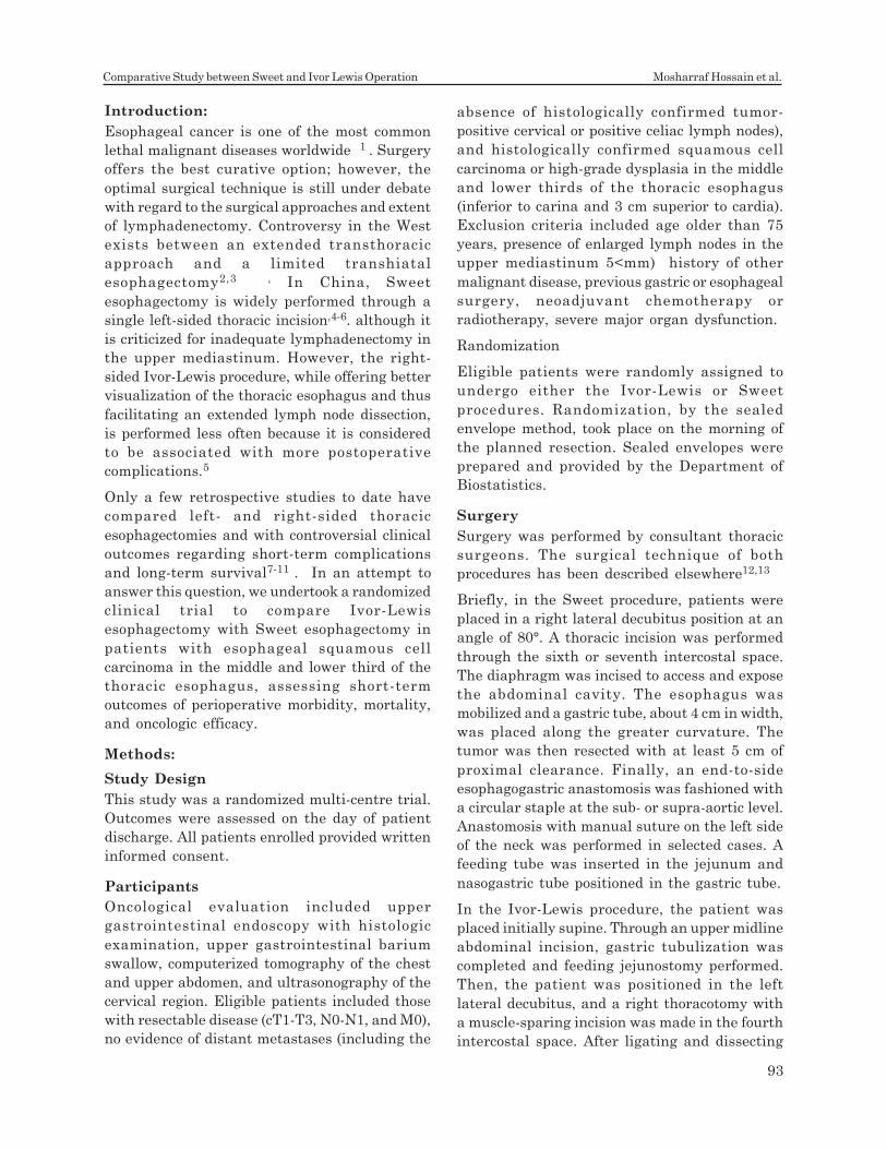
Introduction:
Esophageal cancer is one of the most common
lethal malignant diseases worldwide 1 . Surgery
offers the best curative option; however, the
optimal surgical technique is still under debate
with regard to the surgical approaches and extent
of lymphadenectomy. Controversy in the West
exists between an extended transthoracic
approach and a limited transhiatal
esophagectomy2,3 , In China, Sweet
esophagectomy is widely performed through a
single left-sided thoracic incision,4-6. although it
is criticized for inadequate lymphadenectomy in
the upper mediastinum. However, the right-
sided Ivor-Lewis procedure, while offering better
visualization of the thoracic esophagus and thus
facilitating an extended lymph node dissection,
is performed less often because it is considered
to be associated with more postoperative
complications.5
Only a few retrospective studies to date have
compared left- and right-sided thoracic
esophagectomies and with controversial clinical
outcomes regarding short-term complications
and long-term survival7-11 . In an attempt to
answer this question, we undertook a randomized
clinical trial to compare Ivor-Lewis
esophagectomy with Sweet esophagectomy in
patients with esophageal squamous cell
carcinoma in the middle and lower third of the
thoracic esophagus, assessing short-term
outcomes of perioperative morbidity, mortality,
and oncologic efficacy.
Methods:
Study Design
This study was a randomized multi-centre trial.
Outcomes were assessed on the day of patient
discharge. All patients enrolled provided written
informed consent.
Participants
Oncological evaluation included upper
gastrointestinal endoscopy with histologic
examination, upper gastrointestinal barium
swallow, computerized tomography of the chest
and upper abdomen, and ultrasonography of the
cervical region. Eligible patients included those
with resectable disease (cT1-T3, N0-N1, and M0),
no evidence of distant metastases (including the
absence of histologically confirmed tumor-
positive cervical or positive celiac lymph nodes),
and histologically confirmed squamous cell
carcinoma or high-grade dysplasia in the middle
and lower thirds of the thoracic esophagus
(inferior to carina and 3 cm superior to cardia).
Exclusion criteria included age older than 75
years, presence of enlarged lymph nodes in the
upper mediastinum 5<mm) history of other
malignant disease, previous gastric or esophageal
surgery, neoadjuvant chemotherapy or
radiotherapy, severe major organ dysfunction.
Randomization
Eligible patients were randomly assigned to
undergo either the Ivor-Lewis or Sweet
procedures. Randomization, by the sealed
envelope method, took place on the morning of
the planned resection. Sealed envelopes were
prepared and provided by the Department of
Biostatistics.
Surgery
Surgery was performed by consultant thoracic
surgeons. The surgical technique of both
procedures has been described elsewhere12,13
Briefly, in the Sweet procedure, patients were
placed in a right lateral decubitus position at an
angle of 80°. A thoracic incision was performed
through the sixth or seventh intercostal space.
The diaphragm was incised to access and expose
the abdominal cavity. The esophagus was
mobilized and a gastric tube, about 4 cm in width,
was placed along the greater curvature. The
tumor was then resected with at least 5 cm of
proximal clearance. Finally, an end-to-side
esophagogastric anastomosis was fashioned with
a circular staple at the sub- or supra-aortic level.
Anastomosis with manual suture on the left side
of the neck was performed in selected cases. A
feeding tube was inserted in the jejunum and
nasogastric tube positioned in the gastric tube.
In the Ivor-Lewis procedure, the patient was
placed initially supine. Through an upper midline
abdominal incision, gastric tubulization was
completed and feeding jejunostomy performed.
Then, the patient was positioned in the left
lateral decubitus, and a right thoracotomy with
a muscle-sparing incision was made in the fourth
intercostal space. After ligating and dissecting
93
Comparative Study between Sweet and Ivor Lewis Operation Mosharraf Hossain et al.
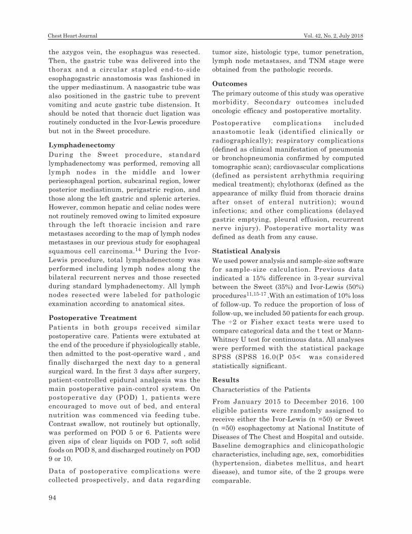
the azygos vein, the esophagus was resected.
Then, the gastric tube was delivered into the
thorax and a circular stapled end-to-side
esophagogastric anastomosis was fashioned in
the upper mediastinum. A nasogastric tube was
also positioned in the gastric tube to prevent
vomiting and acute gastric tube distension. It
should be noted that thoracic duct ligation was
routinely conducted in the Ivor-Lewis procedure
but not in the Sweet procedure.
Lymphadenectomy
During the Sweet procedure, standard
lymphadenectomy was performed, removing all
lymph nodes in the middle and lower
periesophageal portion, subcarinal region, lower
posterior mediastinum, perigastric region, and
those along the left gastric and splenic arteries.
However, common hepatic and celiac nodes were
not routinely removed owing to limited exposure
through the left thoracic incision and rare
metastases according to the map of lymph nodes
metastases in our previous study for esophageal
squamous cell carcinoma.14 During the Ivor-
Lewis procedure, total lymphadenectomy was
performed including lymph nodes along the
bilateral recurrent nerves and those resected
during standard lymphadenectomy. All lymph
nodes resected were labeled for pathologic
examination according to anatomical sites.
Postoperative Treatment
Patients in both groups received similar
postoperative care. Patients were extubated at
the end of the procedure if physiologically stable,
then admitted to the post-operative ward , and
finally discharged the next day to a general
surgical ward. In the first 3 days after surgery,
patient-controlled epidural analgesia was the
main postoperative pain-control system. On
postoperative day (POD) 1, patients were
encouraged to move out of bed, and enteral
nutrition was commenced via feeding tube.
Contrast swallow, not routinely but optionally,
was performed on POD 5 or 6. Patients were
given sips of clear liquids on POD 7, soft solid
foods on POD 8, and discharged routinely on POD
9 or 10.
Data of postoperative complications were
collected prospectively, and data regarding
tumor size, histologic type, tumor penetration,
lymph node metastases, and TNM stage were
obtained from the pathologic records.
Outcomes
The primary outcome of this study was operative
morbidity. Secondary outcomes included
oncologic efficacy and postoperative mortality.
Postoperative complications included
anastomotic leak (identified clinically or
radiographically); respiratory complications
(defined as clinical manifestation of pneumonia
or bronchopneumonia confirmed by computed
tomographic scan); cardiovascular complications
(defined as persistent arrhythmia requiring
medical treatment); chylothorax (defined as the
appearance of milky fluid from thoracic drains
after onset of enteral nutrition); wound
infections; and other complications (delayed
gastric emptying, pleural effusion, recurrent
nerve injury). Postoperative mortality was
defined as death from any cause.
Statistical Analysis
We used power analysis and sample-size software
for sample-size calculation. Previous data
indicated a 15% difference in 3-year survival
between the Sweet (35%) and Ivor-Lewis (50%)
procedures11,15-17 .With an estimation of 10% loss
of follow-up. To reduce the proportion of loss of
follow-up, we included 50 patients for each group.
The ÷2 or Fisher exact tests were used to
compare categorical data and the t test or Mann-
Whitney U test for continuous data. All analyses
were performed with the statistical package
SPSS (SPSS 16.0(P 05< was considered
statistically significant.
Results
Characteristics of the Patients
From January 2015 to December 2016. 100
eligible patients were randomly assigned to
receive either the Ivor-Lewis (n =50) or Sweet
(n =50) esophagectomy at National Institute of
Diseases of The Chest and Hospital and outside.
Baseline demographics and clinicopathologic
characteristics, including age, sex, comorbidities
(hypertension, diabetes mellitus, and heart
disease), and tumor site, of the 2 groups were
comparable.
94
Chest Heart Journal Vol. 42, No. 2, July 2018
Table-I
Basic patients Characteristics and clinical data
Characteristic Group No.(%) P Value
Sweet(n-50) Ivor-Lewis(n-50)
Age, Median(range) 60(39-74) 60(38-74) .56
Sex
Male 41(82) 39(78) .25
Female 9(18) 11(22)
Comorbidity, No(%)
Hypertension 9(18) 13(26) .76
Heart disease 2(4) 1(2) .45
DM 3(6) 4(8) .45
Total 14(28) 18(36) .49
Tumor site
middle 28(56) 32(64) .13
lower 22(44) 18(36)
Histology
High grade Dysplasia 2(4) 1(2)
Squamous cell carcinoma 47(94) 48(96) .30
Small cell carcinoma 1(2) 1(2)
Table-1 shows Basic patients Characteristics and clinical data
Table-II
Histological parameters
Parameter Group No.(%) P Value
Sweet Ivor-Lewis
(n-50) (n-50)
Tumor Staging
Tis 3(6) 1(2)
T1a 2(4) 3(6) .40
T1b 8(16) 8(16)
T2 10(20) 15(30)
T3 24(48) 22(44)
T4 3(6) 1(2)
Nodal Status
N0 27(54) 28(56)
N1 21(42) 20(40) .93
N2 1(2) 2(4)
N3 1(2) 0
TNM Staging
0 2(4)) 1(2)
I 7(14) 9(18) .64
IIA 18(36) 18(36)
IIB 6(12) 8(16)
IIIA 14(28) 11(22)
IIIB 2(4) 2(4)
IV 1(2) 1(2)
Table 2 shows Histological parameters
95
Comparative Study between Sweet and Ivor Lewis Operation Mosharraf Hossain et al.
Morbidity and Mortality
Postoperative mortality did not differ
significantly between the 2 cohorts (2 of 50 [4.0%]
in the Sweet vs 1 of 50 [2%] in the Ivor-Lewis
group. In the Sweet group, 2 patients died of
respiratory failure secondary to pulmonary
infections. In the Ivor-Lewis group, only 1 patient
died of cerebrovascular accident.
Although operating time was significantly longer
in the Ivor-Lewis than in the Sweet groups (mean
[SD], 20238 minutes vs 17435 minutes,
respectively; (P<001) the hospital stay was
significantly shorter for patients who underwent
the Ivor-Lewis esophagectomy (median, 18 days
in the Sweet group vs 16 days in the Ivor-Lewis
group; P = .002). The incidences of anastomotic
leakage, chylothorax, and pulmonary infections
were numerically, but not significantly, higher
in the Sweet group. There was no significant
difference with regard to other postoperative
complications. However, a significantly higher
morbidity rate was found in patients who
underwent Sweet esophagectomy (21 of 50
[42%]) than those who underwent Ivor-Lewis
esophagectomy {15 of 50(30%)}.
Lymphadenectomy
Because we lacked the data on circumferential
involvement, the percentage of R0 resection was
unavailable. Resection without macroscopical
residual (R0/R1) was achieved in 49 of 50 patients
(98%). A significantly higher number of lymph
nodes was retrieved in the Ivor-Lewis group
(median, 22; range, 8-56) compared with the
Sweet group (median, 18; range, 3-51; P <.001).
We further classified lymph node groups
according to dissection area. The Ivor-Lewis
Table-III
Post-operative outcomes
Outcome Group No.(%) P Value
Sweet Ivor-Lewis
(n-50) (n-50)
Operative time, mean(SD), min 174(35) 202(38) <.001
Hospital Stay, Median(Range) 18(10-90) 16(10-60) .002
Post-operative complication
Pulmonary infection 5(10) 4(8) .56
Cardiac complication 7(14) 5(10) .49
Pleural Effusion 3(6) 2(4) .71
Chylothorax 2(4) 1(2) .20
Anastomotic leakage 2(4) 1(2) .10
Table-3 shows Post-operative outcomes
Table-IV
Number of Lymph Node resected.
Region Group No.(%) P Value
Sweet Ivor-Lewis
(n-50) (n-50)
Mediastinum
Upper 0(0-10) 1(0-15)
Middle 2(0-16) 3(0-16) <.001
Lower 1(0-15) 1(0-10)
Middle/Lower 5(0-16) 4(0-15)
Celiac 0(0-10) 1(0-14) <.001
Table 4 shows Number of Lymph Node resected.
96
Chest Heart Journal Vol. 42, No. 2, July 2018
procedure showed superiority in the dissection
of lymph nodes both in the upper mediastinum
and areas around the common hepatic and celiac
arteries, whereas the number of lymph nodes
retrieved in the middle/lower esophagus and
perigastric regions was similar between the 2
groups (Table 4). Consequently, more patients
in the upper mediastinum had positive lymph
nodes following the Ivor-Lewis procedure (06 of
50 (12.0%) than the Sweet procedure 2 of 50 (4%)
(P<.001) Three cases in the Ivor-Lewis group had
positive celiac nodes, although there was no
significant difference in this area (Table-4).
Discussion:
Esophagectomy is among the surgical procedures
with the highest incidence of complications19,20.
Although Ivor-Lewis esophagectomy is advocated
by the Chinese Anti-Cancer Association,18 a left
posterolateral approach with limited
lymphadenectomy remains a priority in China
given the debate on the extent of
lymphadenectomy necessary and, more
importantly, concern about the Ivor-Lewis
esophagectomy being associated with higher
postoperative complications. However, our study
has demonstrated that patients in the Ivor-Lewis
group experienced a lower incidence of in-
hospital morbidity and shorter hospital stay
compared with those in the Sweet group,
although operative time was somewhat longer.
Importantly, our trial showed significantly better
lymph node resection in the Ivor-Lewis
procedure than in the Sweet esophagectomy.
The overall incidence of patients having at least
1 postoperative complication was 35% in our
trial. Although the incidence of each complication
did not differ significantly between the 2 groups,
more patients in the Sweet group did experience
postoperative complications than in the Ivor-
Lewis group. This higher incidence of morbidity
in the Sweet group was associated with a higher
rate of reoperations and longer hospital stay.
A variety of factors, including advanced age,
preexisting poor pulmonary function, poor
performance status, smoking status, and,
notably, surgical approach, were believed to be
related to respiratory problems.21 In our trial,
no patient in the Sweet group underwent surgery
through the combined thoracoabdominal
approach, in consideration of potentially
increased postoperative pain with costal
cartilage incision. This may explain our lower
incidence of pulmonary complications than that
in published reports.22 Although the incidence
was comparable between the 2 groups in our trial,
we prefer the Ivor-Lewis procedure because of
more lymph nodes being resected. During the
Sweet procedure, division of the diaphragm and
1-lung anesthesia throughout the operation may
also contribute to pulmonary problems. Of note,
no Ivor-Lewis group patient died of pulmonary
complication, whereas death was due to
pulmonary complication in 2 following Sweet
esophagectomy.
Anastomotic leakage is an important issue in the
management of surgical complications because
it can be fatal. The rate of leakage in our series
was lower than that in many other series using
a similar intrathoracic stapling technique17,22,23.
High surgeon volume may be an important factor
for this lower incidence.24,25.Although there was
no significant difference, more patients in the
Sweet group experienced anastomotic leakage.
We note that the Ivor-Lewis procedure was our
preferred approach and more widely performed
during the period of this study. The Sweet
approach was our major surgical approach prior
to 2006, during which time our chief surgeons
performed at least 50 Sweet esophagectomies.
Hence, the comparison is valid. Importantly, no
death in this trial was related to anastomotic
leakage. Early identification, clear thoracic and
mediastinal drainage, and sometimes
reoperation for drainage seem to be of most
importance in the avoidance of additional severe
complications.
Cardiovascular events and other minor
complications, including wound infection, pleural
effusion, and delayed gastric emptying, were
also comparable between the 2 groups. Although
the incidence of persistent recurrent nerve
injuries can be biased because none received
laryngoscopy in the postoperative period, strict
criteria of patient selection contributed to this
low incidence. Only patients without enlarged
lymph nodes in the upper mediastinum <5 mm
in diameter were enrolled so that the possibility
of recurrent nerve injury was reduced during
upper mediastinal lymphadenectomy.
97
Comparative Study between Sweet and Ivor Lewis Operation Mosharraf Hossain et al.
Although the distribution of TNM stages was
similar between the 2 groups, stage migration
may still be a consideration owing to inadequate
lymphadenectomy during the Sweet procedure.
Lymph nodes in 2 regions were always omitted
during Sweet esophagectomy: those along
bilateral recurrent nerves in the upper
mediastinum due to anatomical limitation by the
aortic arch and lymph nodes along the common
hepatic and celiac arteries in the upper abdomen
because we chose a single left-sided thoracic
approach for the Sweet procedure, which resulted
in poor exposure in these regions. Although
abdominal lymphatic involvement was rare,
involvement in the upper mediastinum was
common.14 In this study, 06 of 50 (12.0%) patients
in the Ivor-Lewis group showed positive lymph
nodes along bilateral recurrent nerves. This
implies that the more extensive lymph node
resection of the former procedure was needed to
remove more potential positive lymph nodes, thus
offering better tumor staging. With regard to the
current question on the extent of
lymphadenectomy, future follow-up of this trial
may clarify the long-term benefits of the extended
lymphadenectomy using the Ivor-Lewis procedure.
One limitation of this study was that we did not
evaluate postoperative functional status and
therefore cannot comment in detail on quality
of life following surgery. Moreover, pulmonary
complications may be reduced if a left
thoracoabdominal approach is performed with
the diaphragm incised at its periphery, thus
preserving its innervation. However, because of
what is widely done in China and considering
potentially increased wound complication and
postoperative pain, we chose the left-sided
thoracic approach with the diaphragm incised
vertically in this study.
Conclusions:
Our data provide evidence for the superiority of
the Ivor-Lewis esophagectomy over the Sweetprocedure with regard to short-term outcomes
such as lymph node retrieval and overall morbidity
for patients with squamous cell cancer in themiddle and lower third of the thoracic esophagus.
Further follow-up may elucidate whether the Ivor-Lewis procedure also has an advantage in disease
control and long-term survival.
References:
1. Jemal A, Bray F, Center MM, Ferlay J,
Ward E, Forman D. Global cancer
statistics. CA Cancer J Clin 2011;90(2):
61-69.
2. Omloo JM, Lagarde SM, Hulscher JB, et
al. Extended transthoracic resection
compared with limited transhiatal resection
for adenocarcinoma of the mid/distal
esophagus: five-year survival of a
randomized clinical trial. Ann Surg. 2007;
992-1000(6)246.
3. Hulscher JB, van Sandick JW, de Boer
AG, et al. Extended transthoracic
resection compared with limited transhiatal
resection for adenocarcinoma of the
esophagus. N Engl J Med. 2002; 1662-
1669(21)347.
4. Liu JF, Wang QZ, Ping YM, Zhang YD.
Complications after esophagectomy for
cancer: 53-year experience with 20,796
patients. World J Surg. 2008;32(3):395-400.
5. Chen SB, Weng HR, Wang G, et al.
Surgical treatment for early esophageal
squamous cell carcinoma. Asian Pac J
Cancer Prev. 2013;6:3825-3830.
6. Song PI, Liang H, Fan JH, Wei WQ, Wang
GQ, Qiao YL. Long-term survival after
esophagectomy for early esophageal
squamous cell carcinoma in Linxian,
China. J Surg Oncol. 2011;104(2):176-180.
7. Launois B, Paul JL, Lygidakis NJ, et al.
Results of the surgical treatment of
carcinoma of the esophagus. Surg Gynecol
Obstet. 1983;156(6):753-7601.
8. Yu Y, Wang Z, Liu XY, Zhu XF, Chen QF.
Therapeutic efficacy comparison of two
surgical procedures to treat middle thoracic
esophageal carcinoma. World J Surg.
2010;34(2):272-276.
9. Fu SJ, Fang WT, Mao T, Chen WH.
Comparison of surgical outcomes after
different surgical approach for middle or
lower thoracic esophageal squamous cancer
[in Chinese]. Zhonghua Wei Chang Wai Ke
Za Zhi. 2012;15(4):373-376.
10. Suttie SA, Li AG, Quinn M, Park KG. The
impact of operative approach on outcome
of surgery for gastro-oesophageal
tumours. World J Surg Oncol. 2007;5:95.
11. Luo KJ, Fu JH, Hu Y, et al. Efficacy of
surgical resection of left and right
98
Chest Heart Journal Vol. 42, No. 2, July 2018
transthoracic approaches for middle
thoracic esophageal squamous cell
carcinoma [in Chinese]. Ai Zheng.
2009;28(12):1260-1264.
12. Fujita H, Sueyoshi S, Tanaka T, et al.
Optimal lymphadenectomy for squamous
cell carcinoma in the thoracic esophagus:
comparing the short- and long-term
outcome among the four types of
lymphadenectomy. World J Surg.
2003;27(5):571-579.
13. Li B, Guo Q, Yang H, et al. Left
thoracoabdominal approach in surgical
treatment of adenocarcinoma of the
esophagogastric junction in the Northern
Henan Province of China. Dig Surg.
2011;28:2-8.
14. Li B, Chen H, Xiang J, et al. Pattern of
lymphatic spread in thoracic esophageal
squamous cell carcinoma: a single-
institution experience. J Thorac Cardiovasc
Surg. 2012;144(4):778-785.
15. Page RD, Khalil JF, Whyte RI, Kaplan
DK, Donnelly RJ. Esophagogastrectomy
via left thoracophrenotomy. Ann Thorac
Surg. 1990;49(5):763-766.
16. Stilidi I, Davydov M, Bokhyan V,
Suleymanov E. Subtotal esophagectomy
with extended 2-field lymph node dissection
for thoracic esophageal cancer. Eur J
Cardiothorac Surg. 2003;23(3):415-420.
17. Chen G, Wang Z, Liu XY, Liu FY.
Recurrence pattern of squamous cell
carcinoma in the middle thoracic esophagus
after modified Ivor-Lewis esophagectomy.
World J Surg. 2007;31(5):1107-1114.
18. Mao YS, He J, Xue Q, et al. Nationwidespeaking tour of standardized diagnosis andtreatment for esophageal cancer [inChinese]. Zhonghua Wei Chang Wai Ke ZaZhi. 2013;16(9):801-804.
19. Lerut T, Nafteux P, Moons J, et al. Three-field lymphadenectomy forcarcinoma of the esophagus andgastroesophageal junction in 174 R0resections: impact on staging, disease-freesurvival, and outcome: a plea for adaptationof TNM classification in upper-halfesophageal carcinoma. Ann Surg.2004;240(6):962-972.
20. Biere SS, van Berge Henegouwen MI,Maas KW, et al. Minimally invasiveversus open oesophagectomy for patientswith oesophageal cancer: a multicentre,open-label, randomised controlledtrial. Lancet. 2012;379:1887-1892.
21. Ferguson MK, Celauro AD, Prachand V. Prediction of major pulmonarycomplications after esophagectomy. AnnThorac Surg. 2011;91(5):1494-1500.
22. Gillies RS, Simpkin A, Sgromo B, Marshall RE, Maynard ND. Left thoracoabdominalesophagectomy: results from a singlespecialist center. Dis Esophagus. 2011;24(3):138-144.
23. Ott K, Bader FG, Lordick F, Feith M,Bartels H, Siewert JR. Surgical factorsinfluence the outcome after Ivor-Lewisesophagectomy with intrathoracicanastomosis for adenocarcinoma of theesophagogastric junction: a consecutiveseries of 240 patients at an experiencedcenter. Ann Surg Oncol. 2009;16(4):1017-1025.
24. Derogar M, Sadr-Azodi O, Johar A,Lagergren P, Lagergren J. Hospital andsurgeon volume in relation to survival afteresophageal cancer surgery in a population-based study. J Clin Oncol. 2013;31(5):551-557.
25. Lai FC, Chen L, Tu YR, Lin M, Li X.Prevention of chylothorax complicatingextensive esophageal resection by massligation of thoracic duct: a random controlstudy. Ann Thorac Surg. 2011;91(6):1770-1774.
99
Comparative Study between Sweet and Ivor Lewis Operation Mosharraf Hossain et al.
Chest Heart Journal
Vol. 42, No. 2, July 2018
ORIGINAL ARTICLE
Effect of Therapeutic Exercise of Upper
Extremity in COPD patientsPervin S 1, Amin MR2, Kabir J3, Rahman A4, Khan MH5 , Islam MS6 ,
Jahan R7, Habib RB8, Happy BD9
Abstract:
Background and Objective: Chronic obstructive pulmonary disease is one of the major
causes of mortality and morbidity in the world. This study aim to determine the effects of
home based upper extremity exercises in patients with Chronic Obstructive Pulmonary
Diseases.
Methods: Experimental study was performed with 30 COPD Patients, they were selected
based on the inclusion and exclusion criteria and randomly assigned to 2 groups. All the
participants were explained about the study in which Chest expansion measurement
was assessed by using inch tape at 3 levels, dyspnea level was graded by using Modified
Borg scale, Wellbeing and activity of daily life score was assessed by using CAT
questionnaire and those was performed as a pre-test and same tests were repeated as
post-test after the training session for a period of 4 weeks. Group A participants were
taught to perform the exercise and asked to do at home for 5 days a week for total duration
of 4 weeks. Group B - Control group was treated with thoracic mobility exercise, repetition
-3sets/10rep and frequency -7days per week.
Results: Group A: chest expansion measurements at all 3 levels were improved in post-
test compared to pre- test and Group A is higher when compared with Group B. Dyspnea
score were reduced in Group A compared to Group B and wellbeing, activity of daily
living score improved in both groups, Group A was higher compared to Group B (P <
0.05, statistically significant).
Conclusion: This study proven that home based upper extremity exercise are effective in
improving chest expansion, reduced dyspnea and improved quality of wellbeing and
activity of daily living.
Keywords: Chronic obstructive pulmonary disease (COPD), Dyspnea, Therapeutic
exercise.
[Chest Heart Journal 2018; 42(2) : 100-106]
DOI: http://dx.doi.org/10.33316/chab.j.v42i2.2019586
1. Assistant Professor, Department of Physical Medicine & Rehabilitation, NIDCH, Dhaka, Bangladesh.
2. Associate Professor and Head, Department of Physical Medicine & Rehabilitation, NIDCH, Dhaka, Bangladesh.
3. Medical Officer. Gynae & Obs Department, Dhaka Medical College Hospital, Dhaka, Bangladesh.
4. Associate Consultant, Anesthesia Department, Imperial Hospital, Chittagong, Bangladesh.
5. Assistant Professor, Physical Medicine & Rehabilitation Department, NITOR, Dhaka, Bangladesh.
6. Assistant professor & RP, NIDCH, Dhaka, Bangladesh.
7. Professor, Respiratory Medicine, NIDCH, Dhaka, Bangladesh.
8. Epidemiologist, Junior Consultant, Pediatrics, DGHS, Dhaka, Bangladesh.
9. Medical Officer, Physical Medicine & Rehabilitation Department, NINS, Dhaka, Bangladesh.
Correspondence to: Dr. Sanjida Pervin, Assistant Professor, Department of Physical Medicine & Rehabilitation,
NIDCH, Dhaka, Bangladesh. Mobile: +8801911620771.Email: [email protected].
Submission on: 7 May, 2018 Accepted for Publication: 18 June, 2018
Available at http.//www.chabjournal.org
Introduction
Chronic Obstructive Pulmonary Disease (COPD)
represents an important public health challenge
that is both preventable and treatable. Many
people’s suffer from this disease for years, and
die prematurely from it or its complications.
COPD is a lung disease characterized by chronic
airflow limitation of the airways, which can be
prevented but cannot be cured. COPD mainly
caused by cigarette smoking; air pollution is also
considered a risk factor in many developing
countries which contributes the disease to both
gender whereas COPD is more commonly
observed in long term smokers of middle age.
The disease symptoms add on the co-morbidities
with the age and also due to smoking1. COPD is
a major cause of mortality and morbidity in the
world2 and it is currently the 4th leading cause
of death in world3. Globally, the COPD burden
is projected to increase in coming decades
because of continued exposure to COPD risk
factors and aging of population4. Individual with
COPD are illustrated with various other
problems like nutritional deficits, cardiovascular
disease, diabetes, osteoporosis. Whereas disease
progresses with major changes including airway
obstruction, increased dyspnea, hyperinflation,
reduced lung functions5, and peripheral muscle
weakness6 is more predominant which leads to
fatigue of the patient when performing their daily
activities. Increased chest wall resistance and
stiffness causes respiratory muscles tightness
and further contributes to mechanical
disadvantage of respiratory muscles7. Major
complaints of COPD are dyspnoea and fatigue
especially while doing their routine activities with
the use of their upper limbs like combing hair8.
Dyspnoea during these activities may be due to
the irregular and superficial breathing pattern
forced by the mechanical effects of arm during
arm elevation. Physical activity level is lowered
especially when patients use the upper limbs
without any support, as well some upper limb
muscles such as the sternocleidomastoid and
upper trapezius diminish their participation in
ventilation as they are recruited to help with
the postural support of the arms1. Hereby it
alters the mechanics of rib cage and abdominal
compartments. Generally, accessory muscles are
inactive during inspiration at rest in healthy
individuals and they act vigorously during
physical activity in COPD, ultimately during
activities with upper limb, accessory muscles
become ineffective in respiration as they perform
their major action to sustain the shoulder girdle
in position as a consequence, the respiratory
work is shifted to the diaphragm which is already
at a mechanical disadvantage in patients with
COPD9. Usage of upper limbs play a major role
in daily activities, normal subjects usually
perform such activities without noticing the
energy lost, but patients with COPD report
tiredness when performing them1. There is
evidence that physical activity is decreased in
COPD patients10. This leads to a downward spiral
of inactivity which predisposes patients to reduce
quality of life, increased rates of hospitalization
and mortality11-13. Disease creates a burden for
the sufferers and for the society as the natural
history of the disease is progression to disability
and death from respiratory failure at a relatively
early age14. Both upper and lower extremity
muscle are impaired in individual with COPD,
muscle weakness does not affect all the muscles.
As some studies report that quadriceps is more
affected than upper limb muscles due to under
usage of lower limb among COPD. Whereas
strength of upper limb muscles such as pectoralis
major, latissimusdorsi, biceps and middle
deltoids is also impaired restricting the
movement of upper extremity which further
limits the thoracic expansion among the COPD1.
For these reasons upper extremity resistance
training is required to enhance the functional
performance of upper limbs in daily care. Upper
extremity resistance training is also the part of
pulmonary rehabilitation. The basic need for this
study is to provide the ease of treatment for the
patients with COPD; it is difficult for the patients
to travel on regular basis to the hospital for
exercises, for that reason the patients were
taught a simple exercise that enhances their
performance in activity of daily living. Hence
purpose of this study is to provide a home based
upper extremity exercises for the patients with
COPD.
Methods:
Experimental study was performed with 30
Subjects randomly assigned to 2 groups. Group
101
Effect of Therapeutic Exercise of Upper Extremity in COPD patients Pervin S et al.
A (Experimental group) and Group B (Control
Group). Study was performed at National
Institute of Diseases of the Chest & Hospital,
Dhaka. All the participants were explained about
the study by providing information sheet and
Informed consent signed by all the participants
prior to initiation of the study. Both male and
female Subjects with Mild to Moderate COPD
based on GOLD classification, Age group
between 30-65 years, Ex-smoker and those with
dyspnoea are included. Subjects are excluded if
they are known to have Restrictive lung disease,
recent thoracic surgery, unstable vital signs,
unstable hypertension, Musculoskeletal and
neurological disease involving upper limb and
thorax. Both the group participants underwent
assessment, As a pretest they are assessed for
Chest expansion at 3 levels – axillary level, 4th
intercostals level and xiphisternal level using
inch tape and dyspnoea grade was noted using
Borg scale and their wellbeing and activity of
daily life score assessed using CAT
questionnaire. All these tests were repeated for
post test following the treatment period for 4
weeks. Group A- 15 participants were asked to
perform Upper extremity exercises at home. All
the participants were taught to perform the
exercise in OPD and exercise protocol was
clearly explained and they were given an exercise
regimen chart, and recording chart to follow the
frequency of the exercise in which they are asked
to record the date whenever the exercise is
performed. Participant was asked to come to
OPD once in a week along with the recording
chart which was verified. Upper extremity
exercise includes.
Shoulder front raise with weight
Patient was asked to hold 1 liter water bottle in
hands in erect standing posture. Then asked to
raise the hands in front of thighs with palms in
a neutral position, without bending elbows with
co-ordinate breathing exercise. (ie, inspiration
during lifting up the hand and expiration during
lowering the hand down) and then lower the
weights.
Shoulder lateral raise with weight
Patient was asked to hold 1 liter water bottle in
hands in erect standing posture. Then asked to
raise the hands in front of thighs with the top
of each weight pointed away, and then lift arms
up by sides without bending elbows with co-
ordinate breathing exercise.(i.e., inspiration
during lifting up the hand and expiration during
lowering the hand down), then lower the arms.
External rotators with weight
Patient should be positioned in supine lying by
holding 1 liter water bottle with the shoulder
abducted to 900 and the elbow bent to 900, so
the hand points to the roof, patient should rotate
the shoulder joint externally so that the hand
moves backwards and the palm faces the roof
along with co-ordinate breathing exercise.
Internal rotators with weight
Patient should be positioned in supine lying by
holding1 liter water bottle with the shoulder
abducted to 900 and the elbow bent to 900, so the
hand points to the roof. Patient should rotate
the shoulder joint internally so that the hand
moves forwards and the palm faces the floor
along with coordinate breathing exercise.
Shoulder shrugging exercise
Patient should stand upright, arms should be
fully extended to the waist, feet shoulder width
apart and head looking forwards, shrug the
shoulders up as high as possible, and patient
should pause at top of the movement and then
relax back down along with co-ordinate breathing
exercise.
Treatment Protocol
• Resistance\weight - 1 liter water bottle.
• Frequency-5days per week for a period of 4
weeks.
• 1 session per day.
• 8 to 10 repetitions \ 3 sets.
• Intensity - Borg scale, Rate of perceived
exertion of 12-14.
• Total duration for upper extremity exercise
is 30 minutes per day same for all the
exercises.
Group B
Group B - Control group was treated with
thoracic mobility exercise with a repetition of
3sets/10rep and frequency-7days per week.
102
Chest Heart Journal Vol. 42, No. 2, July 2018
Thoracic mobility exercises
Position of the subject: Sitting.
Procedure: the patient should exhale while
bending forward to touch the floor with arms
crossed at the feet then the patient should extend
up while taking a deep inspiration and lift the
arm up with a frequency of 7days per week and
3 sets of 10 repetitions.
Position of the patient: standing.
Procedure: The patient should stand with his
knees straight, the patient instructed to exhale
while bending forward to touch the floor with
arms; then the patient should extend up by lifting
his hands simultaneously taking a deep
inspiration. 10 repetitions/ set – 3 sets with a
frequency of 7 days per week. Both the groups
will be advised to continue their normal activity
and follow their inhalers and medications
prescribed by the chest physicians
Fig.-1: Comparison of pre test and post test
values of Chest expansion measurements at
Axillary level for Group A& Group B.
Fig.-2: Comparison of pre test and post test
values of Chest expansion measurements at 4th
Intercostal level for Group A& Group B.
Fig.-3: Comparison of pre test and post test
values of Chest expansion measurements at
Xiphi sternal level for Group A & Group B.
Fig.-5: Comparison of pretest and post test values
of CAT questionnaire between Group A & Group
B.
Fig.-4: Comparison of pre test and post test
values for Modified Borg scale between Group A
and Group B.
1.353
1.1
1.747
1.14
0
0.5
1
1.5
2
Group-A Group-B
Pre-test
post-test
1.47
1.107
1.693
1.167
0
0.5
1
1.5
2
Group-A Group-B
Pre-test
post-test
1.58
1.1
1.693
1.16
0
0.5
1
1.5
2
Group-A Group-B
Pre-test
post-test
32.83
1.97
2.5
0
0.5
1
1.5
2
2.5
3
3.5
Group-A Group-B
Pre-test
post-test
17.37
15.27
15.67
14.67
13
14
15
16
17
18
Group-A Group-B
Pre-test
post-test
103
Effect of Therapeutic Exercise of Upper Extremity in COPD patients Pervin S et al.
In this unpaired t test df 28 at 5% level of
significance, in the table critical value is 1.70.
Test statistics (2.55) is greater than critical value
(1.70). So, P < 0.05
Table1: Comparison of pre test and post test
values between Group A and Group B for Modified
Borg scale and CAT questionnaire.
Results
The collected data was used for statistical
analysis, Paired ‘t’ test was used to compare the
data of pre and post within the groups for chest
expansion measurement. Wilcoxon rank test was
used to compare the data of pre and post within
the groups for Borg’s scale and CAT
questionnaire. Independent ‘t’ test was used to
compare the data between the groups for chest
expansion measurement. Mann whiteny U test
was used to compare the data between the
groups for Borg scale and CAT questionnaire.
Graph 1 shows the difference in pre test and
post test values between Group A and Group B
for chest expansion measurement at axillary
level. Group A difference in pre and post values
are greater than Group B. Graph 2 shows the
difference between Group A and Group B in pre
test and post test values at 4th Intercostal level
and similar difference was seen in xiphisternal
level shown in Graph 3. Graph 2 & 3 shows
higher difference between pre and post test
values of Group A. Graph 4 given that Dyspnea
score rated with Modified Borg scale was
comparatively lower in post test than pre test
which shows that dyspnea is reduced well in
group A than Group B also seen in Table 1. Graph
5 shows the wellbeing and activity of daily living
score which improved in post test in both groups.
Whereas score is higher in group A compared to
group B, shown in Table 1(P < 0.05).
Discussion
COPD is the commonest heterogeneous
pulmonary condition and it is the major cause of
morbidity and mortality among the pulmonary
patients. This disease is usually characterised
by dyspnea, and a small proportion of excessive
secretions particularly during exacerbation15.
COPD patients also develop peripheral muscle
dysfunction that further contributes to exercise
intolerance. These impairments limit the
independence of the patient in daily life and
simple activities of daily care becomes difficult
and it affects the health related quality of life16.
COPD patients poorly tolerate activities
involving upper limb compared to lower limb.
Upper limb movements are associated with
ventilatory strategies thereby it increases
metabolic demand it has been observed that at
arm elevation at rest significantly decreased
vital capacity and functional residual capacity17.
But during upper limb activities arm fatigue is
reported as a common limiting symptom.
Supported upper limb activity increases the
functional residual capacity due to the passive
stretch of thoracic muscles imposed on the rib
cage when the arms are raised with weights it
strengthens the upper limb muscles1 and
concluded that supported arm activity improves
the endurance and quality of life17.
Stefaniacostie et al stated that unsupported
upper extremity exercise results in ventilator
constraints, concluded that more studies are
recommended to prove the benefits of supported
arm exercise and unsupported upper extremity
exercise, whereas unsupported arm exercise
may determine progressive restriction of the
activities performed with the arm, where the
diaphragm is functionally compromised18.
Thereby our study performed with supported
arm exercises along with co-ordinated breathing
exercise that reduces the rate of dyspnoea and
improves the ability to perform the arm activities
without restriction. Sarah Bernard et al
described the effects of Aerobic and strength
training in patients with COPD and concluded
that the combination of strength training in
Test Group-A Group-B
Mean Standard deviation Mean Standard deviation
Pre Post Pre Post Pre Post Pre Post
Modified Borg 3.00 1.97 1.195 1.157 2.83 2.50 1.358 1.296
Scale
CAT questionnaire 17.33 15.67 3.885 3.395 15.27 14.67 3.173 3.222
104
Chest Heart Journal Vol. 42, No. 2, July 2018
aerobic training showed significant improvement
in patients with COPD19. Unsupported arm
exercise poses a unique challenge for patients
with COPD, because upper limb muscles acts as
accessory muscle for respiration. During arm
activities the participation of accessory muscles
in ventilation decreases and shifts the
respiratory work to diaphragm due to which
activities with upper limb in daily living becomes
difficult. Further it causes
thoracoabdominaldyssynchrony. It has been
proved that upper limb exercise training
improves work capacity and endurance and
reduces oxygen consumption during workload20.
Ganesan et al reviewed the effects of upper
extremity exercises in COPD and reported that
arm strength training should be included in
pulmonary rehabilitation protocol and it showed
that unsupported arm training improved arm
endurance capacity in majority of people with
COPD. And arm training positively influenced
peak arm exercise capacity, metabolic and
ventilatory demands and also improved activities
of daily living21. For these reason this study
focuses on upper extremity exercises which helps
and encourages thoracic movements and
expands the thoracic cage and also strengthens
the upper limb muscles which eases the way to
perform daily activities by reducing the rate of
dyspnoea increasing the chest expansion and
improving the wellbeing and activity of daily
living.
Conclusion:
From the results, this study concludes that home
based upper extremity exercise are effective in
improving chest expansion, reduced dyspnea and
improved quality of wellbeing and activity of daily
living. Thereby this study recommends the need
to involve the upper limb exercises as a part of
rehabilitation program. Pulmonary
Rehabilitation is appropriate for patients with
COPD and it should be considered part of
integrated patient management.
References:
1. Tania Janaudis-Ferreira. Strategies for
exercise assessment and training in patients
with chronic obstructive pulmonary disease.
Umea University Medical Dissertations,
New Series No 1360.2010: 1- 68.
2. Andre Nyberg. Assessing the effect of high
repetitive single limb exercise on exercise
capacity and quality of life in patients with
chronic obstructive pulmonary disease
(COPD): study protocol for randomized
controlled trial. Trial.2012; 13:114.
3. Lozano R, Naghavi M, Foreman K, et al.
Global and regional mortality from 235
causes of death for 20 age groups in 1990
and 2010: a systematic analysis for the
Global Burden of Disease Study 2010.
Lancet 2012; 380(9859): 2095-128.
4. Mathers CD, Loncar D. Projections of global
mortality and burden of disease from 2002
to 2030. PLOS Med 2006; 3(11): e442.
5. Annemarie L Lee, Anne E Holland. Time to
adapt exercise training regimens in
pulmonary rehabilitation – a review of the
literature.International journal of
COPD.2014; 9: 1275-1288.
6. C.J. Clarke, L.M. Cochrane, E. Mackay, B.
Paton. Skeletal muscle strength and
endurance in patients with mild COPD and
the effects of weight training.EurRespir J.
2000; 15: 92-97.
7. Carla Malaguti, Rafaella R Rondelli,
Leandra M de Souza, Marcia Domingues,
Simone Dal Corso. Reliability of chest wall
mobility and its correlation with pulmonary
function in patients with chronic obstructive
pulmonary disease.Respiratory Care. 2009;
54(12): 1703-1711.
8. Marcelo Velloso, Sergio Garcia Stella, Sonia
Cendon, Antonio Carlos Silva, Jose R.
Jardim. Metabolic and ventilator
parameters of four activities of daily living
accomplished with arms in COPD patients.
Chest.2003; 123: 1047-1053.
9. Dirceu Costa, Karina Maria Cancelliero,
Daniela IKE, Thais Lima Laranjeira, Camila
Bianca FalascoPantoni, Audrey Borghi Silva.
Strategy for respiratory exercise pattern
associated with upper limb movements in
COPD patients. CLINICS. 2011; 66(2): 299-
305.
10. Pitta F, Troosters T, Spruit MA, Probst VS,
Decramer M, Gosselink R. Characteristics
105
Effect of Therapeutic Exercise of Upper Extremity in COPD patients Pervin S et al.
of physical activities in daily life in chronic
obstructive pulmonary disease. Am J Respir
Crit Care Med 2005; 171(9): 972-7.
11. Watz H, Pitta F, Rochester CL, et al. An
Official European Respiratory Society
statement on physical activity in COPD. Eur
Respir J 2014; 44(6): 1521-37.
12. Garcia-Aymerich J, Lange P, Benet M,
Schnohr P, Anto JM. Regular physical
activity reduces hospital admission and
mortality in chronic obstructive pulmonary
disease: a population based cohort study.
Thorax 2006; 61(9): 772-8.
13. Yohannes AM, Baldwin RC, Connolly M.
Mortality predictors in disabling chronic
obstructive pulmonary disease in old age.
Age ageing 2002; 31(2): 137-40.
14. Kenji Shibuya, Colin D Mathers, Alan D
Lopez. Chronic Obstructive Pulmonary
Disease (COPD): Consistent Estimates of
Incidence, Prevalence, and Mortality by
WHO Region. Global Programme on
Evidence for Health Policy.World Health
Organization.2001; 1-28.
15. Jane Cross. Chest physiotherapy during an
acute exacerbation of chronic obstructive
pulmonary disease. Chronic respiratory
disease. 2012; 9(2): 73-75.
16. Tania Janaudis Ferreira, Kylie Hill, Roger
S Goldstein, Priscila Robles Ribeiro, Marla
K. Beauchamp, Thomas E Dolmage, et al.
Resistance arm training in patients with
COPD. A randomized controlled trial.Chest.
2011; 139(1): 151-158.
17. Thomas E Dolmage, Luigi Maestro, Monica
A Avendano, Roger S Goldstein. The
Ventilatory response to arm elevation of
patients with chronic obstructive
pulmonary disease.Chest. 1993; 104(4):
1097-1100.
18. Stefaniacosti, Mauro Di Bari, Paolo
Pillastrini, Roberto D’Amico, Ernesto
Crisfulli, CinziaArletti, et al. Short-term
Efficacy of upper extremity exercise
training in patients with chronic airway
obstruction: A systematic review. Physical
Therapy. 2009; 89(5): 443-455.
19. Sarah Bernard, Francois Whittom, Pierre
Leblanc, Jean Jobin, Roger Belleau, Chantal
Berube, et al. Aerobic and strength training
in patients with chronic obstructive
pulmonary disease.Am J RespirCrit Care
Med. 1999; 159: 896-901.
20. Subin, VaishaliRao, V. Prem, Sahoo. Effect
of upper limb, lower limb and combined
training on health related quality of life in
COPD.Lung India. 2010; 27(1): 4-7.
21. GanesanKathiresan, Senthil Kumar
Jeyaraman, JayachandranJaganathan.
Effect of upper extremity exercise in people
with COPD. J Thorac Dis. 2010; 2: 223- 236.
106
Chest Heart Journal Vol. 42, No. 2, July 2018
Introduction:
The concept of artificial respiration was
recognized in the 16th century by Vesalius and it
is the 20th century that mechanical ventilation
became a widely used therapeutic modality1.
Over the past decade, there has been an
explosion of new ventilatory techniques that
present a bewildering array of alternatives for
the treatment of patients with respiratory
failure.
Respiratory support for respiratory failure can
be given through lung in intubated or
tracheostomised patient as positive pressure
ventilation .This support can also be given
through the devices not primarily applied to the
lung, such as extra corporeal membrane
oxygenation (ECMO)2,3, extracorporeal carbon
dioxide removal (ECCO2R)4 or intravascular
blood gas exchanger (IVOX)5
The main objectives of mechanical ventilation
are to support pulmonary gas exchange, to
increase lung volume & to decrease the WOB.
All these necessities can be obtained by setting
up different mode of ventilator & by adjustment
of different parameters appropriate for a
particular clinical setting.
This is a long process consisting of endotracheal
intubation, appropriate use of sedative or
Outcome of Mechanical Ventilation in Criticaly
ILL PatientsMd. Sayedul Islam1, Nihar Ranjan Saha2, Subrata Gain3, Sanjoy Kumar Kar4, Snehashis Nag5
Abstract:
Method: This is a prospective study carried out during the period of Nov” 2006 to Nov
2007 in a modernized private hospital, Dhaka. The patients were admitted to ICU after
primary assessment that they may need organ support or life support and all the patients
fulfilled the complete code of ICU. Patients who were weaned and extubated and
subsequently shifted to ward were considered good outcome and adverse who cannot be
extubated, if they died, or sought discharge against medical advice.
Result: Total number of patient admitted were 844 during this period. Male and female
ratio was 16:9 and the mean age for the ventilated patients was 52±SD (10.25). Among
them, 370 (43.68%) patients needed artificial respiration by mechanical ventilation in
the course of their management. Among the ventilated patients 199(52.45%) has successful
weaning, 5(1.35) needed tracheostomy, 171 (46.25%) expired. The rate of survival from
ventilator support is statistically significant, P value is < .05.
Conclusion: Mechanical ventilation is a very effective measure in the course of
management of critically ill patients when there is definite indication. This single centred
study must be justified with other large scale study from different worldwide.
[Chest Heart Journal 2018; 42(2) : 107-112]
DOI: http://dx.doi.org/10.33316/chab.j.v42i2.2019587
Chest Heart Journal
Vol. 42, No. 2, July 2018
ORIGINAL ARTICLE
1,2. Associate Professor Respiratory Medicine, NIDCH, Bangladesh.
3,4,5 Regitrar, Respiratory Medicine. NIDCH, Bangladesh.
Correspondence to: Dr. Md. Sayedul Islam, Associate Professor Respiratory Medicine, NIDCH, Bangladesh.
Cell: 01552390582, E mail: drsayedul@ gmail.com
Submission on: 11 May, 2018 Accepted for Publication: 17 June, 2018
Available at http.//www.chabjournal.org
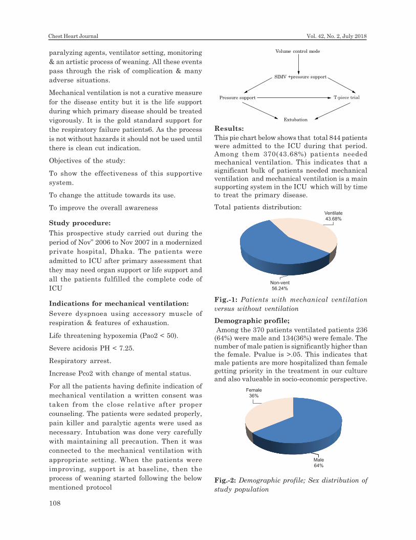
Female
36%
Male
64%
36%
Non-vent
56.24%
Ventilate
43.68%
Fig.-1: Patients with mechanical ventilation
versus without ventilation
paralyzing agents, ventilator setting, monitoring
& an artistic process of weaning. All these events
pass through the risk of complication & many
adverse situations.
Mechanical ventilation is not a curative measure
for the disease entity but it is the life support
during which primary disease should be treated
vigorously. It is the gold standard support for
the respiratory failure patients6. As the process
is not without hazards it should not be used until
there is clean cut indication.
Objectives of the study:
To show the effectiveness of this supportive
system.
To change the attitude towards its use.
To improve the overall awareness
Study procedure:
This prospective study carried out during the
period of Nov” 2006 to Nov 2007 in a modernized
private hospital, Dhaka. The patients were
admitted to ICU after primary assessment that
they may need organ support or life support and
all the patients fulfilled the complete code of
ICU
Indications for mechanical ventilation:
Severe dyspnoea using accessory muscle of
respiration & features of exhaustion.
Life threatening hypoxemia (Pao2 < 50).
Severe acidosis PH < 7.25.
Respiratory arrest.
Increase Pco2 with change of mental status.
For all the patients having definite indication of
mechanical ventilation a written consent was
taken from the close relative after proper
counseling. The patients were sedated properly,
pain killer and paralytic agents were used as
necessary. Intubation was done very carefully
with maintaining all precaution. Then it was
connected to the mechanical ventilation with
appropriate setting. When the patients were
improving, support is at baseline, then the
process of weaning started following the below
mentioned protocol
Results:
This pie chart below shows that total 844 patients
were admitted to the ICU during that period.
Among them 370(43.68%) patients needed
mechanical ventilation. This indicates that a
significant bulk of patients needed mechanical
ventilation and mechanical ventilation is a main
supporting system in the ICU which will by time
to treat the primary disease.
Total patients distribution:
Volume control mode
SIMV + pressure support
Extubation
T-piece trialPressure support
Demographic profile;
Among the 370 patients ventilated patients 236
(64%) were male and 134(36%) were female. The
number of male patien is significantly higher than
the female. Pvalue is >.05. This indicates that
male patients are more hospitalized than female
getting priority in the treatment in our culture
and also valueable in socio-economic perspective.
Fig.-2: Demographic profile; Sex distribution of
study population
108
Chest Heart Journal Vol. 42, No. 2, July 2018
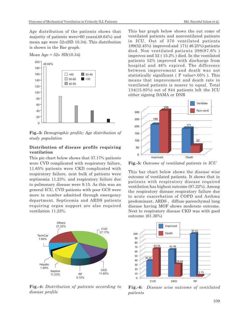
0
50
100
150
200
250
300
199
171
298
52
Improved Death
Ventilate
Non-venti
Distribution of disease profile requiring
ventilation
This pie chart below shows that 37.17% patients
were CVD complicated with respiratory failure,
11.65% patients were CKD complicated with
respiratory failure, next bulk of patients were
septisemia 11.23%. and respiratory failure due
to pulmonary disease were 9.15. As this was an
general ICU, CVD patients with poor GCS were
more in number admitted through emergency
department. Septicemia and ARDS patients
requiring organ support are also required
ventilation 11.23%.
This bar graph below shows the out come of
ventilated patients and nonventilated patients
in ICU. Out of 370 ventilated patients
199(52.45%) improved and 171( 46.25%) patients
died. Non ventilated patients 298(87.6% )
improves and 52 ( 15.2% ) died. In the ventilated
patients 52% improved with discharge from
hospital and 46% expired. The difference
between improvement and death was not
statistically significant ( P value>.05% ). This
means that improvement and death rate in
ventilated patients is nearer to equal. Total
134(15.85%) out of 844 patients left the ICU
either signing DAMA or DNR
Age distribution of the patients shows that
majority of patients were>60 years(48.64%) and
mean age were 52±(SD 10.34). This distribution
is shown in the Bar graph.
Mean Age = 52± SD(10.34)
Fig.-3: Demographic profile; Age distribution of
study population
200
180
160
140
120
100
80
60
40
20
0
>60 30-40
<3050-60
40-50
48.64%
Fig.-4: Distribution of patients according to
disease profile
CVD
37.17%
CKD
11.65%RF
9.15%
Septice
11.23%
Hepatic
1.94%
TermCar
7.60%
Others
21.22%
This bar chart below shows the disease wise
outcome of ventilated patients. It shows that in
patients with respiratory disease required
ventilation has highest outcome (97.22%). Among
the respiratory disease respiratory failure due
to acute exacerbation of COPD and Asthma
predominant. ARDS , diffuse parenchymal lung
disease having MOF shows moderate outcome.
Next to respiratory disease CKD was with good
outcome (61.36%)
Fig.-5: Outcome of ventilated patients in ICU
Fig.-6: Disease wise outcome of ventilated
patients
0
10
20
30
40
50
60
70
80
90
100
36.44
61.36
97.22
63.56
28.64
2.73
CVD DKD RF
Improved
Death
109
Outcome of Mechanical Ventilation in Criticaly ILL Patients Md. Sayedul Islam et al.
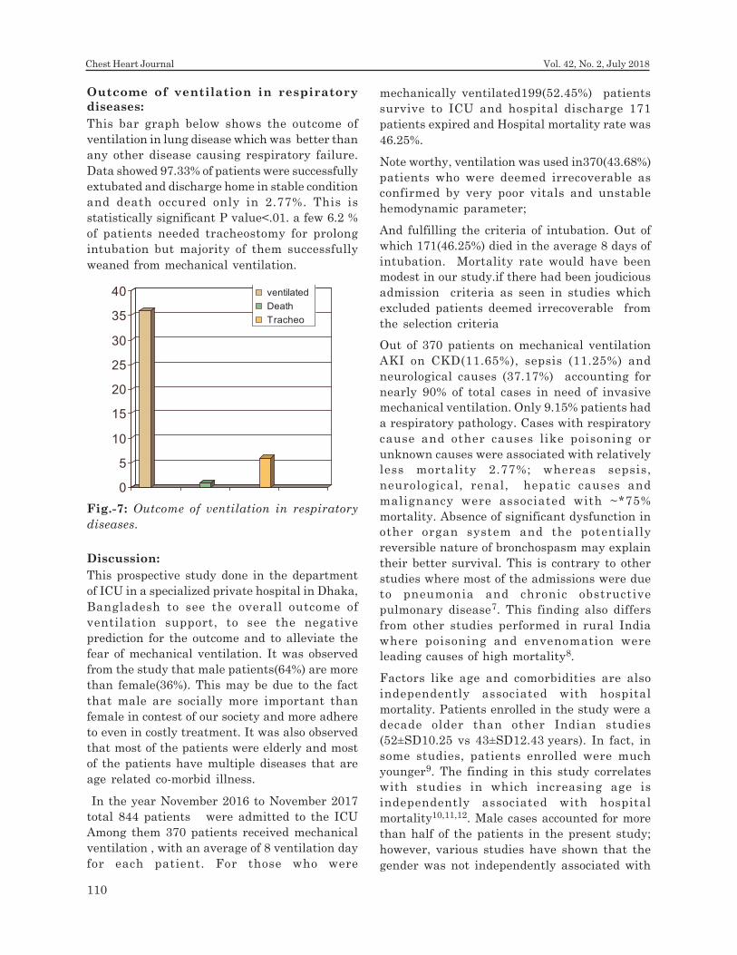
Outcome of ventilation in respiratory
diseases:
This bar graph below shows the outcome of
ventilation in lung disease which was better than
any other disease causing respiratory failure.
Data showed 97.33% of patients were successfully
extubated and discharge home in stable condition
and death occured only in 2.77%. This is
statistically significant P value<.01. a few 6.2 %
of patients needed tracheostomy for prolong
intubation but majority of them successfully
weaned from mechanical ventilation.
mechanically ventilated199(52.45%) patients
survive to ICU and hospital discharge 171
patients expired and Hospital mortality rate was
46.25%.
Note worthy, ventilation was used in370(43.68%)
patients who were deemed irrecoverable as
confirmed by very poor vitals and unstable
hemodynamic parameter;
And fulfilling the criteria of intubation. Out of
which 171(46.25%) died in the average 8 days of
intubation. Mortality rate would have been
modest in our study.if there had been joudicious
admission criteria as seen in studies which
excluded patients deemed irrecoverable from
the selection criteria
Out of 370 patients on mechanical ventilation
AKI on CKD(11.65%), sepsis (11.25%) and
neurological causes (37.17%) accounting for
nearly 90% of total cases in need of invasive
mechanical ventilation. Only 9.15% patients had
a respiratory pathology. Cases with respiratory
cause and other causes like poisoning or
unknown causes were associated with relatively
less mortality 2.77%; whereas sepsis,
neurological, renal, hepatic causes and
malignancy were associated with ~*75%
mortality. Absence of significant dysfunction in
other organ system and the potentially
reversible nature of bronchospasm may explain
their better survival. This is contrary to other
studies where most of the admissions were due
to pneumonia and chronic obstructive
pulmonary disease7. This finding also differs
from other studies performed in rural India
where poisoning and envenomation were
leading causes of high mortality8.
Factors like age and comorbidities are also
independently associated with hospital
mortality. Patients enrolled in the study were a
decade older than other Indian studies
(52±SD10.25 vs 43±SD12.43 years). In fact, in
some studies, patients enrolled were much
younger9. The finding in this study correlates
with studies in which increasing age is
independently associated with hospital
mortality10,11,12. Male cases accounted for more
than half of the patients in the present study;
however, various studies have shown that the
gender was not independently associated with
Fig.-7: Outcome of ventilation in respiratory
diseases.
0
5
10
15
20
25
30
35
40 ventilated
Death
Tracheo
Discussion:
This prospective study done in the department
of ICU in a specialized private hospital in Dhaka,
Bangladesh to see the overall outcome of
ventilation support, to see the negative
prediction for the outcome and to alleviate the
fear of mechanical ventilation. It was observed
from the study that male patients(64%) are more
than female(36%). This may be due to the fact
that male are socially more important than
female in contest of our society and more adhere
to even in costly treatment. It was also observed
that most of the patients were elderly and most
of the patients have multiple diseases that are
age related co-morbid illness.
In the year November 2016 to November 2017
total 844 patients were admitted to the ICU
Among them 370 patients received mechanical
ventilation , with an average of 8 ventilation day
for each patient. For those who were
110
Chest Heart Journal Vol. 42, No. 2, July 2018
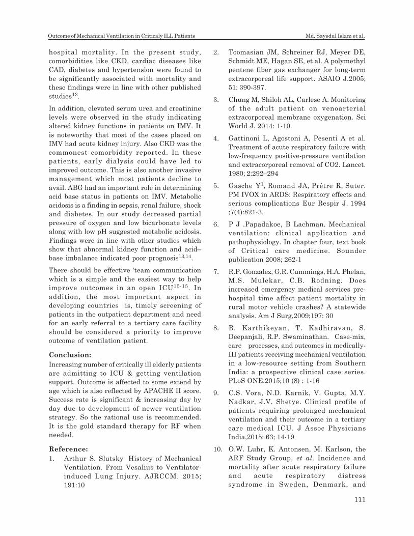
hospital mortality. In the present study,
comorbidities like CKD, cardiac diseases like
CAD, diabetes and hypertension were found to
be significantly associated with mortality and
these findings were in line with other published
studies13.
In addition, elevated serum urea and creatinine
levels were observed in the study indicating
altered kidney functions in patients on IMV. It
is noteworthy that most of the cases placed on
IMV had acute kidney injury. Also CKD was the
commonest comorbidity reported. In these
patients, early dialysis could have led to
improved outcome. This is also another invasive
management which most patients decline to
avail. ABG had an important role in determining
acid base status in patients on IMV. Metabolic
acidosis is a finding in sepsis, renal failure, shock
and diabetes. In our study decreased partial
pressure of oxygen and low bicarbonate levels
along with low pH suggested metabolic acidosis.
Findings were in line with other studies which
show that abnormal kidney function and acid–
base imbalance indicated poor prognosis13,14.
There should be effective ‘team communication
which is a simple and the easiest way to help
improve outcomes in an open ICU15-15. In
addition, the most important aspect in
developing countries is, timely screening of
patients in the outpatient department and need
for an early referral to a tertiary care facility
should be considered a priority to improve
outcome of ventilation patient.
Conclusion:
Increasing number of critically ill elderly patients
are admitting to ICU & getting ventilation
support. Outcome is affected to some extend by
age which is also reflected by APACHE II score.
Success rate is significant & increasing day by
day due to development of newer ventilation
strategy. So the rational use is recommended.
It is the gold standard therapy for RF when
needed.
Reference:
1. Arthur S. Slutsky History of Mechanical
Ventilation. From Vesalius to Ventilator-
induced Lung Injury. AJRCCM. 2015;
191:10
2. Toomasian JM, Schreiner RJ, Meyer DE,
Schmidt ME, Hagan SE, et al. A polymethyl
pentene fiber gas exchanger for long-term
extracorporeal life support. ASAIO J.2005;
51: 390-397.
3. Chung M, Shiloh AL, Carlese A. Monitoring
of the adult patient on venoarterial
extracorporeal membrane oxygenation. Sci
World J. 2014: 1-10.
4. Gattinoni L, Agostoni A, Pesenti A et al.
Treatment of acute respiratory failure with
low-frequency positive-pressure ventilation
and extracorporeal removal of CO2. Lancet.
1980; 2:292–294
5. Gasche Y1, Romand JA, Prêtre R, Suter.
PM IVOX in ARDS: Respiratory effects and
serious complications Eur Respir J. 1994
;7(4):821-3.
6. P J .Papadakoe, B Lachman. Mechanical
ventilation: clinical application and
pathophysiology. In chapter four, text book
of Critical care medicine. Sounder
publication 2008; 262-1
7. R.P. Gonzalez, G.R. Cummings, H.A. Phelan,
M.S. Mulekar, C.B. Rodning. Does
increased emergency medical services pre-
hospital time affect patient mortality in
rural motor vehicle crashes? A statewide
analysis. Am J Surg,2009;197: 30
8. B. Karthikeyan, T. Kadhiravan, S.
Deepanjali, R.P. Swaminathan. Case-mix,
care processes, and outcomes in medically-
III patients receiving mechanical ventilation
in a low-resource setting from Southern
India: a prospective clinical case series.
PLoS ONE.2015;10 (8) : 1-16
9. C.S. Vora, N.D. Karnik, V. Gupta, M.Y.
Nadkar, J.V. Shetye. Clinical profile of
patients requiring prolonged mechanical
ventilation and their outcome in a tertiary
care medical ICU. J Assoc Physicians
India,2015: 63; 14-19
10. O.W. Luhr, K. Antonsen, M. Karlson, the
ARF Study Group, et al. Incidence and
mortality after acute respiratory failure
and acute respiratory distress
syndrome in Sweden, Denmark, and
111
Outcome of Mechanical Ventilation in Criticaly ILL Patients Md. Sayedul Islam et al.
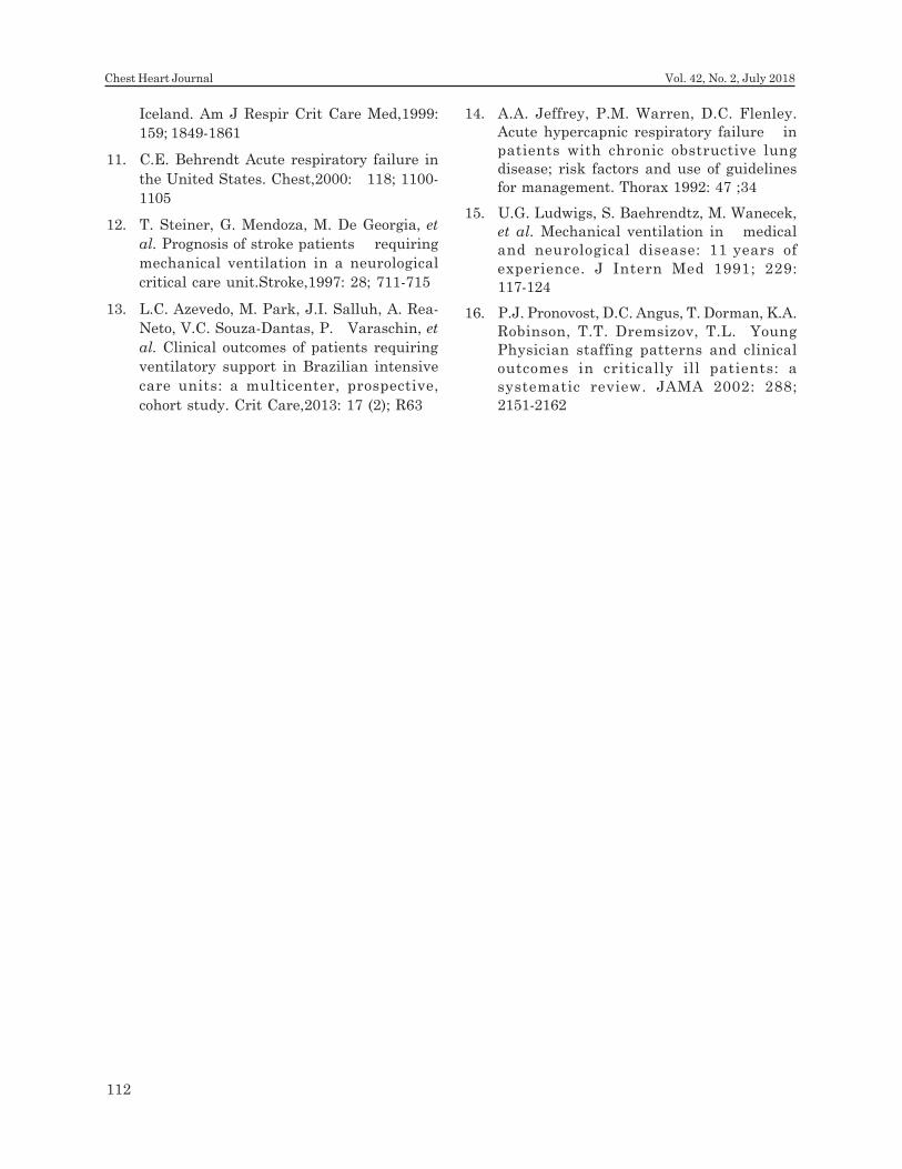
Iceland. Am J Respir Crit Care Med,1999:
159; 1849-1861
11. C.E. Behrendt Acute respiratory failure in
the United States. Chest,2000: 118; 1100-
1105
12. T. Steiner, G. Mendoza, M. De Georgia, et
al. Prognosis of stroke patients requiring
mechanical ventilation in a neurological
critical care unit.Stroke,1997: 28; 711-715
13. L.C. Azevedo, M. Park, J.I. Salluh, A. Rea-
Neto, V.C. Souza-Dantas, P. Varaschin, et
al. Clinical outcomes of patients requiring
ventilatory support in Brazilian intensive
care units: a multicenter, prospective,
cohort study. Crit Care,2013: 17 (2); R63
14. A.A. Jeffrey, P.M. Warren, D.C. Flenley.
Acute hypercapnic respiratory failure in
patients with chronic obstructive lung
disease; risk factors and use of guidelines
for management. Thorax 1992: 47 ;34
15. U.G. Ludwigs, S. Baehrendtz, M. Wanecek,
et al. Mechanical ventilation in medical
and neurological disease: 11 years of
experience. J Intern Med 1991; 229:
117-124
16. P.J. Pronovost, D.C. Angus, T. Dorman, K.A.
Robinson, T.T. Dremsizov, T.L. Young
Physician staffing patterns and clinical
outcomes in critically ill patients: a
systematic review. JAMA 2002: 288;
2151-2162
112
Chest Heart Journal Vol. 42, No. 2, July 2018
Chest Heart Journal
Vol. 42, No. 2, July 2018
ORIGINAL ARTICLE
Blood eosinophils and inhaled corticosteroid/long
acting β β β β β2 agonist efficacy on Quality of life and
COPD exacerbation rate in Stable Chronic
Obstructive Pulmonary DiseasesMd. Shahjada Tabrez1, Md. Mahabubur Rahman2, Mahboba Akther3,
Mohammed Kamrul Hasan4, Md. Ali Hossain5,Bashir Ahmed5, Md. Abdur Rouf5,
Bipul Kanti Biswas6, Liaquat Ali7, Kazi Saifuddin Bennoor8
Abstract:
Background: COPD patients with increased airway eosinophilic inflammation show a
favorable response to inhaled corticosteroids (ICS) in combination with a long-acting
bronchodilator(LABA). Thus, this study investigated the effect of 3-months treatment with
Inhaled Corticosteroid/long-acting beta2-agonist (LABA) in stable COPD patients with
high blood eosinophils with an improvement in quality of life and reduction of COPD
exacerbation rate.
Methods: It was a interventional study conducted at the outpatient department of
Respiratory Medicine in National Institute of Diseases of the Chest and Hospital (NIDCH),
Mohakhali, Dhaka from which 80 stable COPD patients were selected. Baseline blood
eosinophils level was measured of all patients and randomly assigned to 12-weeks
treatment withSalmeterol/fluticasone proprionate inhaler (ICS/LABA) 25/250 mgin
group A and Salmeterol 25mg in group B. Subjects began 3-month ICS/LABA treatment
after washout period. Subjective measurement of symptoms by COPD assessment test
(CAT) score were done in initial visit and during follow up at 4th ,8th and 12th week. Among
all , 29 patients in group A and 28 patients in group B came up to final follow-up.
Results: In this study, Mean COPD Assessment Test (CAT) score change between two
group in first visit was 1.53 (p<0.01), at second visit 1.45 (p<0.05) and at final visit was
2.06 (p<0.05). Differences were statistically significant.CAT score decreases in consecutive
3 follow up than baseline record in both group.CAT score with baseline records between
group A and group B showed statistically significant differences (p=<0.05).Thus, patients
with eosinophilia, ICS-based therapy was associated with significant improvements in
CAT scores and lower incidence of acute exacerbation (3.45% vs 7.14% ) compared with
bronchodilator (BD) therapy alone.
1. Medical Officer, National Institute of Diseases of the Chest & Hospital (NIDCH), Mohakhali, Dhaka,
Bangladesh.
2 . Medical Officer, NIDCH), Mohakhali,Dhaka, Bangladesh.
3. MD Phase B Resident (Neonatology), BSMMU, Shahbag, Dhaka, Bangladesh.
4. MD (Chest Diseases) Final Part Student, NIDCH, Mohakhali, Dhaka, Bangladesh.
5. Professor of Respiratory Medicine, NIDCH, Mohakhali, Dhaka, Bangladesh.
6. Associate Professor, Respiratory Medicine, NIDCH,Mohakhali, Dhaka, Bangladesh.
7. Professor of Biochemistry and Cell Biology, BUHS, Mirpur, Dhaka, Bangladesh.
8. Assistant Professor of Respiratory Medicine, NIDCH, Mohakhali, Dhaka, Bangladesh.
Correspondence to: Dr. Md. Shahjada Tabrez, Medical Officer, MU-7, National Institute of Diseases of the
Chest & Hospital (NIDCH), Mohakhali, Dhaka, Bangladesh. Cell: 01915478978, e-mail: [email protected]
Submission on: 10 May, 2018 Accepted for Publication: 28 June, 2018
Available at http.//www.chabjournal.org
Introduction:
COPD is a progressive inflammatory disorder
that is characterized by persistent airflow
limitation1. It affects approximately 10% of
adults over 40 years of age and is the fourth
leading cause of death worldwide2. Although
most COPD guidelines advocate the use of
inhaled corticosteroids (ICS) for only those with
frequent exacerbations, in real clinical practice,
they are widely used especially in combination
with a long-acting beta2- agonist (LABA)3.
Because only a small fraction of COPD patients
are responsive to ICS-based therapy4,
identifying characteristics associated with ICS-
responsiveness is crucial for clinicians to make
therapeutic decisions. Although neutrophils are
thought to play a prominent role in the
pathogenesis of COPD5, Christenson et al6
recently reported that eosinophilia and T helper
type 2 (Th2) inflammation might also play a
significant role in a subset of patients with COPD
and that approximately 20% of smokers with
COPD have a Th2-high signature. This result is
consistent with previous findings that airway
eosinophilic inflammation is not uncommon, but
is present in approximately 20%–40% of COPD
patients5. Inhaled corticosteroids (ICS) are an
important treatment for COPD1. Exacerbations,
defined as acute worsening of symptoms
necessitating treatment with antibiotics and/or
systemic corticosteroids or hospitalization, are
a key determinant of COPD morbidity, mortality
and healthcare costs7. Compared with placebo,
ICS such as fluticasone propionate (FP) and
budesonide reduce exacerbations by up to 20%
as monotherapy, and up to 30% in combination
with a long-acting β2-agonist (LABA)8. National
and international guidelines on the management
of COPD1 recommend that patients with COPD
at risk of exacerbations receive ICS/LABA
maintenance therapy. A predictive marker for
ICS/LABAeffectiveness in preventing COPD
exacerbations could aid clinical decision-making
by identifying patients likely to gain the most
beneût from ICS-based treatment. Blood
eosinophil count may provide such a marker.
Studies have demonstrated associations between
airway eosinophilia and exacerbations of chronic
bronchitisand COPD9. Exacerbations are
heterogeneous, presenting as one of four distinct
phenotypes, and airway eosinophilia in the stable
state was found to be predictive of subsequentexacerbation phenotype10. The use of systemic
corticosteroids in patients experiencing acute
exacerbations of COPD has shown greaterbeneût in patients with a blood eosinophil level
of ≥2% versus those with <2%11. There is alsoevidence for an association between airway
eosinophilia with response to systemic
corticosteroids for quality of life11. A recentretrospective analysis of data from two parallel
1-year studies of once-daily ICS/LABA,ûuticasone furoate (FF)/vilanterol (VI) in patients
with moderate-to-very severe COPD showed agreater reduction of moderate and severe
exacerbations in patients with a blood eosinophil
level ≥2% vs <2% when treated with ICS/LABAcompared with LABA alone12.Thus this study is
designedto investigate the potential of bloodeosinophil level as a marker for the efûcacy of
ICS/LABA on quality of life and prevention of
exacerbations in stable COPD patients. Therelationship between dyspnea and quality of life
in COPD has previously been indicated in a fewstudies13. Perceived dyspnea was shown to have
a greater impact on health-related quality of lifethan spirometric or functional measurements in
these patients14. In an international study, the
most frequently reported symptom was dyspnea(78%)and the most frequent complaint reported
by patients with COPD was daily activitylimitation15. Hajiro, et al.13 showed that dyspnea
is one of the main determinations of disease
specific health related quality of life, and hasmoderate to strong correlations with
impairments in the health related quality of life
in patients with COPD. On the other hand,
quality of life measurements did not correlate
well with the severity of airflow limitation16.
Conclusions: High blood eosinophils are associated with improved lung function after
3-months ICS/LABA treatment in COPD patients. So, in combination with age and
baseline lung function parameters, blood eosinophils may be a possible biomarker for
identification of COPD patients with favorable response to ICS/ LABA treatment.
Key words: COPD, Eosinophil, Inhaled corticosteroid, COPD Assessment Test (CAT) score.
[Chest Heart Journal 2018; 42(2) : 113-119]
DOI: http://dx.doi.org/10.33316/chab.j.v42i2.2019588
114
Chest Heart Journal Vol. 42, No. 2, July 2018
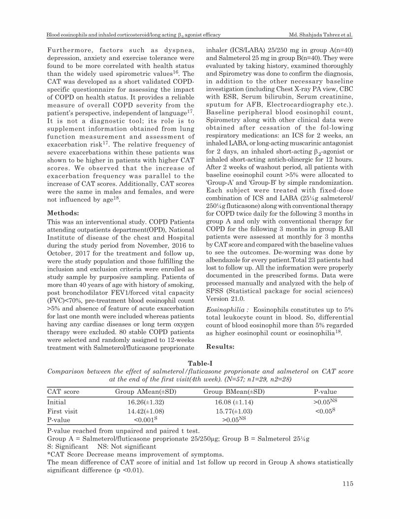
Furthermore, factors such as dyspnea,
depression, anxiety and exercise tolerance were
found to be more correlated with health status
than the widely used spirometric values16. The
CAT was developed as a short validated COPD-
specific questionnaire for assessing the impact
of COPD on health status. It provides a reliable
measure of overall COPD severity from the
patient’s perspective, independent of language17.
It is not a diagnostic tool; its role is to
supplement information obtained from lung
function measurement and assessment of
exacerbation risk17. The relative frequency of
severe exacerbations within these patients was
shown to be higher in patients with higher CAT
scores. We observed that the increase of
exacerbation frequency was parallel to the
increase of CAT scores. Additionally, CAT scores
were the same in males and females, and were
not influenced by age18.
Methods:
This was an interventional study. COPD Patients
attending outpatients department(OPD), National
Institute of disease of the chest and Hospital
during the study period from November, 2016 to
October, 2017 for the treatment and follow up,
were the study population and those fulfilling the
inclusion and exclusion criteria were enrolled as
study sample by purposive sampling. Patients of
more than 40 years of age with history of smoking,
post bronchodilator FEV1/forced vital capacity
(FVC)<70%, pre-treatment blood eosinophil count
>5% and absence of feature of acute exacerbation
for last one month were included whereas patients
having any cardiac diseases or long term oxygen
therapy were excluded. 80 stable COPD patients
were selected and randomly assigned to 12-weeks
treatment with Salmeterol/fluticasone proprionate
inhaler (ICS/LABA) 25/250 mg in group A(n=40)
and Salmeterol 25 mg in group B(n=40). They were
evaluated by taking history, examined thoroughlyand Spirometry was done to confirm the diagnosis,in addition to the other necessary baselineinvestigation (including Chest X-ray PA view, CBCwith ESR, Serum bilirubin, Serum creatinine,
sputum for AFB, Electrocardiography etc.).Baseline peripheral blood eosinophil count,Spirometry along with other clinical data wereobtained after cessation of the followingrespiratory medications: an ICS for 2 weeks, aninhaled LABA, or long-acting muscarinic antagonist
for 2 days, an inhaled short-acting β2-agonist or
inhaled short-acting anticholinergic for 12 hours.After 2 weeks of washout period, all patients withbaseline eosinophil count >5% were allocated to‘Group-A’ and ‘Group-B’ by simple randomization.Each subject were treated with fixed-dose
combination of ICS and LABA (25¼g salmeterol/250¼g fluticasone) along with conventional therapyfor COPD twice daily for the following 3 months ingroup A and only with conventional therapy forCOPD for the following 3 months in group B.Allpatients were assessed at monthly for 3 months
by CAT score and compared with the baseline valuesto see the outcomes. De-worming was done byalbendazole for every patient.Total 23 patients hadlost to follow up. All the information were properlydocumented in the prescribed forms. Data wereprocessed manually and analyzed with the help of
SPSS (Statistical package for social sciences)Version 21.0.
Eosinophilia : Eosinophils constitutes up to 5%total leukocyte count in blood. So, differential
count of blood eosinophil more than 5% regarded
as higher eosinophil count or eosinophilia18.
Results:
Table-I
Comparison between the effect of salmeterol/fluticasone proprionate and salmeterol on CAT score
at the end of the first visit(4th week). (N=57; n1=29, n2=28)
CAT score Group AMean(±SD) Group BMean(±SD) P-value
Initial 16.26(±1.32) 16.08 (±1.14) >0.05NS
First visit 14.42(±1.08) 15.77(±1.03) <0.05S
P-value <0.001S >0.05NS
P-value reached from unpaired and paired t test.
Group A = Salmeterol/fluticasone proprionate 25/250µg; Group B = Salmeterol 25¼g
S: Significant NS: Not significant
*CAT Score Decrease means improvement of symptoms.
The mean difference of CAT score of initial and 1st follow up record in Group A shows statistically
significant difference (p <0.01).
115
Blood eosinophils and inhaled corticosteroid/long acting β2 agonist efficacy Md. Shahjada Tabrez et al.
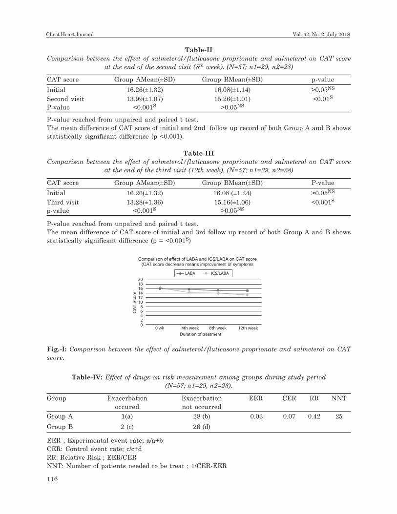
Table-II
Comparison between the effect of salmeterol/fluticasone proprionate and salmeterol on CAT score
at the end of the second visit (8th week). (N=57; n1=29, n2=28)
CAT score Group AMean(±SD) Group BMean(±SD) p-value
Initial 16.26(±1.32) 16.08(±1.14) >0.05NS
Second visit 13.99(±1.07) 15.26(±1.01) <0.01S
P-value <0.001S >0.05NS
P-value reached from unpaired and paired t test.
The mean difference of CAT score of initial and 2nd follow up record of both Group A and B shows
statistically significant difference (p <0.001).
Table-III
Comparison between the effect of salmeterol/fluticasone proprionate and salmeterol on CAT score
at the end of the third visit (12th week). (N=57; n1=29, n2=28)
CAT score Group AMean(±SD) Group BMean(±SD) P-value
Initial 16.26(±1.32) 16.08 (±1.24) >0.05NS
Third visit 13.28(±1.36) 15.16(±1.06) <0.001S
p-value <0.001S >0.05NS
P-value reached from unpaired and paired t test.
The mean difference of CAT score of initial and 3rd follow up record of both Group A and B shows
statistically significant difference (p = <0.001S)
Table-IV: Effect of drugs on risk measurement among groups during study period
(N=57; n1=29, n2=28).
Group Exacerbation Exacerbation EER CER RR NNT
occured not occurred
Group A 1(a) 28 (b) 0.03 0.07 0.42 25
Group B 2 (c) 26 (d)
EER : Experimental event rate; a/a+b
CER: Control event rate; c/c+d
RR: Relative Risk ; EER/CER
NNT: Number of patients needed to be treat ; 1/CER-EER
Fig.-I: Comparison between the effect of salmeterol/fluticasone proprionate and salmeterol on CAT
score.
LABA ICS/LABA
Duration of treatment
0 wk
CA
T S
co
re
4th week 8th week 12th week
20
18
16
14
12
10
8
6
4
2
0
Comparison of effect of LABA and ICS/LABA on CAT score(CAT score decrease means improvement of symptoms
116
Chest Heart Journal Vol. 42, No. 2, July 2018
Discussion
In this study, all patients of COPD were selected
with blood eosinophilia (>5%). Pre-treatment
blood eosinophil count in group A was 5.93±2.16
and group B was 5.84±1.86 which correlates with
the study of in which they aimed to investigate
the potential of blood eosinophil level as a marker
for the preventive efûcacy of ICS/LABA in COPD
patients19.In this study the mean age of the group
A patients was 58.70 ±8.56 years and group B
patients was 57.25±10.03 years. The mean age
difference was not statistically significant.A
similar study20 showed mean age of patients in
both groups were 64.3±18.1 years. The mean age
of the present study was lower can be explained,
as the average life expectancy is comparatively
low in our set up as compared to the Western
world21.Among group A patients, highest
percentage were male (93.1%) and 6.9% female.
Similarly in group B patients, highest percentage
were male (96.4%) and only 3.6% were female.
No statistically significant sex difference was
found between the two groups of patients which
correlated with the study22, in which overall male
were 97% and only 3% were female.In the
present study, COPD Assessment Test (CAT)
score change in 1st visit decreased 1.84(±0.67)
in group A and decreased 0.31(±0.02) in group B
that was statistically significant (p<0.01). Mean
CAT score change in 2nd visit decreased 2.27
(±0.53) in group A and decreased 0.82 (±0.07) in
group B that was statistically significant (p<0.05).
Mean CAT change in 3rd visit decreased 2.98
(±0.38) in group A and decreased 0.92 (±0.13) in
group B that was statistically significant(p<0.05)
. Findings of this study consistent with the result
of the study22, in which their data extend these
findings by demonstrating that high blood
eosinophils were associated with treatment
response (defined as an increase in FEV1 and
decrease in CAT score from baseline) following
ICS and LABA treatment in COPD for 3
months.Few previous studies have evaluated the
use of eosinophil cell counts as a biomarker of
ICS responses in patients with COPD. Leigh et
al.200623observed an association of
sputumeosinophilia(>3% in induced samples)
with significant improvements indyspnea
following treatment with inhaled corticosteroid.
Similar findings were reported for
prednisone24.A post hoc analysis of data from
the Foster 48-week trial to reduce exacerbations
in COPD to examine the role of blood eosinophil
levels on treatment responses and observed that
in the highest eosinophil count group, treatment
with fluticasone plus LABA resulted in
significantly better improvements of quality of
life, measured by decremental CAT score(P-
values <0.001)25. Cheng et al. 2017 used data
from a previous prospective randomized study
and classified patients into higher and lower
eosinophil count groups26. The authors observed
that patients with high plasma eosinophilia have
a significantly greater pulmonary response, a
reduced risk of acute exacerbations and
improved CAT scores when treated with a
combination of ICS- and bronchodilator-based
therapy, compared with bronchodilator therapy
alone. (P<0.05).Lee et al.2016 also evaluated the
effectiveness and safety of high- or medium-dose
ICS when combined with salmeterol for patients
with different blood eosinophil counts in COPD
and found similar results as this study reveals26.
DiSantostefano et al.2013 also found that blood
eosinophil level was the primary driver of
treatment response, with a greater treatment
effect observed in patients with blood eosinophil
levels >2.4% when treated with ICS/LABA versus
LABA alone27.
Conclusions:
This study concludes that ICS with conventional
therapy significantly improves lung functions as
revealed by FEV1 and quality of life as revealed
by CAT score in patients with COPD with higher
blood eosinophils. ICS with conventional therapy
significantly reduce frequency of exacerbation
with improvement of clinical outcomes as
compared to conventional therapy.
Acknowledgement:
Author Contributions: Dr. Md. Shahjada Tabrez
had full access to all data in the study and takes
responsibility for the integrity of the data and
accuracy of the data analysis.
Dr. Md. Shahjada Tabrez contributed to study
conception and design, data collection, analysis
and interpretation of data, drafting of the
manuscript and critical revisions of the article.
117
Blood eosinophils and inhaled corticosteroid/long acting β2 agonist efficacy Md. Shahjada Tabrez et al.
Professor Md. Ali Hossain, Dr. Md. Mahabubur
Rahman, Dr. Mahboba Akther, Dr.
KaziSaifuddinBennoorcontributed to concept and
design of the study and critical revisions of the
manuscript.
Conflict of Interest: The authors of this paper
have declared that there is no conflict of interest
to any of the authors.
Funding Agency: This study has been performed
without any funding from outside else.
References:
1. Global Initiative for Chronic Obstructive
Lung Disease. Global Strategy for the
Diagnosis, Management and Prevention of
Chronic Obstructive Pulmonary Disease;
2015.
2. Lozano R, Naghavi M, Foreman K, et al.
Global and regional mortality from 235
causes of death for 20 age groups in 1990
and 2010: a systematic analysis for the
Global Burden of Disease Study 2010.
Lancet. 2012;380(9859):2095–2128.
3. Price D, West D, Brusselle G, et al.
Management of COPD in the UK primary-
care setting: an analysis of real-life
prescribing patterns. Int J Chron Obstruct
Pulmon Dis. 2014;9:889–904.
4. Miravitlles M, Soler-Cataluna JJ, Calle M,
Soriano JB. Treatment of COPD by clinical
phenotypes: putting old evidence into
clinical practice. Eur Respir J.
2013;41(6):1252–1256.
5. Barnes PJ. Mediators of chronic obstructive
pulmonary disease. Pharmacol Rev.
2004;56(4):515–548.
6. Christenson SA, Steiling K, van den Berge
M, et al. Asthma-COPD overlap. Clinical
relevance of genomic signatures of type 2
inflammation in chronic obstructive
pulmonary disease. Am J Respir Crit Care
Med. 2015;191(7): 758–766.
7. Anzueto A. Impact of exacerbations on
COPD. Eur Respir Rev 2010;19:113–18.
8. Calverley P, Pauwels R, Vestbo J, et al.
Combined salmeterol and ûuticasone in the
treatment of chronic obstructive pulmonary
disease: a randomised controlled trial.
Lancet 2003;361:449–56.
9. Siva R, Green RH, Brightling CE, et al.
Eosinophilic airway inûammation and
exacerbations of COPD: a randomised
controlled trial. Eur Respir J 2007;29:
906–13.
10. Bafadhel M, McKenna S, Terry S, et al.
Acute exacerbations of COPD: identiûcation
of biological clusters and their biomarkers.
Am J Respir Crit Care Med 2011;184:
662–71.
11. Bafadhel M, McKenna S, Terry S, et al.
Blood eosinophils to direct corticosteroid
treatment of exacerbations of chronic
obstructive pulmonary disease: a
randomized placebo-controlled trial. Am J
Respir Crit Care Med 2012;186:48–55.
12. Pascoe S, Locantore N, Dransûeld MT, et
al. Blood eosinophil counts, exacerbations,
and response to the addition of inhaled
ûuticasone furoate to vilanterol in
patientwith chronic obstructive pulmonary
disease: a secondary analysis of data from
two parallel randomised controlled trials.
Lancet Respir Med 2015;3:435–42.
13. Hajiro T, Nishimura K, Tsukino M, Ikeda
A, Oga T, Izumi T. A comparison of the level
of dyspnea vs disease severity in indicating
the health-related quality of life of patients
with COPD. Chest 1999;116:1632-7
14. Von Leupoldt A, Hahn E, Taube K,
Schubert-Heukeshoven S, Magnussen H,
Dahme B. Effects of 3-week outpatient
pulmonary rehabilitation on exercise
capacity, dyspnea, and quality of life in
COPD. Lung 2008;186:387-91.
15. Miravitlles M, Anzueto A, Legnani D,
Forstmeier L, Fargel M. Patient’s
perception of exacerbations of COPD—the
PERCEIVE study. Respir Med 2007;101:
453-60.
16. Mahler DA, Mackowiak JI. Evaluation of the
short-form 36-item questionnaire to
measure health-related quality of life in
patients with COPD. Chest 1995;107:
1585-9.
118
Chest Heart Journal Vol. 42, No. 2, July 2018
17. Jones PW, Brusselle G, Dal Negro RW,
Ferrer M, Kardos P, Levy ML, et al.
Properties of the COPD assessment test in
a cross-sectional European study. Eur
Respir J 2011;38:29-35.
18. Blumenreich MS. The White Blood Cell and
Differential Count. In: Walker HK, Hall WD,
Hurst JW, editors. Clinical Methods: The
History, Physical, and Laboratory
Examinations. 3rd edition. Boston:
Butterworths, Chapter 1990;153:724-727.
19. Pavord ID, Lettis S, Locantore N, et al.
Blood eosinophils and inhaled
corticosteroid/long-acting beta-2 agonist
efficacy in COPD.Thorax, 2016;71:118–125.
20. Cheng S-L, Wang HH, Lin C-H. Effect of
allergic phenotype on treatment response
to inhaled bronchodilators with or without
inhaled corticosteroids in patients with
COPD. International Journal of Chronic
Obstructive Pulmonary Disease, 2017;
12:2231-2238.
21. Iqbal F, Hussain S, Hassan M.
Hypertension, diabetes mellitus and
hypercholesterolemia as risk factors for
stroke. Pak J Med Sci, 2003;42(1):17-22.
22. Park HY, Lee H, Koh W-J, et al. Association
of blood eosinophils and plasma periostin
with FEV1 response after 3-month inhaled
corticosteroid and long-acting beta2-agonist
treatment in stable COPD patients.
International Journal of Chronic
Obstructive Pulmonary Disease, 2016;11:
23-30.
23. Leigh R, Pizzichini MM, Morris MM,
Maltais F, Hargreave FE, Pizzichini E.
Stable COPD:predicting benefit from high-
dose inhaled corticosteroid treatment. Eur
Respir J, 2006;27(5):964–971.
24. Brightling CE, Monteiro W, Ward R, et al.
Sputum eosinophilia and short-term
response to prednisolone in chronic
obstructive pulmonary disease: a
randomized controlled trial. Lancet,
2000;356:1480–1485.
25. Siddiqui SH, Guasconi A, Vestbo Jet al.
Blood eosinophils: a biomarker of response
to extrafine beclomethasone/formoterol in
chronic obstructive pulmonary disease. Am
J Respir Crit Care Med, 2015;192(4):523–525.
26. Lee SY, Park HY, Kim EK, et al.
Combination therapy of inhaled steroids and
long-acting beta2-agonists in asthma-COPD
overlap syndrome. Int J Chron Obstruct
Pulmon Dis, 2016;11:2797–2803.
27. DiSantostefano RL, Li H, Rubin DB, et al.
Which patients with chronic obstructive
pulmonary disease benefit from the addition
of an inhaled corticosteroid to their
bronchodilator? A cluster analysis. BMJ
Open, 2013;3:e001838
119
Blood eosinophils and inhaled corticosteroid/long acting β2 agonist efficacy Md. Shahjada Tabrez et al.
Chest Heart Journal
Vol. 42, No. 2, July 2018
ORIGINAL ARTICLE
Assessing treatment Patterns of acute
Exacerbation of Chronic Bronchitis (AECB) in
Outpatient Settings- BangladeshMahmud Masum Attar1, Iqbal Hasan Mahmood2, Asif Mujtaba Mahmud3,
Abdullah Al-Karim4
Abstract:
Background: Chronic obstructive pulmonary disease (COPD) is a heterogeneous group
of disorder that is broadly defined and encompasses several clinical and pathologic
entities, primarily emphysema and chronic bronchitis. Being a lifelong disease, COPD
patients require constant care and special care is needed for acute exacerbation of their
condition along with the proper assessment of their treatment pattern and medications.
Though a global guideline for management of COPD (GOLD) along with other management
protocols are available in different countries, but in Bangladesh there is no local guideline
and little data regarding the treatment pattern and medications including the use of
antibiotic during the exacerbation phase of COPD. Therefore, this study has been designed
to assess the use of antibiotic during acute exacerbation of chronic bronchitis (AECB) in
outpatient settings- Bangladesh.
Methods: This was a multicenter, observational, prospective study. A total of 525 patients
were planned to be enrolled in outpatient (Male & Female) > 18 years and presenting
with suspected acute exacerbation of chronic bronchitis (AECB) where the physician
intend to treat with antibiotic as well as signed informed consent to meet the inclusion
criteria in this observational study from 20 centers. The physicians were selected
randomly from a list of physicians who have post graduations in Respiratory Medicine
and General practitioner with special training on Respiratory Medicine. Clinical
diagnosis was based on sign, symptoms, radiological evidence and laboratory results as
per routine practices. Patient data were collected by the investigators during baseline
(Visit 1), intermediate (Visit 2) (if decided by the physicians) and at the end of antibiotic
therapy. At visit 2 the effectiveness of the therapy in terms of therapeutic response and
patients’ compliance as per physicians’ choice were assessed. Therapeutic response were
evaluated in terms of Cure, Improvement, failure and relapse by the investigators.
Results: Among the enrolled patients there were 435 (83%) male with age varied from
20~91 years (57.9±13.5). More than 80% of the patients were 50 years and older and only
55 (10.47%) was below 40 years old. The rural resident patients were larger than urban
and semi urban (48.8% vs. 36.4% and 14.9%). 34.5% were employed, 34.9% were previously
employed, 15.4% were retired person and 15.2% were house wife. There were 26.5% of
current smoker, 51% of former smoker, and 22.5% of never smoker. Abnormal breath
1. National Institute of Disease of Chest & Hospital, Dhaka, Bangladesh.
2. M. H. Samarita Medical College & Hospital, Dhaka, Bangladesh.
3. Asgar Ali Hospital, Dhaka, Bangladesh.
4. Sanofi Bangladesh Limited, Dhaka, Bangladesh.
Corresponding to: Dr. Mahmud Masum Attar, National Institute of Disease of Chest & Hospital, Bangladesh.
Submission on: 14 May, 2018 Accepted for Publication: 29 June, 2018
Available at http.//www.chabjournal.org
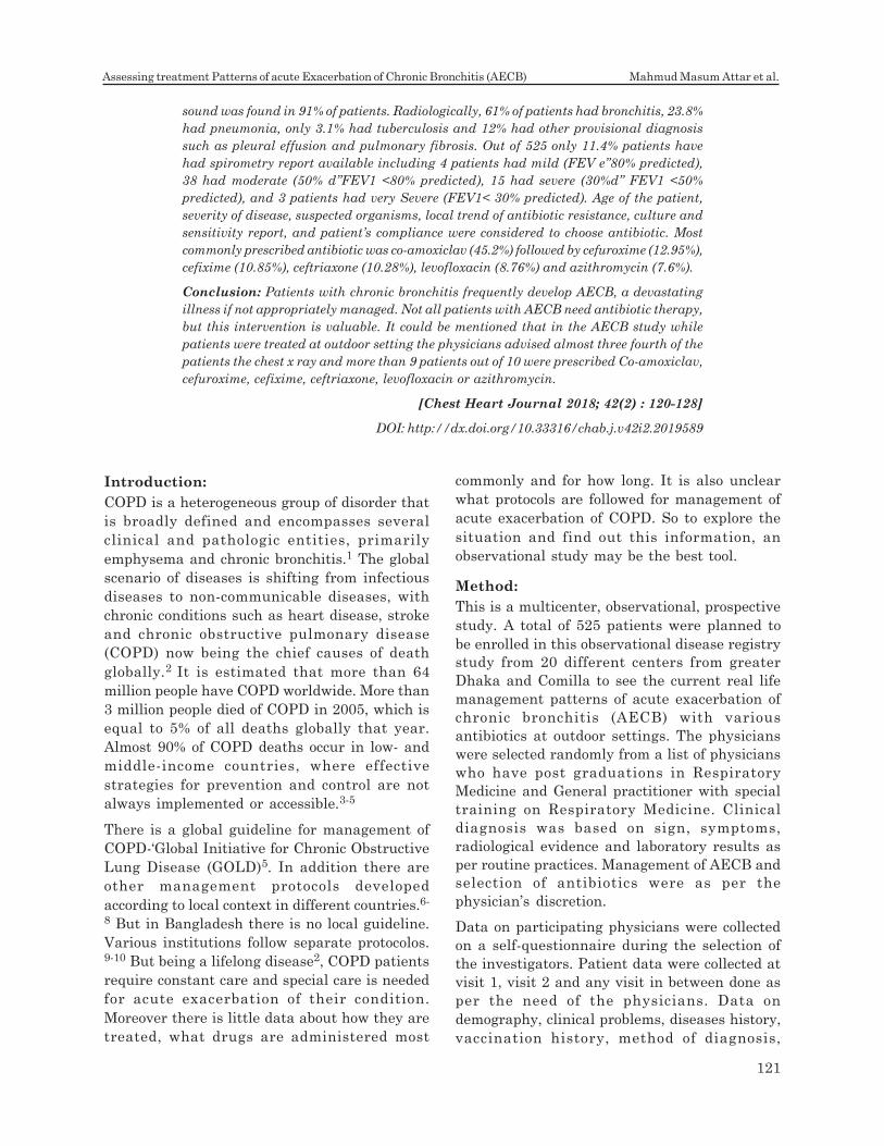
Introduction:
COPD is a heterogeneous group of disorder that
is broadly defined and encompasses several
clinical and pathologic entities, primarily
emphysema and chronic bronchitis.1 The global
scenario of diseases is shifting from infectious
diseases to non-communicable diseases, with
chronic conditions such as heart disease, stroke
and chronic obstructive pulmonary disease
(COPD) now being the chief causes of death
globally.2 It is estimated that more than 64
million people have COPD worldwide. More than
3 million people died of COPD in 2005, which is
equal to 5% of all deaths globally that year.
Almost 90% of COPD deaths occur in low- and
middle-income countries, where effective
strategies for prevention and control are not
always implemented or accessible.3-5
There is a global guideline for management of
COPD-‘Global Initiative for Chronic Obstructive
Lung Disease (GOLD)5. In addition there are
other management protocols developed
according to local context in different countries.6-
8 But in Bangladesh there is no local guideline.
Various institutions follow separate protocolos.9-10 But being a lifelong disease2, COPD patients
require constant care and special care is needed
for acute exacerbation of their condition.
Moreover there is little data about how they are
treated, what drugs are administered most
commonly and for how long. It is also unclear
what protocols are followed for management of
acute exacerbation of COPD. So to explore the
situation and find out this information, an
observational study may be the best tool.
Method:
This is a multicenter, observational, prospective
study. A total of 525 patients were planned to
be enrolled in this observational disease registry
study from 20 different centers from greater
Dhaka and Comilla to see the current real life
management patterns of acute exacerbation of
chronic bronchitis (AECB) with various
antibiotics at outdoor settings. The physicians
were selected randomly from a list of physicians
who have post graduations in Respiratory
Medicine and General practitioner with special
training on Respiratory Medicine. Clinical
diagnosis was based on sign, symptoms,
radiological evidence and laboratory results as
per routine practices. Management of AECB and
selection of antibiotics were as per the
physician’s discretion.
Data on participating physicians were collected
on a self-questionnaire during the selection of
the investigators. Patient data were collected at
visit 1, visit 2 and any visit in between done as
per the need of the physicians. Data on
demography, clinical problems, diseases history,
vaccination history, method of diagnosis,
sound was found in 91% of patients. Radiologically, 61% of patients had bronchitis, 23.8%
had pneumonia, only 3.1% had tuberculosis and 12% had other provisional diagnosis
such as pleural effusion and pulmonary fibrosis. Out of 525 only 11.4% patients have
had spirometry report available including 4 patients had mild (FEV e”80% predicted),
38 had moderate (50% d”FEV1 <80% predicted), 15 had severe (30%d” FEV1 <50%
predicted), and 3 patients had very Severe (FEV1< 30% predicted). Age of the patient,
severity of disease, suspected organisms, local trend of antibiotic resistance, culture and
sensitivity report, and patient’s compliance were considered to choose antibiotic. Most
commonly prescribed antibiotic was co-amoxiclav (45.2%) followed by cefuroxime (12.95%),
cefixime (10.85%), ceftriaxone (10.28%), levofloxacin (8.76%) and azithromycin (7.6%).
Conclusion: Patients with chronic bronchitis frequently develop AECB, a devastating
illness if not appropriately managed. Not all patients with AECB need antibiotic therapy,
but this intervention is valuable. It could be mentioned that in the AECB study while
patients were treated at outdoor setting the physicians advised almost three fourth of the
patients the chest x ray and more than 9 patients out of 10 were prescribed Co-amoxiclav,
cefuroxime, cefixime, ceftriaxone, levofloxacin or azithromycin.
[Chest Heart Journal 2018; 42(2) : 120-128]
DOI: http://dx.doi.org/10.33316/chab.j.v42i2.2019589
121
Assessing treatment Patterns of acute Exacerbation of Chronic Bronchitis (AECB) Mahmud Masum Attar et al.
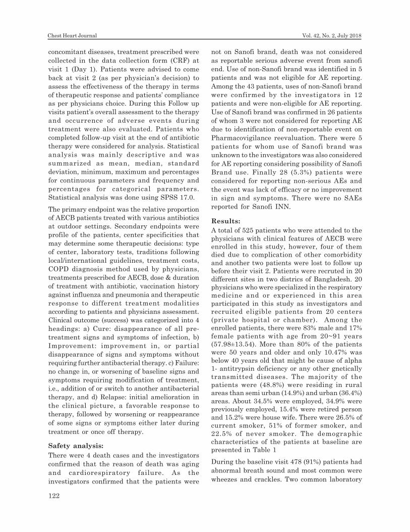
concomitant diseases, treatment prescribed were
collected in the data collection form (CRF) at
visit 1 (Day 1). Patients were advised to come
back at visit 2 (as per physician’s decision) to
assess the effectiveness of the therapy in terms
of therapeutic response and patients’ compliance
as per physicians choice. During this Follow up
visits patient’s overall assessment to the therapy
and occurrence of adverse events during
treatment were also evaluated. Patients who
completed follow-up visit at the end of antibiotic
therapy were considered for analysis. Statistical
analysis was mainly descriptive and was
summarized as mean, median, standard
deviation, minimum, maximum and percentages
for continuous parameters and frequency and
percentages for categorical parameters.
Statistical analysis was done using SPSS 17.0.
The primary endpoint was the relative proportion
of AECB patients treated with various antibiotics
at outdoor settings. Secondary endpoints were
profile of the patients, center specificities that
may determine some therapeutic decisions: type
of center, laboratory tests, traditions following
local/international guidelines, treatment costs,
COPD diagnosis method used by physicians,
treatments prescribed for AECB, dose & duration
of treatment with antibiotic, vaccination history
against influenza and pneumonia and therapeutic
response to different treatment modalities
according to patients and physicians assessment.
Clinical outcome (success) was categorized into 4
headings: a) Cure: disappearance of all pre-
treatment signs and symptoms of infection, b)
Improvement: improvement in, or partial
disappearance of signs and symptoms without
requiring further antibacterial therapy. c) Failure:
no change in, or worsening of baseline signs and
symptoms requiring modification of treatment,
i.e., addition of or switch to another antibacterial
therapy, and d) Relapse: initial amelioration in
the clinical picture, a favorable response to
therapy, followed by worsening or reappearance
of some signs or symptoms either later during
treatment or once off therapy.
Safety analysis:
There were 4 death cases and the investigators
confirmed that the reason of death was aging
and cardiorespiratory failure. As the
investigators confirmed that the patients were
not on Sanofi brand, death was not considered
as reportable serious adverse event from sanofi
end. Use of non-Sanofi brand was identified in 5
patients and was not eligible for AE reporting.
Among the 43 patients, uses of non-Sanofi brand
were confirmed by the investigators in 12
patients and were non-eligible for AE reporting.
Use of Sanofi brand was confirmed in 26 patients
of whom 3 were not considered for reporting AE
due to identification of non-reportable event on
Pharmacovigilance reevaluation. There were 5
patients for whom use of Sanofi brand was
unknown to the investigators was also considered
for AE reporting considering possibility of Sanofi
Brand use. Finally 28 (5.3%) patients were
considered for reporting non-serious AEs and
the event was lack of efficacy or no improvement
in sign and symptoms. There were no SAEs
reported for Sanofi INN.
Results:
A total of 525 patients who were attended to the
physicians with clinical features of AECB were
enrolled in this study, however, four of them
died due to complication of other comorbidity
and another two patients were lost to follow up
before their visit 2. Patients were recruted in 20
different sites in two districs of Bangladesh. 20
physicians who were specialized in the respiratory
medicine and or experienced in this area
participated in this study as investigators and
recruited eligible patients from 20 centers
(private hospital or chamber). Among the
enrolled patients, there were 83% male and 17%
female patients with age from 20~91 years
(57.98±13.54). More than 80% of the patients
were 50 years and older and only 10.47% was
below 40 years old that might be cause of alpha
1- antitrypsin deficiency or any other gnetically
transmitted diseases. The majority of the
patients were (48.8%) were residing in rural
areas than semi urban (14.9%) and urban (36.4%)
areas. About 34.5% were employed, 34.9% were
previously employed, 15.4% were retired person
and 15.2% were house wife. There were 26.5% of
current smoker, 51% of former smoker, and
22.5% of never smoker. The demographic
characteristics of the patients at baseline are
presented in Table 1
During the baseline visit 478 (91%) patients had
abnormal breath sound and most common were
wheezes and crackles. Two common laboratory
122
Chest Heart Journal Vol. 42, No. 2, July 2018
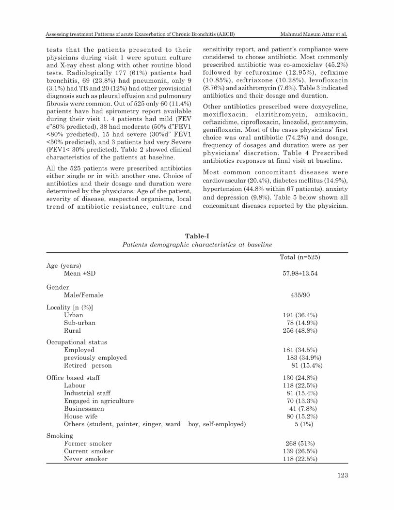
tests that the patients presented to their
physicians during visit 1 were sputum culture
and X-ray chest along with other routine bloodtests. Radiologically 177 (61%) patients had
bronchitis, 69 (23.8%) had pneumonia, only 9
(3.1%) had TB and 20 (12%) had other provisionaldiagnosis such as pleural effusion and pulmonary
fibrosis were common. Out of 525 only 60 (11.4%)patients have had spirometry report available
during their visit 1. 4 patients had mild (FEV
e”80% predicted), 38 had moderate (50% d”FEV1<80% predicted), 15 had severe (30%d” FEV1
<50% predicted), and 3 patients had very Severe(FEV1< 30% predicted). Table 2 showed clinical
characteristics of the patients at baseline.
All the 525 patients were prescribed antibioticseither single or in with another one. Choice of
antibiotics and their dosage and duration weredetermined by the physicians. Age of the patient,
severity of disease, suspected organisms, local
trend of antibiotic resistance, culture and
sensitivity report, and patient’s compliance were
considered to choose antibiotic. Most commonly
prescribed antibiotic was co-amoxiclav (45.2%)
followed by cefuroxime (12.95%), cefixime
(10.85%), ceftriaxone (10.28%), levofloxacin
(8.76%) and azithromycin (7.6%). Table 3 indicated
antibiotics and their dosage and duration.
Other antibiotics prescribed were doxycycline,
moxifloxacin, clarithromycin, amikacin,
ceftazidime, ciprofloxacin, linezolid, gentamycin,
gemifloxacin. Most of the cases physicians’ first
choice was oral antibiotic (74.2%) and dosage,
frequency of dosages and duration were as per
physicians’ discretion. Table 4 Prescribed
antibiotics responses at final visit at baseline.
Most common concomitant diseases were
cardiovascular (20.4%), diabetes mellitus (14.9%),
hypertension (44.8% within 67 patients), anxiety
and depression (9.8%). Table 5 below shown all
concomitant diseases reported by the physician.
Table-I
Patients demographic characteristics at baseline
Total (n=525)
Age (years)
Mean ±SD 57.98±13.54
Gender
Male/Female 435/90
Locality [n (%)]
Urban 191 (36.4%)
Sub-urban 78 (14.9%)
Rural 256 (48.8%)
Occupational status
Employed 181 (34.5%)
previously employed 183 (34.9%)
Retired person 81 (15.4%)
Office based staff 130 (24.8%)
Labour 118 (22.5%)
Industrial staff 81 (15.4%)
Engaged in agriculture 70 (13.3%)
Businessmen 41 (7.8%)
House wife 80 (15.2%)
Others (student, painter, singer, ward boy, self-employed) 5 (1%)
Smoking
Former smoker 268 (51%)
Current smoker 139 (26.5%)
Never smoker 118 (22.5%)
123
Assessing treatment Patterns of acute Exacerbation of Chronic Bronchitis (AECB) Mahmud Masum Attar et al.
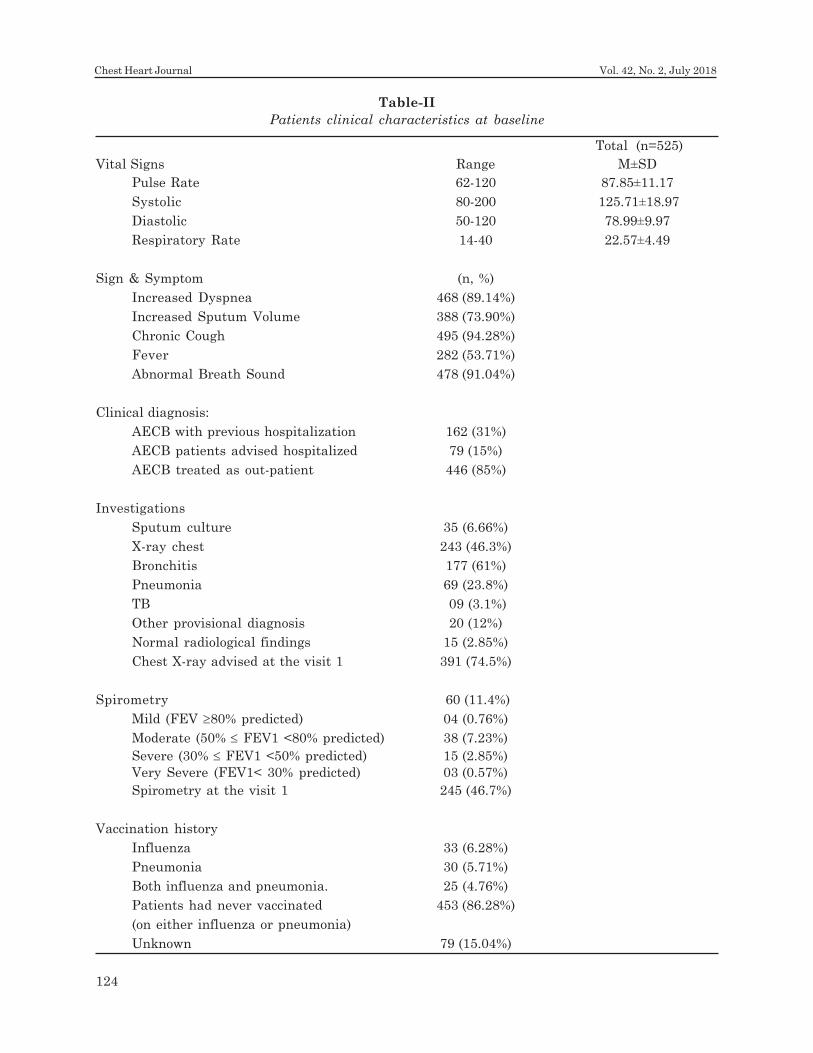
Table-II
Patients clinical characteristics at baseline
Total (n=525)
Vital Signs Range M±SD
Pulse Rate 62-120 87.85±11.17
Systolic 80-200 125.71±18.97
Diastolic 50-120 78.99±9.97
Respiratory Rate 14-40 22.57±4.49
Sign & Symptom (n, %)
Increased Dyspnea 468 (89.14%)
Increased Sputum Volume 388 (73.90%)
Chronic Cough 495 (94.28%)
Fever 282 (53.71%)
Abnormal Breath Sound 478 (91.04%)
Clinical diagnosis:
AECB with previous hospitalization 162 (31%)
AECB patients advised hospitalized 79 (15%)
AECB treated as out-patient 446 (85%)
Investigations
Sputum culture 35 (6.66%)
X-ray chest 243 (46.3%)
Bronchitis 177 (61%)
Pneumonia 69 (23.8%)
TB 09 (3.1%)
Other provisional diagnosis 20 (12%)
Normal radiological findings 15 (2.85%)
Chest X-ray advised at the visit 1 391 (74.5%)
Spirometry 60 (11.4%)
Mild (FEV ≥80% predicted) 04 (0.76%)
Moderate (50% ≤ FEV1 <80% predicted) 38 (7.23%)
Severe (30% ≤ FEV1 <50% predicted) 15 (2.85%)
Very Severe (FEV1< 30% predicted) 03 (0.57%)
Spirometry at the visit 1 245 (46.7%)
Vaccination history
Influenza 33 (6.28%)
Pneumonia 30 (5.71%)
Both influenza and pneumonia. 25 (4.76%)
Patients had never vaccinated 453 (86.28%)
(on either influenza or pneumonia)
Unknown 79 (15.04%)
124
Chest Heart Journal Vol. 42, No. 2, July 2018
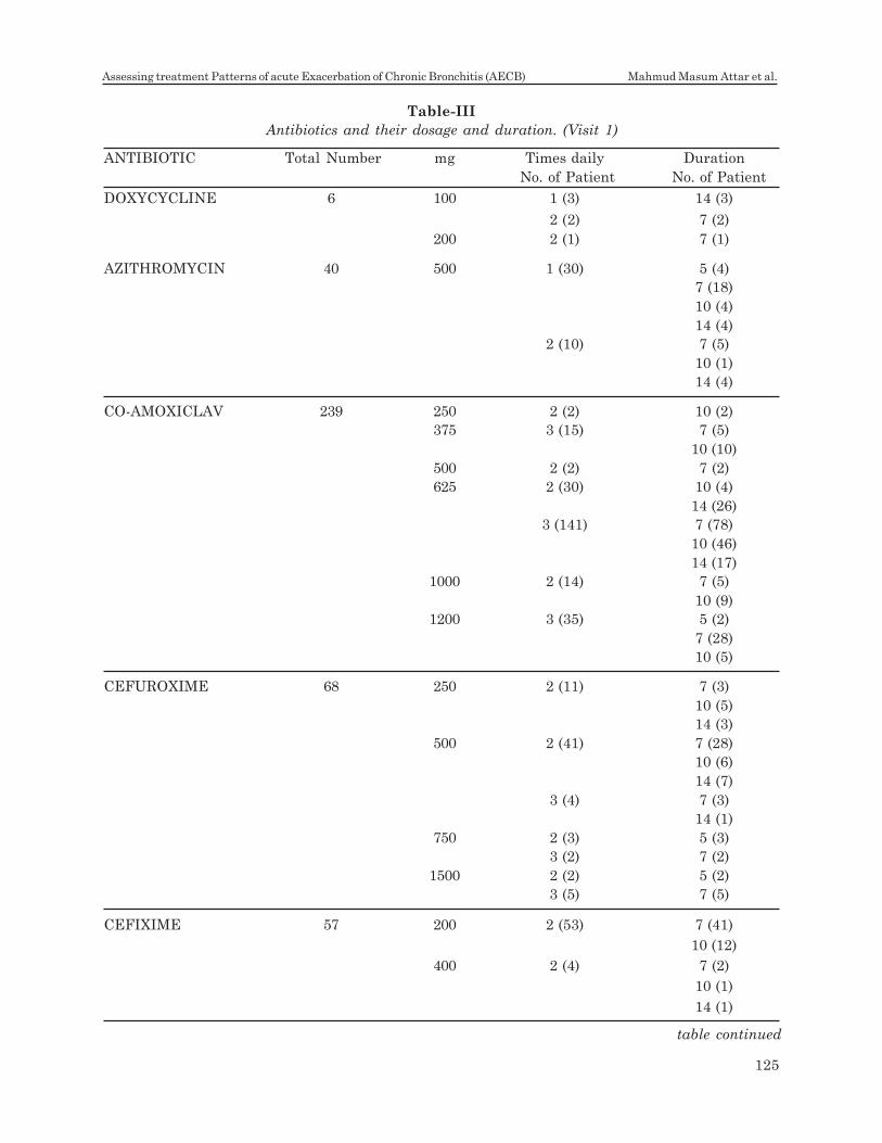
Table-III
Antibiotics and their dosage and duration. (Visit 1)
ANTIBIOTIC Total Number mg Times daily Duration
No. of Patient No. of Patient
DOXYCYCLINE 6 100 1 (3) 14 (3)
2 (2) 7 (2)
200 2 (1) 7 (1)
AZITHROMYCIN 40 500 1 (30) 5 (4)
7 (18)
10 (4)
14 (4)
2 (10) 7 (5)
10 (1)
14 (4)
CO-AMOXICLAV 239 250 2 (2) 10 (2)
375 3 (15) 7 (5)
10 (10)
500 2 (2) 7 (2)
625 2 (30) 10 (4)
14 (26)
3 (141) 7 (78)
10 (46)
14 (17)
1000 2 (14) 7 (5)
10 (9)
1200 3 (35) 5 (2)
7 (28)
10 (5)
CEFUROXIME 68 250 2 (11) 7 (3)
10 (5)
14 (3)
500 2 (41) 7 (28)
10 (6)
14 (7)
3 (4) 7 (3)
14 (1)
750 2 (3) 5 (3)
3 (2) 7 (2)
1500 2 (2) 5 (2)
3 (5) 7 (5)
CEFIXIME 57 200 2 (53) 7 (41)
10 (12)
400 2 (4) 7 (2)
10 (1)
14 (1)
table continued
125
Assessing treatment Patterns of acute Exacerbation of Chronic Bronchitis (AECB) Mahmud Masum Attar et al.
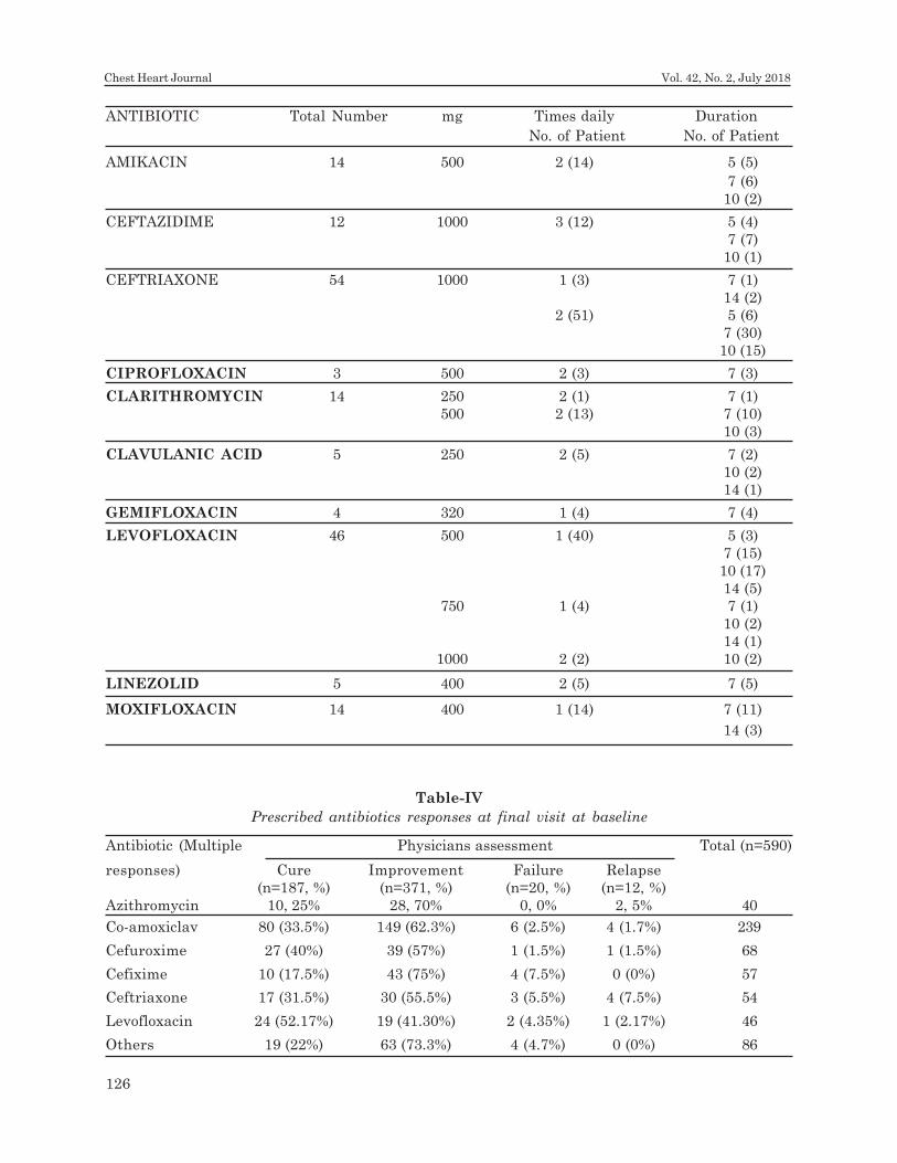
AMIKACIN 14 500 2 (14) 5 (5)
7 (6)
10 (2)
CEFTAZIDIME 12 1000 3 (12) 5 (4)
7 (7)
10 (1)
CEFTRIAXONE 54 1000 1 (3) 7 (1)
14 (2)
2 (51) 5 (6)
7 (30)
10 (15)
CIPROFLOXACIN 3 500 2 (3) 7 (3)
CLARITHROMYCIN 14 250 2 (1) 7 (1)
500 2 (13) 7 (10)
10 (3)
CLAVULANIC ACID 5 250 2 (5) 7 (2)
10 (2)
14 (1)
GEMIFLOXACIN 4 320 1 (4) 7 (4)
LEVOFLOXACIN 46 500 1 (40) 5 (3)
7 (15)
10 (17)
14 (5)
750 1 (4) 7 (1)
10 (2)
14 (1)
1000 2 (2) 10 (2)
LINEZOLID 5 400 2 (5) 7 (5)
MOXIFLOXACIN 14 400 1 (14) 7 (11)
14 (3)
Table-IV
Prescribed antibiotics responses at final visit at baseline
Antibiotic (Multiple Physicians assessment Total (n=590)
responses) Cure Improvement Failure Relapse
(n=187, %) (n=371, %) (n=20, %) (n=12, %)
Azithromycin 10, 25% 28, 70% 0, 0% 2, 5% 40
Co-amoxiclav 80 (33.5%) 149 (62.3%) 6 (2.5%) 4 (1.7%) 239
Cefuroxime 27 (40%) 39 (57%) 1 (1.5%) 1 (1.5%) 68
Cefixime 10 (17.5%) 43 (75%) 4 (7.5%) 0 (0%) 57
Ceftriaxone 17 (31.5%) 30 (55.5%) 3 (5.5%) 4 (7.5%) 54
Levofloxacin 24 (52.17%) 19 (41.30%) 2 (4.35%) 1 (2.17%) 46
Others 19 (22%) 63 (73.3%) 4 (4.7%) 0 (0%) 86
126
Chest Heart Journal Vol. 42, No. 2, July 2018
ANTIBIOTIC Total Number mg Times daily Duration
No. of Patient No. of Patient
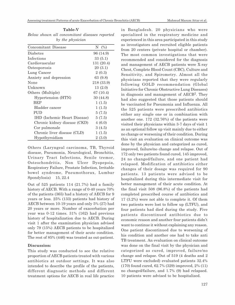
Others (Laryngeal carcinoma, TB, Thyroid
disease, Pneumonia, Neurological, Bronchitis,
Urinary Tract Infections, Senile tremor,
Osteochondritis, Non Ulcer Dyspepsia,
Respiratory Failure, Prostate Infection, Irritable
bowel syndrome, Pneumothorax, Lumbar
Spondylosis) 15, 22.4
Out of 525 patients 114 (21.7%) had a family
history of AECB. With a range of 0-40 years 70%
of the patients (365) had a history of AECB for 9
years or less. 25% (133) patients had history of
AECB between 10-19 years and only 5% (27) had
20 years or more. Number of exacerbation per
year was 0-12 times. 31% (162) had previous
history of hospitalization due to AECB. During
visit 1 after the examination physician advised
only 79 (15%) AECB patients to be hospitalized
for better management of their acute condition.
The rest of 85% (446) was treated as out-patient.
Discussion:
This study was conducted to see the relative
proportion of AECB patients treated with various
antibiotics at outdoor settings. It was also
intended to describe the profile of the patients,
different diagnostic methods and different
treatment options for AECB in real life practice
Table-V
Below shown all concomitant diseases reported
by the physician
Concomitant Disease N (%)
Diabetes 96 (14.9)
Infections 33 (5.1)
Cardiovascular 131 (20.4)
Osteoporosis 20 (3.1)
Lung Cancer 2 (0.3)
Anxiety and depression 63 (9.8)
None 218 (33.9)
Unknown 13 (2.0)
Others (Multiple) 67 (10.4)
Hypertension (HTN) 30 (44.8)
BEP 1 (1.5)
Bladder cancer 1 (1.5)
PUD 5 (7.5)
IHD (Ischemic Heart Disease) 5 (7.5)
Chronic kidney disease (CKD) 4 (6.0)
Cor pulmonale 3 (4.5)
Chronic liver disease (CLD) 1 (1.5)
Hypothyroidism 2 (3.0)
in Bangladesh. 20 physicians who were
specialized in the respiratory medicine and
experienced in this area participated in this study
as investigators and recruited eligible patients
from 20 centers (private hospital or chamber).
The most common investigations that were
recommended and considered for the diagnosis
and management of AECB patients were X-ray
Chest, Complete Blood Count (CBC), Culture and
Sensitivity, and Spirometry. Almost all the
physicians reported that they were regularly
following GOLD recommendation (Global
Initiative for Chronic Obstructive Lung Diseases)
in diagnosis and management of AECB2. They
had also suggested that those patients should
be vaccinated for Pneumonia and Influenza. All
the 525 patients were prescribed antibiotics
either any single one or in combination with
another one. 172 (32.76%) of the patients were
visited their physicians within 3-7 days of visit 1
as an optional follow up visit mainly due to either
no change or worsening of their condition. During
this visit an evaluation on clinical outcome was
done by the physician and categorized as cured,
improved, failure/no change and relapse. Out of
172 only two patients found cured, 145 improved,
24 no changed/failure, and one patient had
relapsed. Modification of antibiotics either
changes of their dosage was required for 90
patients. 13 patients were advised to be
hospitalized during this intermediate visit for
better management of their acute condition. At
the final visit 508 (96.8%) of the patients had
completed prescribed course of antibiotics and
17 (3.2%) were not able to complete it. Of them
two patients were lost to follow up (LTFU), and
four patients had died during the study. Five
patients discontinued antibiotics due to
economic reason and another four patients didn’t
want to continue it without explaining any reason.
One patient discontinued due to worsening of
his condition and another one had to take anti
TB treatment. An evaluation on clinical outcome
was done on the final visit by the physician and
categorized as cured, improved, failure/no
change and relapse. Out of 519 (4 deaths and 2
LTFU were excluded) evaluated patients 32.4%
(170) found cured, 62.7% (329) improved, 2% (11)
no changed/failure, and 1.7% (9) had relapsed.
10 patients were advised to be hospitalized.
127
Assessing treatment Patterns of acute Exacerbation of Chronic Bronchitis (AECB) Mahmud Masum Attar et al.
In this study (AECB) the most common factors
that the physicians considered to prescribe
empirical antibiotics were cardinal symptoms of
the patients followed by guidelines/
recommendations, local sensitivity/resistant
patterns and cost of antibiotic treatment which
were reflected in the responses of the Physician’s
questionnaire. All most all the physicians
participated in this study reported that they were
regularly following GOLD recommendation2
(Global Initiative for Chronic Obstructive Lung
Diseases) in diagnosis and management of AECB.
Although many of these patient related factors
and the causative organism related factors were
considered during taking decision on the selection
of antibiotic regimen by the physician in outdoor
setting in Bangladesh but it could not be confirmed
if the participating physicians systematically
categorize the patient based on their historical
information, lung function and shifting of
associated organisms in the decision making.
This study has been conducted in 20 different
centers from the two districts towns which do
not represent entire population especially
patient from the rural part of Bangladesh. A
longer follow-up could evaluate the effect of
antibiotic therapy in reducing the subsequent
exacerbation in patient with chronic bronchitis.
Conclusion:
Patients with chronic bronchitis frequently
develop AECB, a devastating illness if not
appropriately managed. Not all patients with
AECB need antibiotic therapy, but this
intervention is valuable. It could be mentioned
that in the AECB study while patients were
treated at outdoor setting the physicians advised
almost three fourth of the patients the chest x
ray and more than 9 patients out of 10 were
prescribed Co-amoxiclav, cefuroxime, cefixime,
ceftriaxone, levofloxacin or azithromycin.
Acknowledgement:
This study was sponsored by Sanofi Bangladesh
Limited. The sponsor participated in the study
design and managed all operational aspects of
the study, including monitoring data collection,
statistical analysis and writing of the report. AAK
is an employee of Sanofi Bangladesh Limited &
also the CPL from the sponsor. MMA, IHM &
AMM were investigators in this study. All
authors contributed to the interpretation of the
data and contributed to this publication and
approved the final manuscript for submission to
the target journal. They received no financial
compensation for the authorship.
References:
1. American Thoracic Society/European
Respiratory Society Task Force: Standards
for the diagnosis and management of
patients with COPD. Version 1.2. New York:
American Thoracic Society, 2004. PDF
available for download at thoracic.org/
sections/copd/ (accessed March 20, 2009).
2. Global Initiative for Chronic Obstructive
Lung Disease (GOLD), 2006 MCR VISION,
Inc.
3. World Health Organization, 2014. http://
www.who.int/respiratory/copd/World_
Health_Statistics_2008/en/; accessed at 17/
7/14.
4. The global burden of disease: 2004 update,
2008.
5. World Health Organization, 2008, ‘World
health statistics 2008’, http://www.who.int/
whosis/whostat/EN_WHS08_Full.pdf?ua=1;
accessed at 17/7/14
6. Canadian Thoracic Society Recommendations
for managing chronic obstructive pulmonary
disease-2008 update-highlights for primary
care.
7. NICE 2010, chronic obstructive pulmonary
disease: Management of chronic obstructive
pulmonary disease in adults in primary and
secondary care.
8. American Thoracic Society 2004, ‘COPD
Guideline’.
9. Alam et al, 2010. ‘Prevalence of COPD and
its socioeconomic and lifestyle
determinants in Bangladesh: Preliminary
results from a population based study’.
Primary Care Respiratory Journal 2010;
19(2): A1-A25.
10. Hossain A, Islam K.2009.’ ‘Prevalence and
risk factors of chronic obstructive
pulmonary disease (COPD) in Dhaka city
Bangladesh’. Chest; 136(4_Meeting
Abstracts): 90S-b-91S.
128
Chest Heart Journal Vol. 42, No. 2, July 2018
Chest Heart Journal
Vol. 42, No. 2, July 2018
ORIGINAL ARTICLE
Association between Diabetes Mellitus and
Pulmonary Tuberculosis in AdultsMd Saiful Islam Patwary1, Mohammod Tanvir Islam2, AKM Motiur Rahman Bhuiyan3,
Shamim Ahmed4, Afroja Alam5, Binoy Krishna Tarafder6, Mohammad Kamruzzaman
Mazumder7, Muhammad Jamal Uddin8, Md. Nazim Al – Azad9, Mohammad Farhad10,
Debasish Kumar Ghosh11, Md Zilan Miah Sarker12
Abstract:Background: Tuberculosis (TB) and diabetes mellitus (DM) are national and global
pandemic. Bangladesh is among the top ten countries for both diabetes and tuberculosis
prevalence. The coexistence of both diabetes and tuberculosis has a greater impact on the
disease process and treatment outcome. Hence bidirectional screening for this coexistence
may have a positive role in the better management of both disease conditions.
Methods and Materials: A case-control study was conducted in the Department of
Internal medicine, Bangabandhu Sheikh Mujib Medical University, Dhaka, Bangladesh
among smear positive pulmonary TB patients as cases and non-TB controls. Participants
were tested for fasting plasma glucose and glycosylated hemoglobin (HbA1c) for diagnosis
of diabetes according to ADA guidelines.
Results: Among 142 cases and 142 controls the mean (SD) age was 40.48±14.17 and
42.70± 13.17 years. BMI was <18.5 kg/m² in most of the tuberculosis patients. Among 142
TB cases 45 (31.7%) were diabetic and 74(52.1%) were prediabetic. Tuberculosis was
associated with prediabetes (OR 18.5, CI 5.6 – 60.9) and diabetes (OR 21.2, CI 4.4 -
100).Tuberculosis (TB) cases with Diabetes mellitus were more from urban areas. The
previously diagnosed diabetic cases have poor glycemic control during diagnosis of
tuberculosis.
Conclusion: In this case control study reconfirms the association of pulmonary
tuberculosis and diabetes mellitus. The present study reveals risk of tuberculosis twenty
one times more in diabetes mellitus cases and eighteen times more in prediabetes cases.
This high frequency of diabetes mellitus suggest in adult patient should be tested for
diabetes and patient with cough should be tested for tuberculosis.
Key words: Diabetes mellitus, tuberculosis.
[Chest Heart Journal 2018; 42(2) : 129-136]
DOI: http://dx.doi.org/10.33316/chab.j.v42i2.2019590
1. Medical Officer, UHC, Haimchar, Chandpur, Bangladesh.
2. Associate Professor, Department of Internal Medicine, BSMMU, Bangladesh.
3. Associate Professor , Department of Internal Medicine, BSMMU, Bangladesh.
4. Associate Professor, Respiratory Medicine, BSMMU, Bangladesh.
5. Associate Professor , Department of Internal Medicine, BSMMU, Bangladesh.
6. Associate Professor, Medicine, Colonel Malek Medical college, Manikgonj, Bangladesh.
7. Assistant professor, Medicine, Ibn sina medical college, kollanpur, Dhaka, Bangladesh.
8. OSD, DGHS, Mohakhali, Dhaka, Bangladesh.
9. Junior consultant, Medicine, Mugda Medical College & Hospital, Dhaka, Bangladesh.
10. OSD, DGHS, Mohakhali, Dhaka, Bangladesh.
11. Assistant Professor, (Endocrinology & Metabolism), Khulna Medical College, Khulna, Bangladesh.
12. Professor, Department of Internal Medicine, BSMMU, Bangladesh.
Correspondence to: Dr. Md Saiful Islam Patwary, Medical Officer, UHC, Haimchar, Chandpur,
Mobile: 01711115325, Email: [email protected] on: 10 May, 2018 Accepted for Publication: 10 June, 2018
Available at http.//www.chabjournal.org
Introduction:
Tuberculosis (TB) is a major cause of morbidity
and mortality throughout the world. Bangladesh
stands sixth among the 22 high TB burdened
areas and the annual incidence of TB is 225/
100000 per year in this country. The prevalence
is estimated to be 434 per 100,000 populations.
The estimated TB mortality is 45 per 100,000
populations per year1. Bangladesh, along with
India, China and few other countries are also
overburdened with both Tuberculosis and
Diabetes Mellitus. Number of diabetic patients
around the world is escalating in such a steep
way that by the year 2030, DM will become the
7th leading cause of death around the world2. It
is to be mentioned that, 80% of diabetic death in
the world occurs in the low income countries like
Bangladesh3. Bangladesh remains in the list of
top 10 DM burdened countries around the world
and by the year 2030 the ranking is expected to
go up4. Both diabetes and tuberculosis are global
and national concern.
Recent studies show prevalence of diabetes
mellitus in Bangladesh community prevalence:
30-35% abnormal glucose tolerance of which
diabetes is 8-12%5. Besides 1.75% of adult
population (total 1.58 million) had Impaired
Glucose Tolerance6.
In Bangladesh almost all are type 2 diabetes;
type 1 is rare. About half of all people with
diabetes are unaware of their disease. 80% of
people with diabetes live in low and middle
income countries. Diabetic population shows
8.4% of all cause mortality. Health spending on
diabetes accounts for 10.8% of total health
expenditure worldwide (1).Co-existence of DM
in TB has a greater impact on disease process
and treatment outcome.
Diabetes contributes to the global increase in
incidence of tuberculosis and may become a
threat to the TB control program in coming
days7. DM has been identified as an important
risk factor for development of active TB8. The
incidence of tuberculosis is 2 to 5 times higher
in diabetic patients than the non-diabetic ones8.
A screening program in China showed significant
number of new cases (3%) of DM in TB patients.
Total number of DM cases was also higher in
this population. Moreover they were able to find
7.8% new cases of impaired fasting glucose which
could significantly help in the primary prevention
of diabetes9. Prevalence of DM in TB was found
to be much higher in Kerala (44%), Mexico (36%)
and Texas (39%)10,11.
In China overall prevalence of DM in patients
with TB was 12.4%. Health facilities serving
urban populations and hospitals had a higher
prevalence of DM compared with health facilities
serving rural populations and clinics9.
The innate and adaptive immune responses
necessary for the prevention of tuberculous
proliferation is hampered in diabetic patients8.
TB patients with DM have different
manifestations of the disease and unfavorable
treatment outcome12. It has been found that
these patients have more cavitatory lesions on
chest x-ray, less sputum positivity and a paucity
of clinical presentation13. Both treatment failure
and number of death are high in this group of
people. According to Baker et al they have a
risk ratio (RR) for the combined outcome of
failure and death of 1.69 (95% CI, 1.36 to 2)14.
DM has a 2 fold higher risk of death15. Dooley et
al also showed in the same study that the time
for sputum culture conversion was longer in
pulmonary TB cases with diabetes. Treatment
failure among diabetic patients was also higher
than the non-diabetic TB patients (4.1% vs.
6.7%)15. Diabetic patients are more prone to
develop multidrug-resistant TB and they are
more than 5 times likely to become infected with
drug-resistant strains16.
TB patients with anti-tuberculer drugs in diabetic
patients can be troublesome as drugs like
rifampicin can induce hyperglycemia directly or
by indirect interaction with ongoing oral
hypoglycemic agents17,18. TB infection is also
responsible for reversible impaired glucose
tolerance19. In diabetic patients the plasma
concentrations of isoniazid and rifampicin is
found to be lower20.
This steep rise of the curve of diabetic prevalence
will have an impact on the global prevalence of
tuberculosis also. So detecting diabetes early in
TB cases will be needed for better management
of these “Tubercular diabetic” patients7.
Extensive works have been done in China, India
130
Chest Heart Journal Vol. 42, No. 2, July 2018
and other countries where diabetes has been
screened in TB patients and found to be
higher9,10,21. Diabetic patients were also screened
for TB in these countries and a higher prevalence
was also evident there22,23. Bidirectional
screening was also done for DM and TB in some
places. Currently world health organization
(WHO) also recommends for such bi-directional
screening as detecting early the co-existence of
these diseases can lead to better management
of both cases24.
Methods:
This study was conducted as a case-control study.
Smear positive PTB patients were as cases, non
TB patients were as controls. Cases were
collected from DOTS corner and Outpatients
Department (OPD) of BSMMU and ‘TB control
and training institute’, Chankharpol, Dhaka.
Controls were collected from Medicine OPD,
BSMMU.
This study was carried out during a period of 1st
Jan 2015 to 31st December 2015 in the
Department of Internal Medicine, BSMMU,
Dhaka. A total of 142 patients were cases and
142 were controls. Samples were collected by
convenient sampling technique as per inclusion
and exclusion criteria. PTB was diagnosed by
history ( fever, cough for 3 weeks or more, weight
loss) & bacteriological confirmation by sputum
for Acid Fast Bacilli & GeneXpert. TB was
excluded by history (cough, low grade
intermittent fever, night sweating in past 3
weeks and unexplained weight loss in the past
month), physical examination and relevant
investigations (CBC, CXR, FBS, HbA1c). Study
purpose was explained to the patient and
informed written consent was taken before
collection of data. The findings were entered into
the structured questionnaire and data sheet.
Patient blood samples were obtained and sent
to Biochemistry department to determine the
FBS, HbA1C for both cases and controls.
Complete blood count was done in hematology
and chest x-ray was done in radiology
department for only controls. Later reports were
collected and entered into the data sheet.
Persons fulfilling American Diabetes Association
(ADA) diagnostic criteria 2014 for DM was
enrolled as diabetic. For patients with self-
reported DM, we also measured FBS, HbA1C
level to see current glycaemic status. All
available patients who fulfilled the inclusions
criteria were selectedand analyzed. HbA1c was
measured using the NGSP certified Bio-Rad D-
10TM Hemoglobin A1C Program 220-0101, USA.
All data were expressed as frequencies and mean
(±SD or ± SE). Student’s unpaired t- test for
quantitative data and Chi-Square test for
qualitative data were performed by using the
statistical package for social science (SPSS)
program23. Pearson’s correlation test was used
to see correlation among different variables. P
values ≤0.05 was considered as significant.
Multiple Logistic regression analysis was done
to estimate the role of prediabetes and diabetes
as a risk factor for tuberculosis.
Results:
A total of 284 subjects were enrolled in the study
among which 142 were cases (smear positive
pulmonary TB) and 142 were control. The age
range of the subjects were between 18 and 70
years with the mean age of case group 40.48 ±
14.17 years and control group 42.70±13.17 (table
i). Table I shows the baseline characteristics of
the cases and controls.
There was no statistically significant difference
between the duration of cough and weight loss
among cases. Out of 142 cases previous history
of pulmonary TB was present in 13 (4.6%).Among
cases equal number were smoker and non-
smoker (50%); Among controls non-smokers
(58.50%) were more than smokers though the
difference was not statistically significant. Most
of the cases (60.6%) were under-weight while the
participants of control group had normal weight
(68.3%); differences in BMI among case and
control groups were statistically significant (p
value <0.001). (Table II).
Among 142 TB cases, DM was present in 45
(31.7%) (Previously known DM 32, newly
diagnosed DM 13). 74(52%) cases were
prediabetic. Normal fasting glucose was found
in 23(16.2%). Among 142 controls, DM was found
in 13(9.2%) (Previously known 5, newly diagnosed
8) and prediabetes was found in 30(21.1%), normal
glucose tolerance in 99(69.7%) and were
statistically significant (p<0.001).(Table III).
131
Association between Diabetes Mellitus and Pulmonary Tuberculosis in Adults Md Saiful Islam Patwary et al.
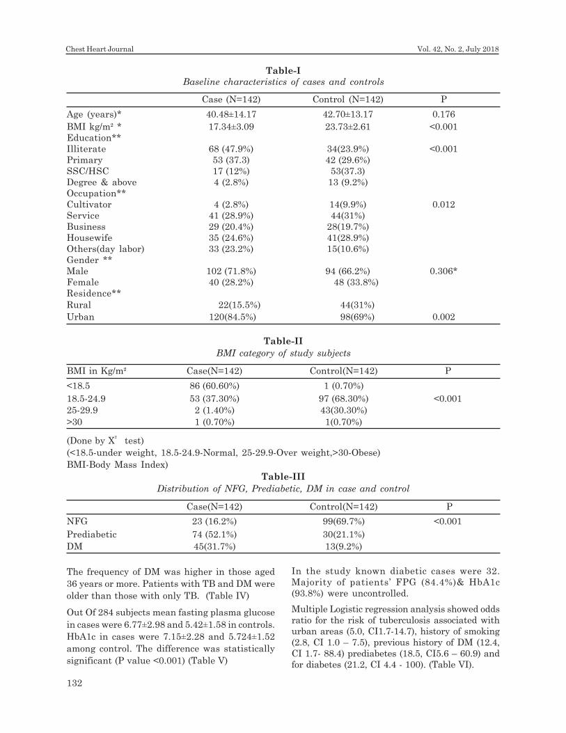
The frequency of DM was higher in those aged
36 years or more. Patients with TB and DM were
older than those with only TB. (Table IV)
Out Of 284 subjects mean fasting plasma glucose
in cases were 6.77±2.98 and 5.42±1.58 in controls.
HbA1c in cases were 7.15±2.28 and 5.724±1.52
among control. The difference was statistically
significant (P value <0.001) (Table V)
In the study known diabetic cases were 32.
Majority of patients’ FPG (84.4%)& HbA1c
(93.8%) were uncontrolled.
Multiple Logistic regression analysis showed odds
ratio for the risk of tuberculosis associated with
urban areas (5.0, CI1.7-14.7), history of smoking
(2.8, CI 1.0 – 7.5), previous history of DM (12.4,
CI 1.7- 88.4) prediabetes (18.5, CI5.6 – 60.9) and
for diabetes (21.2, CI 4.4 - 100). (Table VI).
Table-I
Baseline characteristics of cases and controls
Case (N=142) Control (N=142) P
Age (years)* 40.48±14.17 42.70±13.17 0.176
BMI kg/m² * 17.34±3.09 23.73±2.61 <0.001
Education**
Illiterate 68 (47.9%) 34(23.9%) <0.001
Primary 53 (37.3) 42 (29.6%)
SSC/HSC 17 (12%) 53(37.3)
Degree & above 4 (2.8%) 13 (9.2%)
Occupation**
Cultivator 4 (2.8%) 14(9.9%) 0.012
Service 41 (28.9%) 44(31%)
Business 29 (20.4%) 28(19.7%)
Housewife 35 (24.6%) 41(28.9%)
Others(day labor) 33 (23.2%) 15(10.6%)
Gender **
Male 102 (71.8%) 94 (66.2%) 0.306*
Female 40 (28.2%) 48 (33.8%)
Residence**
Rural 22(15.5%) 44(31%)
Urban 120(84.5%) 98(69%) 0.002
Table-II
BMI category of study subjects
BMI in Kg/m² Case(N=142) Control(N=142) P
<18.5 86 (60.60%) 1 (0.70%)
18.5-24.9 53 (37.30%) 97 (68.30%) <0.001
25-29.9 2 (1.40%) 43(30.30%)
>30 1 (0.70%) 1(0.70%)
(Done by X² test)
(<18.5-under weight, 18.5-24.9-Normal, 25-29.9-Over weight,>30-Obese)
BMI-Body Mass Index)
Table-III
Distribution of NFG, Prediabetic, DM in case and control
Case(N=142) Control(N=142) P
NFG 23 (16.2%) 99(69.7%) <0.001
Prediabetic 74 (52.1%) 30(21.1%)
DM 45(31.7%) 13(9.2%)
132
Chest Heart Journal Vol. 42, No. 2, July 2018
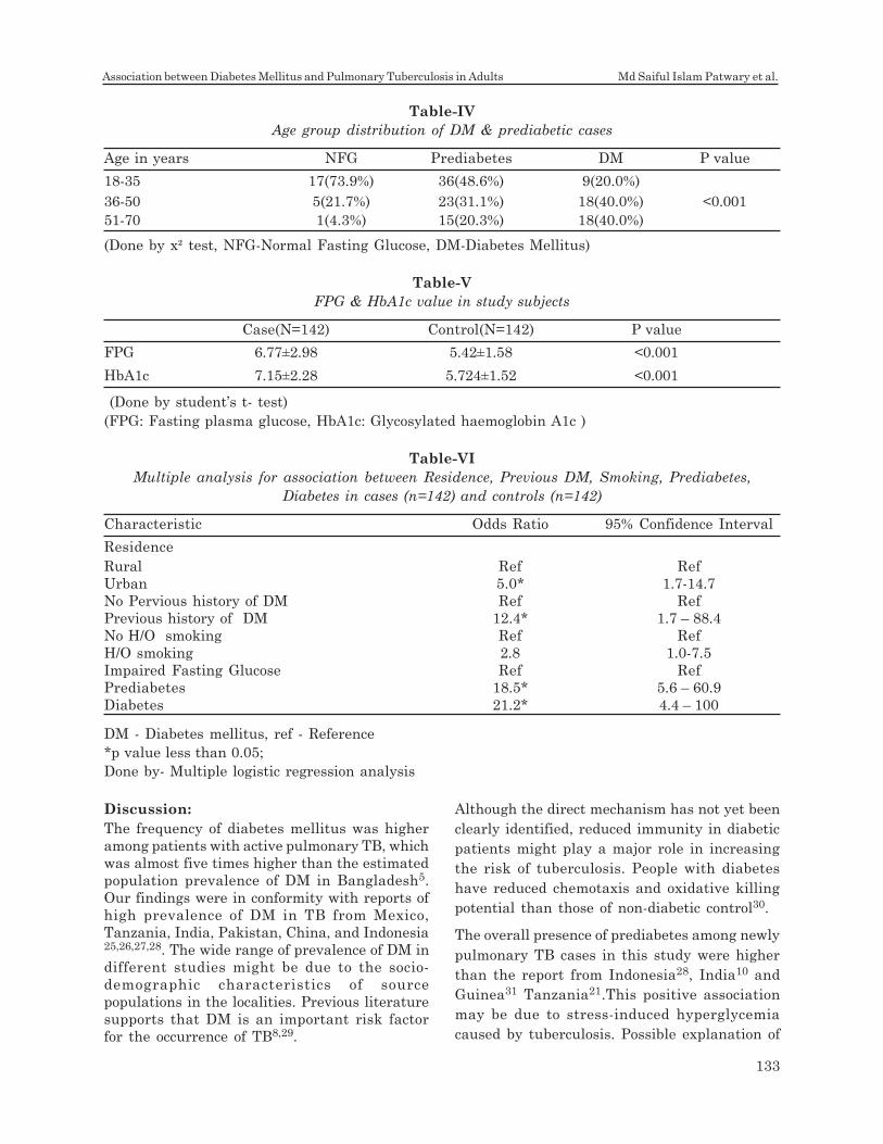
Discussion:
The frequency of diabetes mellitus was higher
among patients with active pulmonary TB, which
was almost five times higher than the estimated
population prevalence of DM in Bangladesh5.
Our findings were in conformity with reports of
high prevalence of DM in TB from Mexico,
Tanzania, India, Pakistan, China, and Indonesia25,26,27,28. The wide range of prevalence of DM in
different studies might be due to the socio-
demographic characteristics of source
populations in the localities. Previous literature
supports that DM is an important risk factor
for the occurrence of TB8,29.
Although the direct mechanism has not yet been
clearly identified, reduced immunity in diabetic
patients might play a major role in increasing
the risk of tuberculosis. People with diabetes
have reduced chemotaxis and oxidative killing
potential than those of non-diabetic control30.
The overall presence of prediabetes among newly
pulmonary TB cases in this study were higher
than the report from Indonesia28, India10 and
Guinea31 Tanzania21.This positive association
may be due to stress-induced hyperglycemia
caused by tuberculosis. Possible explanation of
Table-IV
Age group distribution of DM & prediabetic cases
Age in years NFG Prediabetes DM P value
18-35 17(73.9%) 36(48.6%) 9(20.0%)
36-50 5(21.7%) 23(31.1%) 18(40.0%) <0.001
51-70 1(4.3%) 15(20.3%) 18(40.0%)
(Done by x² test, NFG-Normal Fasting Glucose, DM-Diabetes Mellitus)
Table-V
FPG & HbA1c value in study subjects
Case(N=142) Control(N=142) P value
FPG 6.77±2.98 5.42±1.58 <0.001
HbA1c 7.15±2.28 5.724±1.52 <0.001
(Done by student’s t- test)
(FPG: Fasting plasma glucose, HbA1c: Glycosylated haemoglobin A1c )
Table-VI
Multiple analysis for association between Residence, Previous DM, Smoking, Prediabetes,
Diabetes in cases (n=142) and controls (n=142)
Characteristic Odds Ratio 95% Confidence Interval
Residence
Rural Ref Ref
Urban 5.0* 1.7-14.7
No Pervious history of DM Ref Ref
Previous history of DM 12.4* 1.7 – 88.4
No H/O smoking Ref Ref
H/O smoking 2.8 1.0-7.5
Impaired Fasting Glucose Ref Ref
Prediabetes 18.5* 5.6 – 60.9
Diabetes 21.2* 4.4 – 100
DM - Diabetes mellitus, ref - Reference
*p value less than 0.05;
Done by- Multiple logistic regression analysis
133
Association between Diabetes Mellitus and Pulmonary Tuberculosis in Adults Md Saiful Islam Patwary et al.
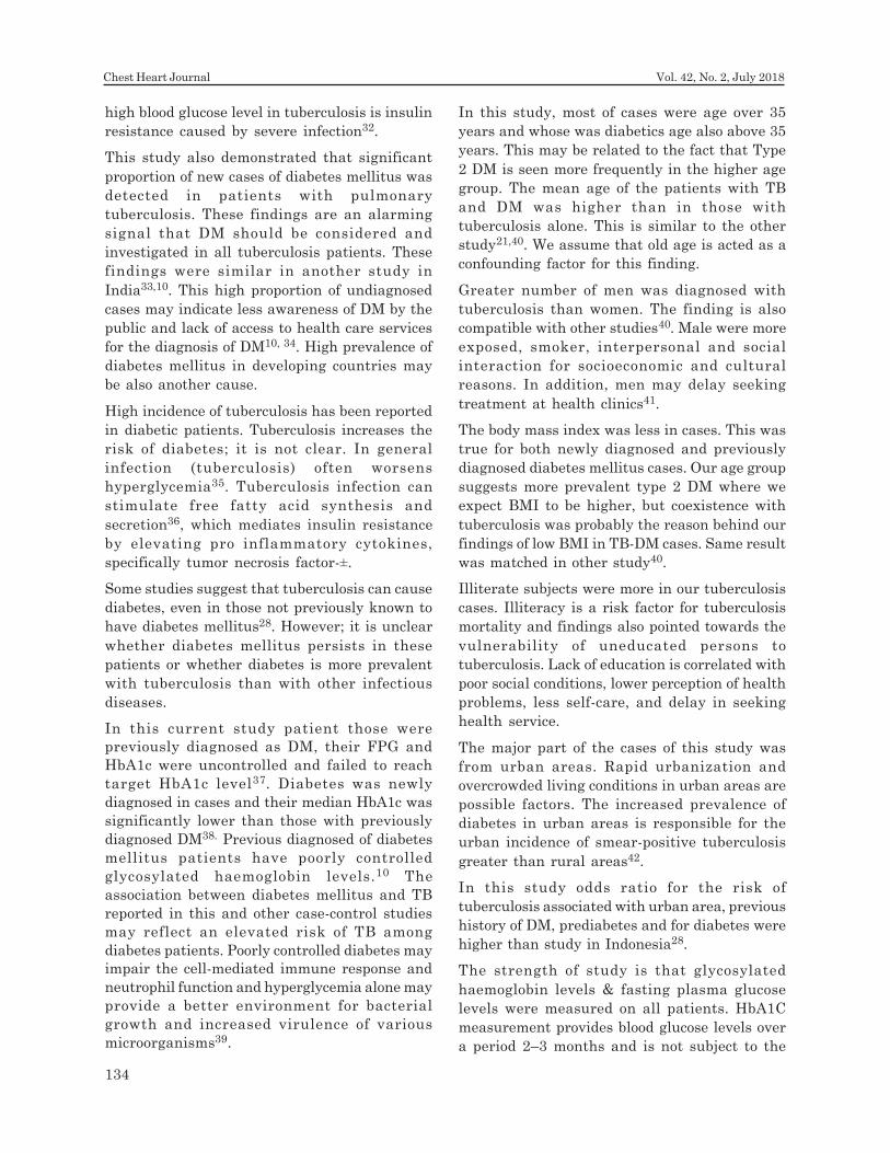
high blood glucose level in tuberculosis is insulin
resistance caused by severe infection32.
This study also demonstrated that significant
proportion of new cases of diabetes mellitus was
detected in patients with pulmonary
tuberculosis. These findings are an alarming
signal that DM should be considered and
investigated in all tuberculosis patients. These
findings were similar in another study in
India33,10. This high proportion of undiagnosed
cases may indicate less awareness of DM by the
public and lack of access to health care services
for the diagnosis of DM10, 34. High prevalence of
diabetes mellitus in developing countries may
be also another cause.
High incidence of tuberculosis has been reported
in diabetic patients. Tuberculosis increases the
risk of diabetes; it is not clear. In general
infection (tuberculosis) often worsens
hyperglycemia35. Tuberculosis infection can
stimulate free fatty acid synthesis and
secretion36, which mediates insulin resistance
by elevating pro inflammatory cytokines,
specifically tumor necrosis factor-±.
Some studies suggest that tuberculosis can cause
diabetes, even in those not previously known to
have diabetes mellitus28. However; it is unclear
whether diabetes mellitus persists in these
patients or whether diabetes is more prevalent
with tuberculosis than with other infectious
diseases.
In this current study patient those were
previously diagnosed as DM, their FPG and
HbA1c were uncontrolled and failed to reach
target HbA1c level37. Diabetes was newly
diagnosed in cases and their median HbA1c was
significantly lower than those with previously
diagnosed DM38. Previous diagnosed of diabetes
mellitus patients have poorly controlled
glycosylated haemoglobin levels.10 The
association between diabetes mellitus and TB
reported in this and other case-control studies
may reflect an elevated risk of TB among
diabetes patients. Poorly controlled diabetes may
impair the cell-mediated immune response and
neutrophil function and hyperglycemia alone may
provide a better environment for bacterial
growth and increased virulence of various
microorganisms39.
In this study, most of cases were age over 35
years and whose was diabetics age also above 35
years. This may be related to the fact that Type
2 DM is seen more frequently in the higher age
group. The mean age of the patients with TB
and DM was higher than in those with
tuberculosis alone. This is similar to the other
study21,40. We assume that old age is acted as a
confounding factor for this finding.
Greater number of men was diagnosed with
tuberculosis than women. The finding is also
compatible with other studies40. Male were more
exposed, smoker, interpersonal and social
interaction for socioeconomic and cultural
reasons. In addition, men may delay seeking
treatment at health clinics41.
The body mass index was less in cases. This was
true for both newly diagnosed and previously
diagnosed diabetes mellitus cases. Our age group
suggests more prevalent type 2 DM where we
expect BMI to be higher, but coexistence with
tuberculosis was probably the reason behind our
findings of low BMI in TB-DM cases. Same result
was matched in other study40.
Illiterate subjects were more in our tuberculosis
cases. Illiteracy is a risk factor for tuberculosis
mortality and findings also pointed towards the
vulnerability of uneducated persons to
tuberculosis. Lack of education is correlated with
poor social conditions, lower perception of health
problems, less self-care, and delay in seeking
health service.
The major part of the cases of this study was
from urban areas. Rapid urbanization and
overcrowded living conditions in urban areas are
possible factors. The increased prevalence of
diabetes in urban areas is responsible for the
urban incidence of smear-positive tuberculosis
greater than rural areas42.
In this study odds ratio for the risk of
tuberculosis associated with urban area, previous
history of DM, prediabetes and for diabetes were
higher than study in Indonesia28.
The strength of study is that glycosylated
haemoglobin levels & fasting plasma glucose
levels were measured on all patients. HbA1C
measurement provides blood glucose levels over
a period 2–3 months and is not subject to the
134
Chest Heart Journal Vol. 42, No. 2, July 2018
rapid swings. Both were separately diagnostic
criteria for diagnosis of diabetes mellitus37.
Conclusion:
Diabetes is a risk factor for tuberculosis. The
increasing diabetes prevalence may be a threat
to TB control. So diabetes prevention and proper
treatment may reduce TB mortality & drug
resistance. Diabetes is an important co-morbid
feature to be sought in patient with TB.
References:
1. National guidelines for the management of
TB –DM . 2014; first edition, 57.
2. Alwan A. Global status report on
noncommunicable diseases 2010; World
Health Organization.
3. Mathers CD, Loncar D. Projections of global
mortality and burden of disease from 2002
to 2030. 2006;. 3( 11) : 442. 318
4. Wild S, Roglic G, Green A, Sicree R, King
H. Global prevalence of diabetes estimates
for the year 2000 and projections for
2030. Diabetes care. 2004; 27(5): 1047-1053.
5. Report on Diabetes camp. 2015; Farida Huq
foundation.
6. International Diabetes Federation. Diabetes
Atlas, 6th edn. Brussels, Belgium:
International Diabetes Federation.2013; 33.
7. Bailey SL, Grant P. ‘The tubercular diabetic’:
the impact of diabetes mellitus on
tuberculosis and its threat to global
tuberculosis control. Clinical Medicine.
2011; 11(4): 344-347.
8. Jeon CY, Murray MB. Diabetes mellitus
increases the risk of active tuberculosis: a
systematic review of 13 observational
studies. 2008; 5( 7): 152.
9. Li L, Lin Y, Mi F, Tan S, et al. Screening of
patients with tuberculosis for diabetes
mellitus in China. Tropical Medicine &
International Health. 2012; 17(10): 1294-
1301.
10. Balakrishnan S, Vijayan S, Nair S, et al.
High diabetes prevalence among
tuberculosis cases in Kerala, India. 2012;
7(10): 502.
11. Restrepo BI, Camerlin AJ, Rahbar M, et al.
Cross-sectional assessment reveals high
diabetes prevalence among newly-diagnosed
tuberculosis cases. Bulletin of the World
Health Organization. 2011; 89(5): 352-359.
12. Wang CS, Yang CJ, Chen HC, et al. Impact
of type 2 diabetes on manifestations and
treatment outcome of pulmonary
tuberculosis, Epidemiology and infection.
2009; 137(02): 203-210.
13. Banerjee S, Banerjee M., Diabetes and
tuberculosis interface.J Indian Med Assoc.
2005; 103: 318, 320, 322 .
14. Baker MA, Harries AD, Jeon CY, et al. The
impact of diabetes on tuberculosis
treatment outcomes: a systematic review,
BMC medicine, 2011; .9 (1 ): 81.
15. Dooley KE, Chaisson RE. Tuberculosis and
diabetes mellitus: convergence of two
epidemics. Lancet Infectious Diseases, 2009;
9: 737–746.
16. Bashar M, Alcabes P, Rom WN, Condos R.
Increased incidence of multidrug-resistant
tuberculosis in diabetic patients on the
Bellevue Chest Service (1987 to 1997). 2001;
120: 1514–9.
17. Takasu N, Yamada T, Miura H et al.
Rifampicin-induced early phase
hyperglycemia in humans. Am Rev Respir
Dis, 1982; 125: 23–7.
18. Niemi M, Backman JT, Fromm MF,
Neuvonen PJ, Kivisto KT. Pharmacokinetic
interactions with rifampicin: clinical
relevance. Clin Pharmacokinet, 2003; 42:
819–50.
19. Oluboyo PO, Erasmus RT. The significance
of glucose intolerance in pulmonary
tuberculosis tubercle, 1990; 71( 2): 135-138.
20. Babalika A, Ulusb I H, Bakircic N, et al.
Plasma Concentrations of Isoniazid and
Rifampin Are Decreased in Adult
Pulmonary Tuberculosis Patients with
Diabetes Mellitus, Antimicrob. Agents
Chemother, 2013; 57(11): 5740-5742.
21. Restrepo BI, Camerlin AJ, Rahbar M, et al.
Cross-sectional assessment reveals high
135
Association between Diabetes Mellitus and Pulmonary Tuberculosis in Adults Md Saiful Islam Patwary et al.
diabetes prevalence among newly-diagnosed
tuberculosis cases. Bulletin of the World
Health Organization, 2011; 89(5): 352-359.
22. Jeon CY, Murray MB. Diabetes mellitus
increases the risk of active tuberculosis: a
systematic review of 13 observational
studies. 2008; 5(7): 152.
23. Jali MV, Mahishale V, Hiremath MB. Screen
all patients with tuberculosis for diab. 24.
World Health organization. 2009. Global
tuberculosis control: a short update to the
report. Geneva: WHO etes. Curr Sci, 2013;
104: 158.
25. Stevenson CR, Forouhi NG, Roglic G,
Williams BG, et al. Diabetes and
tuberculosis: the impact of the diabetes
epidemic on tuberculosisincidence. BMC
Public Health. 2007; 7: 234.
26. Shetty N, Shemko M, Vaz M, D’Souza G.
An epidemiological evaluation of risk factors
for tuberculosis in South India: a matched
case control study. Int J Tuberc Lung Dis.
2006; 10: 80–86.
27. Faurholt-Jepsen D, Range N, Praygod G,
et al. Diabetes is a risk factor for pulmonary
tuberculosis: a case-control study from
Mwanza, Tanzania. 2011; 6( 8): 215
28. Alisjahbana B, van Crevel R, Sahiratmadja
E, et al. Diabetes mellitus is strongly
associated with tuberculosis in Indonesia.
Int J Tuberc Lung Dis, 2006; 10: 696–700.
29. Leung CC, Lam TH, Chan WM, et al.
Diabetic control and risk of tuberculosis: a
cohort study. American Journal of
Epidemiology , 2008; 167(12): 1486-1494.
30. Rayfield EJ, Ault MJ, Keusch GT, et al.
Infection and diabetes: the case for glucose
control. Am J Med, 1982; 72: 439–450.
31. Balde NM, Camara A, Camara LM, et al.
Associated tuberculosis and diabetes in
Conakry, Guinea: prevalence and clinical
characteristics. The international journal of
tuberculosis and lung disease: the official
journal of the International Union against
Tuberculosis and Lung Disease, 2006; 10(9):
1036-1040.
32. Gearhart MM, Parbhoo SK. Hyperglycemia
in the critically ill patient. AACN Clin
Issues, 2006; 17: 50–55.
33. Viswanathan V, Kumpatla S,
Aravindalochanan V, et al. Prevalence of
Diabetes and Pre-Diabetes and Associated
Risk Factors among Tuberculosis Patients
in India. 2012; 7(7): 1367.
34. Hjelm K, Mufunda E. Zimbabwean diabetics’
beliefs about health and illness: an interview
study.BMC International Health and
Human Rights. 2010; 10: 7
35. Wolf G. Serum retinol-binding protein: a link
between obesity, insulin resistance, and type
2 diabetes. Nutr Rev. 2007; 65: 251–256.
36. Podell BK, Ackart DF, Kirk NM, et al. Non-
diabetic hyperglycemia exacerbates disease
severity in Mycobacterium tuberculosis
infected guinea pigs, 2012; 7: 824.
37. American diabetic association. Classification
and Diagnosis of diabetes mellitus. Diabetes
Care. 2014; 38(1): 8-16.
38. Kornfeld H, West K, Kane K, et al. High
Prevalence and Heterogeneity of Diabetes
in TB Patients from South India: A Report
from the Effects of Diabetes on Tuberculosis
Severity (EDOTS) Study, 2016.
39. Geerlings SE, Hoepelman AIM. Immune
dysfunction in patients with diabetes
mellitus (DM). FEMS Immunology &
Medical Microbiology. 1999; 26: 259–265.
40. Blanca I Restrepo, Aulasa J Camerlin,
Mohammad H Rahbar, et al. Bull World
Health Organ. 2011; 89: 352–359.
41. Chan-Yeung M, Noertjojo K, Chan S L, Tam
C M. Sex differences in tuberculosis in Hong
Kong, International Journal of Tuberculosis
and Lung Disease. 2002; 6(1): 11–18.
42. Catherine R, Stevenson, Nita G Forouhi, et
al. Diabetes and tuberculosis: the impact of
the diabetes epidemic on tuberculosis
incidence. BMC Public Health. 2007; 7: 234.
136
Chest Heart Journal Vol. 42, No. 2, July 2018
Chest Heart Journal
Vol. 42, No. 2, July 2018
ORIGINAL ARTICLE
Association of ABO And RH Blood Group with
Hypertension – An Observational StudySobroto Kumar Roy1, Pinaki Ranjan Das 2, Md. Zakir Hossain3, Md. Amzad Hossain4,
AHM Nazmul Ahsan5, Md. Mesbahul Karim Ruble6
Abstract:Objective: The principle aim is to find out the association of ABO and Rh blood group
with hypertension.
Methods: This cross-sectional study was conducted at hypertension and research centre,
Rangpur. A total of 1128 hypertensive patients were included in this study by purposive
sampling method. Staging of hypertension was done according to The JNC 7 Hypertension
Guidelines. ABO and Rh blood group are determined by agglutination method.
Result: This study demonstrated that majority (58.1%) patients were within 40 to 60
years of age. 63.7% patients were male and 36.3% were female. 65% patients were from
rural area and the rest 35% from urban area. Socio-demographic data demonstrate that
48.6% was service holder, businessman 13.5%, farmer 12.2% and others occupation
includes 25.7% like retired person, student etc. Majority (40.7%) patients were poor; middle
class 31.7% and 23%were rich. Maximum observed systolic blood pressure was 170 mm
of Hg and minimum 110 mm of Hg. Maximum diastolic blood pressure was 120 mm of Hg
and minimum 60 mm of Hg. Out of 1128 hypertensive patients 21% patients had normal
BMI, 66.7% overweight, 8.8% moderately obese and 3.5% patients were under weight.
ABO blood group of this hypertensive study subjects showed 41.3% (p<0.001) were blood
group B, group A 32.2% (p=0.25); group O 14.0% (p<0.001) and 12.5% (p<0.001) were
blood group AB. Rh typing of the study patients showed 92.6% (p<0.001) was found Rhesus
positive and only 7.4% (p<0.001) was Rhesus negative. Among the Rh positive group
32.1% (p=0.25) was A positive, 35.5% (p<0.001) B positive, 12.5% (p<0.001) AB positive
and 12.4% (p<0.001) was found O positive respectively. Among Rh negative group 0.1%
(p=0.44) was A negative, 5.8% (p<0.001) B negative, 00%% (p>0.05) AB negative and
1.6% (p=0.001) was found O negative respectively in comparison with another study where
Rh positive blood group were A-21.58%, B-34.58%, AB-8.85%, O- 30.70% and Rh negative
group were A- 0.82%, B-0.96%, AB-0.64%, O-1.87% respectively.
The prevalence of hypertension was more in Rh positive blood group 92.6% (p<0.001),
particularly in Rh B positive 35.5% (p<0.001) and significantly less in A, AB and O blood
group which were found statistically significant.
Conclusion: The prevalence of hypertension is more in Rh B positive and significantly
less in other ABO blood group.
Key Word: Hypertension, ABO blood group, Rh blood group.
[Chest Heart Journal 2018; 42(2) : 137-141]
DOI: http://dx.doi.org/10.33316/chab.j.v42i2.2019591
1. Junior Consultant , Medicine, Sadar Hospital, Gopalgonj, Bangladesh.
2. Assistant Registrar, Cardiology, National Institute of Cardiovascular Disease, Dhaka, Bangladesh.
3. Professor & Head, Department of Medicine, Shaheed Ziaur Rahman Medical College, Bogura, Bangladesh.
4. Assistant Proefssor, Medicine, Shaheed Suhrawardy Medical college, Dhaka, Bangladesh.
5, Associate Proefssor, Medicine, Shaheed Suhrawardy Medical college, Dhaka, Bangladesh.
6. Assistant Proefssor, Respiratory Medicine, Shaheed Suhrawardy Medical college, Dhaka, Bangladesh.
Correspondence to: Dr. Sobroto Kumar Roy, Junior Consultant , Medicine, Sadar Hospital, Gopalgonj,
Bangladesh.Submission on: 14 May, 2018 Accepted for Publication: 29 June, 2018
Available at http.//www.chabjournal.org
Introduction:
Hypertension has proven to be a silent killer
contributing to many deaths and considerably
increasing morbidity worldwide.1 Hypertension
is rapidly emerging as a major public health
problem in developing countries.2 25%of world
adult population is already hypertensive. Almost
three quarters of the hypertensive population
are in developing countries.3 Nationwide survey
on NCD conducted in Bangladesh in 2010
indicated that the prevalence of hypertension is
17.9%.4 Twelve million people suffers from
hypertension in Bangladesh.5 Essential
hypertension generally is regarded as a polygenic
disorder.6
The ABO blood group system was discovered by
Austrian scientist, Karl Landsteiner, who found
three different blood types (A, B and O) in 1900
from serological differences in blood called the
Landsteiner Law.7 In 1902, DesCasterllo and
Sturli discovered the fourth type, AB.8 The genes
of ABO blood group have been determined at
chromosome locus.9-12
The blood group of a person depends upon the
presence or absence of two genes, A and B. In
some study of different parts of the world it has
been shown that there are some correlation
between ABO and Rh blood group with
hypertension. But population based study on
association of hypertension with ABO blood group
and Rh typing lacking in our country.
In most cases the cause of hypertension is
unknown, there may be some genetic influence
though yet not established. ABO blood group and
Rh typing in each individual is also genetically
determined.
So this association might helps in the
identification of hypertension, its early
prevention, treatment and prevention of target
organ damage.
Materials & Methods:
This cross-sectional study was conducted at
Hypertension Research Centre, Rangpur - A
Hypertension care and Research centre from
July, 2012 to December, 2012. A total of 1128
patients were included in this study by purposive
sampling method. All the patients were
diagnosed cases of hypertension (BP >140/90 mm
of Hg). This study included adult patient aged
e”18 years. All cases of secondary hypertension
were excluded by history, physical examination
and relevant investigations. Blood pressure was
measured with a well-calibrated
sphygmomanometer. Staging of hypertension
was done according to The Seventh Report of
the Joint National Committee on Prevention,
detection, Evaluation and treatment of High
Blood Pressure. ABO and Rh blood group of all
hypertensive patients are determined by
agglutination method.
Results:
This study intended to find the association
between hypertension and ABO blood group. The
findings derived from data analyses were
presented below.
Table-I
Distribution of age and sex of the study
subjects (n = 1128)
Age (years) Sex Total
Male Female
18-40 315 (27.92%) 140 (12.41%) 455 (40.3%)
40-60 385 (34.13%) 270 (23.93%) 655 (58.1%)
> 60 18 (1.6%) 00 (00%) 18 (1.6%)
Total 718 (63.7%) 410 (36.3%) 1128 (100%)
In this study, out of 1128 hypertensive patients
majority 655 (58.1%) were between 40-60 year of
age and about 718 (63.7%) were male and 410
(36.3%) were female. (Table-I).
Socio-demographic data demonstrated that
educational status of the study subjects included
majority 314 (27.8%) were graduate and
Occupation comprised majority were Service
holder 548 (48.6%). Most 733 (65%) patients were
from rural areas and the rest (35%) was from
urban areas. Among 1128 hypertensive patients
most of the 751 (66.7%) patients were
overweight.
Majority of the study patients 851(75.4%) had
no family history of hypertension. (Table-II).
Association of ABO and Rh blood group with
hypertension:
In this study among the 1128 subjects 466(41.3%)
belongs to ABO blood group B, 363(32.2%) blood
138
Chest Heart Journal Vol. 42, No. 2, July 2018
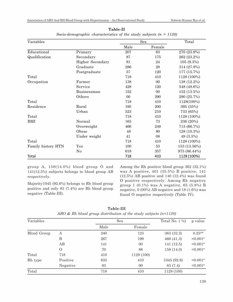
group A, 158(14.0%) blood group O and
141(12.5%) subjects belongs to blood group AB
respectively.
Majority1045 (92.6%) belongs to Rh blood group
positive and only 83 (7.4%) are Rh blood group
negative (Table-III).
Among the Rh positive blood group 362 (32.1%)
was A positive, 401 (35.5%) B positive, 141
(12.5%) AB positive and 140 (12.4%) was found
O positive respectively. Among Rh negative
group 1 (0.1%) was A negative, 65 (5.8%) B
negative, 0 (00%) AB negative and 18 (1.6%) was
found O negative respectively (Table IV).
Table-II
Socio-demographic characteristics of the study subjects (n = 1128)
Variables Sex Total
Male Female
Educational Primary 207 63 270 (23.9%)
Qualification Secondary 87 175 262 (23.2%)
Higher Secondary 81 24 105 (9.3%)
Graduate 286 28 314 (27.8%)
Postgraduate 57 120 177 (15.7%)
Total 718 410 1128 (100%)
Occupation Farmer 138 00 138 (12.2%)
Service 428 120 548 (48.6%)
Businessman 152 00 152 (13.5%)
Others 00 290 290 (25.7%)
Total 718 410 1128(100%)
Residence Rural 195 200 395 (35%)
Urban 523 210 733 (65%)
Total 718 410 1128 (100%)
BMI Normal 163 73 236 (20%)
Overweight 466 249 715 (66.7%)
Obese 48 80 128 (10.3%)
Under weight 41 08 49 (3.5%)
Total 718 410 1128 (100%)
Family history HTN Yes 100 53 153 (13.56%)
No 618 357 975 (86.44%)
Total 718 410 1128 (100%)
Table-III
ABO & Rh blood group distribution of the study subjects (n=1128)
Variables Sex Total No. ( %) p value
Male Female
Blood Group A 240 123 363 (32.2) 0.25ns
B 267 199 466 (41.3) <0.001s
AB 141 00 141 (12.5) <0.001s
O 70 88 158 (14.0) <0.001s
Total 718 410 1128 (100)
Rh type Positive 635 410 1045 (92.6) <0.001s
Negative 83 00 83 (7.4) <0.001s
Total 718 410 1128 (100)
139
Association of ABO And RH Blood Group with Hypertension – An Observational Study Sobroto Kumar Roy et al.

Discussion
In this study among 1128 hypertensive patients
majority (58.1%) were 40 to 60 years of age and
63.7% were male and 36.3% female with Male
female ratio 1.75:1. Hypertension is more
common in men than in women of same age.
Sex difference in the prevalence of
hypertension may be mainly attributed to the
differences in dietary habit, life style choice,
salt intake, Physical activity level and some
genetic polymorphism. 13
Among 1128 hypertensive patients only 13.56%
patients had positive family history and majority
(75.4%) patients had no family history of
hypertension. Majority of the study subjects
66.7% were overweight.
Positive family history is associated with
hypertension prevalence double that found in
patients with negative history and is independent
with weight. When over weight is also present,
however hypertension prevalence is three to four
times as high. 14
In this study among the 1128 subjects blood
group B was found in 41.3% (p<0.001), group A
in 32.2% (p=0.25), group O in 14.0% (p<0.001)
and blood group AB in 12.5% (p<0.001)
respectively (Table III).
The relative frequency of O, A, B and AB blood
group in Western Europe are 46%, 42%, 9% and
3% respectively.15
In the United States, the frequency of O, A, B
and AB blood group is 45%, 41%, 10% and 4%. 16
In a study of our country showed prevalence of
ABO blood group are A - 22.40%. B - 35.54%, AB
– 9.49%, O – 32.57%. 17
This study shows prevalence of hypertension is
high in group B and significantly low in blood
group O. So there was significant association
found between hypertension with blood group B
and O (p value is <0.05 which is statistically
significant).
In this study majority 92.6% (p<0.001) was found
Rhesus positive and only 7.4% (p<0.001) was
Rhesus negative. Among the Rh positive group
32.1% (p=0.25) was A positive, 35.5% (p<0.001)
B positive, 12.5% (p<0.001) AB positive and
12.4% (p<0.001) was found O positive
respectively. Among Rh negative group 0.1%
(p=0.44) was A negative, 5.8% (p<0.001) B
negative, 00% (p>0.05) AB negative and 1.6%
(p=0.001) was found O negative respectively.
Significant association was found in B and O
positive blood group (p value is <0.001).
In a study of Belgium reported an association
between the ABO blood group and blood pressure
among 42, 000 Belgian men. 18 They found that
those with ABO blood type AB had the highest
values of SBP and DBP.
Conclusion & Recommendation:
The prevalence of hypertension is more in Rh B
positive and significantly less in other ABO &
Rh blood group.
The limitation of the present study is data were
collected from single center. Further multi-
Table-IV
ABO & Rh blood group distribution of the study subjects (n=1128)
Blood group with Rh type Sex Total No. ( %) p value
Male Female
A (+ve) 239 123 362 (32.1) 0.25ns
A (-ve) 1 0 1 (0.1) 0.44ns
B (+ve) 202 199 401 (35.5) <0.001s
B (-ve) 65 0 65 (5.8) <0.001s
AB(+ve) 141 0 141 (12.5) <0.001s
O (+ve) 52 88 140 (12.4) <0.001s
O (-ve) 18 0 18 (1.6) 0.001s
Total 718 410 1128 (100)
140
Chest Heart Journal Vol. 42, No. 2, July 2018
center study was recommended to validate the
finding of the present study.
References:
1. Kotchen TA, Hypertensive Vascular
Disease; In: Longo DL, Fauci SA, Kasper
DL, Hauser SL, Jameson JL, Loscalzo J.
Harrison’s Principles of Internal Medicine
,18 th edition. New York: McGrawHill
publishers, 2012; 2042-59.
2. Hypertension Study Group. Prevalence,
awareness, treatment and control of
hypertension among the elderly in
Bangladesh and India: A multicentre study.
Bull World Health Organ. 2001; 79:490–500.
3. Kearny PM, Global burden of Hypertension:
analysis of worldwide data. Lancet 2005;
365:217-23.
4. Rahman M, Chowdhury MAJ et al.NCD Risk
Factor Survey. BSM 2010;1- 35.
5. Sultana M H. Non-adherence to drug
treatment in patients of essential
hypertension. BMRC Bull; 2009; 35: 76-78.
6. McKusick V. Genetics and the nature of
essential hypertension. Circulation
1960;22:857-863.
7. Landsteiner K: Zurr Kenntnis der
antifermentativen, lytischen und
agglutinierenden Wirkungenm des
Blutserums und der Lymphe. Zentralbl
Bacteriol 1900; 27: 357-362.
8. von Decastello A, Struli A: “Ueber die
Isoagglurtinine im serum gesunder und
kranker Menschen”. Mfinch men Wschr
1902; 49: 1090-1095.
9. Watkins WM: Biochemistry and genetics of
the ABO, Lewis, and P blood group system,
In: Advances in Human Genetics (Harris H
& Hirschhorn K eds.). Plenum Press, New
York, 1980, 136-136.
10. Larsen RD, Ernst RK, Nair RP, Lowe JB.
Molecular cloning, sequence, and expression
of a human GDP-L-fucose: b-D-galactoside
2-alpha-L-fucosyltransferase cDNA that can
form the H blood group antigen. Proc Natl
Acad Sci USA 1990: 87 (17):6674-6678.
11. Yamamoto F, Hakomori S. Sugar-nucleotide
donor specificity of histo-blood group A and
B transferases is based on amino acid
substitution. J Biol Chem 1990;265 (31) :
19257-19262.
12. Bennett EP, Steffensen R, Clausen H,
weghuis DO, Geurts van kessel A : Genomic
cloning of the human histo-blood group and
locus.Biochem Biophys Res Commun
1995;206 (1) :318-325.
13. Ruixin Y, Jinzhen W, Shangling P, Weixiong
L, Dezhai Y, Yuming C. Sex differences in
environmental and genetic factors for
hypertension. The American journal of
medicine 2008;121(9):811-819.
14. Stamler R, Stamler J, Reidlinger WF,
Algera G, Roberts RH. Family (Parentaral)
history and prevalence of hypertension.
JAMA 1979;241(1):43-46.
15. Guyton AC, Hall JE. Blood groups;
transfusion;Tissue and Organ
transplantation. In: Textbook of medical
physiology. 10th ed. New Delhi: Prism
Saunders; 2000. 413-7.
16. Ganong WF. Blood types. In: Review of
medical physiology. 21st ed. New York:
McGraw-Hill; 2003. p. 539-42.
17. Pathan AH, Apu As, Jamaluddin ATM,
Asaduzzaman M, Rahman MZ, Rahman A,
et al. Bangladesh J. Life Sci.2008;20(2): 131-
135.
18. Kesteloot H, Van Houte O. An epidemiologic
survey of arterial blood pressure in a large
male population group. Am J Epidemiol.
1974; 99:14-29.
141
Association of ABO And RH Blood Group with Hypertension – An Observational Study Sobroto Kumar Roy et al.
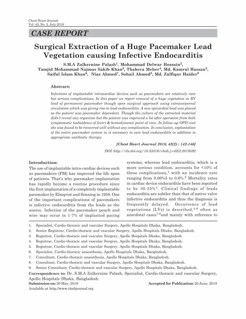
Introduction:
The use of implantable intra-cardiac devices such
as pacemakers (PM) has improved the life span
of patients. That’s why pacemaker implantation
has rapidly become a routine procedure since
the first implantation of a completely implantable
pacemaker by Elmqvist and Senning in 1958. One
of the important complications of pacemakers
is infective endocarditis from the leads as the
source. Infection of the pacemaker pouch and
wire may occur in 1-7% of implanted pacing
systems, whereas lead endocarditis, which is a
more serious condition, accounts for <10% of
these complications,1 with an incidence rate
ranging from 0.06%5 to 0.6%.2 Mortality rates
in cardiac device endocarditis have been reported
to be 30-35%3. Clinical findings of leads
endocarditis are subtler than that of native valve
infective endocarditis and thus the diagnosis is
frequently delayed. Occurrence of lead
vegetations (LVs) is described,4-6 often as
anecdotal cases7-8and mainly with reference to
Surgical Extraction of a Huge Pacemaker Lead
Vegetation causing Infective EndocarditisS.M.A Zulkernine Palash1, Mohammad Delwar Hossain2,
Tamjid Mohammad Najmus Sakib Khan3, Thahera Meher4, Md. Kamrul Hassan5,
Saiful Islam Khan6, Niaz Ahmed7, Sohail Ahmed8, Md. Zulfiqur Haider9
Abstract:
Infections of implantable intracardiac devices such as pacemakers are relatively rare
but serious complications. In this paper we report removal of a huge vegetation in RV
lead of permanent pacemaker though open surgical approach using extracorporeal
circulation which was giving rise to lead endocarditis. A new epicardial lead was placed
as the patient was pacemaker dependent. Though the culture of the extracted material
didn’t reveal any organism but the patient was improved a lot after operation from both
symptomatic (subsidence of fever) & hemodynamic point of view. In follow-up OPD visit
she was found to be recovered well without any complication. In conclusion, explantation
of the entire pacemaker system in is necessary to cure lead endocarditis in addition to
appropriate antibiotic therapy.
[Chest Heart Journal 2018; 42(2) : 142-146]
DOI: http://dx.doi.org/10.33316/chab.j.v42i2.2019592
1. Specialist, Cardio-thoracic and vascular Surgery, Apollo Hospitals Dhaka, Bangladesh.
2. Senior Registrar, Cardio-thoracic and vascular Surgery, Apollo Hospitals Dhaka, Bangladesh.
3. Registrar, Cardio-thoracic and vascular Surgery, Apollo Hospitals Dhaka, Bangladesh.
4. Registrar, Cardio-thoracic and vascular Surgery, Apollo Hospitals Dhaka, Bangladesh.
5. Registrar, Cardio-thoracic and vascular Surgery, Apollo Hospitals Dhaka, Bangladesh.
6. Specialist, Cardio-thoracic anaesthesia, Apollo Hospitals Dhaka, Bangladesh.
7. Consultant, Cardio-thoracic anaesthesia, Apollo Hospitals Dhaka, Bangladesh.
8. Consultant, Cardio-thoracic and vascular Surgery, Apollo Hospitals Dhaka, Bangladesh.
9. Senior Consultant, Cardio-thoracic and vascular Surgery, Apollo Hospitals Dhaka, Bangladesh.
Correspondence to: Dr. S.M.A Zulkernine Palash, Specialist, Cardio-thoracic and vascular Surgery,
Apollo Hospitals Dhaka, Bangladesh.Submission on: 20 May, 2018 Accepted for Publication: 20 June, 2018
Available at http.//www.chabjournal.org
Chest Heart Journal
Vol. 42, No. 2, July 2018
CASE REPORT
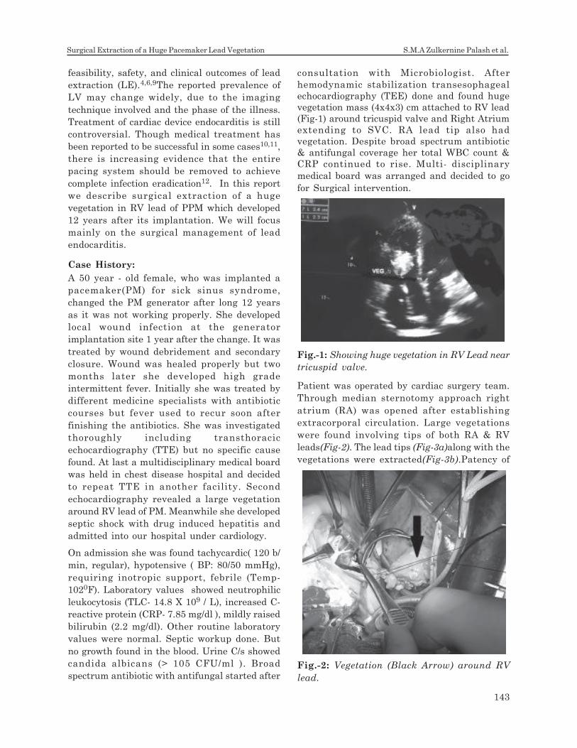
feasibility, safety, and clinical outcomes of lead
extraction (LE).4,6,9The reported prevalence of
LV may change widely, due to the imaging
technique involved and the phase of the illness.
Treatment of cardiac device endocarditis is still
controversial. Though medical treatment has
been reported to be successful in some cases10,11,
there is increasing evidence that the entire
pacing system should be removed to achieve
complete infection eradication12. In this report
we describe surgical extraction of a huge
vegetation in RV lead of PPM which developed
12 years after its implantation. We will focus
mainly on the surgical management of lead
endocarditis.
Case History:
A 50 year - old female, who was implanted a
pacemaker(PM) for sick sinus syndrome,
changed the PM generator after long 12 years
as it was not working properly. She developed
local wound infection at the generator
implantation site 1 year after the change. It was
treated by wound debridement and secondary
closure. Wound was healed properly but two
months later she developed high grade
intermittent fever. Initially she was treated by
different medicine specialists with antibiotic
courses but fever used to recur soon after
finishing the antibiotics. She was investigated
thoroughly including transthoracic
echocardiography (TTE) but no specific cause
found. At last a multidisciplinary medical board
was held in chest disease hospital and decided
to repeat TTE in another facility. Second
echocardiography revealed a large vegetation
around RV lead of PM. Meanwhile she developed
septic shock with drug induced hepatitis and
admitted into our hospital under cardiology.
On admission she was found tachycardic( 120 b/
min, regular), hypotensive ( BP: 80/50 mmHg),
requiring inotropic support, febrile (Temp-
1020F). Laboratory values showed neutrophilic
leukocytosis (TLC- 14.8 X 10⁹ / L), increased C-
reactive protein (CRP- 7.85 mg/dl ), mildly raised
bilirubin (2.2 mg/dl). Other routine laboratory
values were normal. Septic workup done. But
no growth found in the blood. Urine C/s showed
candida albicans (> 105 CFU/ml ). Broad
spectrum antibiotic with antifungal started after
Patient was operated by cardiac surgery team.
Through median sternotomy approach right
atrium (RA) was opened after establishing
extracorporal circulation. Large vegetations
were found involving tips of both RA & RV
leads(Fig-2). The lead tips (Fig-3a)along with the
vegetations were extracted(Fig-3b).Patency of
consultation with Microbiologist. After
hemodynamic stabilization transesophageal
echocardiography (TEE) done and found huge
vegetation mass (4x4x3) cm attached to RV lead
(Fig-1) around tricuspid valve and Right Atrium
extending to SVC. RA lead tip also had
vegetation. Despite broad spectrum antibiotic
& antifungal coverage her total WBC count &
CRP continued to rise. Multi- disciplinary
medical board was arranged and decided to go
for Surgical intervention.
Fig.-1: Showing huge vegetation in RV Lead near
tricuspid valve.
Fig.-2: Vegetation (Black Arrow) around RV
lead.
143
Surgical Extraction of a Huge Pacemaker Lead Vegetation S.M.A Zulkernine Palash et al.
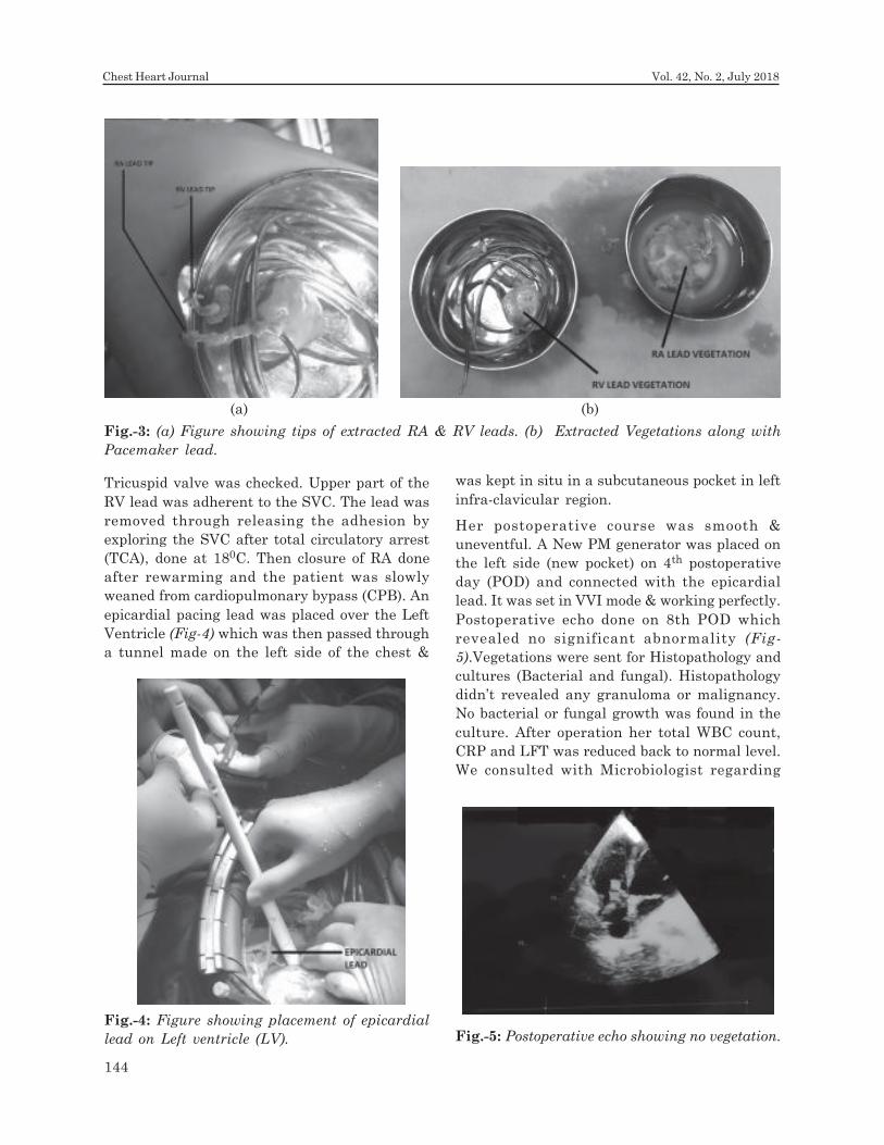
Tricuspid valve was checked. Upper part of the
RV lead was adherent to the SVC. The lead was
removed through releasing the adhesion by
exploring the SVC after total circulatory arrest
(TCA), done at 180C. Then closure of RA done
after rewarming and the patient was slowly
weaned from cardiopulmonary bypass (CPB). An
epicardial pacing lead was placed over the Left
Ventricle (Fig-4) which was then passed through
a tunnel made on the left side of the chest &
was kept in situ in a subcutaneous pocket in left
infra-clavicular region.
Her postoperative course was smooth &
uneventful. A New PM generator was placed on
the left side (new pocket) on 4th postoperative
day (POD) and connected with the epicardial
lead. It was set in VVI mode & working perfectly.
Postoperative echo done on 8th POD which
revealed no significant abnormality (Fig-
5).Vegetations were sent for Histopathology and
cultures (Bacterial and fungal). Histopathology
didn’t revealed any granuloma or malignancy.
No bacterial or fungal growth was found in the
culture. After operation her total WBC count,
CRP and LFT was reduced back to normal level.
We consulted with Microbiologist regarding
Fig.-3: (a) Figure showing tips of extracted RA & RV leads. (b) Extracted Vegetations along with
Pacemaker lead.
(a) (b)
Fig.-5: Postoperative echo showing no vegetation.Fig.-4: Figure showing placement of epicardial
lead on Left ventricle (LV).
144
Chest Heart Journal Vol. 42, No. 2, July 2018
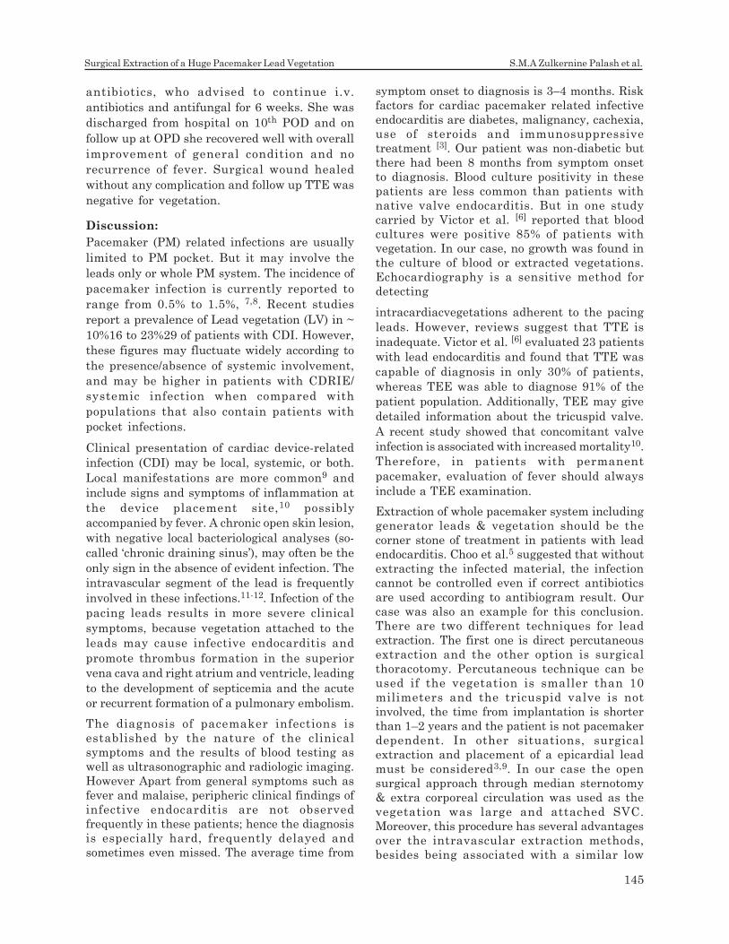
antibiotics, who advised to continue i.v.
antibiotics and antifungal for 6 weeks. She was
discharged from hospital on 10th POD and on
follow up at OPD she recovered well with overall
improvement of general condition and no
recurrence of fever. Surgical wound healed
without any complication and follow up TTE was
negative for vegetation.
Discussion:
Pacemaker (PM) related infections are usually
limited to PM pocket. But it may involve the
leads only or whole PM system. The incidence of
pacemaker infection is currently reported to
range from 0.5% to 1.5%, 7,8. Recent studies
report a prevalence of Lead vegetation (LV) in ~
10%16 to 23%29 of patients with CDI. However,
these figures may fluctuate widely according to
the presence/absence of systemic involvement,
and may be higher in patients with CDRIE/
systemic infection when compared with
populations that also contain patients with
pocket infections.
Clinical presentation of cardiac device-related
infection (CDI) may be local, systemic, or both.
Local manifestations are more common9 and
include signs and symptoms of inflammation at
the device placement site,10 possibly
accompanied by fever. A chronic open skin lesion,
with negative local bacteriological analyses (so-
called ‘chronic draining sinus’), may often be the
only sign in the absence of evident infection. The
intravascular segment of the lead is frequently
involved in these infections.11-12. Infection of the
pacing leads results in more severe clinical
symptoms, because vegetation attached to the
leads may cause infective endocarditis and
promote thrombus formation in the superior
vena cava and right atrium and ventricle, leading
to the development of septicemia and the acute
or recurrent formation of a pulmonary embolism.
The diagnosis of pacemaker infections is
established by the nature of the clinical
symptoms and the results of blood testing as
well as ultrasonographic and radiologic imaging.
However Apart from general symptoms such as
fever and malaise, peripheric clinical findings of
infective endocarditis are not observed
frequently in these patients; hence the diagnosis
is especially hard, frequently delayed and
sometimes even missed. The average time from
symptom onset to diagnosis is 3–4 months. Risk
factors for cardiac pacemaker related infective
endocarditis are diabetes, malignancy, cachexia,
use of steroids and immunosuppressive
treatment [3]. Our patient was non-diabetic but
there had been 8 months from symptom onset
to diagnosis. Blood culture positivity in these
patients are less common than patients with
native valve endocarditis. But in one study
carried by Victor et al. [6] reported that blood
cultures were positive 85% of patients with
vegetation. In our case, no growth was found in
the culture of blood or extracted vegetations.
Echocardiography is a sensitive method for
detecting
intracardiacvegetations adherent to the pacing
leads. However, reviews suggest that TTE is
inadequate. Victor et al. [6] evaluated 23 patients
with lead endocarditis and found that TTE was
capable of diagnosis in only 30% of patients,
whereas TEE was able to diagnose 91% of the
patient population. Additionally, TEE may give
detailed information about the tricuspid valve.
A recent study showed that concomitant valve
infection is associated with increased mortality10.
Therefore, in patients with permanent
pacemaker, evaluation of fever should always
include a TEE examination.
Extraction of whole pacemaker system including
generator leads & vegetation should be the
corner stone of treatment in patients with lead
endocarditis. Choo et al.5 suggested that without
extracting the infected material, the infection
cannot be controlled even if correct antibiotics
are used according to antibiogram result. Our
case was also an example for this conclusion.
There are two different techniques for lead
extraction. The first one is direct percutaneous
extraction and the other option is surgical
thoracotomy. Percutaneous technique can be
used if the vegetation is smaller than 10
milimeters and the tricuspid valve is not
involved, the time from implantation is shorter
than 1–2 years and the patient is not pacemaker
dependent. In other situations, surgical
extraction and placement of a epicardial lead
must be considered3,9. In our case the open
surgical approach through median sternotomy
& extra corporeal circulation was used as the
vegetation was large and attached SVC.
Moreover, this procedure has several advantages
over the intravascular extraction methods,
besides being associated with a similar low
145
Surgical Extraction of a Huge Pacemaker Lead Vegetation S.M.A Zulkernine Palash et al.
overall risk. Specifically, the risk of mechanical
injury to cardiac structures and dissemination
of vegetations is less because the leads are not
exposed to mechanical stress and blood
circulation through the right heart is interrupted
during the procedure. However, the extraction
of infected leads through a purse-string suture
with the heart beating, as described by
Niederh•auser and associates10, still carries the
risk of incompletely removing vegetations, with
subsequent dissemination. An open heart
operation also allows additional surgical
procedures to be performed in the right heart,
such as reconstruction of the tricuspid valve or
the removal of intra-cardiac vegetations and
thrombi, which was necessary in all patients.
The preoperatively started antimicrobial therapy
should be continued for at least 4-6 weeks after
implantation of the new pacemaker. The new
pacemaker was implanted through a different
implantation site, usually the contra lateral
pectoralis region or the upper abdominal
area11,12. In our case both strategy was followed
and patient remained free of infection on
subsequent OPD visit.
Conclusion:
In patients with pacemakers and risk factors for
endocarditis, fever must suggest lead
endocarditis and TEE must be performed for
accurate diagnosis. In addition to appropriate
antibiotic therapy, extraction of infected material
is needed for the cure. The removal of infected
leads during extracorporeal circulation prevents
mechanical injury as well as the spread of
infection and allows additional intra-cardiac
procedures to be performed, if necessary.
Complications were rare, except in patients who
present lately in a critically ill condition and
septic shock.
References:
1. Arber N, Pras E, Copperman Y, Schapiro
JM, Meiner V, Lossos IS et al. Pacemaker
endocarditis. Report of 44 cases and review
of the literature. Medicine (Baltimore)
1994;73:299–305.
2. del Rio A, Anguera I, Miro JM, Mont L,
Fowler VG Jr, Azqueta M et al. Surgical
treatment of pacemaker and defibrillator
lead endocarditis: the impact of electrode
lead extraction on outcome. Chest
2003;124:1451–9.
3. Bluhm G. Pacemaker infections: a clinical
study with special reference to prophylactic
use of some isoxazolylpenicillins. Acta Med
Scand1985;699:1e62.
4. Massoure PL, Reuter S, Lafitte S,
Laborderie J, Bordachard P, Clementy J et
al. Pacemaker endocarditis: clinical features
and management of 60 consecutive cases.
Pacing ClinElectrophysiol2007;30:12–9.
5. Catanchin A, Murdock CJ, Athan E.
Pacemaker infections: a 10-year experience.Heart Lung Circ2007;16:434–9.
6. Grammes JA, Schulze CM, Al-Bataineh M,Yesenosky GA, Saari CS, Vrabel MJ et al.
Percutaneous pacemaker and implantablecardioverter-defibrillator lead extractionin100 patients with intracardiacvegetationsdefined by transesophageal echocardiogram.J Am CollCardiol2010;55:886–94.
7. Kerut EK, Hanawalt C, Everson CT. Role of
the echocardiography laboratory in diagnosisand management of pacemaker andimplantable cardiac defibrillator infection.Echocardiography 2007;24:1008–12.
8. 18. Calton R, Cameron D, Cusimano RJ,Merchant N, Chauhan V. Successful
laserassisted removal of an infected ICDlead with a large vegetation. PacingClinElectrophysiol 2006;29:910–3.
9. Sohail MR, Uslan DZ, Khan AH, FriedmanPA, Hayes DL, Wilson WR et al. Infectiveendocarditis complicating permanent
pacemaker and implantable cardioverter-defibrillator infection. Mayo Clin Proc2008;83:46–53.
10. Lee JH, Geha AS, Ratehalli NM, Cmolik BL,Johnson NJ, Biblo LA, et al. Salvage ofinfected ICDs: management without
removal. Pacing ClinElectrophysiol1996;19:437e42.
11. Turkisher V, Priel I, San M. Successfulmanagement of an infected implantablecardioverter defibrillator with oral
antibiotics and without removal of thedevice. Pacing ClinElectrophysiol 1997;20:2268e70.
12. Myers MR, Parsonnet V, Bernstein AD.
Extraction of implanted transvenous pacing
leads: a review of a persistent clinical
problem. Am Heart J 1990;121:881-8.
146
Chest Heart Journal Vol. 42, No. 2, July 2018