بروتينات الغذاء
TRANSCRIPT
بروتينات الغذاء
Food Proteins
Prof. Dr. Mohamed Fawzy Ramadan HassanienZagazig University, Egypt

-Amino Acid Sequence -Protein Conformation -Levels of Protein Structure-Primary structure -Secondary structure -Tertiary structure -Quaternary structure-Classification of Proteins-Denaturation of Protein
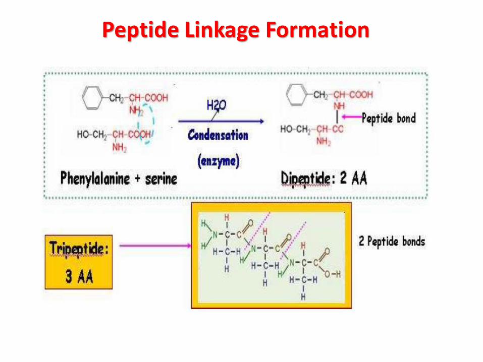
Peptide Linkage Formation
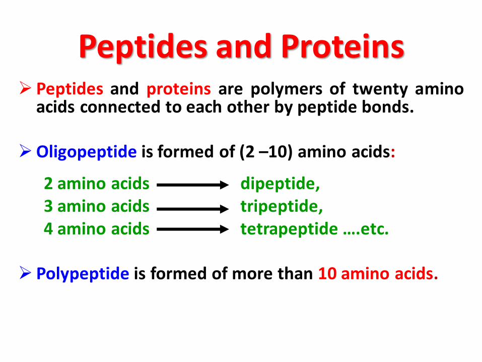
Peptides and Proteins➢ Peptides and proteins are polymers of twenty amino
acids connected to each other by peptide bonds.
➢Oligopeptide is formed of (2 –10) amino acids:
2 amino acids dipeptide, 3 amino acids tripeptide, 4 amino acids tetrapeptide ….etc.
➢ Polypeptide is formed of more than 10 amino acids.

In proteins,
almost all carboxyl and amino groups
are
combined in peptide linkage
and
not available for chemical reaction
(except for hydrogen bond formation).

-Like peptides, proteins are formed from amino acids throughamide linkages.
-Covalently bound hetero constituents can also be incorporatedinto proteins. For example, phosphoproteins such as milk casein orphosvitin of egg yolk contain phosphoric acid esters of serine andthreonine residues.
-The structure of a protein is dependent on the amino acidsequence (the primary structure) which determines the molecularconformation (secondary and tertiary structures).
-Proteins sometimes occur as molecular aggregates which arearranged in an orderly geometric fashion (quaternary structure).
-The sequences and conformations of a large number of proteinshave been elucidated and recorded in several data bases.
Food Proteins

-Glycoproteins,such as casein, variouscomponents of egg whiteand egg yolk, collagenfrom connective tissueand serum proteins ofsome species of fish,contain one or moremonosaccharide oroligosaccharide unitsbound O-glycosidically toserine, threonine orhydroxylysine or N-glycosidically toasparagine.
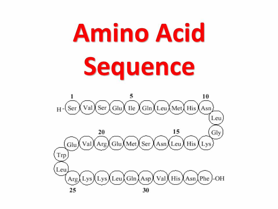
Amino Acid Sequence

-Sequence analysis can only be conducted on a pure protein.
-First, the amino acid composition is determined after acidic hydrolysis.
-The procedure (separation on a single cation-exchange resin column and colordevelopment with ninhydrin reagent) has been standardized and automated(amino acid analyzers).
-As an alternative to these established methods, the derivatization of aminoacids with the subsequent separation and detection of derivatives is possible(pre-column derivatization).
Various derivatization reagents can be selected, such as:
• 9-Fluorenylmethylchloroformate (FMOC)• Phenylisothiocyanate (PITC)• Dimethylaminoazobenzenesulfonylchloride (DABS-Cl)• Dimethylaminonaphthalenesulfonylchloride (DANS-Cl)• 7-Fluoro-4-nitrobenzo-2-oxa-1,3-diazole (NBDF)• 7-Chloro-4-nitrobenzo-2-oxa-1,3-diazole (NBDCl)• o-Phthaldialdehyde (OPA)
1-Amino Acid Composition, Subunits
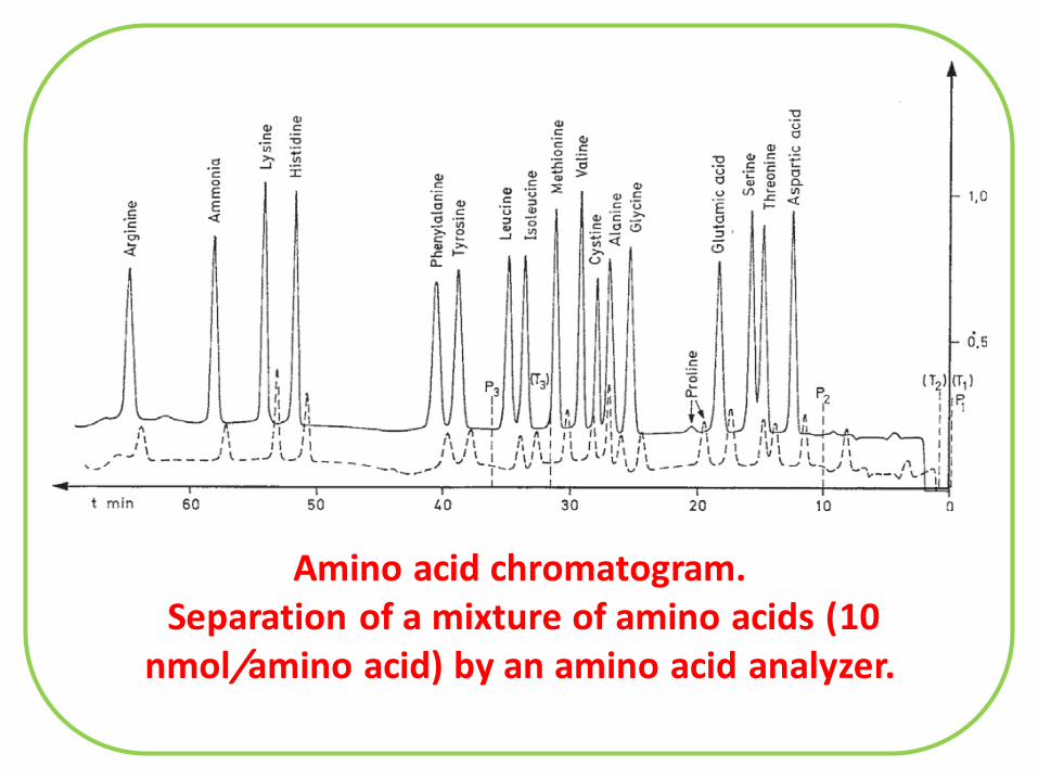
Amino acid chromatogram.Separation of a mixture of amino acids (10
nmol/amino acid) by an amino acid analyzer.
-It is also necessary to know the molecular weight (MW) of the protein.
-MW could be determined by gel column chromatography, ultracentrifugationor electrophoresis.
-It is necessary to determine whether the protein is a single molecule orconsists of a number of different polypeptide chains (subunits) associatedthrough disulfide bonds or non-covalent forces.
-Dissociation into subunits can be accomplished by a change in pH, by chemicalmodification of the protein, such as succinylation, or with denaturing agents(urea, guanidine hydrochloride, sodium dodecyl sulfate SDS).
-Disulfide bonds, which are also found in proteins which consist of only onepeptide chain, can be cleaved by oxidation of cystine to cysteic acid or byreduction to cysteine with subsequent alkylation of thiol group to prevent re-oxidation.
-Separation of subunits is achieved by chromatographic or electrophoreticmethods.
Amino Acid Composition, Subunits
-N-terminal amino acids can be determined by treating a proteinwith l-fluoro-2,4-dinitrobenzene (Sanger’s reagent) or 5-dimethylaminonaphthalene-1-sulfonyl chloride (dansyl chloride).
-Another possibility is the reaction with cyanate, followed byelimination of the N-terminal amino acid in the form of hydantoin,and separation and recovery of the amino acid by cleavage of thehydantoin .
-The N-terminal amino acid (and the amino acid sequence close tothe N-terminal) is accessible by hydrolysis with aminopeptidase, inwhich case it should be remembered that the hydrolysis rate isdependent on amino acid side chains and that proline residues arenot cleaved.
-A special procedure is required when the N-terminal residue isacylated (N-formyl- or N-acetyl amino acids).
2-Terminal Groups

-Determination of C-terminal amino acids is possible via thehydrazinolysis procedure recommended by Akabori:
-The C-terminal amino acid could be then separated from theamino acid hydrazides by a cation exchange resin.

-The C-terminal amino acids can be removedenzymatically by
- Carboxypeptidase A which cleaves amino acids witharomatic and large aliphatic side chains,
- Carboxypeptidase B which cleaves lysine, arginine andamino acids with neutral side chains or
- Carboxypeptidase C which cleaves with less specificitybut cleaves proline.
Terminal Groups

-Long peptide chains are usually fragmented. The fragments are then analyzedfor amino acid sequences.
-Selective enzymatic cleavage of peptide bonds is accomplished primarily withTrypsin, which cleaves exclusively Lys-X- and Arg-X-bonds, andChymotrypsin, which cleaves peptide bonds with less specificity (Tyr-X, Phe-X,Trp-X and Leu-X).
-The enzymatic attack can be influenced by modification of protein. For example,-Acylation of the amino group of lysine limits tryptic hydrolysis to Arg-X,-Substitution of the SH-group of cysteine residue with an aminoethyl groupintroduces a new cleavage position for trypsin into the molecule “pseudolysineresidue”
3- Partial Hydrolysis

-Also suited for the specificenzymatic hydrolysis ofpeptide chains is theendoproteinase Glu-C fromStaphylococcus aureus. Itcleaves Glu-X bonds as wellas Glu-X plus Asp-X bonds.
-The most importantchemical method forselective cleavage usescyanogen bromide (BrCN)to attack Met-X-linkages.
Partial Hydrolysis
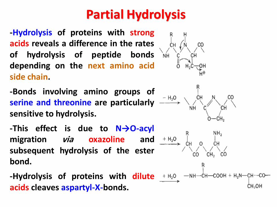
-Hydrolysis of proteins with strongacids reveals a difference in the ratesof hydrolysis of peptide bondsdepending on the next amino acidside chain.
-Bonds involving amino groups ofserine and threonine are particularlysensitive to hydrolysis.
-This effect is due to N→O-acylmigration via oxazoline andsubsequent hydrolysis of the esterbond.
-Hydrolysis of proteins with diluteacids cleaves aspartyl-X-bonds.
Partial Hydrolysis
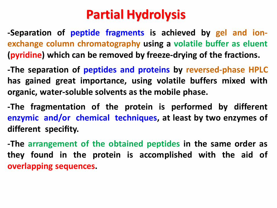
-Separation of peptide fragments is achieved by gel and ion-exchange column chromatography using a volatile buffer as eluent(pyridine) which can be removed by freeze-drying of the fractions.
-The separation of peptides and proteins by reversed-phase HPLChas gained great importance, using volatile buffers mixed withorganic, water-soluble solvents as the mobile phase.
-The fragmentation of the protein is performed by differentenzymic and/or chemical techniques, at least by two enzymes ofdifferent specifity.
-The arrangement of the obtained peptides in the same order asthey found in the protein is accomplished with the aid ofoverlapping sequences.
Partial Hydrolysis

-The classical method is the Edman degradation reaction.
-It involves stepwise degradation of peptides withphenylisothiocyanate.
-The resultant phenylthiohydantoin is identified directly.
-The stepwise reactions are performed in solution or on peptidebound to a carrier, i. e. to a solid phase.
-Both approaches have been automated (“sequencer”). Carriersused include resins containing amino groups (e.g., aminopolystyrene) or glass beads treated with amino alkylsiloxane:
4- Sequence Analysis

-The peptides are then attached to the carrier bycarboxyl groups (activation with carbodiimide asin peptide synthesis) or by amino groups.
-For example, a peptide segment from thehydrolysis of protein by trypsin has lysine as itsC-terminal amino acid. It is attached to thecarrier with phenylene-diisothiocyanate throughamino groups.
-Mild acidic treatment of the carrier underconditions of the Edman degradation splits thefirst peptide bond.
-The Edman procedure is then performed onthe shortened peptide through second, third andsubsequent repetitive reactions:
Sequence Analysis
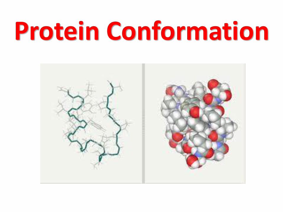
Protein Conformation
Protein molecule can be formed of
one or more
polypeptide chains
which may vary in the number
and sequence of amino acid residues.

-Information about conformation is availablethrough X-ray crystallographic analysis ofprotein crystals and by measuring the distancebetween selected protons of the peptide chainby means of H-NMR spectroscopy in solution.
-X-ray structural analysis of a fully extendedpeptide chain reveal the lengths and angles ofbonds
-The peptide bond has partial (40%) doublebond character with electrons shared betweenthe C-O and C-N bonds.
-The resonance energy is about 83.6 kJ/mole
Structure of an elongated peptide chain.
Extended Peptide Chains
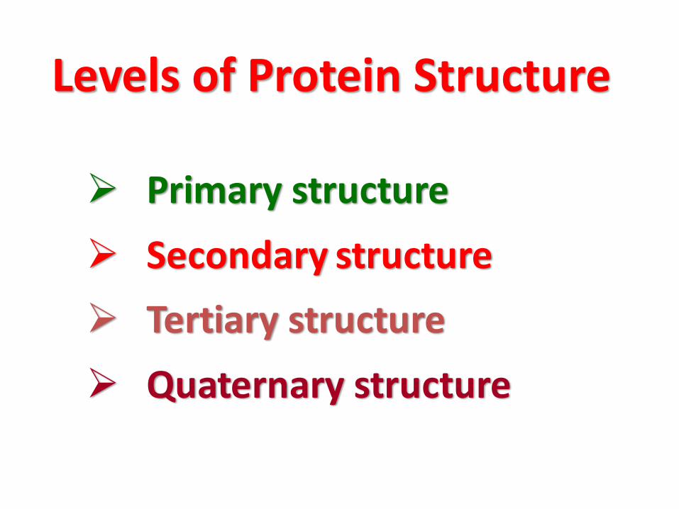
Levels of Protein Structure
➢ Primary structure
➢ Secondary structure
➢ Tertiary structure
➢ Quaternary structure

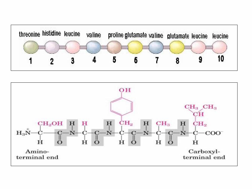
➢ It is the amino acid sequence of the polypeptide chainlinked by peptide bonds.
➢ It is characteristic for every protein.
➢ All proteins have an
➢ N-terminal end (with a free amino group) and
➢ C-terminal end (with a free carboxyl group).
➢ Polypeptide chain sequence is written according tothe sequence of amino acid residues from the N to Cterminus amino acids.
Primary structure

➢ Is the local spatial arrangement of the polypeptide’sbackbone (peptide bond) atoms without regard to theconformations of its side chains.
➢ Peptide bonds contain polar amide hydrogen atoms(with a partial positive charge) and polar carbonyloxygen atoms (with a partial negative charge).
➢ This allows hydrogen bonds to form between peptidebonds in different parts of the chain.
➢ The polypeptide chain can take different shapes orpatterns in different parts of the chain, and thesepatterns are called the secondary protein structure.
➢ There are 2 types of secondary structure:
▪Alpha helix (α-helix)
▪Beta-pleated sheet (β-pleated sheet).
Secondary structure

Alpha helix• A spiral, compact, rod like structure
• Mostly right handed α-helix, with Rgroups protruding outside
• Stabilized by numerous hydrogen bondswhich are formed between carbonyloxygen (C=O, hydrogen acceptor) andpeptide nitrogen (NH, hydrogen donor).
• Forms about 100% of fibrous protein
-keratin
-80% of the globular protein; hemoglobin.
Secondary structure

Alpha helix
Alpha helix is disrupted by:
• Proline: its imino group is notgeometrically compatible withα- helix.
• Large numbers of bulky aminoacids e.g. tryptophan because ofsteric interference.
• Large numbers of branchedamino acids e.g. valine andisoleucine because of stericinterference.
• Large numbers of acidic andbasic amino acids because theyform ionic bonds or electricallyrepel each other.

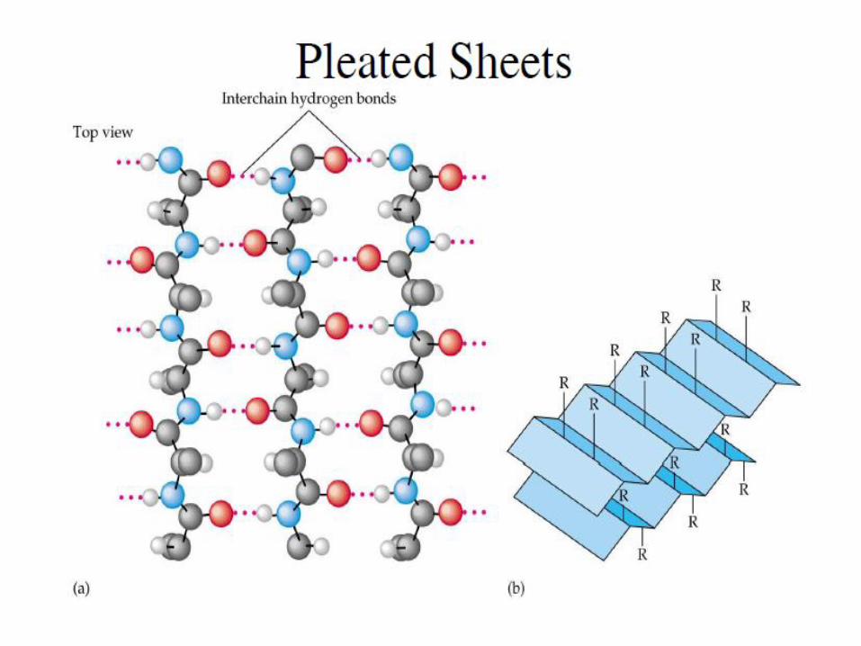
β- PLEATED SHEET
• Almost fully extended and its
surface appear pleated.
• Found in fibrous and globular
protein.
• Formed of 1 or more
polypeptide chains.
• Stabilized by hydrogen bonds
between peptide bonds.

Types of β-PLEATED SHEET
1. Parallel β-pleated sheet: formedof 2 or more polypeptide chainsrunning in the same direction (N-terminals are on the same side)
2. Anti-parallel β-pleated sheet:formed of one or morepolypeptide chains running inopposite directions (N and Cterminals are alternating).

Comparison of -helix and -sheet
-helix -sheet
Structure 1 polypeptide chain 1 or more polypeptide chains
polypeptide Coiled Almost fully extended
Hydrogen bonds
- Formed between 2peptide bonds of 4 amino
acids apart in the primary
structure.
- Parallel to the axis ofpolypeptide chain.
- Formed between amino acidswhich has no relation in primary
structure.
- Perpendicular to the axis of
polypeptide chain.
R groups - Protrude outside the helix - Project above and below the plane of the sheet

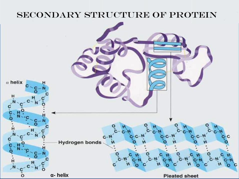
SECONDARY STRUCTURE OF PROTEIN
α- helix

Tertiary structure
• Is the three dimensional structure of a single polypeptide chain giving protein its characteristic shape.
I- Globular proteins (enzymes)• Approximately spherical shape- water
Soluble.
II- Fibrous proteins (structural proteins)
• Rod-like shape
• Poor water solubility.
• Cross links and bonds in 3ry structure:
• S-S bond, Ionic, Hydrophobic interactions and H-bonding.
Fibrous protein
Globular protein

Tertiary structure
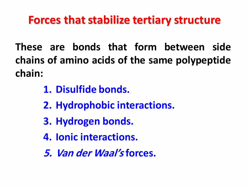
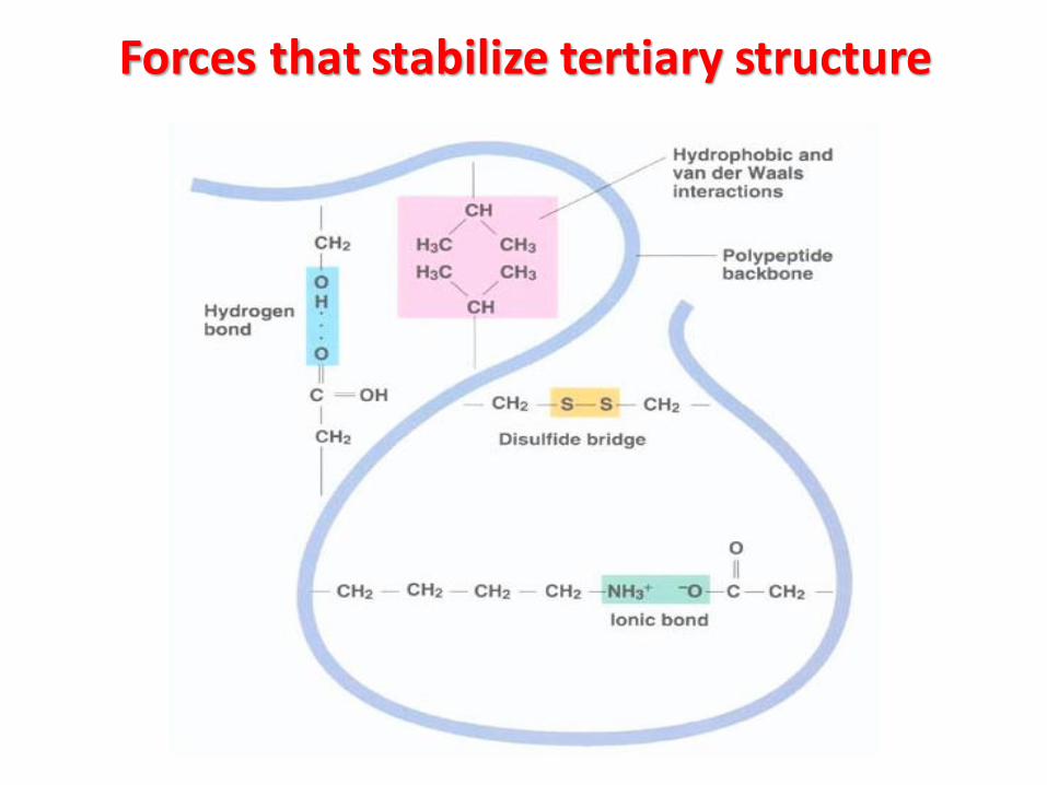
Forces that stabilize tertiary structure
These are bonds that form between sidechains of amino acids of the same polypeptidechain:
1. Disulfide bonds.
2. Hydrophobic interactions.
3. Hydrogen bonds.
4. Ionic interactions.
5. Van der Waal’s forces.

Disulfide bonds:covalent bond between 2 SH groups of 2 cysteine residues forming an S~S
bond of cystine residue.
Hydrophobic interaction:non covalent bonds between amino acids with non-polar side chains that are
located in the interior of polytpeptide chain away from water.
Hydrogen bonds:non covalent bond between a hydrogen atom attached to nitrogen or oxygen
and another oxygen or nitrogen atom.
Ionic interaction:non covalent bonds between negatively charged groups in acidic amino acids
(as carboxilic group in the side chain of aspartate or glutamate) andpositively charged groups in basic amino acids (as amino group in theside chain of lysine)
Van der Waal’s forces:non covalent bonds occurring when two adjacent atoms come into closer
distance.
Forces that stabilize tertiary structure
Forces that stabilize tertiary structure
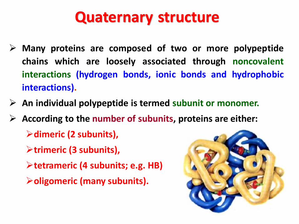
➢ Many proteins are composed of two or more polypeptide
chains which are loosely associated through noncovalent
interactions (hydrogen bonds, ionic bonds and hydrophobic
interactions).
➢ An individual polypeptide is termed subunit or monomer.
➢ According to the number of subunits, proteins are either:
➢dimeric (2 subunits),
➢trimeric (3 subunits),
➢tetrameric (4 subunits; e.g. HB)
➢oligomeric (many subunits).
Quaternary structure

Examples of globular proteins

Classification of Proteins
Simple Conjugated Derived proteins proteins proteins
1. Albumin2. Globulins3. Histones
1. Phosphoproteins2. Glycoproteins3. Chromoproteins4. Lipoproteins5. Nucleoproteins6. Metalloproteins
Results from
denaturation
or cleavage of
native proteins by
the action of
acids, alkali or
enzymes.

Proteins can be modified to include other chemical groups
“prosthetic groups” besides amino acids:
Class Prosthetic group (s) Example
•Lipoproteins
•Glycoproteins
•Phosphoproteins
•hemoproteins
•Lipids
•Carbohydrates
•Phosphate groups
•Heme (iron porphyrin)
•VLDL
•Immunoglobulin G
•Casinogen of milk
•Hemoglobin
Conjugated Proteins
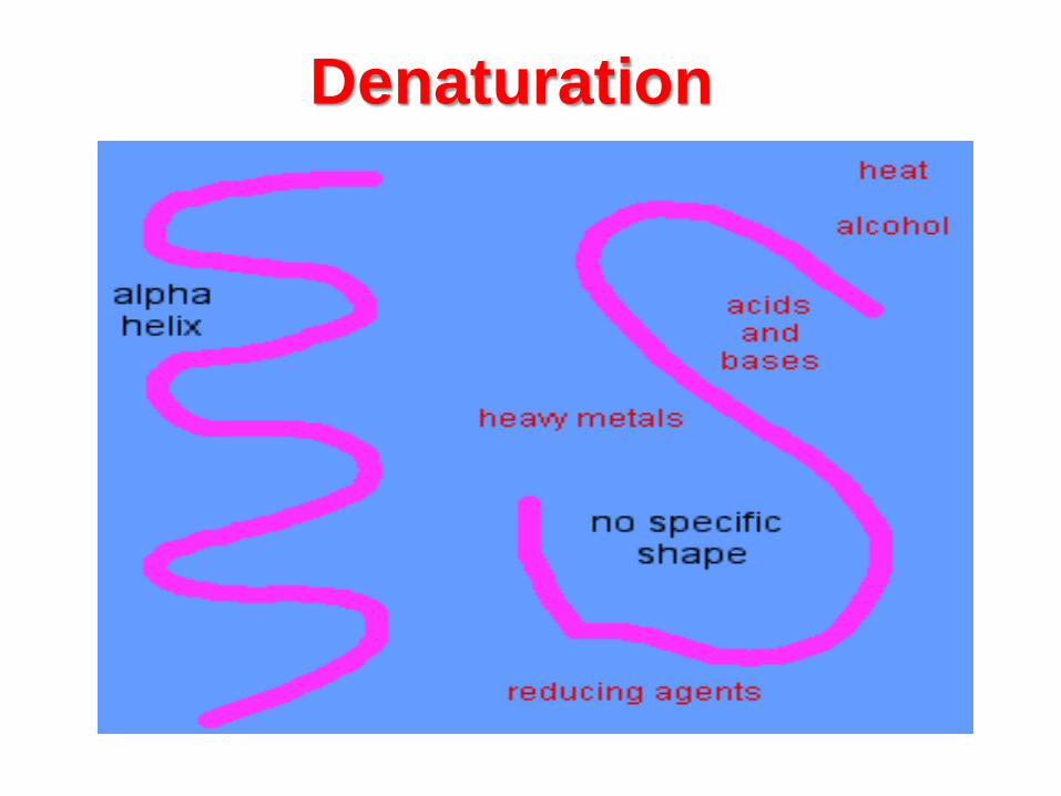
Denaturation of Protein
Denaturation of Protein
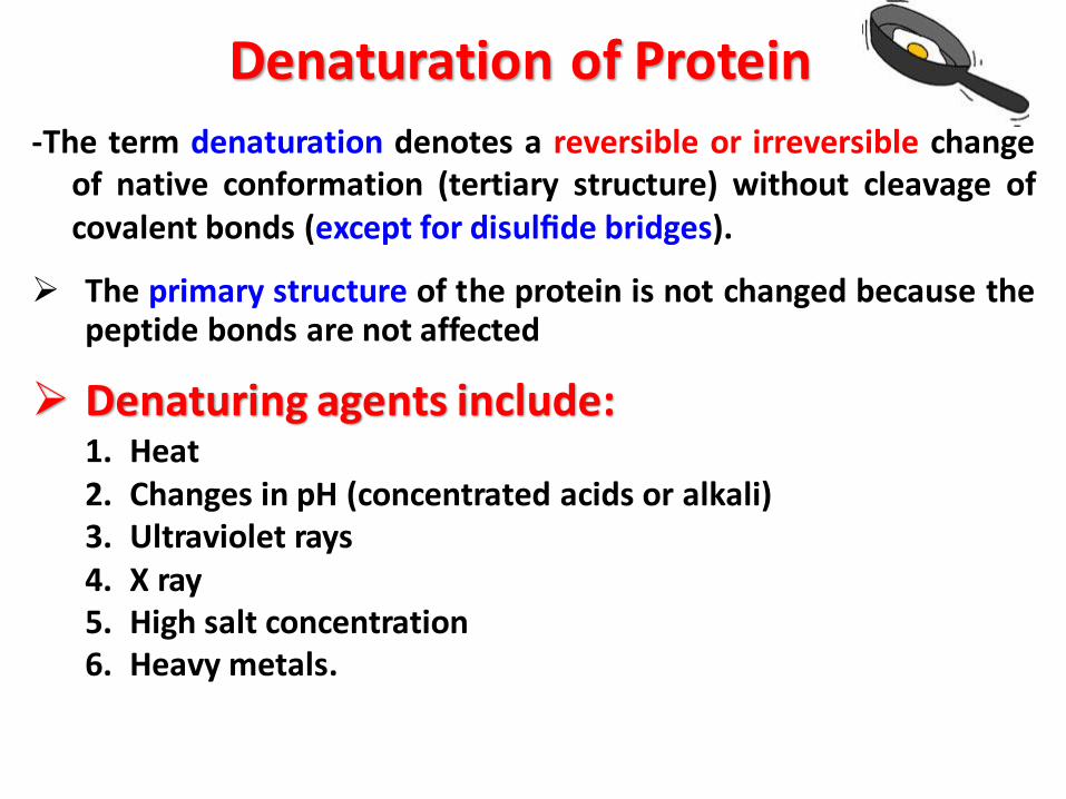
-The term denaturation denotes a reversible or irreversible changeof native conformation (tertiary structure) without cleavage ofcovalent bonds (except for disulfide bridges).
➢ The primary structure of the protein is not changed because thepeptide bonds are not affected
➢ Denaturing agents include:1. Heat2. Changes in pH (concentrated acids or alkali)3. Ultraviolet rays4. X ray5. High salt concentration6. Heavy metals.

Denaturation-Denaturation is possible with any treatment that cleaveshydrogen bridges, ionic or hydrophobic bonds. This can beaccomplished by: changing the temperature, adjusting the pH,increasing the interface area, or adding organic solvents, salts,urea, or detergents such as sodium dodecyl sulfate.
-Denaturation is generally reversible when the peptide chain isstabilized in its unfolded state by the denaturing agent and nativeconformation can be re-established after removal of the agent.
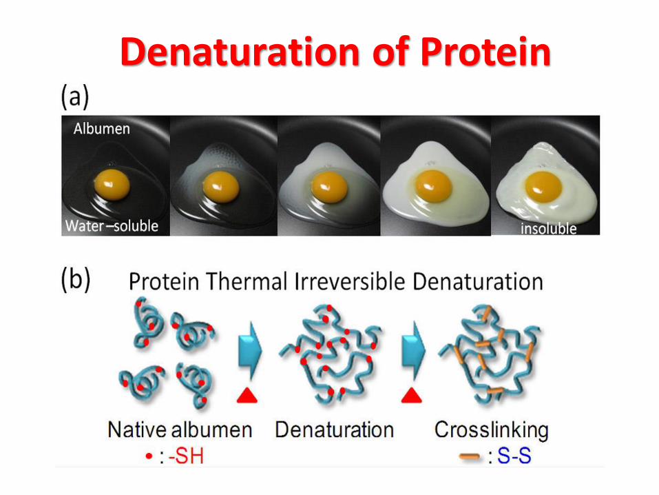
-Irreversible denaturation occurs when the unfolded peptide chainis stabilized by interaction with other chains (as occurs for instancewith egg proteins during boiling). During unfolding reactive groups,such as thiol groups, that were blocked, may be exposed. Theirparticipation in the formation of disulfide bonds may also cause anirreversible denaturation.
Denaturation
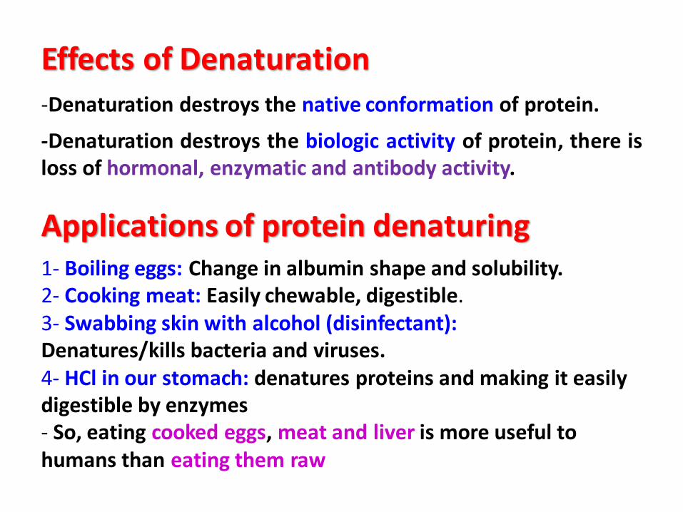
Effects of Denaturation-Denaturation destroys the native conformation of protein.
-Denaturation destroys the biologic activity of protein, there isloss of hormonal, enzymatic and antibody activity.
Applications of protein denaturing1- Boiling eggs: Change in albumin shape and solubility.2- Cooking meat: Easily chewable, digestible.3- Swabbing skin with alcohol (disinfectant):Denatures/kills bacteria and viruses.4- HCl in our stomach: denatures proteins and making it easily digestible by enzymes- So, eating cooked eggs, meat and liver is more useful to humans than eating them raw

-An aggregation of the peptidechains caused by the folding ofglobular proteins is connected withreduced solubility or swellability.
-Thus the part of wheat gluten thatis soluble in acetic acid diminishesas heat stress increases.
-As a result of the reduced risingcapacity of gluten caused by thepre-treatment, the volume ofbread made of recombined floursis smaller.
Solubility of gluten (wheat) in diluted acetic
acid after various forms of thermal stress
Denaturation of Protein: Examples in Food
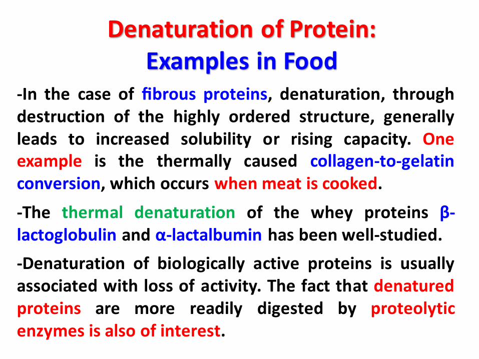
-In the case of fibrous proteins, denaturation, throughdestruction of the highly ordered structure, generallyleads to increased solubility or rising capacity. Oneexample is the thermally caused collagen-to-gelatinconversion, which occurs when meat is cooked.
-The thermal denaturation of the whey proteins β-lactoglobulin and α-lactalbumin has been well-studied.
-Denaturation of biologically active proteins is usuallyassociated with loss of activity. The fact that denaturedproteins are more readily digested by proteolyticenzymes is also of interest.
Denaturation of Protein: Examples in Food