pirh2 mediates the sensitivity of myeloma cells to ... · multiple myeloma treatment center &...
TRANSCRIPT
RESEARCH ARTICLE
Pirh2 mediates the sensitivity of myeloma cellsto bortezomib via canonical NF-κB signalingpathway
Li Yang, Jing Chen, Xiaoyan Han, Enfan Zhang, Xi Huang, Xing Guo, Qingxiao Chen, Wenjun Wu,Gaofeng Zheng, Donghua He, Yi Zhao, Yang Yang, Jingsong He, Zhen Cai&
Multiple Myeloma Treatment Center & Bone Marrow Transplantation Center, The First Affiliated Hospital, College of Medicine,Zhejiang University, Hangzhou 310003, China& Correspondence: [email protected] (Z. Cai)
Received October 4, 2017 Accepted December 8, 2017
ABSTRACT
Clinical success of the proteasome inhibitor establishedbortezomib as one of the most effective drugs in treat-ment of multiple myeloma (MM). While survival benefit ofbortezomib generated new treatment strategies, theprimary and secondary resistance of MM cells tobortezomib remains a clinical concern. This study aimedto highlight the role of p53-induced RING-H2 (Pirh2) inthe acquisition of bortezomib resistance in MM and toclarify the function and mechanism of action of Pirh2 inMM cell growth and resistance, thereby providing thebasis for new therapeutic targets for MM. The protea-some inhibitor bortezomib has been established as oneof the most effective drugs for treating MM. Wedemonstrated that bortezomib resistance in MM cellsresulted from a reduction in Pirh2 protein levels. Pirh2overexpression overcame bortezomib resistance andrestored the sensitivity of myeloma cells to bortezomib,while a reduction in Pirh2 levels was correlated withbortezomib resistance. The levels of nuclear factor-kappaB (NF-κB) p65, pp65, pIKBa, and IKKa were higherin bortezomib-resistant cells than those in parental cells.Pirh2 overexpression reduced the levels of pIKBa andIKKa, while the knockdown of Pirh2 via short hairpinRNAs increased the expression of NF-κB p65, pIKBa,and IKKa. Therefore, Pirh2 suppressed the canonical NF-κB signaling pathway by inhibiting the phosphorylation
and subsequent degradation of IKBa to overcomeacquired bortezomib resistance in MM cells.
KEYWORDS bortezomib, drug resistance, multiplemyeloma, NF-κB, Pirh2
INTRODUCTION
The proteasome inhibitor bortezomib is effective at treatingmultiple myeloma (MM) because the efficacy of bortezomib-based chemotherapy regimens is as high as 80%–90%(Rajkumar, 2016), However, drug resistance limits its repe-ated use, although the mechanisms are not fully understood(Malard et al., 2017). The mechanism of acquisition ofbortezomib resistance and the ways to overcome thisresistance are important clinical issues (Chao and Wang,2016). Hence, the molecular mechanisms of bortezomibresistance need to be urgently explored to enhance the useof existing treatments and to define more effective single orcombination therapies. The current established molecularmechanisms underlying resistance to proteasome inhibitorsinvolve constitutive and immunoproteasomes, mutated pro-teasome subunits, unfolded protein response (UPR) medi-ators, multidrug efflux transporters, aggresomes, autophago-somes, pro-survival signaling pathway mediators, or bonemarrow microenvironmental components (Niewerth et al.,2015). The ubiquitin-proteasome system is the main mech-anism controlling protein turnover and maintaining cellularprotein homeostasis. E3 ubiquitin ligases determine thespecificity of protein degradation, and these ligases havebeen shown to be closely related to cancer occurrence,development, transfer, and drug resistance (Masumoto andLi Yang and Jing Chen have authors contributed equally to this work.
© The Author(s) 2018. This article is an open access publication
Protein Cell 2018, 9(9):770–784https://doi.org/10.1007/s13238-017-0500-9 Protein&Cell
Protein
&Cell
Kitagawa, 2016). Several lines of evidence link E3 ligaseswith MM and associated drug resistance (Lub et al., 2016).
p53-induced RING-H2 (Pirh2) is a newly discovered E3ubiquitin ligase induced by p53 activation that has anintrinsic ubiquitin-protein ligase activity for polyubiquitinationand subsequent proteasomal degradation. Pirh2 was initiallythought to be similar to HDM2, the human counterpart ofMDM2 in mice (Halaby et al., 2013). Recent studies haveshown that Pirh2 regulates cellular homeostasis in both p53-dependent and p53-independent cellular contexts. More-over, based on its substrates, the association of Pirh2 withthe occurrence and prognosis of malignant tumors has beenconfirmed (Jung et al., 2012, Daks et al., 2016, Yang et al.,2016). Pirh2 ubiquitinates substrate proteins and directsthem through degradation pathways involved in apoptosisinduction, cell cycle regulation, and DNA repair. However,the role of Pirh2 in proliferation, invasion, and drug resis-tance of tumors still needs further investigation.Recently,data from publicly available databases have been used tocorrelate gene expression in myeloma tumor cells with clin-ical responses to bortezomib. A reduced expression of Pirh2was observed in bortezomib-resistant cells using an Affy-metrix GeneChip Human Transcriptome Array 2.0 (Huanget al., 2017). These findings helped further elucidating therole of the E3 ligase Pirh2 in the bortezomib resistance ofMM, thereby facilitating bortezomib retreatment as a moreeffective and rational therapeutic strategy in the clinic.
RESULTS
Reduced levels of Pirh2 were correlatedwith bortezomib resistance
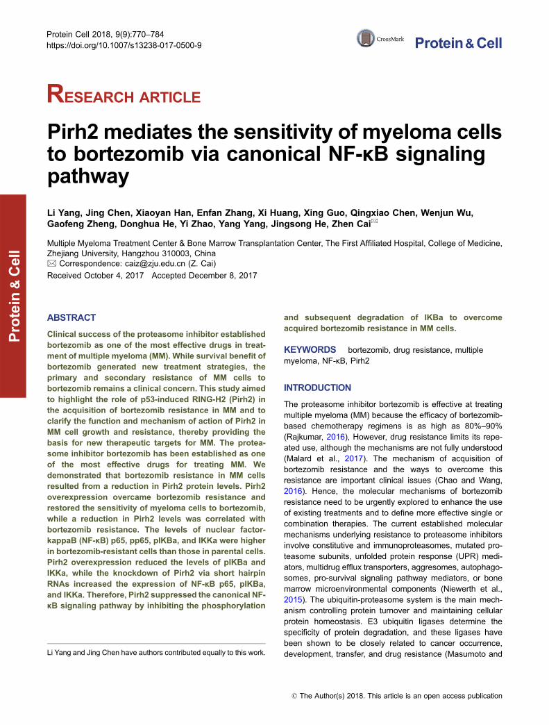
A decreased expression of Pirh2 was observed in thebortezomib-resistant cells RPMI8226.BR and OPM-2.BRand their controls using Affymetrix HTA 2.0. The expressionof Pirh2 protein and mRNA was confirmed in the MM celllines RPMI8226, ARP-1, ARK, MM.1S, MM.1R, NCI-H929,OPM-2, and LP-1. Immunofluorescence revealed that Pirh2was primarily expressed in the cytoplasm (Fig. 1).
The bortezomib-resistant cell line NCI-H929.BR wasestablished, and changes in Pirh2 expression were identifiedto evaluate the role of the E3 ubiquitin ligase Pirh2 inbortezomib resistance in MM. As detected with the CCK-8assay, the IC50 of NCI-H929 and NCI-H929.BR cells treatedwith bortezomib for 24 h was 17.62 ± 1.92 nmol/L and 234.30± 6.02 nmol/L, respectively (Fig. 2A); the resistance ratiowas 13.30 (P < 0.05). Growth curve (Fig. 2B) and flowcytometry results (Fig. 2C) indicated the lack of a significantdifference between the bortezomib-resistance and borte-zomib-sensitive cells (P > 0.05). Compared with that inparental cells, Pirh2 expression was reduced in the borte-zomib-resistant cell lines RPMI8226.BR and OPM-2.BR(Fig. 2D). Pirh2 expression levels were also found to grad-ually decrease over 1–3 months in parental NCI-H929 cellsin response to increasing drug concentrations (Fig. 2E).
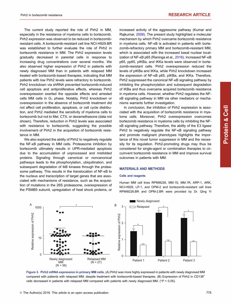
Pirh2 was more highly expressed in patients with newlydiagnosed MM than in patients with relapsed MM
Pirh2 mRNA expression was also determined in bone mar-row samples obtained from patients with MM. Pirh2expression was found to be higher in patients with newlydiagnosed MM than in patients with relapsed MM treatedwith bortezomib plus dexamethasone and cyclophos-phamide (CTX) (Fig. 3A, P < 0.05). Next, CD138+ MM cellswere isolated from three patients. Pirh2 expression wascompared in samples from the same patient at differentstages of disease. Pirh2 expression in CD138+ cells waslower in patients with relapsed MM than in patients withnewly diagnosed MM (Fig. 3B, P < 0.05).
Pirh2 knockdown prevented bortezomib-induced cellapoptosis and antiproliferative effects
Western blotting and qRT-PCR were performed to verifytransfection efficiency in the myeloma cell lines RPMI8226-shPirh2, OPM-2-shPirh2, NCI-H929-shPirh2 and their cor-responding controls (Fig. 4A and 4B). Growth curve and cellcycle analysis demonstrated the lack of significant differencebetween cells with Pirh2 knockdown and controls withoutbortezomib treatment (Fig. 4C and 4D, P > 0.05). However,Pirh2 knockdown enabled the transition of MM cells from G1
phase to S and G2 phases in the presence of bortezomib(Fig. 4D) and weakened the inhibition of cell proliferation bybortezomib (Fig. 4E). The percentage of cells in G1 phase invarious groups was as follows: RPMI8226-shPirh2 vs.RPMI8226-ctl, 39.03% ± 3.20% vs. 52.84% ± 42.89%; OPM-2-shPirh2 vs. OPM-2-ctl, 42.40% ± 5.84% vs. 57.00% ±6.23%; and NCI-H929-shPirh2 vs. NCI-H929-ctl, 23.37% ±2.12% vs. 42.91% ± 1.89% (Fig. 4F, P < 0.05). In addition,Pirh2 knockdown reduced bortezomib-induced apoptosis inMM cells. The percentage of apoptotic cells in variousgroups was as follows: RPMI8226-shPirh2 vs. RPMI8226-ctl, 47.90% ± 1.63% vs. 55.60% ± 2.86%; OPM-2-shPirh2vs. OPM-2-ctl, 48.30% ± 1.17% vs. 63.60% ± 1.24%; andNCI-H929-shPirh2 vs. NCI-H929-ctl, 20.28% ± 0.98% vs.38.37% ± 1.34% (Fig. 4G, P < 0.05).
Pirh2 overexpression enhanced bortezomib-inducedcell apoptosis and antiproliferative effects and resultedin G1 phase cell cycle arrest in MM cells
Pirh2-overexpressing myeloma cell lines ARP-1-Pirh2, ARK-Pirh2, LP-1-Pirh2 and their corresponding controls wereestablished as described earlier. Western blotting and qRT-PCR were performed to verify transfection efficiency (Fig. 5Aand 5B). Growth curve and cell cycle analysis demonstratedthe lack of significant difference between Pirh2-overex-pressing cells and controls (Fig. 5C and 5D, P > 0.05).However, Pirh2 overexpression increased the inhibition ofcell proliferation by bortezomib (Fig. 5E, P < 0.05) andarrested MM cells in G1 phase. The percentage of cells in G1
Pirh2 in bortezomib resistance RESEARCH ARTICLE
© The Author(s) 2018. This article is an open access publication 771
Protein
&Cell
C
Pirh2
β-Actin
RPMI 8226
ARP- 1
ARK
MM1S
MM1R
NCI- H929
LP-1
OPM- 2
RPMI 8226 ARP- 1ARK MM1S MM1RNCI- H 929LP- 1 OPM- 2
Pirh 2 DAPI Merge
A B
6
8
Positive control
RPMI 8226ARP-1ARK
MM1SMM1R
NCI- H929LP- 1
OPM- 2
4
2
0
Rel
atlv
e ex
pres
sion
of P
irh2
RESEARCH ARTICLE Li Yang et al.
772 © The Author(s) 2018. This article is an open access publication
Protein
&Cell

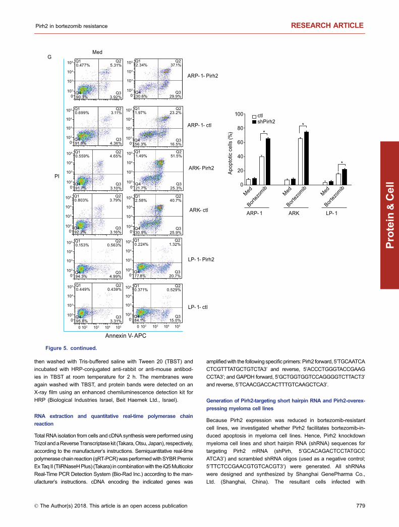
phase in various groups was as follows: ARP-1-Pirh2 vs.ARP-1-ctl, 51.56% ± 3.91% vs. 40.88% ± 2.09%; ARK-Pirh2vs. ARK-ctl, 49.10% ± 4.32% vs. 31.90% ± 3.98%; and LP-1-Pirh2 vs. LP-1-ctl, 58.90% ± 4.06% vs. 32.40% ± 2.76%(Fig. 5F, P < 0.05) and Furthermore, Pirh2 overexpressionincreased bortezomib-induced apoptosis in MM cells. Thepercentage of apoptotic cells in various groups was as fol-lows: ARP-1-Pirh2 vs. ARP-1-ctl, 66.90% ± 3.73% vs.41.70% ± 1.86%; ARK-Pirh2 vs. ARK-ctl, 76.80% ± 4.17%vs. 66.60% ± 3.24%; and LP-1-Pirh2 vs. LP-1-ctl, 22.02% ±1.23% vs. 15.53% ± 2.03% (Fig. 5G, P < 0.05).
Pirh2 mediated the sensitivity of myeloma cellsto bortezomib but not to CTX or melphalan
The CCK-8 assay demonstrated that Pirh2 knockdownweakened the inhibition of cell proliferation by bortezomib(Fig. 6, P < 0.05) but did not affect the antiproliferative effectof CTX or melphalan (Mel) on the cell lines RPMI8226-shPirh2, OPM-2-shPirh2, and NCI-H929-shPirh2 (Fig. 6, P >0.05).
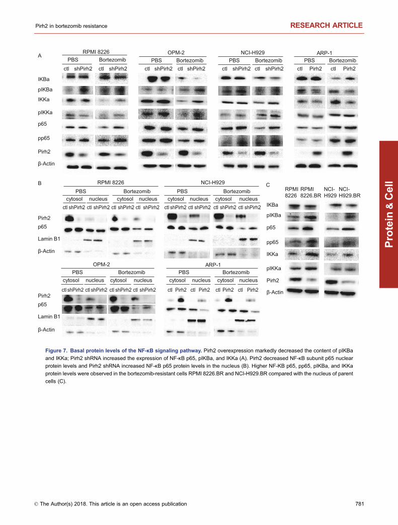
Pirh2 suppressed the nuclear factor-kappaB (NF-κB)signaling pathway in bortezomib-resistant myelomacells
Increased nuclear factor-kappaB (NF-κB) expression hasbeen observed in patients with refractory primary myeloma,which is thought to be linked to drug resistance (Turner et al.,2016). Thus, we investigated whether Pirh2 overcomesbortezomib resistance in myeloma cells by inhibiting the NF-κB pathway. The degradation of IKBa and the release of NF-κB subunits were preceded by the phosphorylation of IKBa.Additionally, Pirh2 overexpression markedly decreased thelevels of pIKBa and IKKa, while Pirh2 knockdown via shRNAincreased the expression of NF-κB p65, pIKBa, and IKKa(Fig. 7A). Pirh2 overexpression decreased the nuclear pro-tein levels of NF-κB subunit p65, whereas Pirh2 knockdownexerted the opposite effect (Fig. 7B). Higher protein levels ofNF-κB p65, pp65, pIKBa, and IKKa were observed in thenuclei of bortezomib-resistant cells RPMI8226.BR and NCI-H929.BR than in the nuclei of the parental cells (Fig. 7C).
Therefore, Pirh2 likely mediates the sensitivity of mye-loma cells to bortezomib via the canonical NF-κB signalingpathway (Fig. 8).
DISCUSSION
The proteasome inhibitor bortezomib is usually effectiveagainst MM. However, drug resistance limits its repeateduse, although the mechanisms are not fully understood. Theubiquitin-proteasome pathway (UPP) plays a crucial role inmaintaining steady-state protein levels and regulating manybiological processes. Increasing evidence indicates that theUPP plays an important role in oncogenesis, cancer devel-opment, disease progression, and chemoresistance (Caoand Mao, 2011; Tu et al., 2012; Micel et al., 2013; Yerlikayaand Yontem, 2013; Gandhi et al., 2014; Lu et al., 2014;Brinkmann et al., 2015; Wu et al., 2015). For example, thecellular proteasome is an important molecular target in MMtherapy, clinical trial development, and the application ofproteasome inhibitors. Therefore, inhibiting the UPP may bean effective approach to treat MM (Landis-Piwowar, 2012).E3 ligases carry out the final step in the ubiquitination cas-cade and determine the substrate protein to be ubiquitylatedbefore mediating the transfer of ubiquitin residues from anE2 enzyme to a lysine on the target. Thus, E3 ligases are ofinterest as drug targets because of their ability to regulateprotein stability and functions (Liu et al., 2014), especially inoncogenesis (Jung et al., 2012, Hsieh et al., 2013, Severeet al., 2013, Sharma and Nag, 2014, Zhang et al., 2014, Haoand Huang, 2015, Yin et al., 2015), cancer progression (Louand Wang, 2014, Sun and Denko, 2014, Goka and Lippman,2015), metastasis (Wang et al., 2012), disease prognosis(Bielskiene et al., 2015, Hou and Deng, 2015) andchemotherapy resistance (Nelson et al., 2016, Petzold et al.,2016). A growing number of E3 ligases and their substrateproteins have emerged as crucial players in drug resistance,and new insights have been obtained into the roles of E3ubiquitin ligases in bortezomib resistance (Malek et al.,2016). Jones and his colleagues (Jones et al., 2012) havefound that the E3 ubiquitin ligase HDM2 played crucial rolesin the cross-resistance of the myeloma cell line NCI-H929 tobortezomib, doxorubicin, cisplatin, and Mel. Researchershave also found that HDM2 inhibition by bortezomib canenhance cellular sensitivity to bortezomib and overcomebortezomib resistance (Ooi et al., 2009). Meanwhile, Chau-han and colleagues indicated that the inhibitor of the deu-biquitylating enzyme USP7 could induce apoptosis inmyeloma cells that are resistant to conventional therapies,including bortezomib, by inhibiting HDM2 and p21 (Chauhanet al., 2012).
The E3 ligase Pirh2 regulates the turnover and function ofproteins involved in cell proliferation and differentiation, cellcycle checkpoint regulation, and cell death (Halaby et al.,2013). Previous studies have demonstrated the role of Pirh2as a tumor suppressor and prognostic marker in varioushuman cancer subtypes (Hakem et al., 2011, Halaby et al.,2013). However, human Pirh2 also has been reported to beoverexpressed in cancers, such as breast cancer, and wasfound to be highly associated with tumor grade, size, and Ki-67 expression (Yang et al., 2016).
Figure 1. Pirh2 expression in MM cell lines. (A) Analysis of
Pirh2 expression in human MM cell lines RPMI8226, ARP-1,
ARK, MM1S, MM1R, NCI-H929, OPM-2, and LP-1 using
Western blotting and (B) The levels of Pirh2 mRNA by qRT-
PCR of MM cell lines and MCF-7 cell as positive control. A
immunofluorescence showed that Pirh2 in MM cells was
expressed mainly in the cytoplasm (C, 400×).
b
Pirh2 in bortezomib resistance RESEARCH ARTICLE
© The Author(s) 2018. This article is an open access publication 773
Protein
&Cell

Pirh2
β-Actin
Pirh2
β-Actin
NCI-H929 + Bortezomib0 1M 2M 3M
1.0 1.5 2.0 2.5 3.0 0
20
40
60
80
100
00 30 60 90 120 150
200
400
600
800
00 30 60 90 120 150
200
400
600
800
1000
NCI-H929.BR
NCI-H929.BR NCI-H929.BRChannels (PI-A) Channels (PI-A)
Num
ber
NCI-H929NCI-H929.BRNCI-H929
Log (Bortezomib nmol/L)
Via
ble
cells
(%)
Days (d)
Cel
l con
c (1
× 1
04 /mL)
A B
C
D
Pirh
2/β-
Act
in
NCI-H929 (+Bortezomib)
RPMI 8226 RPMI 8226.BR OPM-2 OPM-2.BR
*
E
*
G1 37.58% G1 36.81%
S 54.42%
S 55.19%
0
20
40
60
80
100
0 1 2 3 4 5
01
345
6 7
Pirh
2/β-
Act
in
2
0.00 1M 2M 3M
0.5
1.5
2.0
2.5
1.0
RPMI 8226
RPMI 8226.BROPM-2
OPM-2.BR
Figure 2. Establishment of bortezomib-resistant cell line NCI-H929.BR. (A) CCK-8 assay showed that the IC50 of NCI-H929 and
NCI-H929.BR treated with bortezomib for 24 h was 17.62 ± 1.92 nmol/L vs. 234.30 ± 6.02 nmol/L; the resistance ratio was 13.30 (P <
0.05). (B) Growth curve and (C) flow cytometry results showed no significant difference between the two (P > 0.05). (D) Pirh2 expression
decreased in bortezomib-resistant cell lines RPMI8226.BR and OPM-2.BR compared with their parental cells. OPM-2.BR shows a
corresponding decrease in Pirh2 protein levels compared with OPM-2 (by > 2.0-fold) (n = 3). (E) Pirh2 expression levels were found to
decline gradually by exposing parental cells NCI-H929 to serially increased drug concentrations for 1–3 months. (*P < 0.05; **P < 0.05,
NCI-H929 exposing in bortezomib for 3 months vs. parental cells NCI-H929).
RESEARCH ARTICLE Li Yang et al.
774 © The Author(s) 2018. This article is an open access publication
Protein
&Cell
The current study reported the role of Pirh2 in MM,especially in the resistance of myeloma cells to bortezomib.Pirh2 expression was observed to be reduced in bortezomib-resistant cells. A bortezomib-resistant cell line NCI-H929.BRwas established to further evaluate the role of Pirh2 inbortezomib resistance in MM. The Pirh2 expression levelsgradually decreased in parental cells in response toincreasing drug concentrations over several months. Wealso observed higher expression of Pirh2 in patients withnewly diagnosed MM than in patients with relapsed MMtreated with bortezomib-based therapies, indicating that MMpatients with low Pirh2 levels were refractory to bortezomib.Pirh2 knockdown via shRNA prevented bortezomib-inducedcell apoptosis and antiproliferative effects, whereas Pirh2overexpression exerted the opposite effects and arrestedcells MM cells in G1 phase. Notably, Pirh2 knockdown oroverexpression in the absence of bortezomib treatment didnot affect cell proliferation, apoptosis, or cell cycle distribu-tion, and Pirh2 mediated the sensitivity of myeloma cells tobortezomib but not to Mel, CTX, or dexamethasone (data notshown). Therefore, reduction in Pirh2 levels was associatedwith resistance to bortezomib, suggesting the possibleinvolvement of Pirh2 in the acquisition of bortezomib resis-tance in MM.
We also explored the ability of Pirh2 to negatively regulatethe NF-κB pathway in MM cells. Proteasome inhibition bybortezomib ultimately results in UPR-mediated apoptosisdue to the accumulation of unprocessed and misfoldedproteins. Signaling through canonical or noncanonicalpathways leads to the phosphorylation, ubiquitination, andsubsequent degradation of IkB kinases through the protea-some pathway. This results in the translocation of NF-κB tothe nucleus and transcription of target genes that are asso-ciated with mechanisms of resistance, such as the acquisi-tion of mutations in the 26S proteasome, overexpression ofthe PSMB5 subunit, upregulation of heat shock proteins, or
increased activity of the aggresome pathway (Kumar andRajkumar, 2008). The present study highlighted a molecularmechanism by which Pirh2 overcame bortezomib resistancein myeloma cells. NF-κB is activated in patients with borte-zomib-refractory primary MM and bortezomib-resistant MM,which is associated with the increased basal nuclear local-ization of NF-κB p65 (Raninga et al., 2016). Increased NF-κBp65, pp65, pIKBa, and IKKa levels were observed in borte-zomib-resistant cells. Pirh2 overexpression reduced thelevels of pIKBa and IKKa, while Pirh2 knockdown increasedthe expression of NF-κB p65, pIKBa, and IKKa. Therefore,Pirh2 suppressed the canonical NF-κB signaling pathway byinhibiting the phosphorylation and subsequent degradationof IKBa and thus overcame acquired bortezomib resistancein myeloma cells. However, whether Pirh2 regulates the NF-κB signaling pathway in MM via other mediators or mecha-nisms warrants further investigation.
In conclusion, the inhibition of Pirh2 expression is asso-ciated with the acquisition of bortezomib resistance in mye-loma cells. Moreover, Pirh2 overexpression overcomesbortezomib resistance in myeloma cells by inhibiting the NF-κB signaling pathway. Therefore, the ability of the E3 ligasePirh2 to negatively regulate the NF-κB signaling pathwayand promote malignant phenotypes highlights the impor-tance of this novel tumor suppressor in MM and the neces-sity for its regulation. Pirh2-promoting drugs may thus beconsidered for single-agent or combination therapies to cir-cumvent bortezomib resistance in MM and improve survivaloutcomes in patients with MM.
MATERIALS AND METHODS
Cells and reagents
Human MM cell lines RPMI8226, MM.1S, MM.1R, ARP-1, ARK,
NCI-H929, LP-1, and OPM-2 and bortezomib-resistant cell lines
RPMI8226.BR and OPM-2.BR were provided by Dr. Qing Yi
Relapsed MM Patient 1 Patient 2 Patient 3(N = 27)Newly diagnosed
MM(N = 56)
0
1000
10
*
Rel
ativ
e ex
pres
sion
of P
irh2
0
1
2
3
Newly diagnosed
Relapsed
*
**
Rel
ativ
e ex
pres
sion
of P
irh2
A B
Figure 3. Pirh2 mRNA expression in primary MM cells. (A) Pirh2 was more highly expressed in patients with newly diagnosed MM
compared with patients with relapsed MM, despite treatment with bortezomib-based therapies. (B) Expression of Pirh2 in CD138+
cells decreased in patients with relapsed MM compared with patients with newly diagnosed MM. (*P < 0.05).
Pirh2 in bortezomib resistance RESEARCH ARTICLE
© The Author(s) 2018. This article is an open access publication 775
Protein
&Cell

(Department of Cancer Biology, Lerner Research Institute,
Cleveland Clinic, OH, USA). The bortezomib-resistant cell line
NCI-H929.BR was developed by exposing parental cells to sub-
lethal concentrations of bortezomib. Primary CD138+ cells from
the bone marrow of MM patients and peripheral blood mononu-
clear cells from healthy individuals were obtained after approval
from the ethics committee of the First Affiliated Hospital, Zhejiang
University School of Medicine, China, and informed consent was
obtained from the participants. CD138+ cells were collected using
positive selection with CD138 microbeads (Miltenyi Biotech, CA,
USA). Dimethyl sulfoxide and propidium iodide (PI) were procured
from Sigma-Aldrich (MO, USA). Bortezomib was obtained from
Millennium Pharmaceuticals, Inc. (MA, USA). The Annexin V
Apoptosis Detection Kit and Fluorescein Isothiocyanate (FITC)/PI
were purchased from eBioscience (CA, USA). Primary antibodies
against IKBa, pIKBa, p65, pp65, IKKa, and pIKKa were procured
from Cell Signaling Technology (MA, USA). Primary antibodies
against Pirh2 [EPR14980 and 1H10] were purchased from
Abcam. β-Actin was obtained from Sigma-Aldrich (MO, USA).
Horseradish peroxidase-conjugated anti-mouse and anti-rabbit
antibodies were procured from Jackson ImmunoResearch Labo-
ratories (PA, USA).
OPM- 2RPMI8226 NCI- H929
Pirh2
β- Actin
ctl ctl ctl shPirh2 shPirh2 shPirh2 A B
C D
F E
0 5 10 20 400.00.20.40.60.81.0
RPMI 8226- ctlRPMI 8226- shPirh2
*
Rel
ativ
e ce
ll vi
abilit
y
0 5 10 20 400.00.20.40.60.81.0
OPM- 2- ctlOPM- 2- shPirh2
NCI- H929- ctlNCI- H929- shPirh2
**
*
Rel
ativ
e ce
ll vi
abilit
y
0 7.5 15 30 600.00.20.40.60.81.0
Rel
ativ
e ce
ll vi
abilit
y
0
50
100
150
Cel
ls (%
)
0
50
100
150
ctlshPirh2
Rel
atlv
e ex
pres
elon
of P
irh2
Cel
ls (%
)
0 1 2 3 4 5 6 70
20406080
100120
RPMI 8226RPMI 8226- shPirh2RPMI 8226- ctl
Days (d) Days (d) Days (d)
Cel
l con
c (1
× 1
04 /mL)
Cel
l con
c (1
× 1
04 /mL)
Cel
l con
c (1
× 1
04 /mL)
0 1 2 3 4 5 6 70
20406080
100120
OPM- 2OPM- 2- shPirh2OPM- 2- ctl
0 1 2 3 4 5 6 70
20406080
100120 NCI- H929
NCI- H929-shPirh2NCI- H929-ctl
RPMI 822
6
OPM- 2
NCI- H92
9
RPMI 822
6-ctl
RPMI 822
6-sh
Pirh2
OPM- 2- c
tl
OPM- 2- s
hPirh
2NCI- H
929-
ctl
NCI- H92
9-sh
Pirh2
RPMI 822
6-ctl
RPMI 822
6-sh
Pirh2
OPM- 2- c
tl
OPM- 2- s
hPirh
2NCI- H
929-
ctl
NCI- H92
9-sh
Pirh2
* **
Bortezomib (nmol/L) Bortezomib (nmol/L) Bortezomib (nmol/L)
02468
10
G1SG2
G1SG2
Figure 4. Characteristics of Pirh2 shRNA cells. Pirh2 knockdown myeloma cell lines RPMI 8226-shPirh2, OPM-2-shPirh2,
and NCI-H929-shPirh2 and their controls were established. (A) Western blot and (B) qRT-PCR were performed to verify
transfection efficiency (*P < 0.05). (C) Growth curve using CCK8 and (D) cell cycle analysis by flow cytometry showed no significant
difference between cells with Pirh2 knockdown and controls (P > 0.05). (E) CCK-8 assay showed that Pirh2 knockdown weakened
the inhibition of cell proliferation ability of bortezomib (*P < 0.05). (F) Flow cytometry results showed that the percentages of G1 phase
in groups with bortezomib treated for RPMI 8226 (10 nmol/L), OPM-2 (10 nmol/L), and NCI-H929 (15 nmol/L) were as follows: RPMI
8226-shPirh2 vs. RPMI 8226-ctl, 39.03% ± 3.20% vs. 52.84% ± 42.89%; OPM-2-shPirh2 vs. OPM-2-ctl, 42.40% ± 5.84% vs. 57.00%
± 6.23%; NCI-H929-shPirh2 vs. NCI-H929-ctl, 23.37% ± 2.12% vs. 42.91% ± 1.89%. Pirh2 shRNA prevented bortezomib-induced cell
apoptosis. (G) The percentage of apoptotic cells in groups treated with bortezomib or Med for 24 h and the results of experiments
repeated three times for RPMI 8226 (10 nmol/L), OPM-2 (10 nmol/L), and NCI-H929 (15 nmol/L).
RESEARCH ARTICLE Li Yang et al.
776 © The Author(s) 2018. This article is an open access publication
Protein
&Cell

Cell culture
Human MM cell lines RPMI8226, MM.1S, MM.1R, ARP-1, ARK,
NCI-H929, LP-1, and OPM-2; bortezomib-resistant cell lines
RPMI8226.BR, OPM-2.BR, and NCI-H929.BR; and primary cells
were all cultured in RPMI 1640 medium (Thermo Scientific,
HyClone) supplemented with 10% fetal bovine serum (Thermo Sci-
entific, HyClone) and 1% L-glutamine at 37°C in a humidified
atmosphere with 5% CO2.
Establishment of bortezomib-resistant cell lines
The cell line NCI-H929.BR was generated by exposing parental NCI-
H929 cells to sublethal concentrations of bortezomib for 6 months as
reported (Zhu et al., 2009). Bortezomib resistance was confirmed by
measuring cell proliferation in response to bortezomib exposure.
Cell proliferation assay
The cell counting kit 8 (CCK-8) assay was used to assess MM cell
proliferation. A total of 1–2 × 10⁴/well MM cells were seeded in
96-well plates and incubated at 37°C in a humidified atmosphere
with 5% CO2. Each cell line was assessed in triplicate. CCK-8
solution (10 μL) was added into each well 2 h before measuring the
absorbance. Absorbance at 450 nm was measured using the
ELX800 microplate reader (BioTek, USA).
Cell cycle analysis
Cells were cultured (5 × 10⁵/well) with or without 10 mmol/L borte-
zomib in 6-well plates for 24 h. Then, the cells were collected and
permeabilized with precooled 75% ethanol at 4°C overnight. The
next day, the cells were washed with phosphate-buffered saline
G
NCI- H929- ctl
OPM- 2- ctl
OPM- 2- shPirh2
RPMI 8226- ctl
RPMI 8226- shPirh2Q3
24.8%
Q223.1%
Q11.51%
Q450.5%0
102
103
104
105
NCI- H929- shPirh2
Med Bortezomib
Annexin V- APC
PI
ctlshPirh2
Apop
totic
cel
ls (%
)
Q318.9%
Q238.7%
Q11.84%
Q440.6%0
102
103
104
105
Q33.35%
Q22.25%
Q10.105%
Q494.3%0
102
103
104
105
Q32.75%
Q22.00%
Q10.109%
Q495.1%0
102
103
104
105
Q325.3%
Q223.0%
Q15.27%
Q447.5%0
102
103
104
105
Q35.37%
Q24.08%
Q10.372%
Q490.2%0
102
103
104
105
Q318.9%
Q238.7%
Q11.84%
Q440.6%0
102
103
104
105
Q34.33%
Q23.67%
Q10.721%
Q491.3%0
102
103
104
105
Q318.3%
Q21.98%
Q10.290%
Q470.4%0
102
103
104
105
Q31.80%
Q21.59%
Q10.108%
Q496.5%0
102
103
104
105
Q334.7%
Q23.67%
Q10.402%
Q461.3%0
102
103
104
105
Q32.69%
Q21.85%
Q10.319%
Q495.1%0
0
102
103
104
105
102 103 104 105 0 102 103 104 105
**
*
0
20
40
60
80
100
Bortez
omib
Med
RPMI 8226
Bortez
omib
Med
OPM-2
Bortez
omib
Med
NCI-H929
Figure 4. continued.
Pirh2 in bortezomib resistance RESEARCH ARTICLE
© The Author(s) 2018. This article is an open access publication 777
Protein
&Cell

(PBS) and incubated with 0.01% RNase A for 30 min at 37°C. Next,
the cells were incubated with 0.5% PI in the dark for 30 min. DNA
content was measured by flow cytometry (BD Biosciences, CA,
USA). The data were analyzed using ModFit software (version 3.2,
Verity Software House).
Assessment of apoptosis
MM cells (2 × 10⁵/mL) were cultured in 24-well plates at 37°C in a
humidified atmosphere with 5% CO2 for 24 h with or without borte-
zomib to identify apoptotic cells. The cells were then harvested,
washed twice with PBS, resuspended in 200–300 μL of staining
buffer, and stained with Annexin V-FITC/PI according to the
manufacturer’s instructions. The cells were subjected to flow
cytometry, and the data were analyzed using FlowJo version 7.6.1.
Western blot analysis
Cells were collected and extracted with lysis buffer to detect
changes in cellular protein levels. Supernatants containing total
cellular protein were collected for Western blotting. Equal amounts
of proteins (40–60 μg, depending on the protein) were separated
by 8%–12% sodium dodecyl sulfate-polyacrylamide gel elec-
trophoresis and transferred onto polyvinylidene difluoride mem-
branes (Merck Millipore, Germany). The membranes were
incubated with the corresponding primary antibodies overnight at
4°C after blocking with 5% non-fat milk. The membranes were
A
C D
FE
ctl Pirh2Pirh2 ctlctlPirh2ARK ARP- 1 LP- 1
Pirh2
β- Actin
0
50
100
150
Cel
ls (%
)
0 1 2 3 4 5 6 70
20406080
100120
ARKARK- Pirh2ARK- ctl
0 1 2 3 4 5 6 70
20406080
100120
ARP- 1ARP- 1- Pirh2ARP- 1- ctl
0 1 2 3 4 5 6 70
20406080
100120
LP- 1LP- 1- Pirh2LP- 1- ctl
0 5 10 20 400.00.20.40.60.81.0
ARK- ctlARK- Pirh2
*
*
Rel
ativ
e ce
ll vi
abilit
y
0 5 10 20 400.00.20.40.60.81.0
ARP- 1- ctlARP- 1- Pirh2
*
Rel
ativ
e ce
ll vi
abilit
y
0 5 10 20 400.00.20.40.60.81.0
LP- 1- ctlLP- 1- Pirh2
*
*R
elat
ive
cell
viab
ility
0
50
100
150
Cel
ls (%
)
BctlshPirh2
Rela
tlve
expr
esel
on o
f Pirh
2
ARKARP- 1
LP- 1
** *
02468
10
Cel
l con
c (1
× 1
04 /mL)
Cel
l con
c (1
× 1
04 /mL)
Cel
l con
c (1
× 1
04 /mL) G1S
G2
G1SG2
Days (d) Days (d) Days (d)
Bortezomib (nmol/L) Bortezomib (nmol/L) Bortezomib (nmol/L)
ARK- ctl
ARK-Pirh
2ARP- 1
- ctl
ARP- 1- P
irh2
LP- 1
- ctl
LP-1-
Pirh2
ARK- ctl
ARK-Pirh
2ARP- 1
- ctl
ARP- 1- P
irh2
LP- 1
- ctl
LP-1-
Pirh2
Figure 5. Characteristics of Pirh2-overexpression cells. Pirh2-overexpression myeloma cell lines ARP-1-Pirh2, ARK-Pirh2, and
LP-1-Pirh2 and their controls were established. (A) Western blot and (B) qRT-PCR were performed to verify transfection efficiency
(*P < 0.05). (C) Growth curve using CCK8 and (D) cell cycle analysis by flow cytometry showed no significant difference between
cells with Pirh2 overexpression and controls (P > 0.05). (E) CCK-8 assay showed that Pirh2 overexpression increased the inhibition
of cell proliferation ability of bortezomib (*P < 0.05). (F) Pirh2 overexpression made MM cell cycle arrest in G1 phase with bortezomib
treated for ARP-1 (10 nmol/L), ARK (10 nmol/L), and LP-1 (10 nmol/L). The percentages of G1 phase in groups were as follows: ARP-
1-Pirh2 vs. ARP-1-ctl, 51.56% ± 3.91% vs. 40.88% ± 2.09%; ARK-Pirh2 vs. ARK-ctl, 49.10% ± 4.32% vs. 31.90% ± 3.98%; LP-1-
Pirh2 vs. LP-1-ctl, 58.90% ± 4.06% vs. 32.40% ± 2.76% (P < 0.05). (G) Pirh2 overexpression sensitized bortezomib-induced cell
apoptosis. The percentage of apoptosis cells in groups treated with bortezomib or Med for 24 h and the results of experiments
repeated three times for ARP-1 (10 nmol/L), ARK (10 nmol/L), and LP-1 (10 nmol/L) (*P < 0.05).
RESEARCH ARTICLE Li Yang et al.
778 © The Author(s) 2018. This article is an open access publication
Protein
&Cell
then washed with Tris-buffered saline with Tween 20 (TBST) and
incubated with HRP-conjugated anti-rabbit or anti-mouse antibod-
ies in TBST at room temperature for 2 h. The membranes were
again washed with TBST, and protein bands were detected on an
X-ray film using an enhanced chemiluminescence detection kit for
HRP (Biological Industries Israel, Beit Haemek Ltd., Israel).
RNA extraction and quantitative real-time polymerase chain
reaction
Total RNA isolation from cells and cDNA synthesis were performed using
Trizol andaReverseTranscriptasekit (Takara,Otsu, Japan), respectively,
according to the manufacturer’s instructions. Semiquantitative real-time
polymerase chain reaction (qRT-PCR)wasperformedwithSYBRPremix
ExTaq II (TliRNaseHPlus) (Takara) in combinationwith the iQ5Multicolor
Real-Time PCR Detection System (Bio-Rad Inc.) according to the man-
ufacturer’s instructions. cDNA encoding the indicated genes was
amplifiedwith the following specific primers: Pirh2 forward, 5′TGCAATCA
CTCGTTTATGCTGTCTA3′ and reverse, 5′ACCCTGGGTACCGAAG
CCTA3′; and GAPDH forward, 5′GCTGGTGGTCCAGGGGTCTTACT3′
and reverse, 5′TCAACGACCACTTTGTCAAGCTCA3′.
Generation of Pirh2-targeting short hairpin RNA and Pirh2-overex-
pressing myeloma cell lines
Because Pirh2 expression was reduced in bortezomib-resistant
cell lines, we investigated whether Pirh2 facilitates bortezomib-in-
duced apoptosis in myeloma cell lines. Hence, Pirh2 knockdown
myeloma cell lines and short hairpin RNA (shRNA) sequences for
targeting Pirh2 mRNA (shPirh, 5′GCACAGACTCCTATGCC
ATCA3′) and scrambled shRNA oligos (used as a negative control;
5′TTCTCCGAACGTGTCACGT3′) were generated. All shRNAs
were designed and synthesized by Shanghai GenePharma Co.,
Ltd. (Shanghai, China). The resultant cells infected with
GMed
ARP- 1- Pirh2
ARP- 1- ctl
ARK- Pirh2
ARK- ctl
LP- 1- Pirh2
LP- 1- ctl
PI Apop
totic
cel
ls (%
)
Annexin V- APC
Q33.92%
Q25.31%
Q10.477%
Q490.3%0
102
103
104
105
Q329.9%
Q237.1%
Q12.34%
Q430.6%0
102
103
104
105
Q34.36%
Q23.11%
Q10.699%
Q491.8%0
102
103
104
105
Q316.5%
Q223.2%
Q11.97%
Q456.3%0
102
103
104
105
Q33.10%
Q24.65%
Q10.559%
Q491.7%0
102
103
104
105
Q325.3%
Q251.5%
Q11.49%
Q421.7%0
102
103
104
105
Q33.16%
Q23.79%
Q10.803%
Q492.2%0
102
103
104
105
Q325.9%
Q240.7%
Q12.58%
Q430.9%0
102
103
104
105
Q34.99%
Q20.563%
Q10.153%
Q494.3%0
102
103
104
105
Q320.7%
Q21.32%
Q10.224%
Q477.8%0
102
103
104
105
Q33.31%
Q20.439%
Q10.449%
Q495.8%0
102
103
104
105
Q315.0%
Q20.529%
Q10.371%
Q484.1%0
102
103
104
105
0 102 103 104 105 0 102 103 104 105
ctlshPirh2
**
*
0
20
40
60
80
100
Bortez
omib
Med
ARP- 1
Bortez
omib
Med
ARK
Bortez
omib
Med
LP- 1
Figure 5. continued.
Pirh2 in bortezomib resistance RESEARCH ARTICLE
© The Author(s) 2018. This article is an open access publication 779
Protein
&Cell

lentiviruses containing Pirh2-targeting shRNA sequences were
referred to as RPMI8226-shPirh2, OPM-2-shPirh2, and NCI-H929-
shPirh2, and the cells infected with the corresponding controls
were referred to as RPMI8226-ctl, OPM-2-ctl, and NCI-H929-ctl.
For the stable overexpression of Pirh2, cells were infected with
lentiviral particles harboring the Pirh2 expression vector LV5-EF1a-
GFP/Puro-Pirh2 with full-length cDNA sequence from GenBank
(Accession Number: BC047393; purchased from GenePharma,
Shanghai, China). The Pirh2-overexpressing myeloma cell lines
were named ARP-1-Pirh2, ARK-Pirh2 and LP-1-Pirh2, and their
corresponding controls were named ARP-1-ctl, ARK-ctl and LP-1-ctl.
Target cells were infected with lentiviruses for 24–48 h. The optimal
infection efficacy was determined by assaying different multiplicity of
infections with or without 2 μg/mL puromycin according to the
manufacturer’s protocol.
A
B
C
0.00 10
Viab
ility
20 30Bortezomib (nmol/L)
RPMI8226-ctlRPMI8226-shPirh2
40 50
0.2
0.4
0.6
0.8
1.0
0.00 50
Viab
ility
100 150Bortezomib (nmol/L)
NCI-H929-ctlNCI-H929-shPirh2
200 250
0.2
0.4
0.6
0.8
1.0
0.00 100
Viab
ility
200 300CTX (μg/mL)
NCI-H929-ctlNCI-H929-shPirh2
400
0.2
0.4
0.6
0.8
1.0
0.00 50
Viab
ility
100 150MEL (μmol/L)
NCI-H929-ctlNCI-H929-shPirh2
200
50 100 150 200
0.2
0.4
0.6
0.8
1.0
0.00 50
Viab
ility
100 150Bortezomib (nmol/L)
OPM-2-ctlOPM-2-shPirh2
200
0.2
0.4
0.6
0.8
1.0
0.00 50
Viab
ility
100 150CTX (μg/mL)
OPM-2-ctlOPM-2-shPirh2
0.2
0.4
0.6
0.8
1.0
0.00 50
Viab
ility
100 150MEL (μmol/L)
OPM-2-ctlOPM-2-shPirh2
0.2
0.4
0.6
0.8
1.0
0.00 100
Viab
ility
200 300CTX (μg/mL)
RPMI8226-ctlRPMI8226-shPirh2
400
0.2
0.4
0.6
0.8
1.0
0.00
Viab
ility
MEL (μmol/L)
RPMI8226-ctlRPMI8226-shPirh2
0.2
0.4
0.6
0.8
1.0
Figure 6. Pirh2 mediated the sensitivity of myeloma cells to bortezomib but not to CTX and Mel. The CCK-8 assay showed
that Pirh2 knockdown weakened the inhibition of cell proliferation ability of bortezomib (P < 0.05) but did not affect the antiproliferative
effect of CTX and Mel (P > 0.05) in cell lines RPMI 8226-shPirh2 (A), OPM-2-shPirh2 (B), and NCI-H929-shPirh2 (C).
RESEARCH ARTICLE Li Yang et al.
780 © The Author(s) 2018. This article is an open access publication
Protein
&Cell
RPMI 8226
RPMI 8226
OPM-2PBS Bortezomib
ctl ctlshPirh2 shPirh2PBS
ctl ctlshPirh2 shPirh2
IKBa
p65
pIKKa
pIKBa
pp65
Pirh2
β-Actin
β-Actin
β-Actin
IKKa
IKBa
p65
pIKBa
pp65
Pirh2
β-Actin
NCI-H929
NCI-H929
PBSctl ctlshPirh2 shPirh2
ARP-1PBS
ctl ctlPirh2 Pirh2Bortezomib Bortezomib Bortezomib
PBS Bortezomibcytosol nucleus cytosol nucleus
ctl ctl ctl ctlshPirh2 shPirh2 shPirh2 shPirh2
PBS Bortezomibcytosol nucleus cytosol nucleus
ctl ctl ctl ctlshPirh2 shPirh2 shPirh2 shPirh2
PBS Bortezomib
ctl ctl ctl ctlshPirh2 shPirh2 shPirh2 shPirh2
PBS Bortezomib
ctl ctl ctl ctlPirh2 Pirh2 Pirh2 Pirh2
p65
Lamin B1
Pirh2
p65
Lamin B1
Pirh2
NCI-H929
NCI-H929.BR
RPMI8226
RPMI8226.BR
B
A
C
OPM-2 ARP-1
cytosol nucleus cytosol nucleus cytosol nucleus cytosol nucleus
pIKKa
IKKa
Figure 7. Basal protein levels of the NF-κB signaling pathway. Pirh2 overexpression markedly decreased the content of pIKBa
and IKKa; Pirh2 shRNA increased the expression of NF-κB p65, pIKBa, and IKKa (A). Pirh2 decreased NF-κB subunit p65 nuclear
protein levels and Pirh2 shRNA increased NF-κB p65 protein levels in the nucleus (B). Higher NF-KB p65, pp65, pIKBa, and IKKa
protein levels were observed in the bortezomib-resistant cells RPMI 8226.BR and NCI-H929.BR compared with the nucleus of parent
cells (C).
Pirh2 in bortezomib resistance RESEARCH ARTICLE
© The Author(s) 2018. This article is an open access publication 781
Protein
&Cell

Statistical analysis
Data were presented as the mean ± standard deviation. Two-tailed
Student’s t test was used to determine significant differences
between two groups, and one-way analysis of variance was used to
estimate differences between three or more groups. P values lower
than 0.05 were considered significant. All analyses were performed
using GraphPad Prism 5.0 (GraphPad Software, CA, USA).
ACKNOWLEDGMENTS
This work was supported by the National Natural Science Founda-
tion of China (Grant Nos. 31371380, 91429302, and 81641131).
ABBREVIATIONS
CCK-8, the cell counting kit 8; CTX, cyclophosphamide; Mel,
melphalan; MM, multiple myeloma; NF-κB, nuclear factor-kappaB;
PBS, phosphate-buffered saline; Pirh2, p53-induced RING-H2;
TBST, Tris-buffered saline with Tween 20; UPP, the ubiquitin-
proteasome pathway; UPR, unfolded protein response;
COMPLIANCE WITH ETHICAL GUIDELINES
Li Yang, Jing Chen, Xiaoyan Han, Enfan Zhang, Xi Huang, Xing
Guo, Qingxiao Chen, Wenjun Wu, Gaofeng Zheng, Donghua He, Yi
Zhao, Yang Yang, Jingsong He, and Zhen Cai declare that they have
no conflict of interest. All procedures followed were in accordance
with the ethical standards of the responsible committee on human
experimentation (Ethics Committee of the First Affiliated Hospital,
College of Medicine, Zhejiang University, Hangzhou, China).
Informed consent was obtained from all patients for being included
in the study.
AUTHORS’ CONTRIBUTIONS
ZC initiated the study and designed the experiments. LY and JC per-
formed the majority of the experiments and wrote the manuscript.
EFZ, XH, XG, QXC, and HYperformed the experiments and analyzed
the data. WJW, XYH, GFZ, YZ, DHH, YY and YL collected primary
samples for the study. JSH and ZC supervised the experiments.
OPEN ACCESS
This article is distributed under the terms of the Creative Commons
Attribution 4.0 International License (http://creativecommons.org/
licenses/by/4.0/), which permits unrestricted use, distribution, and
reproduction in any medium, provided you give appropriate credit to
the original author(s) and the source, provide a link to the Creative
Commons license, and indicate if changes were made.
Cytoplasm
Nucleus
Cytomembrane
Targeted protein
Ub + ATP + E1
E2
E3
26S
Bortezomib
Protein homeostasis
Ubiquitylation
proteasome
Degradation NF-κB activatestranscription
IKBaBap50
p65
NF-κB
NF-κB
protein
E3
IKBa
Bp50
p65
IKK
pIKBa
B activat
Pirh2
Pirh2
Figure 8. Pirh2 mediate the sensitivity of myeloma cells to bortezomib via the canonical NF-κB signaling pathway.
RESEARCH ARTICLE Li Yang et al.
782 © The Author(s) 2018. This article is an open access publication
Protein
&Cell
REFERENCES
Bielskiene K, Bagdoniene L, Mozuraitiene J, Kazbariene B, Jan-
ulionis E (2015) E3 ubiquitin ligases as drug targets and
prognostic biomarkers in melanoma. Medicina (Kaunas) 51:1–9Brinkmann K, Schell M, Hoppe T, Kashkar H (2015) Regulation of
the DNA damage response by ubiquitin conjugation. Front Genet
6:98
Cao B, Mao X (2011) The ubiquitin-proteasomal system is critical for
multiple myeloma: implications in drug discovery. Am J Blood
Res 1:46–56Chao A, Wang TH (2016) Molecular mechanisms for synergistic
effect of proteasome inhibitors with platinum-based therapy in
solid tumors. Taiwan J Obstet Gynecol 55:3–8Chauhan D, Tian Z, Nicholson B, Kumar KG, Zhou B, Carrasco R,
McDermott JL, Leach CA, Fulcinniti M, Kodrasov MP et al (2012)
A small molecule inhibitor of ubiquitin-specific protease-7 induces
apoptosis in multiple myeloma cells and overcomes bortezomib
resistance. Cancer Cell 22:345–358Daks A, Petukhov A, Fedorova O, Shuvalov O, Merkulov V, Vasileva
E, Antonov A, Barlev NA (2016) E3 ubiquitin ligase Pirh2
enhances tumorigenic properties of human non-small cell lung
carcinoma cells. Genes Cancer 7:383–393Gandhi AK, Kang J, Havens CG, Conklin T, Ning Y, Wu L, Ito T, Ando
H, Waldman MF, Thakurta A et al (2014) Immunomodulatory
agents lenalidomide and pomalidomide co-stimulate T cells by
inducing degradation of T cell repressors Ikaros and Aiolos via
modulation of the E3 ubiquitin ligase complex CRL4(CRBN.). Br J
Haematol 164:811–821Goka ET, Lippman ME (2015) Loss of the E3 ubiquitin ligase HACE1
results in enhanced Rac1 signaling contributing to breast cancer
progression. Oncogene 34:5395–5405Hakem A, Bohgaki M, Lemmers B, Tai E, Salmena L, Matysiak-
Zablocki E, Jung YS, Karaskova J, Kaustov L, Duan S et al
(2011) Role of Pirh2 in mediating the regulation of p53 and c-Myc.
PLoS Genet 7:e1002360
Halaby MJ, Hakem R, Hakem A (2013) Pirh2: an E3 ligase with
central roles in the regulation of cell cycle, DNA damage
response, and differentiation. Cell Cycle 12:2733–2737Hao Z, Huang S (2015) E3 ubiquitin ligase Skp2 as an attractive
target in cancer therapy. Front Biosci (Landmark Ed) 20:474–490Hou YC, Deng JY (2015) Role of E3 ubiquitin ligases in gastric
cancer. World J Gastroenterol 21:786–793Hsieh SC, Kuo SN, Zheng YH, Tsai MH, Lin YS, Lin JH (2013) The
E3 ubiquitin ligase SIAH2 is a prosurvival factor overexpressed in
oral cancer. Anticancer Res 33:4965–4973Huang YH, Li SC, Huang LH, Chen PC, Lin YY, Lin CC, Kuo HC
(2017) Identifying genetic hypomethylation and upregulation of
toll-like receptors in Kawasaki disease. Oncotarget. 8(7):11249
Jones RJ, Bjorklund CC, Baladandayuthapani V, Kuhn DJ, Orlowski
RZ (2012) Drug resistance to inhibitors of the human double
minute-2 E3 ligase is mediated by point mutations of p53, but can
be overcome with the p53 targeting agent RITA. Mol Cancer Ther
11:2243–2253Jung YS, Qian Y, Chen X (2012) Pirh2 RING-finger E3 ubiquitin
ligase: its role in tumorigenesis and cancer therapy. FEBS Lett
586:1397–1402
Kumar S, Rajkumar SV (2008) Many facets of bortezomib resis-
tance/susceptibility. Blood 112:2177–2178Landis-Piwowar KR (2012) Proteasome inhibitors in cancer therapy:
a novel approach to a ubiquitous problem. Clin Lab Sci 25:38–44Liu J, Shaik S, Dai X, Wu Q, Zhou X, Wang Z, Wei W (2014)
Targeting the ubiquitin pathway for cancer treatment. Biochim
Biophys Acta 1855:50–60Lou Z, Wang S (2014) E3 ubiquitin ligases and human papillo-
mavirus-induced carcinogenesis. J Int Med Res 42:247–260Lu G, Middleton RE, Sun H, Naniong M, Ott CJ, Mitsiades CS, Wong
KK, Bradner JE, Kaelin WG Jr (2014) The myeloma drug
lenalidomide promotes the cereblon-dependent destruction of
Ikaros proteins. Science 343:305–309Lub S, Maes K, Menu E, De Bruyne E, Vanderkerken K, Van
Valckenborgh E (2016) Novel strategies to target the ubiquitin
proteasome system in multiple myeloma. Oncotarget 7:6521–6537
Malard F, Harousseau JL, Mohty M (2017) Multiple myeloma
treatment at relapse after autologous stem cell transplantation:
a practical analysis. Cancer Treat Rev 52:41–47Malek E, Abdel-Malek MA, Jagannathan S, Vad N, Karns R, Jegga
AG, Broyl A, van Duin M, Sonneveld P, Cottini F et al (2016)
Pharmacogenomics and chemical library screens reveal a novel
SCFSKP2 inhibitor that overcomes Bortezomib resistance in
multiple myeloma. Leukemia. 31(3):645–653Masumoto K, Kitagawa M (2016) E3 ubiquitin ligases as molecular
targets in human oral cancers. Curr Cancer Drug Targets 16:130–135
Micel LN, Tentler JJ, Smith PG, Eckhardt GS (2013) Role of ubiquitin
ligases and the proteasome in oncogenesis: novel targets for
anticancer therapies. J Clin Oncol 31:1231–1238Nelson JK, Cook EC, Loregger A, Hoeksema MA, Scheij S,
Kovacevic I, Hordijk PL, Ovaa H, Zelcer N (2016) Deubiquitylase
inhibition reveals liver X receptor-independent transcriptional
regulation of the E3 Ubiquitin Ligase IDOL and lipoprotein
uptake. J Biol Chem 291:4813–4825Niewerth D, Jansen G, Assaraf YG, Zweegman S, Kaspers GJ,
Cloos J (2015) Molecular basis of resistance to proteasome
inhibitors in hematological malignancies. Drug Resist Updat
18:18–35Ooi MG, Hayden PJ, Kotoula V, McMillin DW, Charalambous E,
Daskalaki E, Raje NS, Munshi NC, Chauhan D, Hideshima Tet al
(2009) Interactions of the Hdm2/p53 and proteasome pathways
may enhance the antitumor activity of bortezomib. Clin Cancer
Res 15:7153–7160Petzold G, Fischer ES, Thoma NH (2016) Structural basis of
lenalidomide-induced CK1alpha degradation by the CRL4 ubiq-
uitin ligase. Nature 532(7597):127–130Rajkumar SV (2016) Myeloma today: disease definitions and
treatment advances. Am J Hematol 91:90–100Raninga PV, Di Trapani G, Vuckovic S, Tonissen KF (2016) TrxR1
inhibition overcomes both hypoxia-induced and acquired borte-
zomib resistance in multiple myeloma through NF-small ka,
Cyrillicbeta inhibition. Cell Cycle 15:559–572Severe N, Dieudonne FX, Marie PJ (2013) E3 ubiquitin ligase-
mediated regulation of bone formation and tumorigenesis. Cell
Death Dis 4:e463
Pirh2 in bortezomib resistance RESEARCH ARTICLE
© The Author(s) 2018. This article is an open access publication 783
Protein
&Cell
Sharma P, Nag A (2014) CUL4A ubiquitin ligase: a promising drug
target for cancer and other human diseases. Open Biol 4:130217
Sun RC, Denko NC (2014) Hypoxic regulation of glutamine
metabolism through HIF1 and SIAH2 supports lipid synthesis
that is necessary for tumor growth. Cell Metab 19:285–292Tu Y, Chen C, Pan J, Xu J, Zhou ZG, Wang CY (2012) The Ubiquitin
Proteasome Pathway (UPP) in the regulation of cell cycle control
and DNA damage repair and its implication in tumorigenesis. Int J
Clin Exp Pathol 5:726–738Turner JG, Kashyap T, Dawson JL, Gomez J, Bauer AA, Grant S,
Dai Y, Shain KH, Meads M, Landesman Y, Sullivan DM (2016)
XPO1 inhibitor combination therapy with bortezomib or carfil-
zomib induces nuclear localization of IkappaBalpha and over-
comes acquired proteasome inhibitor resistance in human
multiple myeloma. Oncotarget 7:78896–78909Wang G, Chan CH, Gao Y, Lin HK (2012) Novel roles of Skp2 E3
ligase in cellular senescence, cancer progression, and metasta-
sis. Chin J Cancer 31:169–177Wu B, Chu X, Feng C, Hou J, Fan H, Liu N, Li C, Kong X, Ye X,
Meng S (2015) Heat shock protein gp96 decreases p53 stability
by regulating Mdm2 E3 ligase activity in liver cancer. Cancer Lett
359:325–334Yang S, Chen Y, Sun F, Ni Q, Wang H, Huang Y, Zhang C, Liu K,
Wang S, Qiu J et al (2016) Downregulated PIRH2 can decrease
the proliferation of breast cancer cells. Arch Med Res 47:186–195
Yerlikaya A, Yontem M (2013) The significance of ubiquitin protea-
some pathway in cancer development. Recent Pat Anticancer
Drug Discov 8:298–309Yin J, Zhu JM, Shen XZ (2015) The role and therapeutic implications
of RING-finger E3 ubiquitin ligases in hepatocellular carcinoma.
Int J Cancer 136:249–257Zhang J, Wan L, Dai X, Sun Y, Wei W (2014) Functional
characterization of Anaphase Promoting Complex/Cyclosome
(APC/C) E3 ubiquitin ligases in tumorigenesis. Biochim Biophys
Acta 1845:277–293Zhu R, Xi H, Li YH, Jiang H, Zou JF, Hou J (2009) Establishment of a
bortezomib-resistant myeloma cell line and differential proteins
analysis by MALDI-OF-MS. Zhejiang Da Xue Xue Bao Yi Xue
Ban 38:445–452
RESEARCH ARTICLE Li Yang et al.
784 © The Author(s) 2018. This article is an open access publication
Protein
&Cell